RAPID RISK ASSESSMENT - Rift Valley fever outbreak in Mayotte, France
←
→
Page content transcription
If your browser does not render page correctly, please read the page content below
RAPID RISK ASSESSMENT

     Rift Valley fever outbreak in Mayotte, France

                                                                                                         7 March 2019

     Main conclusions and options for response
     Between the end of November 2018–1 March 2019, regional authorities reported 82 human Rift Valley fever
     (RVF) cases across Mayotte. These cases are concentrated mainly in the Centre-West and North of the island,
     affecting mainly rural areas. No fatalities have been reported to date.
     Travellers to and residents of Mayotte are at very low risk of infection if they apply appropriate preventive
     measures. However, those who are in contact with potentially infected animals (e.g. veterinarians and those
     involved in livestock farming, butchering and slaughtering of animals in RVF-affected areas) have an increased
     risk of infection with RVF virus (RVFV) and should therefore handle potentially infected animals in a secure
     manner by practising safe animal husbandry and slaughtering practices. In affected areas, consumption of raw
     milk and eating animal products that have not been thoroughly cooked should be avoided. In addition, as a
     precautionary measure, personal protective measures against mosquito bites should be applied. Transmission of
     the virus through contact with blood or infected materials in healthcare settings can be prevented by applying
     the measures defined in WHO’s ‘Standard precautions in health care’ aide-memoire.
     The occurrence of travel-related cases returning to the continental EU/EEA is not new as RVF is endemic in
     many African countries. Indeed, sporadic importation of human RVF cases into the EU/EEA has occurred in
     the past. Importation of RVF human cases from Mayotte to the EU/EEA cannot be excluded, particularly to
     connected EU Outermost Regions in the Indian Ocean (Réunion). Accordingly, EU Member States should
     maintain awareness of the epidemiological situation of RVF in Africa, including in the Outermost Regions in
     the Indian Ocean, and the countries of the Arabian Peninsula and continue to include RVF in their differential
     diagnosis for sick returning travellers. Should the virus be imported through a human case, the likelihood of
     further sustained transmission to humans is very low, as direct human-to-human sustained transmission has
     not been described for RVF. As a precautionary measure to reduce the risk of potential vector-borne
     transmission between humans, imported viraemic RVF cases should be advised to apply personal protective
     measures against mosquito bites in areas with competent and active vectors.
     Overall, the current outbreak in Mayotte probably poses a very low risk for EU/EEA countries in terms of
     introduction of RVF through the animal trade as imports into the continental part of the EU of live animals and
     their meat and milk from Mayotte are probably very rare, due to the distance. Should the virus be introduced
     into continental EU/EEA countries through imported infected animals from an RVF area in Africa, the likelihood
     of further vector-borne transmission among animals remains very low during the winter season due to the
     low-level abundance and activity of competent mosquito vector populations in continental EU/EEA countries,
     although it cannot be excluded. Similarly, another risk that cannot be excluded could be the illegal transport of
     fresh meat and unpasteurised milk and untreated products from infected ruminants in personal luggage.
     The risk of transmission through infected substances of human origin (SoHO) is very low. ECDC will monitor
     the situation in Mayotte and update its assessment should the risk for the EU change.

Suggested citation: European Centre for Disease Prevention and Control. Rift Valley fever outbreak in Mayotte, France –
7 March 2019. Stockholm: ECDC; 2019.

© European Centre for Disease Prevention and Control, Stockholm, 2019
RAPID RISK ASSESSMENT                           Rift Valley fever outbreak in Mayotte, France – 7 March 2019

Source and date of request
ECDC Internal Decision, 25 February 2019.

Public health issue
This rapid risk assessment (RRA) addresses the risk of importation of Rift Valley fever (RVF) virus (RVFV) and
further spread of the virus within the European Union/European Economic Area (EU/EEA) in relation to the recent
increase in cases reported on Mayotte, France.

Consulted experts
ECDC experts (in alphabetical order): Olivier Briet, Caroline Daamen, Dragoslav Domanovic, Laura Espinosa, Joana
Haussig, Otilia Mårdh, Bertrand Sudre and Johanna Young.
External experts: Ewelina Czwienczek, Sofie Dhollander, Andrey Gogin, Andrea Gervelmeyer and Yves Van der
Stede (European Food Safety Authority – EFSA); Marie-Claire Paty and Harold Noel (Santé publique France);
Youssouf Hassani and Christine Larsen (Santé publique France, Cellule d’intervention en région Océan Indien);
Martin Groschup (EVD-LabNet, FLI, Germany) and Chantal Reusken (EVD-LabNet, RIVM, EMC, the Netherlands).

Disease background information
Rift Valley fever (RVF) is an acute febrile zoonotic disease that primarily affects animals, but can also cause illness
in humans. It is a well known disease in livestock in Africa associated with epizootic events with high mortality and
abortion rates in domestic ruminants. The pathogenesis of the disease varies depending on the animal species and
age. Newborn lambs, kids and calves frequently develop an acute form of the disease with high mortality (up to
100%) compared to older animals. More information is available in EFSA’s story map on RVF [1].
The disease is caused by Rift Valley fever phlebovirus species, genus Phlebovirus, family Phenuiviridae [2]. RVFV
was first isolated in 1931 during an epidemic among sheep in the Rift Valley in Kenya [3]. RVFV circulation in most
African countries and certain Middle Eastern countries has been documented by serological surveys, animal and
human cases and outbreak reports [4–7]. Since 2000, epizootic events associated with human cases have been
reported in Saudi Arabia and Yemen (2000–2001), Egypt (2003), Kenya, Tanzania and Somalia (2006–2007);
Sudan (2006-2007); Madagascar (2008–2009), South Africa and Namibia (2009–2011); Mauritania (2010, 2012–
2015); Senegal (2013–2014); Uganda (2016); Niger (2016); Kenya (2018) and South Sudan (2018) [8-14].
In Mayotte, human infections were detected for the first time in 2007–2008 [15]. Since 2008, every patient
presenting with a dengue-like syndrome undergoes systematic laboratory investigation for dengue fever,
chikungunya virus disease, RVF and leptospirosis [16]. From July 2007–July 2009, 13 laboratory-confirmed RVF
cases were reported [17]. Further molecular analysis demonstrated that RVFV was genetically closely linked to the
2006–2007 isolates from Kenya [18]. Prior to the current outbreak, no human cases had been reported in Mayotte
since 2013 [16].
RVFV circulation among livestock in Mayotte has been retrospectively identified since 2004 based on
serosurveys [19]. A serosurvey based on cattle serum collected from June 2007–May 2008 in Mayotte showed a
RVF island-wide seroprevalence of 10.6% (95% CI 7%–14%) [19]. Annual prevalence (IgG and IgM) in animals
was observed to have increased to 36% in 2008–2009 (95% CI 17%–55%) [20]. Subsequently, a continuous
decrease in annual seroprevalence of IgG against RVFV among domestic animals was observed in Mayotte from
2009–2015 [20,21].

Transmission
Among animals, RVFV is primarily transmitted by the bite of infected mosquitoes mainly belonging to the Aedes
and Culex genera (but also of Anopheles and other mosquito species) [7,22-24]. The virus circulates between
animals within an enzootic cycle with long inter-epizootic periods. Vertical transmission also takes place in ruminants
[25,26] and vectors [24]. There is limited evidence for direct contact transmission from infected ruminants to healthy
[27-29].
The eco-epidemiology of RVF is complex and transmission patterns differ across ecosystems, involving multiple
competent mosquito vectors and animal species [22,28,30]. Epizootics can occur when environmental conditions
favour vector population growth, such as irrigated and riverine ecosystems and pastoral ecosystems after heavy
rainfalls, supporting subsequent virus amplification or when the virus is introduced into naïve animal populations by
livestock movements associated with short or long distance trade [6,7,31,32].
In general, spillover to humans occurs concomitantly to epizootic events. The main route of infection of humans is
through direct or indirect contact with the blood, body fluids, tissues and organs of infected animals and aborted

                                                           2
RAPID RISK ASSESSMENT Rift Valley fever outbreak in Mayotte, France – 7 March 2019
animal foetuses. Infection can occur through inoculation (e.g. contact with abraded/broken skin or wound needle-
stick injuries) and inhalation of aerosolised infectious body fluids. Consequently, transmission to humans is
associated with various occupational and exposure hazards, particularly activities related to slaughtering, skinning
or butchering of infected animals and veterinary practices (e.g. assistance during calving/lambing or abortions due
to contact with blood or any other infected animal tissue). Dietary exposure (e.g. raw or unpasteurised milk) has
been identified as a risk factor for RVFV infection [33,34]. Mosquito bites have been identified as a risk factor for
RVF in humans [8,35]. To date, there is no documented direct horizontal human-to-human transmission [8,28],
although vertical transmission of RVFV from mothers to their newborn children has been occasionally reported
[36,37].
Incubation period and symptoms
The incubation period in humans varies from two to six days [38]. Viraemia is usually seen during the first three
days of fever [39]. Approximately 50% of infected humans remain asymptomatic, while the majority of
symptomatic cases develop a relatively mild and non-specific flu-like disease with fever lasting around four to
seven days. A small percentage of clinical cases may develop more severe medical conditions: haemorrhagic fever
associated with a hepatotropic disease (around 1%), delayed neurological disease (around 5% of cases) or ocular
complications (around 1%) with haemorrhagic retinal lesions [8,28,40].
Clinical manifestations in animals vary depending on age and animal species [28,41]. After a short incubation
period of one to six days, signs may range from mild to severe, including haemorrhagic fever [42]. Abortions are
frequent and are often the first indication of development of epizootics in livestock. Mortality is high in newborn
and young susceptible animals [43].
At-risk groups
The groups at greatest risk of infection in RVF-affected countries are veterinarians and people involved in livestock
farming, butchering and slaughtering. People living or visiting areas where the virus is circulating are at risk of
infection through the transmission routes mentioned above [8]. A cross-sectional study among febrile pregnant
women in an area endemic for Rift Valley fever in Sudan identified an acute Rift Valley fever virus infection as a
significant predictor of having a miscarriage (adjusted odds ratio 7.4, 95%
CI 2.7%–20.1%) [44].
Diagnostics and diagnostic capacity in EU/EEA
The combination of serological and molecular assays is essential to provide laboratory confirmation of RVFV
infection.
RVFV can be detected in human blood specimens up to days 4–5 post onset of symptoms by reverse transcription
polymerase chain reaction (RT-PCR) and/or virus isolation. Late detection by RT-PCR using the whole blood
specimen has been reported and might support enlarging the window for the use of molecular tools for RVF
diagnosis [45,46].
Serology is usually by antigen-capture enzyme-linked immunosorbent assay (ELISA). IgM response is measurable
from days 5–6 and usually lasts for 2–3 months while IgG typically can be detected from day 8 onwards and
persists for several years.
A few commercial diagnostic tests are available for humans and animals. More background on laboratory aspects
of RVFV diagnosis can be found in a review by Mansfield and colleagues [47]. A complete overview of RVFV
diagnostic capacity in the EU/EEA can be found in the EVD-LabNet directory [48].
Rift Valley fever virus is a Risk Group 3 pathogen according to National Institutes of Health (NIH) guidelines
(Appendix B-III-D, Risk Group 3, RG3) and a Group 3 biological agent according to Directive 2000/54/EC of the
European Parliament and of the Council [49]. RG3 pathogen and samples from suspected cases should be handled
accordingly following appropriate Biosafety Levels (BSL 3) [28,50].
Surveillance in EU
RVF in animals is a notifiable disease in the EU in accordance with Council Directive 82/894/EEC [51] and measures
to prevent and control RVF are laid down in Council Directive 92/119/EEC [52]. The importation of live animals
from neighbouring countries into the EU is regulated [53]. RVF is also a notifiable disease for the World
Organisation for Animal Health [54].
RVF in humans is a notifiable disease at the EU/EEA level under the ‘viral haemorrhagic fevers’ case definition in
accordance with Commission Decision 2018/945/EC. To date, no autochthonous cases or outbreaks of RVF have
been reported in continental EU/EEA countries.
3RAPID RISK ASSESSMENT Rift Valley fever outbreak in Mayotte, France – 7 March 2019
From 2012–2017, EU/EEA countries reported seven travel-related RVF cases. Two cases were reported for 2012 by
France and the United Kingdom, probably infected in Comoros and Egypt respectively [55]. For 2013, the United
Kingdom reported one case, probably infected in Uganda [56]. For 2015, France reported one case, probably
infected in Mali. For 2016, France reported three cases, two of which were probably infected in Mali and one in
Ghana. For 2014 and 2017, EU/EEA countries did not report any cases of RVF [56].
Prevention and treatment
Control measures to prevent the spread of the virus from animals to humans include sanitary restrictions relating
to products of animal origin, such as meat and milk, appropriate disposal of dead animals, information campaigns
about at-risk practices (see above) and prevention measures especially targeting risk groups involved in animal
husbandry and slaughtering practices (e.g. practicing hand hygiene and use of appropriate individual protective
equipment when slaughtering animals or handling sick animals or infectious tissues).
In epizootic areas, all animal products should be thoroughly cooked before consumption and applying personal
protective measures against mosquito bites is advisable [8,57]. In healthcare settings, the application of standard
infection prevention and control measures described in WHO’s aide-memoire ‘Standard precautions in health care’
are effective for preventing transmission [8,58]. There is no licenced vaccine for humans.
In the EU, any suspicion of disease in animals is subject to restrictions placed on the entire holding until RVF can
be ruled out. Confirmation of disease results in culling and destroying the entire herd affected and establishment of
a protection and surveillance zone around the holding. Prevention relies on strict import controls. Vaccination is
currently prohibited in the EU, but may be authorised under special conditions. Vaccination against RVF has been
used to reduce infection in ruminants. Mathematical models developed by Chamchod et al. support the theory that
vaccination helps to reduce the severity of RVF outbreaks and quick implementation of vaccination is crucial in
reducing the magnitude of the outbreak and endemic number of RVFV [59].
There is no specific treatment for humans or animals.
Event background information
Epidemiological situation of human cases
Between the end of November 2018–1 March 2019, regional authorities reported 82 human RVF cases across
Mayotte (Figure 1) [16,60-62]. Among those, 63 were laboratory-confirmed. No fatalities have been reported to
date. As of 21 February 2019, the majority of the cases were male (with a male-to-female ratio of 4:1) and a
median age of 38 years (age range from 10–74 years) [61]. Since the beginning of the outbreak, 36 cases have
been investigated: 26 cases are reported to have had direct or indirect contact with animals, 15 cases had
consumed raw or curdled milk, while five cases were unaware of any direct or indirect contact with livestock [61].
4RAPID RISK ASSESSMENT Rift Valley fever outbreak in Mayotte, France – 7 March 2019
Figure 1. Distribution of confirmed human cases of RVF in Mayotte (n=63) by week of laboratory
analysis request, 22 November 2018–21 February 2019
Source: adapted from Agence de Santé Océan Indien Communiqué de presse 1 mars 2019 [62].
Human cases are concentrated mainly in the Centre-West and North of Mayotte (Figure 2). Commune of residence
is available for 34 of the confirmed cases as follows: Chiconi (n=10), Tsingoni (n=6), M’tsangamouji (n=6),
Bandraboua (n=5), Ouangani (n=4), Mtsamboro (n=2) and Sada (n=1) [61]. The investigations of 36 confirmed
human cases showed a possible direct or indirect link with the Ourovéni area, which is an agricultural area
between the villages of Combani and Kahani with residents owning fields and livestock. This area is well known for
hiking activities [61].
Information on environment and type of accommodation was available for 29 cases: confirmed human cases
declared living in rural (10 cases), semi-urban (14) and urban areas (5). Results from captures of mosquitoes in
living areas was available for 8 confirmed human cases showing different mosquito species present in the domicile
with a predominance of Culex spp. [61].
5RAPID RISK ASSESSMENT Rift Valley fever outbreak in Mayotte, France – 7 March 2019
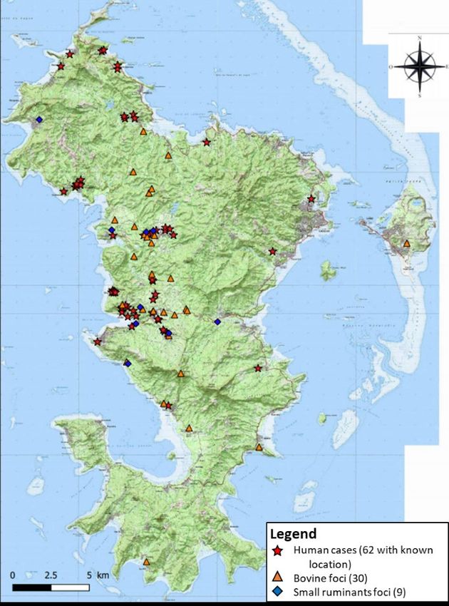
Figure 2. Geographic distribution of human cases (stars), animal outbreaks in cattle (triangles) and
small ruminants (diamonds) of RVF in Mayotte, 22 November 2018–1 March 2019
Ouroveni area
Source: adapted from Agence de Santé Océan Indien Communiqué de presse 1 mars 2019 [62].
Animal health situation
Samples taken by veterinarians between 22 November 2018–1 March 2019 from sick animals or abortions have
identified 39 epizootic foci in Mayotte, each comprising one to six animals, including bovines (30 foci) and small
ruminants (9 foci; Figure 2) [62].
According to Mayotte’s livestock cooperative, CIRAD and previous published serosurveys, the seroprevalence of
RVF among ruminants in Mayotte decreased steadily between 2008–2010 and 2017 [20,21] , then increased in
2017–2018 (3.6%, 95% CI 2.3%–5.6%) and 2018–2019 (10.1%, 95% CI 6.5%–15.3%) [61].
Risk reduction measures
According to local public health authorities in Mayotte, farmers are required to promptly report any sick animals or
abortions occurring in their animals to veterinarians in order to take samples for RVF laboratory investigation. To
prevent disease, farmers and people who practice slaughtering are invited to wear a mask, gloves and glasses
when slaughtering and wash their hands with soap after handling animal or animal tissues. Recommendations have
been issued to the population to cook the meat, boil the milk and protect themselves against mosquito bites. In
addition, as of 27 February 2019, selling raw milk has been prohibited for a period of three months [63].
ECDC threat assessment for EU/EEA
An epizootic of RVF in Mayotte is an unusual event but not unexpected, as the virus has circulated among animals
on several islands of the Indian Ocean, including on Mayotte, since 2004 [64]. Recent serosurveys and animal case
reports indicate an increased circulation of RVFV in the livestock of Mayotte at the end of 2018 [20,61]. The
sustained viral circulation on Mayotte is possibly due to the continuous presence of susceptible animals, existence
of competent vectors maintaining transmission locally and possible regular reintroduction of the virus from nearby
countries through illegal domestic animal movements [20,21]. Several mosquito species that have been identified
as possible RVF vectors are present on Mayotte [64–66] and current weather conditions are favourable for the
vector population (rainy season from December to March). Therefore, further transmission among animals on
Mayotte is likely and the associated risk for human infection remains.
6RAPID RISK ASSESSMENT Rift Valley fever outbreak in Mayotte, France – 7 March 2019
Risk for EU/EEA citizens residing in/travelling to affected
areas
The occurrence of human cases with known risk behaviour during an epizootic of RVF, such as reported during this
outbreak in Mayotte, is consistent with the literature [28,60].
Travellers and residents applying infection prevention measures are at very low risk of infection. Those who are in
contact with potentially infected animals, e.g. veterinarians and those involved in livestock farming, butchering and
slaughtering of animals in RVF affected areas, however, have an increased risk of infection with RVFV and should
therefore handle potentially infected animals in a secure manner by practicing safe animal husbandry and
slaughtering practices. In affected areas, consumption of raw milk and of animal products that have not been
thoroughly cooked should be avoided. In addition, as a precautionary measure, personal protective measures
against mosquito bites should be applied [8,57]. Transmission of the virus through contact with blood or infected
material in healthcare settings can be prevented by applying the measures defined in WHO’s ‘Standard precautions
in health care’ aide-memoire [58].
Risk of importation to and further spread within EU
Outermost Regions in the Indian Ocean
Between 2018–February 2019, there were no official movements of cattle, sheep or goats towards the EU/EEA
from the region of the Comoros Islands (Comoros and Mayotte) and Madagascar. Nevertheless, the RVF-free
islands of the Indian Ocean Area (Réunion, Mauritius and Seychelles) remain at risk of introduction through illegally
imported viraemic animals originating from areas with RVFV circulation, such as countries from eastern Africa and
islands of the Indian Ocean (Comoros and Madagascar). As such, early detection of RVF among animals remains a
priority in the RVF-free islands of the Indian Ocean Area [14]. Should the virus be introduced, further transmission
remains possible due to the presence of naïve livestock and a competent and active mosquito vector population in
Réunion [14].
Importation of human cases from Mayotte to Réunion cannot be excluded due to the regular flight connection
between the two islands. According to the International Air Transport Association (IATA), there were 67 000
travellers from Mayotte to Réunion in 2017. However, should the virus be imported, the likelihood of further spread
by direct human-to-human transmission is very low as sustained direct human-to-human transmission has not
been described for RVF. As a precautionary measure, imported viraemic RVF cases should be advised to apply
personal protective measures against mosquito bites in areas with competent and active vectors [8,57].
Risk of importation to and further spread within continental
parts of EU/EEA
According to EFSA, RVFV can be introduced by illegally imported infected animals and less likely through infected
vectors or illegally imported contaminated animal products [22,67]. Imports into continental EU countries of live
animals and their meat and milk from Mayotte are probably very rare due to distance, so the risk of introduction of
RVF through such trade is probably very low.
Potential RVFV vectors are present in parts of the continental EU/EEA, raising concern about the transmission
potential of RVF in Europe [22,68–74]. Overall, further localised transmission between domesticated animals and
subsequent RVF cases in humans after virus introduction would require adequate temperatures for vector
development and pathogen replication, favourable environmental conditions for sufficient abundance of competent
vectors and a large population of susceptible ruminants [24,68]. To date, the likelihood of further spread in
continental EU/EEA countries is very low due to both a low probability of importing viraemic animals and the
current low winter abundance of competent mosquito vectors, although it cannot be completely excluded.
According to IATA data, there were almost 40 000 travellers from Mayotte to continental Europe in 2017, the vast
majority of them to France (97%). Therefore, the importation of human cases from Mayotte cannot be excluded.
The occurrence of travel-related cases returning to continental EU/EEA countries is not new as RVF is endemic in
many African countries. Sporadic importation of RVF human cases into the EU/EEA has occurred in recent
years [46,56]. To date, no autochthonous human cases of RVF have been reported in continental parts of the
EU/EEA.
The risk for secondary cases in the EU/EEA through direct human-to-human transmission is negligible, including
any secondary transmission from viraemic passengers travelling in airplanes. The probability of further
vector-borne transmission within the human population is very low. Transmission of the virus through contact with
blood or infected materials in healthcare settings may occur, but can be prevented by applying the measures
defined in WHO’s ‘Standard precautions in health care’ aide-memoire [58] and in ECDC’s technical report on core
competencies for infection control and hospital hygiene professionals in the EU [75].
7RAPID RISK ASSESSMENT Rift Valley fever outbreak in Mayotte, France – 7 March 2019
Rift Valley fever and safety of SoHO
Transmission of RVF through transfusion and/or transplantation has not been reported. Nonetheless, the
transmission of RVFV through substances of human origin (SoHO) donated by an asymptomatic viraemic donor
cannot be excluded. Since Mayotte is endemic for malaria [76], the deferral period for donors returning from areas
affected by malaria is sufficient to prevent the donation of RVF-virus-infectious SoHO. Therefore, the risk of SoHO
donation by an RVFV-infected traveller from Mayotte can be considered to be very low. Local blood safety
authorities in the Indian Ocean (Établissement Français du Sang, Réunion) ensure the supply of labile blood
products from Réunion to the Mayotte hospital depot because there are no blood donations due to the presence of
malaria [28].
Disclaimer
ECDC issues this risk assessment document based on an internal decision and in accordance with Article 10 of
Decision No. 1082/13/EC and Article 7.1 of Regulation (EC) No 851/2004 establishing a European centre for
disease prevention and control (ECDC). In the framework of ECDC’s mandate, the specific purpose of an ECDC risk
assessment is to present different options on a certain matter with their respective advantages and disadvantages.
The responsibility on the choice of which option to pursue and action to take, including the adoption of mandatory
rules or guidelines, lies exclusively with EU/EEA Member States. In its activities, ECDC strives to ensure its
independence, high scientific quality, transparency and efficiency. This report was written with the coordination
and assistance of an Internal Response Team at the ECDC. All data published in this risk assessment are correct to
the best of our knowledge at the time of publication. Maps and figures published do not represent a statement on
the part of ECDC or its partners on the legal or border status of the countries and territories shown.
8RAPID RISK ASSESSMENT Rift Valley fever outbreak in Mayotte, France – 7 March 2019
References
1. European Food and Safety Authority. Characterisation of Rift Valley fever (RVF) for scientific opinion on
vector-borne disease [Internet]. Parma: EFSA; 2017 [cited 1 March 2019]. Available from:
http://efsa.maps.arcgis.com/apps/MapJournal/index.html?appid=5caa3b6f07684ce881301ea2326bc811
2. International Committee on Taxonomy of Viruses. Virus Taxonomy: 2018 Release [Internet].
Washington:ICTV; 2018 [cited 1 March 2019]. Available from: http://talk.ictvonline.org/taxonomy
3. Daubney R, Hudson JR, Garnham PC. Enzootic hepatitis or Rift Valley fever. An undescribed virus disease of
sheep cattle and man from east Africa. J Pathol Bacteriol. 1931;34:545-79.
4. World Health Organization. Geographic distribution of Rift Valley fever outbreaks [Internet]. Geneva:
WHO; 2009 [cited 27 February 2019]. Available from: http://www.who.int/emergencies/diseases/rift-valley-
fever/Global_RVF_20090908.png
5. Clements AC, Pfeiffer DU, Martin V. Application of knowledge-driven spatial modelling approaches and
uncertainty management to a study of Rift Valley fever in Africa. Int J Health Geogr. 2006 Dec 10;5:57.
6. Redding DW, Tiedt S, Lo Iacono G, Bett B, Jones KE. Spatial, seasonal and climatic predictive models of Rift
Valley fever disease across Africa. Philos Trans R Soc Lond B Biol Sci. 2017 Jul 19;372(1725).
7. Nanyingi MO, Munyua P, Kiama SG, Muchemi GM, Thumbi SM, Bitek AO, et al. A systematic review of Rift
Valley Fever epidemiology 1931-2014. Infect Ecol Epidemiol. 2015 Jul 31;5:28024.
8. World Health Organization. Rift Valley fever [Internet]. Geneva: WHO; 2019 [cited 26 February 2019].
Available from: http://www.who.int/news-room/fact-sheets/detail/rift-valley-fever
9. Nyakarahuka L, Balinandi S, Mulei S, Kyondo J, Tumusiime A, Klena J, et al. Ten outbreaks of rift valley
fever in Uganda 2016-2018: epidemiological and laboratory findings. Int J Infect Dise. 2019;79:4.
10. McMillen CM, Hartman AL. Rift Valley fever in animals and humans: Current perspectives. Antiviral Res.
2018 Aug;156:29-37.
11. World Health Organization. Rift Valley fever – Kenya – Disease outbreak news [Internet]. Geneva: WHO;
2018 [cited 27 February 2019]. Available from: http://www.who.int/csr/don/18-june-2018-rift-valley-fever-
kenya
12. Centers for Disease Control and Prevention. Rift Valley Fever - Outbreak Summaries [Internet]. Atlanta:
CDC; 2017 [cited 27 February 2017]. Available from: http://www.cdc.gov/vhf/rvf/outbreaks/summaries.html
13. Andriamandimby SF, Randrianarivo-Solofoniaina AE, Jeanmaire EM, Ravololomanana L,
Razafimanantsoa LT, Rakotojoelinandrasana T, et al. Rift Valley Fever during Rainy Seasons, Madagascar,
2008 and 2009. Emerg Infect Dis. 2010 Jun;16(6):963-70.
14. Balenghien T, Cardinale E, Chevalier V, Elissa N, Failloux AB, Jean Jose Nipomichene TN, et al. Towards a
better understanding of Rift Valley fever epidemiology in the south-west of the Indian Ocean. Vet Res.
2013 Sep 09;44:78.
15. Sissoko D, Giry C, Gabrie P, Tarantola A, Pettinelli F, Collet L, et al. Rift Valley Fever, Mayotte, 2007-2008.
Emerg Infect Dise. 2009 Apr;15(4):568-70.
16. Santé Publique France. Points Épidémiologiques - Surveillance de la fièvre de la vallée du Rift à Mayotte.
Point épidémiologique au 30 janvier 2019. [Internet]. Paris: Santé publique France; 2019 [cited 31 January
2019]. Available from: http://invs.santepubliquefrance.fr/Publications-et-outils/Points-
epidemiologiques/Tous-les-numeros/Ocean-Indien/2019/Surveillance-de-la-fievre-de-la-vallee-du-Rift-a-
Mayotte.-Point-epidemiologique-au-30-janvier-2019
17. Santé publique France. Bulletin de veille sanitaire de la Réunion et Mayotte – N° 02 / November 2009 –
Surveillance de la circulation de la fièvre de la vallée du Rift à Mayotte. Paris: Santé publique France; 2009
Available from:
http://invs.santepubliquefrance.fr/publications/bvs/reunion_mayotte/2009/bvs_rm_11_2009.pdf
18. Cêtre-Sossah C, Zeller H, Grandadam M, Caro V, Pettinelli F, Bouloy M, et al. Genome Analysis of Rift Valley
Fever Virus, Mayotte. Emerg Infect Dis. 2012 Jun;18(6):969-71.
19. Cêtre-Sossah C, Pédarrieu A, Guis H, Defernez C, Bouloy M, Favre J, et al. Prevalence of Rift Valley Fever
among Ruminants, Mayotte. Emerg Infect Dis. 2012 Jun;18(6):972-5.
20. Métras R, Cavalerie L, Dommergues L, Mérot P, Edmunds WJ, Keeling MJ, et al. The Epidemiology of Rift
Valley Fever in Mayotte: Insights and Perspectives from 11 Years of Data. PLoS Negl Trop Dis. 2016
Jun;10(6):e0004783.
9RAPID RISK ASSESSMENT Rift Valley fever outbreak in Mayotte, France – 7 March 2019
21. Métras R, Fournié G, Dommergues L, Camacho A, Cavalerie L, Mérot P, et al. Drivers for Rift Valley fever
emergence in Mayotte: A Bayesian modelling approach. PLoS Negl Trop Dis. 2017 Jul;11(7):e0005767.
22. European Food and Safety Authority Panel on Animal Health and Welfare. Scientific Opinion on Rift Valley
fever.. EFSA Journal. 2013;11(4):3180. Available from:
http://efsa.onlinelibrary.wiley.com/doi/epdf/10.2903/j.efsa.2013.3180
23. Tantely LM, Boyer S, Fontenille D. A Review of Mosquitoes Associated with Rift Valley Fever Virus in
Madagascar. Ame J Trop Med Hyg. 2015 Apr;92(4):722-9.
24. Lumley S, Horton DL, Hernandez-Triana LLM, Johnson N, Fooks AR, Hewson R. Rift Valley fever virus:
strategies for maintenance, survival and vertical transmission in mosquitoes. J Gen Virol.
2017 May;98(5):875-887.
25. Antonis AF, Kortekaas J, Kant J, Vloet RP, Vogel-Brink A, Stockhofe N, et al. Vertical Transmission of Rift
Valley Fever Virus Without Detectable Maternal Viremia. Vector Borne Zoonotic Dis. 2013 Aug;13(8):601-6.
26. Davies FG, Karstad L. Experimental infection of the African buffalo with the virus of Rift Valley fever. Trop
Anim Health Prod. 1981 Nov;13(4):185-8.
27. Gerdes GH. Rift Valley fever. Rev Sci Tech. 2004 Aug;23(2):613-23.
28. Hartman A. Rift Valley Fever. Clin Lab Med. 2017 Jun;37(2):285-301.
29. Yedloutschnig RJ, Dardiri AH, Walker JS. Rift Valley fever Infection in sheep by contact exposure. Contri
Epidemiol Biostat. 1981;3:53-9.
30. Jansen van Vuren P, Kgaladi J, Msimang V, Paweska JT. Rift Valley Fever Reemergence after 7 Years of
Quiescence, South Africa, May 2018. Emerg Infect Dis. 2019 Feb;25(2):338-41.
31. Rostal MK, Liang JE, Zimmermann D, Bengis R, Paweska J, Karesh WB. Rift Valley Fever: Does Wildlife Play
a Role? ILAR J. 2017 Dec 15;58(3):359-70.
32. Pedro SA, Abelman S, Tonnang HE. Predicting Rift Valley Fever Inter-epidemic Activities and Outbreak
Patterns: Insights from a Stochastic Host-Vector Model. PLoS Negl Trop Dis. 2016 Dec 21;10(12):e0005167.
33. Nicholas DE, Jacobsen KH, Waters NM. Risk factors associated with human Rift Valley fever infection:
systematic review and meta-analysis. Trop Med Int Health. 2014 Dec;19(12):1420-9.
34. LaBeaud AD, Muiruri S, Sutherland LJ, Dahir S, Gildengorin G, Morrill J, et al. Postepidemic Analysis of Rift
Valley ever Virus Transmission in Northeastern Kenya: A Village Cohort Study. PLoS Negl Trop Dis. 2011
Aug;5(8):e1265.
35. Madani TA, Al-Mazrou YY, Al-Jeffri MH, Mishkhas AA, Al-Rabeah AM, Turkistani AM, et al. Rift Valley Fever
Epidemic in Saudi Arabia: Epidemiological, Clinical, and Laboratory Characteristics. Clin Infect Dis.
2003 Oct 15;37(8):1084-92.
36. Adam I, Karsany MS. Case report: Rift Valley Fever with vertical transmission in a pregnant Sudanese
woman. J Med Virol. 2008 May;80(5):929.
37. Arishi HM, Aqeel AY, Al Hazmi MM. Vertical transmission of fatal Rift Valley fever in a newborn. Ann Trop
Paediatr. 2006 Sep;26(3):251-3.
38. Rudolph KE, Lessler J, Moloney RM, Kmush B, Cummings DA. Incubation periods of mosquito-borne viral
infections: a systematic review. Am J Trop Med Hyg. 2014 May;90(5):882-91.
39. Iowa State University College of Veterinary Medicine. Rift Valley Fever [Internet]. Ames: ISU; 2003 [cited 27
February 2019]. Available from: http://www.state.nj.us/agriculture/divisions/ah/diseases/riftvalley.html
40. Msimang V, Thompson PN, Jansen van Vuren P, Tempia S, Cordel C, Kgaladi J, et al. Rift Valley Fever Virus
Exposure amongst Farmers, Farm Workers, and Veterinary Professionals in Central South Africa. Viruses.
2019 Feb 7;11(2).
41. World Organization for Animal Health. Rift Valley Fever (RVF) – General Disease Information Sheet. Paris:
OIE; 2016. Available from: http://www.oie.int/doc/ged/D13962.PDF.
42. World Organization for Animal Health. Rift Valley Fever. Paris: OIE; 2009. Available from:
http://www.oie.int/fileadmin/Home/eng/Animal_Health_in_the_World/docs/pdf/Disease_cards/RIFT_VALLE
Y_FEVER.pdf
43. Gerdes GH. Rift valley fever. Vet Clin North Am Food Anim Pract. 2002 Nov;18(3):549-55.
44. Baudin M, Jumaa AM, Jomma HJE, Karsany MS, Bucht G, Naslund J, et al. Association of Rift Valley fever
virus infection with miscarriage in Sudanese women: a cross-sectional study. Lancet Glob Health. 2016
Nov;4(11):e864-e71.
45. Grolla A, Mehedi M, Lindsay R, Bosio C, Duse A, Feldmann H. Enhanced Detection of Rift Valley fever Virus
using Molecular Assays on Whole Blood Samples. J Clin Virol. 2012 Aug;54(4):313-7.
10RAPID RISK ASSESSMENT Rift Valley fever outbreak in Mayotte, France – 7 March 2019
46. Tong C, Javelle E, Grard G, Dia A, Lacrosse C, Fourie T, et al. Tracking Rift Valley fever: From Mali to
Europe and other countries, 2016. Euro Surveill. 2019 Feb;24(8). Available from:
http://www.eurosurveillance.org/content/10.2807/1560-7917.ES.2019.24.8.1800213
47. Mansfield KL, Banyard AC, McElhinney L, Johnson N, Horton DL, Hernández-Triana LM, et al. Rift Valley
fever virus: A review of diagnosis and vaccination, and implications for emergence in Europe. Vaccine.
2015 Oct 13;33(42):5520-5331.
48. EVD-LabNet. EVD-LabNet directory search [Internet]. Rotterdam: ErasmusMC; 2016. Available from:
http://www.evd-labnet.eu/evd-labnet-directory-search
49. National Institutes of Health. NIH Guidelines for Research Involving Recombinant or Synthetic Nucleic Acid
Molecules (NIH Guidelines) –April 2016 [Internet]. Bethesda: NIH; 2016 [cited 27 February 2019]. Available
from: http://osp.od.nih.gov/wp-content/uploads/NIH_Guidelines.html
50. Centers for Disease Control and Prevention. Biosafety in Microbiological and Biomedical Laboratories (BMBL)
5th Edition. Atlanta: CDC; 2009. Available from: http://www.cdc.gov/labs/BMBL.html
51. The Council of the European Communities. Council Directive of 21 December 1982 on the notification of
animal diseases within the Community (82/894/EEC). Brussels: Council of the European Communities; 1982.
Available from: http://eur-lex.europa.eu/legal-content/EN/TXT/?uri=CELEX%3A31982L0894
52. The Council of the European Communities. Council Directive 92/119/EEC of 17 December 1992 introducing
general Community measures for the control of certain animal diseases and specific measures relating to
swine vesicular disease. Brussels: Council of the European Communities; 1992. Available from: http://eur-
lex.europa.eu/legal-content/en/ALL/?uri=CELEX:31992L0119
53. European Commission. Commission Regulation (EU) No 206/2010 of 12 March 2010 laying down lists of
third countries, territories or parts thereof authorised for the introduction into the European Union of certain
animals and fresh meat and the veterinary certification requirements (Text with EEA relevance).
Brussels: European Commission; 2010. Available from: https://eur-lex.europa.eu/legal-
content/EN/TXT/PDF/?uri=CELEX:32010R0206
54. World Organization for Animal Health. Animal Health in the World – Overview [Internet]. Paris: OIE; 2016
[cited 27 February 2019]. Available from: http://www.oie.int/animal-health-in-the-world/oie-listed-diseases-
2016
55. European Centre for Disease Prevention and Control. Rift Valley fever. In: ECDC. Annual epidemiological
report for 2015. Stockholm: ECDC, 2017. Available from: http://ecdc.europa.eu/publications-data/rift-valley-
fever-annual-epidemiological-report-2015
56. European Centre for Disease Prevention and Control. Rift Valley fever. In: ECDC. Annual epidemiological
report for 2017. Stockholm: ECDC, 2019. Available from: http://ecdc.europa.eu/publications-data/rift-valley-
fever-annual-epidemiological-report-2017
57. European Centre for Disease Prevention and Control . Personal protective measures against mosquito bites
[Internet]. Stockholm: ECDC; 2008 [cited 27 February 2019]. Available from:
http://ecdc.europa.eu/disease-vectors/prevention-and-control/protective-measures-mosquitoes
58. World Health Organization. Standard precautions in health care – Aide-memoire – October 2007 infection
control. Geneva: WHO; 2007 [cited 3 April 2018]. Available from:
http://www.who.int/csr/resources/publications/standardprecautions
59. Chamchod F, Cosner C, Cantrell RS, Beier JC, Ruan S. Transmission Dynamics of Rift Valley Fever Virus:
Effects of Live and Killed Vaccines on Epizootic Outbreaks and Enzootic Maintenance. Front Microbiol.
2016 Feb 1;6:1568.
60. Agence de Santé Océan Indien. Fièvre de la Vallée du Rift à Mayotte : point de situation au 22 février
[Internet]. 2019 [cited 22 February 2019]. Saint-Denis: ARS; 2019. Available from: http://www.ocean-
indien.ars.sante.fr/fievre-de-la-vallee-du-rift-mayotte-point-de-situation-au-22-fevrier
61. Santé publique France. Points Épidémiologiques - Surveillance de la fièvre de la vallée du Rift à Mayotte.
Point épidémiologique au 21 février 2019 [Internet]. Paris: Santé publique France; 2019
[cited 25 February 2019]. Available from: http://invs.santepubliquefrance.fr/Publications-et-outils/Points-
epidemiologiques/Tous-les-numeros/Ocean-Indien/2019/Surveillance-de-la-fievre-de-la-vallee-du-Rift-a-
Mayotte.-Point-epidemiologique-au-21-fevrier-2019
62. Agence de Santé Océan Indien. Communiqué de Presse – Fièvre de la Vallée du Rift á Mayotte : point de
situation au 1er mars. Mamoudzou: ARS; 2019. Available from: http://www.ocean-
indien.ars.sante.fr/system/files/2019-03/CPFVR%2001%2003%2019-V1.pdf
11RAPID RISK ASSESSMENT Rift Valley fever outbreak in Mayotte, France – 7 March 2019
63. La 1ère. Fièvre de la vallée du Rift à Mayotte, la commercialisation du lait cru interdite 2019 [Internet].
Paris: France Télévisions; 2019 [cited 1 March 2019]. Available from: http://la1ere.francetvinfo.fr/fievre-
vallee-du-rift-mayotte-commercialisation-du-lait-cru-interdite-684962.html
64. Lernout T, Cardinale E, Jego M, Despres P, Collet L, Zumbo B, et al. Rift Valley Fever in Humans and
Animals in Mayotte, an Endemic Situation? PLoS One. 2013 Sep 30;8(9):e74192.
65. Le Goff G, Goodman SM, Elguero E, Robert V. Survey of the Mosquitoes (Diptera: Culicidae) of Mayotte.
PLoS One. 2014;9(7):e100696.
66. Turell MJ, Linthicum KJ, Patrican LA, Davies FG, Kairo A, Bailey CL. Vector Competence of Selected African
Mosquito (Diptera: Culicidae) Species for Rift Valley Fever Virus. J Med Entomol. 2008 Jan;45(1):102-8.
67. European Food and Safety Authority. The risk of Rift Valley fever incursion and its persistence within the
Community. EFSA Journal. 2005 Oct 11;238:1-128.
68. Chevalier V, Pépin M, Plée L, Lancelot R. Rift Valley fever--a threat for Europe? Euro Surveill. 2010 Mar
11;15(10):19506. Available from: http://www.eurosurveillance.org/content/10.2807/ese.15.10.19506-en
69. Brustolin M, Talavera S, Nuñez A, Santamaria C, Rivas R, Pujol N, et al. Rift Valley fever virus and European
mosquitoes: vector competence of Culex pipiens and Stegomyia albopicta (= Aedes albopictus). Med Vet
Entomol. 2017 Dec;31(4):365-372.
70. Vloet RPM, Vogels CBF, Koenraadt CJM, Pijlman GP, Eiden M, Gonzales JL, et al. Transmission of Rift Valley
fever virus from European-breed lambs to Culex pipiens mosquitoes. PLoS Negl Trop Dis.
2017 Dec;11(12):e0006145.
71. Talavera S, Birnberg L, Nuñez AI, Muñoz-Muñoz F, Vázquez A, Busquets N. Culex flavivirus infection in a
Culex pipiens mosquito colony and its effects on vector competence for Rift Valley fever phlebovirus. Parasit
Vectors. 2018 May 23;11(1):310.
72. Fischer EA, Boender GJ, Nodelijk G, de Koeijer AA, van Roermund HJ. The transmission potential of Rift
Valley fever virus among livestock in the Netherlands: a modelling study. Vet Res. 2013 Jul 22;44:58.
73. Sánchez-Vizcaíno F, Martínez-López B, Sánchez-Vizcaíno JM. Identification of suitable areas for the
occurrence of Rift Valley fever outbreaks in Spain using a multiple criteria decision framework. Vet Microbiol.
2013 Jul 26;165(1-2):71-8.
74. Lumley S, Hernández-Triana LM, Horton DL, Fernández de Marco MDM, Medlock JM, Hewson R, et al.
Competence of mosquitoes native to the United Kingdom to support replication and transmission of Rift
Valley fever virus. Parasit Vectors. 2018 May 18;11(1):308.
75. European Centre for Disease Prevention and Control. Core competencies for infection control and hospital
hygiene professionals in the European Union. Stockholm: ECDC; 2013. Available from:
http://ecdc.europa.eu/publications-data/core-competencies-infection-control-and-hospital-hygiene-
professionals-european
76. World Health Organization. World malaria report 2018. Geneva: WHO; 2018. Available from:
https://www.who.int/malaria/publications/world-malaria-report-2018
12You can also read