Co-subsistence of avian influenza virus subtypes of low and high pathogenicity in Bangladesh: Challenges for diagnosis, risk assessment and ...
←
→
Page content transcription
If your browser does not render page correctly, please read the page content below
www.nature.com/scientificreports

                        OPEN       Co-subsistence of avian influenza
                                   virus subtypes of low and high
                                   pathogenicity in Bangladesh:
Received: 14 March 2019
Accepted: 11 May 2019              Challenges for diagnosis, risk
                                   assessment and control
Published: xx xx xxxx

                                   Rokshana Parvin1,2, Jahan Ara Begum2, Emadadul Haque Chowdhury2, Mohammed
                                   Rafiqul Islam 2, Martin Beer1 & Timm Harder1
                                   Endemic co-circulation of potentially zoonotic avian influenza viruses (AIV) of subtypes H5N1 and H9N2
                                   (G1 lineage) in poultry in Bangladesh accelerated diversifying evolution. Two clinical samples from
                                   poultry obtained in 2016 yielded five different subtypes (highly pathogenic [HP] H5N1, HP H5N2, HP
                                   H7N1, HP H7N2, H9N2) and eight genotypes of AIV by plaque purification. H5 sequences grouped with
                                   clade 2.3.2.1a viruses while N1 was related to an older, preceding clade, 2.2.2. The internal genome
                                   segments of the plaque-purified viruses originated from clade 2.2.2 of H5N1 or from G1/H9N2 viruses.
                                   H9 and N2 segments clustered with contemporary H9N2 strains. In addition, HP H7 sequences were
                                   detected for the first time in samples and linked to Pakistani HP H7N3 viruses of 2003. The unexpected
                                   findings of mixtures of reassorted HP H5N1 and G1-like H9N2 viruses, which carry genome segments
                                   of older clades in association with the detection of HP H7 HA segments calls for confirmation of these
                                   results by targeted surveillance in the area of origin of the investigated samples. Hidden niches and
                                   obscured transmission pathways may exist that retain or re-introduce genome segments of older
                                   viruses or reassortants thereof which causes additional challenges for diagnosis, risk assessment and
                                   disease control.

                                   Avian influenza viruses (AIVs) remain important pathogens for wild birds, domestic poultry and humans. AIVs
                                   group into 16 hemagglutinin (HA) and 9 neuraminidase (NA) subtypes each revealing various lineages and
                                   host-specific adaptations1. Based on the pathogenic potential in experimentally infected chickens, phenotypes of
                                   low and high pathogenicity are distinguished (LP/HPAIV). Several AIV subtypes and lineages exhibiting explicit
                                   zoonotic propensity have emerged as a public health threat2. Notably, these are goose/Guangdong (gs/GD) line-
                                   age derived HPAI H5 viruses and 2013-origin Chinese LPAI H7N9 viruses, but also further viruses such as the G1
                                   lineage of subtype H9N2 have caused sporadic human cases3,4. Within the last decade, gs/GD HPAI H5 and LPAI
                                   H9N2 viruses have become endemic in poultry in several Asian and African countries causing huge economic
                                   losses4,5. Domestic duck populations play an important role in establishing and maintaining endemicity as they
                                   often subclinically shed and spread these viruses1,6,7. Long term co-circulation of multiple influenza virus lineages
                                   in different species of poultry and inconsequent control attempts including fragmentary vaccination coverage has
                                   facilitated diversifying evolution by genetic drift and reassortment8–10.
                                       Bangladesh, a south Asian country with a high density of poultry and human populations is no exception in
                                   this respect. Potentially zoonotic HPAI H5N1 and H9N2 viruses co-circulate endemically in commercial and
                                   backyard poultry shuttling between different poultry species and causing sporadic spill-overs into wild bird pop-
                                   ulations since early 200711–14. Several reassortants between HPAI H5N1, H9N2 and HPAI H7N3 viruses have
                                   been recorded in the country although some reassortments took place before incursion into Bangladesh11–13.

                                   1
                                    Federal Research Institute for Animal Health, Friedrich-Loeffler-Institut (FLI), Suedufer 10, 17493, Greifswald-Insel
                                   Riems, Germany. 2Department of Pathology, Faculty of Veterinary Science, Bangladesh Agricultural University,
                                   Mymensingh 2202, Mymensingh, Bangladesh. Correspondence and requests for materials should be addressed to
                                   T.H. (email: Timm.Harder@fli.de)

        Scientific Reports |   (2019) 9:8306 | https://doi.org/10.1038/s41598-019-44220-4                                                              1
www.nature.com/scientificreports/                                                               www.nature.com/scientificreports

                           Figure 1. Gross and microscopic lesions in chicken, upper row, (a) congested lung; (b) petechial and
                           ecchymotic haemorrhages on the air sac and epicardium; (c) pancreas with dark red areas along its length and
                           congested blood vessels at the wall of the intestine; (d) mucosal haemorrhages in proventriculus; lower row, (e)
                           severe congestion and edema in lung tissue; (f) mild cardiac myodegeneration; (g) necrosis of pancreatic islets;
                           (h) haemorrhages in duodenal mucosa.

                           In addition, Bangladesh suffered from successive incursions and replacements of three different gs/GD HPAIV
                           H5N1 clades, 2.2.2, 2.3.2.1, and 2.3.4.214. Since 2012, clade 2.3.2.1a viruses have replaced previously co-circulating
                           gs/GD viruses15,16. Exposure of humans to these viruses is emphasized by nine and three cases of human infection
                           with HPAIV H5N1 (including one death) and H9N2, respectively, till to date17–19.
                               Here we illustrate the intricate epidemiological situation and the evolutionary potential of co-circulating AIV
                           subtypes of different sub-, geno- and phenotypes by molecular analyses of clinically overt AI outbreaks in com-
                           mercial and backyard poultry in Tangail district, one of the most crowded districts for both poultry and human
                           populations at the northern perimeter of Dhaka, the capital of Bangladesh.

                           Results
                           Clinical and pathological observation. Limited HPAI syndrome surveillance was carried out in Tangail
                           district throughout January to May 2016 in commercial medium size layer chicken farms (so-called sector 3
                           according to the Food and Agriculture Organization of the United Nations [FAO]) reporting respiratory illness.
                           Carcasses of four chickens (VP02-VP05) were sampled at four different farms keeping grower and layer chickens
                           6 to 84 weeks old, cage-reared and putatively vaccinated against H5 viruses (unknown source). From neighboring
                           backyard poultry holding the carcass of a duck (VP06) was collected as well. The sampled sector 3 chicken layer
                           farms, each of up to 2000 birds reported morbidity of up to 90% dominated by respiratory disease and reduced
                           egg production. Cumulative mortality approaching 35% at these farms was substantial but still low compared to
                           previously recorded HPAI outbreaks in chickens in Bangladesh20. Clinical presentation and epidemiological con-
                           siderations thus raised the suspicion of AIV H9N2 infections. This suspicion was corroborated by contemporary,
                           laboratory-confirmed findings of H9N2 cases with similar clinical presentation in the same area (unpublished).
                           This prompted submission of samples directly to FVS-BAU while samples from HPAI suspect cases are to be
                           filed to the National Reference Laboratory at Bangladesh Livestock Research Institute. At necropsy, however, at
                           FVS-BAU, severe congestion in lung and trachea along with severely congested blood vessels at intestinal walls,
                           petechial to ecchymotic haemorrhages at the epicardium, air sacs, intestine, proventricular glands and pan-
                           creas dominated (Fig. 1a–d). Histopathologically, widespread haemorrhages, congestion and massive edema in
                           lungs, mild cardiac myo-degeneration, hemorrhages in the lamina propria of intestine and necrosis of pancreatic
                           islets were recorded (Fig. 1e–h) and evoked the impression of a systemic infection. AIV RNA was detected by
                           M-specific conventional RT-PCR while samples were negative for Newcastle disease virus (NDV).
                               Additional samples, came from a commercial chicken farm (C1/2016) and a backyard duck (D1/2016) sam-
                           pled in November 2016 in Mymensingh district bordering Tangail. Similar clinical signs and lesions as described
                           above were recorded in the respective chickens and ducks (data not shown). However, mortality in the commer-
                           cial layer chicken farm of C1/2016 was higher (70–75%). In this farm, in contrast to the flocks in Tangail district,
                           an HPAIV H5N1 mono-infection was diagnosed. Mortality in Tangail farms was lower (35%) and, initially, an
                           H9N2 infection was diagnosed that later turned out to be a mixed infection comprising HPAIV H5 and H7.

                           Viral sequence and phylogenetic analyses directly from clinical samples. At FVS-BAU, all eight
                           AIV genome segments were successfully amplified and full-length genome sequences obtained from clinical sam-
                           ples of four layer chicken farms (VP02-VP05) and the backyard duck (VP06) from Tangail district. Sequence
                           analysis suggested presence of H9N2 viruses with a dibasic HA cleavage site motif (KSKR/GLF) in all samples.
                           HA H9 and NA N2 phylogenetic analyses revealed that all sequences were similar to circulating Bangladeshi
                           H9N2 viruses of the G1 lineage (Fig. 2, H9, N2, green color). Sequences obtained from chicken samples but not
                           the duck sample revealed the Q234L substitution (H9 numbering) known to extend HA binding to alpha 2,6 sialic
                           acid ligands21,22 (Supplementary Data; Table S1). Depending on the sample at least one or more of the internal

Scientific Reports |   (2019) 9:8306 | https://doi.org/10.1038/s41598-019-44220-4                                                              2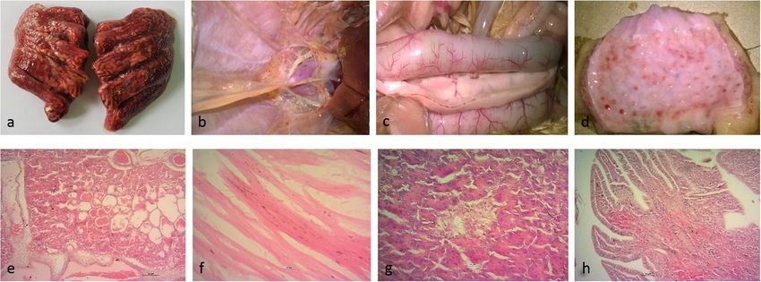
www.nature.com/scientificreports/                                                          www.nature.com/scientificreports

                           Figure 2. Phylogenetic trees of HA H5, H7, H9, and NA N2 and N1 of sequences established from clinical
                           samples (green) and plaque-purified viruses (red) from Tangail district. Sequences of samples from
                           Mymensingh district are shown in blue. Maximum likelihood trees were established using the IQ-Tree software.
                           Amino acid alignments of the HA1 and the full length NA proteins were used for calculation based on the
                           specific FLU substitution model recommended by Dang et al.44. Robustness of branching orders was tested by
                           an ultrafast bootstrap approximation algorithm38. Trees are drawn to scale and clading information is shown to
                           the right of each tree.

Scientific Reports |   (2019) 9:8306 | https://doi.org/10.1038/s41598-019-44220-4                                                       3
www.nature.com/scientificreports/                                                            www.nature.com/scientificreports

                           Figure 3. Sub- and genotype characterization of avian influenza viruses detected in original clinical samples
                           (a) and following virus isolation and plaque purification (b,c) from selected chicken and duck origin samples,
                           respectively. Genetic relationship/origin of segments is color-coded, G1/H9N2 - green, 2.2.2/H5N1 - red,
                           2.3.2.1a/H5N1 – pink, Pak/H7N3- blue.

                           genes appeared to derive from reassortment with HPAIV H5N1, clade 2.2.2 (Figs 3a, 4 & Supplementary Data;
                           Table S2). Samples C1/2016 and D1/2016 from Mymensingh district were identified as contemporary gs/GD
                           H5N1 viruses of clade 2.3.2.1a (Fig. 2, H5, N1, blue color).

                           Genotypic and phenotypic characterization of co-isolated AI viruses. Direct Sanger sequenc-
                           ing of clinical samples (VP02-VP06) suggested presence of reassorted H9N2 viruses. In contrast, full range
                           RT-qPCR-based subtyping by the “Riems influenza A typing assay” (RITA) carried out on RNA extracted from
                           allantoic fluid of embryonated chicken egg (ECE) passage detected further AIV HA and NA subtypes H5, H7,
                           H9, N1 or N2 for samples VP02-06 from Tangail district. No further co-infection was detected for H5N1 positive
                           samples C1/2016 and D1/2016 obtained from Mymensingh district (Table 1). Virus isolation and plaque purifica-
                           tion flanked by RITA was used exemplarily for samples ck/VP02 and dk/VP06 to confirm co-mingling of several
                           subtypes in the same sample. To avoid cross contamination, samples were treated separately on different days.
                               From a total of 300 plaques obtained at three consecutive passages without amplification between each plaque
                           round, ck/VP02 yielded pure plaques of subtypes H5N1, H5N2, and H9N2 of five different genotypes (Fig. 3b).
                           Four subtypes (H5N1, H5N2, H7N1, H7N2) and five different genotypes were rescued from the backyard duck
                           isolate dk/VP06 (Fig. 3c).
                               H5 HA genes of plaque-purified H5N1 or H5N2 (Purified from VP02 and VP06 sample originating from
                           Tangail district) clustered with HPAIV clade 2.3.2.1a (HA cleavage site PQRERRRKR/GLF) maintaining a close
                           relationship with H5N1 viruses described in Bangladesh during 2012–2013 but distinct from recently circulating
                           H5N1 sample C1/2016 and D1/2016 originating from Mymensingh district in late 2016 (Fig. 2, H5). The HA gene
                           of purified H7 viruses (H7N1 or H7N2) of the duck sample (VP06) surprisingly clustered with HPAIV isolated
                           during the early 2000s in Pakistan (Pak/H7N3) (Fig. 2, H7). The presence of a polybasic HA cleavage site motif
                           PETPKRRKR/GLF confirmed the HP character of plaque-purified H7 clones. The H9 HA gene of plaque-purified
                           H9N2 viruses was very similar to the ones generated directly from the clinical samples (Fig. 2, H9).
                               The NA N1 segment in both H5N1 and H7N1 plaques was closely related to H5N1 viruses from Bangladesh
                           and Pakistan which were detected during 2006–2008 and belonged to gs/GD clade 2.2.2 (Fig. 2, N1), clearly dis-
                           tinct from the N1 of clade 2.3.2.1a. NA N2 in H5N2, H7N2 or H9N2 derived from circulating G1/H9N2 viruses
                           (Fig. 2, N2).

Scientific Reports |   (2019) 9:8306 | https://doi.org/10.1038/s41598-019-44220-4                                                           4
www.nature.com/scientificreports/ www.nature.com/scientificreports
Figure 4. Phylogenetic trees of PB2, PB1, PA, NP, M and NS gene segment sequences established from clinical
poultry samples from Tangail district. Maximum likelihood trees were established using the IQ-Tree software.
Nucleotide sequence alignments of the respective internal genome segments were used for choosing the best-
fitted model according to Bayesian information criterion analysed by ModelFinder (embedded in IQ-Tree)45.
Robustness of branching orders was tested by an ultrafast bootstrap approximation algorithm38. Trees are drawn
to scale and clading information is shown to the right of each tree. G1/H9N2-like segments are depicted in
green and gs/Gd 2.2.2-like segments are in red color. Sequences of the H7N1 and H7N2 plaque-purified viruses
are presented in blue.
Scientific Reports | (2019) 9:8306 | https://doi.org/10.1038/s41598-019-44220-4 5www.nature.com/scientificreports/ www.nature.com/scientificreports
Sample Origin M H5 H7 H9 N1 N2
A/layer chicken/BD/VP02/2016 Tangail 13.07 19.4 27.1 14.24 20.46 14.35
A/layer chicken/BD/VP03/2016 Tangail 16.38 17.45 29.82 19.03 18.19 19.26
A/layer chicken/BD/VP04/2016 Tangail 16.34 19.71 Neg 17.17 19.72 18.39
A/layer chicken/BD/VP05/2016 Tangail 30.25 31.86 Neg 34.54 32.3 32.32
A/duck/BD/VP06/2016 Tangail 15.57 17.69 18.91 19.86 17.55 17.61
A/chicken/BD/C1/2016 Mymensingh 20.23 13.24 Neg Neg 14.04 Neg
A/duck/BD/D1/2016 Mymensingh 18.02 16.82 Neg Neg 17.28 Neg
Table 1. Co-infection with up to three HA subtypes detected in poultry samples from Tangail but not from
Mymensingh district, Bangladesh, by use of arrayed subtype-specific RT-qPCRs (RITA, 23). The results are
presented as RT-qPCR Cq values; Neg – Cq > 40.
The genotype of the plaque-purified viruses was determined by sequencing-independent SYBR-Green
RT-qPCRs assigning the internal gene segments to either the G1/H9N2 or 2.2.2/H5N1 origin (Fig. 3b,c). None
of the internal genes of the clinical samples (VP02-VP06) and of the plaque-purified viruses was found similar to
the currently circulating 2.3.2.1a/H5N1.
The rescued reassortant subtypes exhibited distinct plaque morphology. The H5N1 subtype produced the
largest plaques at approximately 4.5 mm in diameter. Plaque area measurements using ImageJ23 revealed smaller
plaques compared to H5N1 for H7N1 (55%), H9N2 (16%), H5N2 and H7N2 (1.0–1.8%). HP H5N1, H5N2,
H7N1 and H7N2 produced higher virus yields (TCID50 of 107 to 109 per mL allantoic fluid) and a shortened
mean-death time (MDT, 48 hours post inoculation) compared to purified H9N2 (TCID50 of 106 per mL; MDT
80 hours post inoculation).
Approximation of viral fitness using an in ovo competition model. Given the co-presence of up
to three different AIV subtypes of LP and HP phenotypes with a seemingly dominance of G1/H9N2 in the clin-
ical samples, the relative fitness of different sub- and genotypes was examined in ECE co-infected with up to
three distinct sub- or genotypes at the same TCID50 of 103/0.2 mL. Independent of the sub- and genotype none
of the co-inoculated clones was outcompeted during three consecutive blind passages. In particular, seemingly
unconstrained growth of H9N2 clones was noted even in the presence of H5 or H7 HPAI viruses. In compe-
titions between HP H5N1 and HP H5N2, the N2 gene became dominant, i.e. Cq-values for N1 and N2 were
similar in the inoculum at passage 0 but differed by 7–9 Cq values at passage 3 with N2 taking advantage over
N1 (Supplementary Data; Table S3). Also in another competition between HPAIV H5N1 and H9N2, N2 fully
superseded N1. In triple co-infection experiments (H5N1, H7N1 and H9N2) equal amplification of the three HA
and the two NA segments were evident over three passages (Supplementary Data; Table S3). The origin of internal
gene segments (H9N2 versus H5 2.2.2) did not reveal clear advantages of certain segments.
Discussion
In total, five subtypes comprising eight different genotypes were discernable by RT-qPCR subtyping (RITA), virus
isolation and after plaque purification from two selected clinical poultry samples (ck/VP02 and dk/VP06) of AI
outbreaks. The same samples were previously diagnosed based on RT-PCR and direct Sanger sequencing of clin-
ical material, to be due to infection with reassorted H9N2 viruses. Despite presence of gene segments of several
sub- and genotypes in the same sample, the corresponding sequence chromatograms showed a high quality; this
is probably due to the high subtype specificity of primers used for cycle sequencing which grossly favored a single
subtype over the rest. H5N1, H5N2 and H9N2 viruses were purified from ck/VP02 whereas H5N1, H5N2, H7N1
and H7N2 subtypes were purified from dk/VP06. Since plaque purification could not be carried out directly from
clinical material, the five subtypes and eight different genotypes may not have been present in this constellation
in the clinical samples. Some of the sub- and genotypes detected following plaque purification could have been
generated during virus isolation in ECE and/or plaque-passaging. Surprisingly, no replicating H9N2 virus was
rescued during plaque purification from dk/VP06 although RITA confirmed the presence of this subtype in this
sample as well (Table 1). We speculate that this might have been due to the interference of the different viruses in
this sample, and a higher replication efficiency of the numerous H5 and H7 viruses which might have outcom-
peted the H9 virus in our cloning approach.
In depth molecular analyses revealed that the rescued H5N1 and H5N2 viruses derived their HA H5 segment
from clade 2.3.2.1a viruses of the years 2012–2014 (HA, Fig. 2, H5), while the N1 segment along with six inter-
nal genes most likely originated from clade 2.2.2/H5N1 (Fig. 2, N1). However, the H5N1 viruses of this clade
became apparently extinct from Bangladesh after 201114,15. Continued circulation of genome segments of that
clade therefore was unexpected and currently stands as a solitary but important finding. As such, the possibility
of undetected circulating clade 2.2.2 HPAI viruses or gene segments thereof in other reassortant subtypes must be
considered. Chicken and duck samples (C1/2016 and D1/2016) obtained in the Mymensingh district in late 2016
and treated similarly in our labs proved to contain each a single AIV strain which carried contemporary 2.3.2.1a
HA and NA genome segments (Fig. 2, H5).
Even more surprising was the finding of an HPAI H7 virus having an HA gene similar to that of HPAI Pak/
H7N3 viruses that had caused outbreaks in Pakistan during 2000–200324, but which had not been detected at that
or any time in Bangladesh. Although subtype H7 viruses have been detected in backyard and free ranging ducks
Scientific Reports | (2019) 9:8306 | https://doi.org/10.1038/s41598-019-44220-4 6www.nature.com/scientificreports/ www.nature.com/scientificreports
in Bangladesh, they were of low pathogenicity and their HA segment was of a Eurasian wild bird lineage unrelated
to the HP H7 Pakistan viruses7,25 (Fig. 2, H7).
A generic explanation for the detection of genome segments of older HP H5 and HP H7 strains in these
samples would be by contamination in the laboratory. However, this can be rejected as a single diagnostic lab-
oratory in Bangladesh (FVS-BAU) undertook all sample manipulations including virus isolation and this labo-
ratory did never have access to replication-competent HP Pak/H7 and Bangladeshi HP H5N1 viruses of clade
2.2.2. Likewise, none of these viruses were available in the laboratory at FLI where subsequent RITA assays, virus
amplification and plaque purification experiments were carried out. Unfortunately, original tissues of these cases
were no longer retrievable for confirmation. Thus, the true sources of the HP H7 and HP H5 2.2.2 viruses remain
elusive.
HA and NA segments of purified H9N2 clones were similar to circulating contemporary G1/H9N2 viruses
although their internal genes originated from clade 2.2.2/H5N1. Previously, it was also reported that Bangladeshi
H9N2 viruses shared internal genes with HP H5N1 (PB1, clade 2.3.2.1a) and with the HP Pak/H7N311–13. These
findings at least indicate independent of our findings that there must have been opportunities of reassortment
between HP H5N1 and the Pakistani H7 viruses although it remained unclear where and when such events took
place. In turn, our findings could provide a very comprehensible explanation for those observations. Also in
Cambodia, extensive co-circulation of and reassortment among H5, H7 and H9 AIV in poultry has recently been
pointed out26.
The potential of sustained replication in ECE of G1/H9N2 viruses even in competition with HPAI H5 and H7
viruses was demonstrated during a limited number of passages in co-infected ECE. Although no prognosis for
the next 10 or 50 passages can be made, it seems conceivable that sets of different AIV sub- and genotypes may be
co-transmitted across at least several passages also in vivo. Yet, it is not possible to extrapolate these results to the
field where additional transmission bottlenecks may exist. Stable replication in chickens of an LPAI H7N7 virus
even in presence of its HP mutant descendant virus has recently been reported27.
From a diagnostic perspective, the presence of minor viral populations within extended mixtures as described
here would only be detected by using highly sensitive and multiple subtype-discerning diagnostic tools such as
RITA. In contrast, more global diagnostic approaches including direct Sanger sequencing based on universal
“Hoffmann” primers and strain-specific sequencing primers might yield incomplete and even false conclusions
depending on the specificity of the primers. This was initially the case here, when different reassorted H9N2
viruses were diagnosed on basis of full-length genome Sanger sequencing. In addition, also high throughput
sequencing approaches might give more insights for those samples in the future.
Presence of HPAIV phenotypes in G1/H9N2 mixtures would be expected to influence pathogenesis and clin-
ical picture. This was evident also here with the detection of signs of systemic infection and lesions in multi-
ple organs (Fig. 1), but comparatively low cumulative mortality in affected flocks. Viral population size within
those mixture and, hence, clinical sequelae, would vary according to the specific immune status of the hosts. In
the reported cases, the affected layer chickens probably had received H5-specific vaccination. Recently, chickens
and ducks sold in live bird market in Bangladesh were found positive for HPAIV H5N1 but appeared appar-
ently healthy when sampled although the vaccination status of those birds was unknown28. Potential masking of
HPAIV related clinical signs in vaccinated or LPAIV co-infected poultry should be considered when analyzing
spread of HPAIV along trading networks.
The much unexpected findings of mixtures of reassorted HP H5N1 and G1-like H9N2 viruses which carry
genome segments of older gs/GD clades and the detection of HP H7 HA segments previously not reported from
Bangladesh, calls for confirmation of these results by targeted surveillance in the area of origin of the investigated
samples. Virus strains or reassorted gene segments thereof believed to be extinct may yet still reside in small
regional pockets or populations. In general, our findings emphasize the need of a broader and deeper analysis of
(AI) viral populations in clinical samples and corresponding viral isolates originating from AI outbreaks. While
next-generation sequencing techniques provide ultimate tools for this approach, available multiplexed or arrayed
RT-qPCR applications allow for a rapid screening at more sensible costs and of higher sample numbers.
Methods
Sample origin, pathology and virus propagation. Necropsy, histopathology and virological investiga-
tions as previously described29 were carried out upon arrival of poultry carcasses at the Department of Pathology,
Faculty of Veterinary Science, Bangladesh Agricultural University (FVS-BAU). In addition, presence of AIV and
Newcastle Disease virus (NDV) RNA, respectively, in lung and trachea tissue was investigated by conventional
RT-PCR targeting fragments of the matrix (M) gene of both AIV and NDV30,31. Full-length genome sequences
were established using the Hoffmann primers32 as detailed below. Tissue homogenates (lung and trachea, other
tissues or sera were not made available) were inoculated into 10-day old embryonated chicken eggs (ECE) follow-
ing standard procedures33. Allantoic fluid of this egg passage was sent to the Friedrich-Loeffler-Institute (FLI) for
further investigations. At FLI, passaged samples were HA- and NA-subtyped using the Riems influenza A typing
assay (RITA)34.
Plaque purification in Madin-Darby canine kidney (MDCK-II) cell cultures. Based on the RITA
results three rounds of plaque purification were conducted for two selected egg-grown virus isolates (ck/VP02
and dk/VP06) to obtain pure virus clones. Briefly, 100 µL of 10-fold serial dilutions of each isolate were incu-
® ™
bated on confluent MDCK-II (ATCC CRL-2936 ) cell monolayers in six well plates for 1 h at 37 °C. Cells were
carefully washed twice with phosphate buffered saline (PBS) and then overlaid with modified Eagle’s medium
containing 1.8% agar, 3.5% bovine serum albumin and 1 μg/mL N-tosyl L-phenylalanine chloromethyl-ketone
(TPCK)-treated trypsin (Sigma T1426). After three days of incubation at 37 °C, 5% CO2, viral plaque formation
was confirmed and individual clones were selected under the light microscope. Virus clones selected were not
Scientific Reports | (2019) 9:8306 | https://doi.org/10.1038/s41598-019-44220-4 7www.nature.com/scientificreports/ www.nature.com/scientificreports
amplified between three plaque passages. Selected virus clones of the third plaque passage were finally amplified
in ECE for production of virus stocks. Plaque clone harvest at different passages and their selection for further
analyses are shown in Supplementary Data; Table S4. Plaque morphology of these stocks was recorded after 10%
formalin fixation and 1% crystal violet staining.
Full-genome sequencing and phylogenetic analyses. At FVS-BAU, universal influenza primer
sets32 for each genome segment were used for full-length amplification of RNA extracted from clinical samples
VP02-VP06. Specific primer sets were used for cycle sequencing. At FLI, Sanger sequencing of full-length HA
and NA was also performed for all plaque-purified viruses. In addition, whole-genome sequences were estab-
lished for the H7N1 and H7N2 plaque-purified clones. For the H5 and H9 plaque-purified clones no full-length
sequences of internal genes were generated due to the excessive amount of sequencing work. Instead, as shown
below, a SYBR-Green-based RT-qPCR technique was used to define their backbone provenance (either H5 or
H9). RT-PCR, amplificate purification and cycle sequencing were carried out as described previously35. Sequences
were assembled and edited using the Geneious software, version 10.2.336. Alignment and identity matrix analyses
were performed using MAFFT37. Sequences generated in this study were deposited in NCBI GenBank and in the
Global Initiative on Sharing All Influenza Data (GISAID) database (accession numbers, Supplementary Data;
Table S5). Sequences of other AIV required for the analyses were extracted from public database. Maximum
likelihood phylogenetic analysis of manually edited alignments of full-length open reading frames was carried
out using the IQ Tree software, version 1.6.738,39. Trees were viewed and edited using the FigTree v1.4.2 software
(http://tree.bio.ed.ac.uk/software/figtree/) and Inkscape 0.92.
Genotype characterization by segment-specific SYBR-Green-based RT-qPCR assays. For rapid
genotyping, segment-specific SYBR-Green RT-qPCR assays were established. Based on full-length genome data
of clinical isolates and some of the plaque-purified viruses, two sets of SYBR-Green RT-qPCRs were designed;
one specific for internal segments of the H5N1 clade 2.2.2 lineage, the other specific for the G1/H9N2 lineage.
Primers were selected using Oligo Calculator, online version 3.26 (Supplementary Data; Table S6)40. Adjusting
™
the assays and validation of specificity using the qScript One-Step SYBR-Green kit (Quanta BioSciences, USA)
was carried out with homologous and heterologous gene segments of fully sequenced reference isolates. Melting
point analysis of the amplificates was used as an additional specificity control as described41.
Calculation of TCID50 and Mean Death Time (MDT). The infectivity titer of viruses was determined
by microtitration in MDCK-II cells as described before42. Virus-induced cytopathic effects were analysed after
an incubation period of 72 hrs and titers were calculated by an adapted Kärber-Spearman method43. For mean
death time (MDT) measurements of plaque-purified viruses, five ECE per virus were inoculated each with a fixed
concentration of 103 TCID50 and candled twice daily.
In ovo virus competition assay. Co-inoculation into 14-days old embryonated chicken egg (ECE) at 103
TCID50 per inoculated virus was used to compare viral fitness of the different sub- and genotypes in 16 combina-
tions each of two viruses obtained during plaque purification. In these combinations all five subtypes and eight
genotypes were included. In an additional co-inoculation experiment, three viruses were combined, HP H5N1,
HP H7N2 and H9N2. Three consecutive blind passages each of 48 hours in ECE were carried out using 200 µL of
allantoic fluid diluted at 10−4 as inoculum for onward passages. RNA was extracted at passages 0 (P0) and 3 (P3)
for sub- and genotyping using RT-qPCRs as detailed above selecting PCRs that are specific for those segments
that distinguished between the two co-inoculated virus clones. Cq-values were determined as a relative measure
of the proportion of the distinct segments in the mixture.
Ethical statement. We state that none of the work carried out in this study required approval from ethics
committees as neither human samples nor animal experiments were employed. All experiments and methods
were performed with approval and in accordance with the respective relevant legal guidelines and regulations of
Bangladesh Agricultural University, Bangladesh, and the Friedrich-Loeffler-Institut, Germany.
Data Availability
The accession numbers of all the sequences generated and deposited in NCBI GenBank and in the Global Initia-
tive on Sharing All Influenza Data (GISAID) database, all designed primer used in this study and other datasets
analyzed during this study are provided in the Supplementary Data of this article.
References
1. Alexander, D. J. An overview of the epidemiology of avian influenza. Vaccine. 25, 5637– 44 PMID, 17126960. (2007).
2. Mostafa, A., Abdelwhab, E. M., Mettenleiter, T. C. & Pleschka, S. Zoonotic Potential of Influenza A Viruses, A Comprehensive
Overview. Viruses. 10, 497 (2018).
3. García-Sastre, A. & Schmolke, M. Avian influenza A H10N8? A virus on the verge? Lancet. 383, 676–677 (2014).
4. Wu, Z. Q. et al. Comparative Epidemiology of Human Fatal Infections with Novel, High (H5N6 and H5N1) and Low (H7N9 and
H9N2) Pathogenicity Avian Influenza A Viruses. Int J Environ Res Public Health. 14, 263 (2017).
5. Fasanmi, O. G., Odetokun, I. A., Balogun, F. A. & Fasina, F. O. Public health concerns of highly pathogenic avian influenza H5N1
endemicity in Africa. Veterinary world 10(10), 1194–1204 (2017).
6. Pantin-Jackwood, M. J. et al. Role of poultry in the spread of novel H7N9 influenza virus in China. J Virol. 88, 5381–5390 (2014).
7. Barman, S. et al. Role of domestic ducks in the emergence of a new genotype of highly pathogenic H5N1 avian influenza A viruses
in Bangladesh. Emerg Microbes Infect. 6, e72 (2017).
8. Xu, K. M. et al. The Genesis and Evolution of H9N2 Influenza Viruses in Poultry from Southern China, 2000 to 2005. J. Virol. 81,
10389-10401 (2007).
Scientific Reports | (2019) 9:8306 | https://doi.org/10.1038/s41598-019-44220-4 8www.nature.com/scientificreports/ www.nature.com/scientificreports
9. Capua, I. & Alexander, D. J. Avian Influenza infection in birds, a challenge and opportunity for the poultry veterinarian. Poultry
Science. 88, 842–846 (2009).
10. Ghedin, E. et al. Mixed Infection and the Genesis of Influenza Virus Diversity. J Virol. 83, 8832–8841 (2009).
11. Monne, I. et al. Reassortant avian influenza A(H5N1) viruses with H9N2-PB1 gene in poultry, Bangladesh. Emerg Infect Dis. 19,
1630–1634 (2013).
12. Parvin, R. et al. Full-genome analysis of avian influenza virus H9N2 from Bangladesh reveals internal gene reassortments with two
distinct highly pathogenic avian influenza viruses. Arch. Virol. 159, 1651–1661 (2014).
13. Shanmuganatham, K. et al. Genesis of avian influenza H9N2 in Bangladesh. Emerg Microbes Infect. 3, e88 https://doi.org/10.1038/
emi.2014.84. Epub 2014 Dec 17 (2014).
14. Parvin, R. et al. Review analysis and impact of co-circulating H5N1 and H9N2 avian influenza viruses in Bangladesh. Epidemiol
Infect. 146, 1259–1266 (2018).
15. Islam, M. R. et al. New introduction of clade 2.3.2.1 avian influenza virus (H5N1) into Bangladesh. Transbound Emerg Dis. 59,
460–463 (2012).
16. Haque, M. E., Giasuddin, M., Chowdhury, E. H. & Islam, M. R. Molecular evolution of H5N1 highly pathogenic avian influenza
viruses in Bangladesh between 2007 and 2012. Avian Pathol. 43, 183–194 (2014).
17. World Health Organization (WHO). Cumulative number of confirmed human cases for avian influenza A(H5N1) reported to
WHO, 2003-2018. Available at, http,//www.who.int/influenza/human_animal_interface/2018_03_02_tableH5N1.pdf (accessed 17
October, 2018).
18. International Centre for Diarrhoeal Diseases Research, Bangladesh (ICDDRB). Outbreak of mild respiratory disease caused by
H5N1 and H9N2 infections among young children in Dhaka, Bangladesh. Health Sci. Bull. 9, 5–12 (2011).
19. International Centre for Diarrhoeal Diseases Research, Bangladesh (ICDDRB). The first fatal human infection with highly
pathogenic avian influenza A (H5N1) virus detected in Bangladesh. Health Sci. Bull. 11, 1–6 (2013).
20. Biswas, P. K. et al. Mortality rate and clinical features of highly pathogenic avian influenza in naturally infected chickens in
Bangladesh. Rev Sci Tech. 30, 871–878 (2011).
21. Matrosovich, M. N., Krauss, S. & Webster, R. G. H9N2 influenza A viruses from poultry in Asia have human virus-like receptor
specificity. Virology. 281, 156–162 (2001).
22. Wan, H. & Perez, D. R. Amino acid 226 in the hemagglutinin of H9N2 influenza viruses determines cell tropism and replication in
human airway epithelial cells. J. Virol. 81, 5181–5191 (2007).
23. Schneider, C. A., Rasband, W. S. & Eliceiri, K. W. NIH Image to ImageJ, 25 years of image analysis. Nat Methods. 9, 671–5 (2012).
24. Aamir, U. B. et al. Zoonotic potential of highly pathogenic avian H7N3 influenza viruses from Pakistan. Virology. 390, 212–20
(2009).
25. Sarker, R. D., Giasuddin, M., Chowdhury, E. H. & Islam, M. R. Serological and virological surveillance of avian influenza virus in
domestic ducks of the north-east region of Bangladesh. BMC Vet Res. 13, 180, https://doi.org/10.1186/s12917-017-1104-6 (2017).
26. Suttie, A. et al. Influenza A (H5N1) viruses with A (H9N2) single gene (matrix or PB1) reassortment isolated from Cambodian live
bird markets. Virology. 523, 22–26 (2018).
27. Graaf, A. et al. A viral race for primacy, Co-infection of a natural pair of low and highly pathogenic H7N7 avian influenza viruses in
chickens and embryonated chicken eggs. Emerg Microbes Infect. 7, 204, https://doi.org/10.1038/s41426-018-0204-0 (2018).
28. Turner, J. C. et al. Insight into live bird markets of Bangladesh, an overview of the dynamics of transmission of H5N1 and H9N2
avian influenza viruses. Emerg Microbes Infect. 6(3), e12, https://doi.org/10.1038/emi.2016.142. (2017).
29. Nooruzzaman, M., Haque, M. E., Chowdhury, E. H. & Islam, M. R. Pathology of clade 2.3.2.1 avian influenza virus (H5N1) infection
in quails and ducks in Bangladesh. Avian Pathol. 10, 1–7 (2018).
30. World Health Organization (WHO). Recommendations and laboratory procedures for detection of avian influenza A (H5N1) virus
in specimens from suspected human cases, http,//www.who.int/csr/disease/avian_influenza/guidelines/RecAIlabtestsAug07.pdf
(accessed 12 October, 2018) (2007).
31. Nooruzzaman, M. et al. Pathotypic and genotypic characterization of two Bangladeshi isolates of Newcastle disease virus of chicken
and pigeon origin. Transbound Emerg Dis. 62, 102–107, https://doi.org/10.1111/tbed.12086 (2015).
32. Hoffmann, E., Stech, J., Guan, Y., Webster, R. G. & Perez, D. R. Universal primer set for the full-length amplification of all influenza
A viruses. Arch Virol. 146, 2275–2289 (2001).
33. World Organization for Animal Health (OIE). OIE manual of diagnostic tests and vaccines for terrestrial animals, http,//www.oie.
int/downld/AVIAN%20INFLUENZA/MANUAL%20CHAP.pdf (accessed 29 October, 2018). (2004).
34. Hoffmann, B., Hoffmann, D., Henritzi, D., Beer, M. & Harder, T. C. Riems influenza a typing array (RITA), An RT-qPCR-based low
density array for subtyping avian and mammalian influenza a viruses. Sci Rep. 6, 27211, https://doi.org/10.1038/srep27211 (2016).
35. Naguib, M. M. et al. Insights into genetic diversity and biological propensities of potentially zoonotic avian influenza H9N2 viruses
circulating in Egypt. Virology. 511, 165–174 (2017).
36. Kearse, M. et al. Geneious Basic, an integrated and extendable desktop software platform for the organization and analysis of
sequence data. Bioinformatics. 28, 1647–1649 (2012).
37. Katoh, K. & Standley, D. M. MAFFT multiple sequence alignment software version 7, improvements in performance and usability.
Mol Biol Evol. 30, 772–780 (2013).
38. Minh, B. Q., Anh, M., Nguyen, T. & von Haeseler, A. Ultrafast Approximation for Phylogenetic Bootstrap. Mol Biol Evol. 30,
1188–1195 (2013).
39. Nguyen, L. T., Schmidt, H. A., von Haeseler, A. & Minh, B. Q. IQ-TREE, a fast and effective stochastic algorithm for estimating
maximum-likelihood phylogenies. Mol Biol Evol. 32, 268–74 (2014).
40. Kibbe, W. A. OligoCalc, an online oligonucleotide properties calculator. Nucleic Acids Res. 35, W43–W46 (2007).
41. Naguib, M. M. et al. Evolutionary trajectories and diagnostic challenges of potentially zoonotic avian influenza viruses H5N1 and
H9N2 co-circulating in Egypt. Infect Genet Evol. 34, 278–291 (2015).
42. Parvin, R. et al. Differential replication properties among H9N2 avian influenza viruses of Eurasian origin. Vet Res. 46, 75 (2015).
43. Ramakrishnan, M. A. Determination of 50% endpoint titer using a simple formula. World J Virol. 5, 85–86 (2016).
44. Dang, C. C., Le, Q. S., Gascuel, O. & Le, V. S. FLU, an amino acid substitution model for influenza proteins. BMC Evol Biol. 10, 99
(2010).
45. Kalyaanamoorthy, S., Minh, B. Q., Wong, T. K. F., von Haeseler, A. & Jermiin, L. S. ModelFinder, fast model selection for accurate
phylogenetic estimates. Nat Methods. 14, 587–589 (2017).
Acknowledgements
The study was supported by a grant to RP from the Alexander-von-Humboldt Foundation, Germany (Grant
Number 3.5-1191460-GD F-P) and in part by the “DELTA-FLU” project, funded by the European Union´s
Horizon 2020 research and innovation program under Grant Agreement No. 727922. We are grateful to Md.
Fayezur Rahman, Azmary Hasnat, Md. Mizanur Rahman for collecting samples in Bangladesh, and to Aline
Maksimov and Sina Ramcke for excellent technical support at FLI.
Scientific Reports | (2019) 9:8306 | https://doi.org/10.1038/s41598-019-44220-4 9www.nature.com/scientificreports/ www.nature.com/scientificreports
Author Contributions
R.P. and T.C.H. designed the study and drafted the manuscript. R.P. performed the experiments. J.A.B. helped
with sample processing and sequencing done in Bangladesh. E.H.C., M.R.I. and M.B. evaluated and contributed
to writing the manuscript. All authors reviewed and approved the final manuscript.
Additional Information
Supplementary information accompanies this paper at https://doi.org/10.1038/s41598-019-44220-4.
Competing Interests: The authors declare no competing interests.
Publisher’s note: Springer Nature remains neutral with regard to jurisdictional claims in published maps and
institutional affiliations.
Open Access This article is licensed under a Creative Commons Attribution 4.0 International
License, which permits use, sharing, adaptation, distribution and reproduction in any medium or
format, as long as you give appropriate credit to the original author(s) and the source, provide a link to the Cre-
ative Commons license, and indicate if changes were made. The images or other third party material in this
article are included in the article’s Creative Commons license, unless indicated otherwise in a credit line to the
material. If material is not included in the article’s Creative Commons license and your intended use is not per-
mitted by statutory regulation or exceeds the permitted use, you will need to obtain permission directly from the
copyright holder. To view a copy of this license, visit http://creativecommons.org/licenses/by/4.0/.
© The Author(s) 2019
Scientific Reports | (2019) 9:8306 | https://doi.org/10.1038/s41598-019-44220-4 10You can also read