Post-injury protective astrogenesis from SVZ niche is controlled by Notch modulator Thbs4
←
→
Page content transcription
If your browser does not render page correctly, please read the page content below

Post-injury protective astrogenesis from SVZ niche is controlled by Notch
                                modulator Thbs4

Eric J. Benner1, Dominic Luciano2,3, Rebecca Jo1,2, Khadar Abdi2, Patricia Paez-Gonzalez2,
Huaxin Sheng4, David Warner4, Chunlei Liu5, Cagla Eroglu2,6 & Chay T. Kuo1,2,3,6,7

1
  George and Jean Brumley Neonatal-Perinatal Research Institute, Department of Pediatrics,
2
  Department of Cell Biology,
3
  Department of Neurobiology,
4
  Department of Anesthesiology,
5
  Department of Radiology,
6
  Duke Institute for Brain Sciences,
7
  Preston Robert Tisch Brain Tumor Center,
Duke University School of Medicine, Durham, NC 27710, USA

Running Title: SVZ production of Thbs4hi astrocytes after cortical injury.
Key Words: subventricular zone, astrogenesis, brain injury, Thbs4, Notch, tsp

Correspondence:
Chay T. Kuo
Departments of Cell Biology, Neurobiology, Pediatrics
Duke University School of Medicine
366 Nanaline Duke Bldg. Box 3709
307 Research Drive
Durham, NC 27710
email: chay.kuo@duke.edu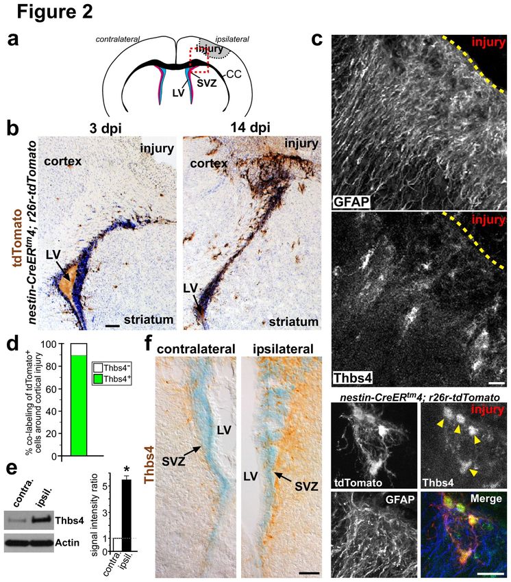
ABSTRACT

Postnatal/adult neural stem cells (NSCs) within the rodent subventricular/subependymal
zone (SVZ/SEZ) generate Doublecortin (DCX)+ neuroblasts that migrate and integrate into
olfactory bulb circuitry1,2. Continuous production of neuroblasts is controlled by SVZ
microenvironmental niche3,4. It is generally believed that enhancing neurogenic activities of
endogenous NSCs may provide needed therapeutic options for disease states and after
brain injury. However, SVZ NSCs can also differentiate into astrocytes. It remains unclear
if there are conditions that favor astrogenesis over neurogenesis in the SVZ niche, and if
astrocytes produced there exhibit different properties from others in the brain. We have
uncovered that SVZ-generated astrocytes express high levels of Thrombospondin-4
(Thbs4)5,6, a secreted homopentameric glycoprotein, in contrast to cortical astrocytes which
are Thbs4low. We found that localized photothrombotic/ischemic cortical injury initiates a
marked increase in Thbs4hi astrocyte production from the postnatal SVZ niche.
Tamoxifen-inducible nestin-CreERtm4 lineage-tracing demonstrated that it is these SVZ-
generated Thbs4hi astrocytes, and not DCX+ neuroblasts, that home-in on the injured
cortex. This robust post-injury astrogenic response required SVZ Notch activation,
modulated by Thbs4 via direct Notch1 receptor binding and endocytosis to activate
downstream signals, including increased Nfia transcription factor expression important for
glia production7. Consequently, Thbs4KO/KO animals showed severe defects in cortical
injury-induced SVZ astrogenesis, instead producing cells expressing DCX from SVZ to the
injury sites. These alterations in cellular responses resulted in abnormal glial scar
formation after injury, and significantly increased microvascular hemorrhage into the
brain parenchyma of Thbs4KO/KO animals. Taken together, these findings have significant
implications for post-injury applications of endogenous and transplanted NSCs in the
therapeutic setting, as well as disease states where Thbs family members play important
roles8,9.

                                             2
The robust neuronal regenerative capacity of the adult rodent brain brings exciting possibilities

for cell-replacement therapies following injuries and neurodegeneration10. However, the utility of

these adult NSCs may hinge on our ability to control the balance between neuron versus

astrocyte production. Currently, the decisions controlling neuro- vs. astrogenesis in the

postnatal/adult SVZ niche are unclear. To address this question, we set out to characterize SVZ-

generated astrocytes. CNS astrocytes secrete Thrombospondins (Thbs), a family of homotrimeric

and homopentameric proteins that mediate various nervous system functions6. We asked whether

SVZ-generated astrocytes express different Thbs protein(s) from their cortical counterparts.

       We started by first establishing primary SVZ adherent cultures from nestin-CreERtm4;

rosa26reporter-tdTomato (r26r-tdTomato) animals, after postnatal day 7 (P7) tamoxifen

injection in vivo to induce tdTomato-reporter expression. We showed previously that this nestin-

CreERtm4 line can efficiently lineage-trace progeny of postnatal/adult SVZ NSCs4,11, generating

migrating neuroblasts that functionally integrate into olfactory bulb circuitry (Supplementary

Fig. 1 and Mov 1). This lineage-tracing of migrating neuroblasts in the rostral migratory stream

continues for several weeks after single tamoxifen injection11, consistent with CreER-mediated

recombination in SVZ type “B” astrocytes, as type “C” transiently amplifying neural progenitors

divide rapidly and are short-lived12. Following 5 days of in vitro differentiation of these cultures,

we found that tdTomato+ lineage-traced GFAP+ astrocytes showed strong immunohistochemical

(IHC) staining for Thbs4 (Fig. 1a). To confirm the specificity of this Thbs4 antibody staining, we

performed the same culture experiment using Thbs4KO/KO mutant animals13. These cells revealed

no Thbs4 immunofluorescence under identical experimental and imaging conditions (Fig. 1b),

and Western blot analyses confirmed these findings (Fig. 1c). Comparison of SVZ astrocyte

cultures to primary astrocytes harvested from the cortex showed selective Thbs4 expression by

                                                 3
differentiated SVZ astrocytes (Fig. 1d). This difference in Thbs4 expression was further

demonstrated by quantitative PCR (qPCR) analyses of FACS-purified GFP+ cortical vs. SVZ

astrocytes from GFAP-GFP transgenic animals (Fig. 1e). IHC staining on P14 brain sections

indicated that Thbs4, while co-labeling with SVZ GFAP+ astrocytes, did not co-localize

with Mash1+ transiently amplifying progenitors, DCX+ neuroblasts, or NG2+, Olig2+

populations in the SVZ niche (Supplementary Fig. 2a - e).

       We next tested whether SVZ-generated GFAP+ astrocytes are fated to express Thbs4 in

vivo. We transplanted second passage SVZ NSC cultures harvested from tamoxifen-induced

nestin-CreERtm4; r26r-tdTomato animals into the cerebral cortex of WT non-transgenic animals

(Fig. 1f). Unlike transplantations into the SVZ, which generated neuroblasts that migrate to the

olfactory bulb4,14 (Supplementary Fig. 3), tdTomato+ cells transplanted into the cortex after 2 to 4

weeks routinely gave rise to GFAP+Thbs4hi astrocytes, contrasting with endogenous cortical

astrocytes which were Thbs4low (Fig. 1e). Of the 402 nestin-CreERtm4 lineage-traced tdTomato+

cells counted over multiple experiments, 96.8% (389) showed GFAP expression. And we

detected strong Thbs4+ IHC co-staining in 98.2% (382) of these GFAP+ cells.

       We wondered under which conditions the SVZ niche favored Thbs4hi astrocyte

production in vivo. Postnatal/adult SVZ NSCs are thought to respond to middle cerebral artery

occlusion and striatal injuries by producing DCX+ neuroblasts that migrate into the striatum15.

Although some have also reported potential for gliogenic responses after injury16,17. To generate

precise location of cortical injuries directly over the SVZ niche we used photothrombosis18, a

well-defined ischemic injury model (Fig. 2a). We performed these injuries using nestin-

CreERtm4; r26r-tdTomato animals after P6 tamoxifen injection. This timing of tamoxifen

injection allowed us to specifically target / lineage-trace SVZ NSCs and their progeny, while

                                                 4
minimizing Cre-reporter labeling of cortical cells11. We induced cortical injuries at P12, and
examined the SVZ responses at 3 and 14 days post injury (dpi), using 3,3’-Diaminobenzidine
(DAB) IHC staining against tdTomato, which allowed simultaneously visualization of tdTomato-
reporter expression and brain tissue histology. At 3 dpi, we did not see significant morphological
changes to lineage-traced tdTomato+ cells around the SVZ in response to cortical injury (Fig.
2b). However, by 14 dpi, we consistently observed robust populations of tdTomato+ cells
between the SVZ and the cortical injury site (Fig. 2b). This delayed reaction was specific to SVZ
ipsilateral to the injury site, as we did not detect obvious changes to tdTomato+ cells in the
contralateral side, nor did we see changes in the ipsilateral cortex in sham-treated control brains
(Supplementary Fig. 4a, b). We did not observe tdTomato+ cells at the injury site in mice with
superficial cortical lesions, or after cortical injection of media used for transplantation,
suggesting a correlation between severity of injury and SVZ astrogenic response (data not
shown). As a further control, we did not detect tdTomato+ cells in the injured cortex of nestin-
CreERtm4; r26r-tdTomato animals without tamoxifen injection (Supplementary Fig. 4c). All
analyses were performed on injuries that did not breach the corpus callosum (determined by
tissue sectioning through the entire injured area), to exclude possible secondary effects caused by
direct injuries to the SVZ niche11.
To determine the cell types lineage-traced from the SVZ 14 dpi, IHC staining showed
that the majority of tdTomato+ cells next to the injury site had complex morphology, labeled
strongly with anti-Thbs4 antibody, and co-stained with GFAP (Fig. 2c, d). Although we did not
observe lineage-traced tdTomato+ cells at the injury site 3 dpi, Western blot analyses of SVZ
tissue and IHC staining of brain sections showed that, at this early time point, the ipsilateral SVZ
had upregulated Thbs4 expression as compared to the contralateral side (Fig. 2e, f). This Thbs4
5expression remained localized to GFAP+ astrocytes (Supplementary Fig. 2f). To determine
whether the increased ipsilateral SVZ Thbs4 protein levels 3 dpi represented upregulated Thbs4-
expression in astrocytes and/or production of more Thbs4hi astrocytes, we performed qPCR
analyses on FACS sorted GFAP-GFP+ SVZ astrocytes 3 dpi, which showed no significant
differences in Thbs4 transcript levels between ipsil- vs. contralateral SVZ samples (data not
shown). This is consistent with the notion that Thbs4hi astrocyte production is induced shortly
after cortical injury, followed by migration to the injury site, contributing to the observed delay
in SVZ response (Fig. 2b). We performed the same cortical injury experiments using GFAP-Cre;
r26r-tdTomato animals and observed similarly robust tdTomato+ lineage-traced cells around the
injury site 14 dpi (Supplementary Fig. 5a, b). Though they likely represent a heterogeneous cell
population, since GFAP-Cre driver also efficiently lineage-traced tdTomato+ cortical cells
(Supplementary Fig. 5a). To determine whether cortical astrocytes may convert from Thbs4low to
a Thbs4hi phenotype (levels seen in SVZ-derived astrocytes) after injury, we performed qPCR
analyses 3 dpi on FACS-sorted GFP+ cortical vs. SVZ astrocytes ipsilateral to injury from
GFAP-GFP animals (prior to significant migration from SVZ), which showed that SVZ
astrocytes remained the Thbs4hi population (Supplementary Fig. 6a - c).
What are the molecular mechanisms controlling this cortical injury-induced Thbs4hi
astrocyte production from SVZ niche? The Notch pathway is an important regulator of SVZ
NSC and niche function11,19, as well as neurogenesis vs. gliogenesis choice20. To determine if
Notch signaling is required for Thbs4hi astrocyte generation after cortical injury, we inducibly
deleted RBPJ21, the Notch Intra-Cellular Domain (NICD) co-transcriptional activator, from the
SVZ using the nestin-CreERtm4 transgene. We crossed nestin-CreERtm4; RBPJKO/+ mice to
RBPJFlox/Flox; r26r-tdTomatoFlox/Flox mice and treated the new mouse litters with tamoxifen at
6P6. We then compared lineage-traced tdTomato+ cells around the cortical injury site between
nestin-CreERtm4; RBPJFlox/+; r26r-tdTomatoFlox/+ (control) and nestin-CreERtm4; RBPJKO/Flox;
r26r-tdTomatoFlox/+ inducible mutant (iKO) animals at 14 dpi. DAB IHC staining against
tdTomato revealed that while there was a robust tdTomato+ cell population between the SVZ and
the cortical injury site in the controls (Fig. 3a), this cellular response was blunted in RBPJ iKO
animals (Fig. 3a). Quantification of tdTomato+ cells above the corpus callosum in sections from
multiple experiments confirmed this reduction in numbers (Fig. 3b). We next asked if increased
Notch signaling results in an enhancement of SVZ response after cortical injury. Using nestin-
CreERtm4; r26r-NICDFlox/+; r26r-tdTomatoFlox/+ animals to over-express NICD in lineage-traced
SVZ cells after tamoxifen injection, we repeated the cortical injury experiments. DAB IHC
staining of brain sections 14 dpi showed a significant increase in lineage-traced tdTomato+ cells
at the injury site in NICD over-expressing animals (Fig. 3a). Quantification of tdTomato+ cells
above the corpus callosum over multiple experiments showed this upregulation in response (Fig.
3b). Consistent with these results, IHC staining of injured brain sections showed increased
Notch1 receptor expression in the ipsilateral SVZ niche when compared to the contralateral side
2 dpi (Supplementary Fig. 7a, b). And Western blot analyses of SVZ tissues 3 dpi showed a
consistent increase in NICD protein in the ipsilateral vs. contralateral SVZ (Fig. 3c).
SVZ ependymal cells, labeled/lineage-traced by FOXJ1-based drivers, have been
reported to respond to injuries by generating progeny that migrate to the injury site22. Since our
nestin-CreERtm4 line, in addition to SVZ NSCs, can also target and lineage-trace potential
ependymal progeny after injury11, we performed identical cortical injuries in FOXJ1-CreERt2;
r26r-tdTomato animals after P6 tamoxifen injection. In repeated experiments we did not observe
tdTomato+ populations migrate from the SVZ to the cortical injury site either early (3 dpi) or late
7(14 dpi) (Supplementary Fig. 8a and data not shown). This makes it unlikely that Thbs4hi
astrocytes are derived from ependymal cells post cortical injury. The FOXJ1-CreERt2 line also
labeled mature cortical neurons, but their tdTomato expression is independent from injury-
induced cell proliferation (Supplementary Fig. 8a and data not shown). To rule out the possibility
that nestin-CreERtm4 modification of the Notch signaling pathway in ependymal cells
subsequently allowed ependymal cells to generate lineage-traced progeny responding to cortical
injury, we performed identical RBPJ iKO and NICD over-expression injury experiments using
the same FOXJ1-CreERt2 driver line. In both genetic backgrounds and experiments we did not
observe tdTomato+ cells migrate from the SVZ to the cortical injury site (Supplementary Fig.
8b).
Although thrombospondins have not been previously linked to Notch function in vivo,
Thbs2 homotrimer can interact with the Notch3 receptor in vitro23. Thus we wondered if Thbs4,
as an astrocyte-secreted molecule6, plays a direct role in modulating Notch activity after cortical
injury. To test this, we first differentiated early passage primary SVZ NSC cultures in the
presence of immobilized Jagged1-Fc, as transient Notch activation leads to decreased
neurogenesis20. As expected, this resulted in decreased neuronal production compared with
control cultures without Jagged1-Fc (Fig. 3d, f). To check whether Thbs4 can mimic Jagged1’s
activity, we repeated this in vitro differentiation with the addition of recombinant Thbs4 alone in
the medium, and observed a similarly robust decrease in neuroblast production (Fig. 3d, f). This
was furthered confirmed by Western blot analyses of DCX protein levels after 5 days of
differentiation (Fig. 3e, f). Interestingly, the effects of treatment with Thbs4 and immobilized
Jagged1-Fc together were not additive, suggesting that they may act in the same signaling
pathway (data not shown). DAPT or DBZ, commonly used Notch pathway inhibitors, conversely
8increased production of neuroblasts from differentiating primary SVZ cultures (Supplementary
Fig. 9 and data not shown). Although adding Thbs4 to the cultures was unable to blunt
DAPT or DBZ-mediated effects (Supplementary Fig. 9a, b), further suggesting that Thbs4
function in this setting required Notch activation.
Thbs4 is a homopentameric protein with multiple EGF-like globular domains6, and SVZ
NSCs express the Notch1 receptor19. We therefore asked whether Thbs4 and Notch1 can directly
interact. Using varied concentrations of purified Thbs4 protein and recombinant Notch1-Fc
fragment (including the first 13 EGF-like repeats within the extracellular domain of Notch1),
pull-down experiments showed direct Thbs4/Notch1-Fc interactions in vitro (Fig. 3g). mIgG2a,
an isotype-specific control for Notch1-Fc, did not show this interaction (Fig. 3g). Purified Thbs2
protein also did not show Notch1-FC interactions (Fig. 3g), consistent with previously observed
differential effects of Thbs family members23. We next harvested cellular extracts from primary
SVZ NSC cultures with or without addition of Thbs4 in the medium. Immunoprecipitation (IP)
with Notch1 antibody was able to pull-down Thbs4 from the culture lysate, consistent with direct
Thbs4 and Notch1 receptor interactions on the surface of SVZ cells (Supplementary Fig. 10a).
Finally, while under physiological conditions in vivo, IP from freshly isolated SVZ tissue
extracts using Notch1 antibody did not detect significant Thbs4 pull-down (Supplementary
Fig. 10b), similar SVZ Notch1 IP experiments 3 dpi revealed Notch1/Thbs4 interactions
(Fig. 3h).
Efficient Notch signaling requires receptor-endocytosis into signaling endosomes24, and
Thbs4 as a pentameric protein may bind multiple Notch1 molecules to promote their endocytosis
and activation. To test this possibility, we cultured primary SVZ NSCs in the presence of both
Thbs4 and Dynasore25, a late endocytosis blocker. Addition of Thbs4 alone resulted in robust
9NICD upregulation, as detected by Western blotting 15 minutes after incubation, and lasting for
several hours thereafter (Fig. 3i, j). This increase in NICD production was blocked by Dynasore
(Fig. 3j). By 36 hours after stimulation, Thbs4-induced NICD upregulation had subsided, and as
expected with Dynasore added there was low NICD with or without Thbs4 in the culture media
(Supplementary Fig. 10c). The transcription factor Nuclear Factor I A (Nfia) is an important
regulator of gliogenesis downstream of Notch signaling7,26. Following Thbs4 addition
during in vitro SVZ NSC differentiation, we detected increased Nfia expression over
control conditions (Supplementary Fig. 10d). Western blot analyses of SVZ tissues 3 dpi
showed similarly increased Nfia protein level in the ipsilateral vs. contralateral SVZ (Fig.
3k), consistent with our observed injury-induced Thbs4hi astrogenic response (Fig. 2f, e).
Our genetic and biochemical findings raised the intriguing possibility that Thbs4,
following cortical injury, is itself necessary for Notch-mediated SVZ astrogenesis. To test this,
we first performed Western blot analyses on SVZ tissue from Thbs4KO/KO animals 3 dpi. While
under physiological conditions we did not detect noticeable differences in SVZ NICD
expression between control and Thbs4KO/KO mutant animals (Supplementary Fig. 10e), the
mutant animals lacked NICD increase in the ipsilateral SVZ 3 dpi (Fig. 4a) unlike in the WT
conditions (Fig. 3c). Further, Thbs4KO/KO animals did not show appreciable increase in Nfia
expression in the ipsilateral SVZ 3 dpi (Fig. 4a). We next tamoxifen-induced nestin-CreERtm4;
Thbs4KO/KO; r26r-tdTomatoFlox/+ animals to lineage-trace responding SVZ-derived tdTomato+
cells after injury, according to the same protocol described above. At 14 dpi, although we
observed Nestin-CreERtm4 lineage-traced tdTomato+ cells between the ipsilateral SVZ and the
cortical injury site in Thbs4KO/KO mutants, these cells showed a surprising neuroblast-like
morphology (Fig. 4b, c). IHC staining against GFAP and DCX revealed that these tdTomato+
10cells next to the injury site in Thbs4KO/KO mutants mostly co-labeled with DCX, but not GFAP
(Fig. 4b – d and data not shown). This is in striking contrast to cortical injuries in control
animals, where little DCX+ cells were detected next to the injury site (with SVZ neuroblasts on
the same brain sections as internal staining controls for DCX antibody, Fig. 4d, Supplementary
Fig. 11a, b). The observed mixture of tdTomato- and tdTomato+ DCX+ cells at the cortical injury
site is consistent with the clonal nature of Nestin-CreERtm4-mediated recombination following
tamoxifen induction4,11 (Fig. 4b, c). Thus, loss of Thbs4 can convert cortical injury-induced SVZ
astrogenesis into neurogenesis.
We next wanted to understand whether this change from astrogenesis to neurogenesis in
Thbs4KO/KO animals had significant effects on injury-induced sequelae. In preparing the injured
brains from Thbs4KO/KO mutants for cryo-sectioning, we often noticed significant hemorrhage
around the injured cortical areas. To study this further, we performed brain magnetic resonance
imaging (MRI) analyses on control (Thbs4KO/+) and Thbs4KO/KO littermates. Using 3D structural
MRI on perfused brains in intact skulls, we can identify the location and extent of cortical
injuries (Supplementary Fig. 12a, b). In isotropic diffusion-weighted images, the boundaries of
lesions were hyperintense in both control and Thbs4KO/KO animals (Supplementary Fig. 12a, b),
demonstrating classical characteristics of stroke infarct27. In contrast, the interiors of the lesions
appeared hypointense as a result of increased edema, and quantitative maps of diffusion
coefficients confirmed hindered molecular diffusion in the infarcted, but faster diffusion in the
edematous areas (Supplementary Fig. 12a, b). We did not detect significant Mean Diffusivity
measurement difference between littermate control and Thbs4KO/KO animals in the infarcted or
edematous cortical areas, either within 2 dpi (data not shown) or 8 dpi (Fig. 4f, Supplementary
Fig. 12d).
11Iron / blood deoxyhemoglobin are paramagnetic, thus increase tissue magnetic
susceptibility in the presence of hemorrhage28. Using 3D spoiled-gradient-recalled (SPGR)
sequence to measure cortical magnetic properties, in the edematous areas Thbs4KO/KO animals
showed both faster image intensity decay, and faster R2* relaxation rate (rate of decay) as
compared to littermate controls (Fig. 4e, Supplementary Fig. 12c). Furthermore, Thbs4KO/KO
animals exhibited significantly increased areas of magnetic susceptibility in the injured cortex as
compared to the uninjured contralateral side, in contrast to control animals which showed only
well-defined hyperintense boundaries surrounding the resolving cortical lesions (Fig. 4e, g,
Supplementary Fig. 12c, e). While we did not detect these cortical magnetic property differences
early on at 2 dpi between control and Thbs4KO/KO animals (data not shown), the faster R2* rate
and increased magnetic susceptibility in Thbs4KO/KO animals at the later time point indicated
presence of extensive, unresolved hemorrhage in their injured cortical regions.
Compromise in cortical vasculature can be detected by using biotinylated fixable dextran
as tracer29. We perfused animals 7 dpi with biotinylated dextran and examined their injured
cortical areas by fixed brain sections. IHC analyses of GFAP+ astrocytes showed that while
cortical glial scars can be readily identified in littermate controls, they were poorly formed and
disorganized in Thbs4KO/KO animals (Fig. 4h), consistent with observed defects in SVZ-mediated
post-injury astrogenesis in these mice (Fig. 4b – d). Whereas in control animals, adjacent to
injured areas, biotinylated dextran was retained inside the vasculature, we detected widespread
parenchymal biotinylated dextran next to injured areas in Thbs4KO/KO animals (Fig. 4i), indicative
of continued microvascular bleeds. These results are consistent with the MRI findings above,
and showed an outcome where SVZ-mediated astrogenesis may be beneficial over
neurogenesis after cortical injury, in agreement with potentially protective roles for
12astrocytes during the recovery process30,31.
The regulation of postnatal SVZ astrogenesis is poorly understood. Here, we have
uncovered that SVZ-derived astrocytes differ in their Thbs4 expression from their cortical
counterparts. Although under physiological conditions the postnatal/adult rodent SVZ niche
produces mostly neuroblasts that migrate to the olfactory bulb, we found that cortical injury
induces SVZ production of Thbs4hi astrocytes migrating to the injury site. This process is
regulated by Thbs4 binding to and modulating the Notch signaling pathway through receptor
endocytosis to promote SVZ astrogenesis. Thbs4KO/KO animals lacked this robust post-injury
astrogenesis response from the SVZ, and exhibited unresolved microvascular bleeds after
cortical injury. We believe these results will have important implications for therapeutic
intervention using transplanted and endogenous neural stem cells after brain injury32, as well as
astrocyte-lineage brain tumors that can arise from the SVZ33.
13Acknowledgements
We thank D. Melton (Harvard) for rosa26r-NICD mice; T. Honjo (Kyoto) for RBPJ-flox mice;
F. Wang for rosa26r-tdTomato mice; E. Rawlins for FOXJ1-CreERt2 mice; B. Deneen (BCM),
S. Singh for helpful discussions; G. Lyons, W. Li, R. Andersen, P. Heine, D. Fromme, and S.
Collins for project assistance; Duke Flow Cytometry Facility for help with FACS samples; Duke
Center for In Vivo Microscopy/Brain Imaging for MRI analyses; members of the Kuo
laboratory, A. West, T. Lechler, and B. Hogan for comments on manuscript. This work was
supported by National Biomedical Technology Resource Center Grant P41 RR005959 (C.L.);
George and Jean Brumley Endowment, Sontag Foundation, David & Lucile Packard Foundation,
and N.I.H. Director’s New Innovator Award 1 DP2 OD004453-01 (C.T.K.).
Author Contributions
E.J.B. designed and performed injury and culture experiments; D.L. designed and performed
FACS, gene expression, and live-imaging experiments; R.J., H.S., D.W. assisted with cortical
injuries and their analyses; K.A., P.P.-G. performed SVZ niche pathway analyses; C.L.
performed MRI scanning/analyses; C.E. provided recombinant Thbs4 protein and advice; C.T.K.
performed transplantations and conceived the project. E.J.B., D.L., C.T.K. designed figures and
wrote the paper. All authors discussed the results and commented on the manuscript.
Author Information
The authors declare no competing financial interests. Correspondence and requests for materials
should be addressed to C.T.K. (chay.kuo@duke.edu).
14References
1 Kriegstein, A. & Alvarez-Buylla, A. The glial nature of embryonic and adult neural stem
cells. Annu. Rev. Neurosci. 32, 149-184, (2009).
2 Kelsch, W., Sim, S. & Lois, C. Watching synaptogenesis in the adult brain. Annu. Rev.
Neurosci. 33, 131-149, (2010).
3 Ihrie, R. A. & Alvarez-Buylla, A. Lake-front property: a unique germinal niche by the
lateral ventricles of the adult brain. Neuron. 70, 674-686, (2011).
4 Paez-Gonzalez, P. et al. Ank3-dependent SVZ niche assembly is required for the
continued production of new neurons. Neuron. 71, 61-75, (2011).
5 Lawler, J. et al. Identification and characterization of thrombospondin-4, a new member
of the thrombospondin gene family. J. Cell. Biol. 120, 1059-1067, (1993).
6 Eroglu, C. The role of astrocyte-secreted matricellular proteins in central nervous system
development and function. J. Cell. Commun. Signal. 3, 167-176, (2009).
7 Deneen, B. et al. The transcription factor NFIA controls the onset of gliogenesis in the
developing spinal cord. Neuron. 52, 953-968, (2006).
8 Liauw, J. et al. Thrombospondins 1 and 2 are necessary for synaptic plasticity and
functional recovery after stroke. J. Cereb. Blood Flow Metab. 28, 1722-1732, (2008).
9 Adams, J. C. & Lawler, J. The thrombospondins. Cold Spring Harb. Perspect. Biol. 3,
a009712, (2011).
10 Aboody, K., Capela, A., Niazi, N., Stern, J. H. & Temple, S. Translating stem cell studies
to the clinic for CNS repair: current state of the art and the need for a Rosetta Stone.
Neuron. 70, 597-613, (2011).
11 Kuo, C. T. et al. Postnatal deletion of Numb/Numblike reveals repair and remodeling
capacity in the subventricular neurogenic niche. Cell. 127, 1253-1264, (2006).
12 Doetsch, F., Caille, I., Lim, D. A., Garcia-Verdugo, J. M. & Alvarez-Buylla, A.
Subventricular zone astrocytes are neural stem cells in the adult mammalian brain. Cell.
97, 703-716, (1999).
13 Frolova, E. G. et al. Thrombospondin-4 regulates vascular inflammation and
atherogenesis. Circ. Res. 107, 1313-1325, (2010).
14 Merkle, F. T., Mirzadeh, Z. & Alvarez-Buylla, A. Mosaic organization of neural stem
cells in the adult brain. Science. 317, 381-384, (2007).
15 Thored, P. et al. Persistent production of neurons from adult brain stem cells during
recovery after stroke. Stem Cells. 24, 739-747, (2006).
16 Givogri, M. I. et al. Notch signaling in astrocytes and neuroblasts of the adult
subventricular zone in health and after cortical injury. Dev. Neurosci. 28, 81-91, (2006).
17 Li, L. et al. Focal cerebral ischemia induces a multilineage cytogenic response from adult
subventricular zone that is predominantly gliogenic. Glia. 58, 1610-1619, (2010).
18 Maxwell, K. A. & Dyck, R. H. Induction of reproducible focal ischemic lesions in
neonatal mice by photothrombosis. Dev. Neurosci. 27, 121-126, (2005).
19 Aguirre, A., Rubio, M. E. & Gallo, V. Notch and EGFR pathway interaction regulates
neural stem cell number and self-renewal. Nature. 467, 323-327, (2010).
20 Morrison, S. J. et al. Transient Notch activation initiates an irreversible switch from
neurogenesis to gliogenesis by neural crest stem cells. Cell. 101, 499-510, (2000).
1521 Han, H. et al. Inducible gene knockout of transcription factor recombination signal
binding protein-J reveals its essential role in T versus B lineage decision. Int. Immunol.
14, 637-645, (2002).
22 Carlen, M. et al. Forebrain ependymal cells are Notch-dependent and generate
neuroblasts and astrocytes after stroke. Nat. Neurosci. 12, 259-267, (2009).
23 Meng, H., Zhang, X., Hankenson, K. D. & Wang, M. M. Thrombospondin 2 potentiates
notch3/jagged1 signaling. J. Biol. Chem. 284, 7866-7874, (2009).
24 Yamamoto, S., Charng, W. L. & Bellen, H. J. Endocytosis and intracellular trafficking of
Notch and its ligands. Curr. Top. Dev. Biol. 92, 165-200, (2010).
25 Macia, E. et al. Dynasore, a cell-permeable inhibitor of dynamin. Dev. Cell. 10, 839-850,
(2006).
26 Namihira, M. et al. Committed neuronal precursors confer astrocytic potential on residual
neural precursor cells. Dev. Cell. 16, 245-255, (2009).
27 Moseley, M. E., de Crespigny, A. J., Roberts, T. P., Kozniewska, E. & Kucharczyk, J.
Early detection of regional cerebral ischemia using high-speed MRI. Stroke. 24, I60-65,
(1993).
28 Patel, M. R., Edelman, R. R. & Warach, S. Detection of hyperacute primary
intraparenchymal hemorrhage by magnetic resonance imaging. Stroke. 27, 2321-2324,
(1996).
29 Daneman, R., Zhou, L., Kebede, A. A. & Barres, B. A. Pericytes are required for blood-
brain barrier integrity during embryogenesis. Nature. 468, 562-566, (2010).
30 Faulkner, J. R. et al. Reactive astrocytes protect tissue and preserve function after spinal
cord injury. J. Neurosci. 24, 2143-2155, (2004).
31 Okada, S. et al. Conditional ablation of Stat3 or Socs3 discloses a dual role for reactive
astrocytes after spinal cord injury. Nat. Medicine. 12, 829-834, (2006).
32 Robel, S., Berninger, B. & Gotz, M. The stem cell potential of glia: lessons from reactive
gliosis. Nat. Rev. Neurosci. 12, 88-104, (2011).
33 Alcantara Llaguno, S. et al. Malignant astrocytomas originate from neural
stem/progenitor cells in a somatic tumor suppressor mouse model. Cancer Cell. 15, 45-
56, (2009).
16Figure Legends
Figure 1 │ SVZ generation of Thbs4hi astrocytes. a, In vitro differentiation of primary SVZ
adherent neural stem cell culture from nestin-CreERtm4; r26r-tdTomato animal, showing Thbs4
staining in tdTomato+GFAP+ astrocytes. b, c Control Thbs4 antibody staining in primary
differentiated SVZ adherent NSC cultures from Thbs4KO/KO animals, confirmed by Western blot
analysis. d, Western blot analysis of Thbs4 protein levels in differentiated primary SVZ vs.
cortical (ctx) astrocyte cultures. e, Quantitative PCR analyses of Thbs4 levels in FACS sorted
SVZ vs. ctx GFP+ astrocytes from GFAP-GFP transgenic mice, * p < 0.001, n = 5, Student’s t-
test; error bars = sem. f, Schematic representation of cortical transplantation strategy. Thbs4,
tdTomato, GFAP IHC antibody staining of brain sections 4 weeks after animals were
transplanted with lineage-traced primary SVZ NSC cultures derived from tamoxifen induced
nestin-CreERtm4; r26r-tdTomato animals, showing co-localization between tdTomato and Thbs4
(arrowheads). Close-up images shown in right hand panels. Scale bars: 20 m (a, b, f).
Figure 2 │ Induction of SVZ Thbs4hi astrocyte production after cortical injury. a,
Schematic representation of photothrombosis cortical injury model, with dashed box indicating
region of imaging in (b). b, DAB IHC staining for tdTomato expression from nestin-CreERtm4;
r26r-tdTomato animals induced with tamoxifen in vivo, showing a delayed (not at 3 dpi) and
robust activation of lineage-traced tdTomato+ cells to brain injury in 14 dpi sections. LV = lateral
ventricle. c, Coronal sections of cortical injury site 14 dpi, IHC stained with GFAP, Thbs4, and
tdTomato antibodies, showing lineage-traced tdTomato+ cells next to the injury site (dashed
lines) are Thbs4hiGFAP+ astrocytes (arrowheads). d, Quantitative analyses of total tdTomato+
17lineage-traced cells at injury site expressing Thbs4 14 dpi (88.10 ± 1.99% stdev, n = 3 animals).
e, Western blot and quantitative analyses of Thbs4 protein levels in SVZ tissues harvested 3dpi,
showing Thbs4 upregulation in the ipsilateral versus the contralateral side from the same brain. *
p < 0.001, n = 5, Student’s t-test, error bar = sem. f, Coronal sections of SVZ, 3 dpi, stained with
Thbs4 antibody via DAB IHC, counterstained with Nissl. Ipsilateral and contralateral SVZ from
the same brain section, imaged under identical conditions, are shown. Scale bars: 100 m (b), 20
m (c), 50 m (f).
Figure 3 │ Notch pathway regulation of injury-induced SVZ astrogenesis. a,
Photothrombosis cortical injury model, with areas of imaging indicated by dashed boxes. DAB
IHC staining for tdTomato expression from nestin-CreERtm4; RBPJkFlox/+; r26r-tdTomato
(control) animals induced with tamoxifen in vivo, showing robust activation of lineage-traced
tdTomato+ cells to brain injury (ipsilateral) in 14 dpi sections. DAB IHC staining for tdTomato
expression from nestin-CreERtm4; RBPJkKO/Flox; r26r-tdTomato (RBPJkKO/Flox) and nestin-
CreERtm4; r26r-NICD; r26r-tdTomato (r26r-NICD) animals induced with tamoxifen in vivo, 14
dpi. LV = lateral ventricle. b, Quantification of tdTomato+ cells above the corpus callosum in
each genetic backgrounds 14 dpi, * p < 0.05 (n = 6), ** p < 0.001 (WT: n = 8; NICD: n = 5),
Student’s t-test; error bars = sem. c, Western blot analyses of Notch Intra-cellular Domain
(NICD) protein levels in SVZ tissues harvested 3dpi, showing NICD upregulation in the
ipsilateral over the contralateral side from the same brain. * p < 0.005, n = 5, Student’s t-test,
error bar = sem. d, In vitro differentiation of primary SVZ adherent neural stem cell cultures,
with or without Jagged-Fc, and/or Thbs4 added. e, Western blot analyses comparing DCX
protein levels after 5 days of in vitro differentiation of primary SVZ cultures, with or without
18Thbs4 addition. f, Quantification of DCX levels on Western blots after differentiation. *, ** p <
0.001, n = 5, Student’s t-test, error bar = sem. g, In vitro binding assays between purified Thbs4
or Thbs2 proteins, with Notch1-Fc (arrows). Purified mIgG2a was used as isotype control for
Notch1-Fc. h, IP experiment using freshly isolated SVZ tissue extract 3 dpi, IP with control
beads or -Notch1 antibody, and blotted with -Thbs4 or -Notch1 antibodies. Note the
detection of Thbs4/Notch1 interactions (arrow). i, Thbs4 modulation of NICD generation in
primary SVZ adherent cultures. * p < 0.005, n = 4, Student’s t-test, error bar = sem. j, Thbs4
modulation of NICD generation with or without Dynasore, 12 hrs post stimulation. k, Western
blot analyses of Nfia protein levels in SVZ tissues harvested 3 dpi, showing upregulation in the
ipsilateral over the contralateral side from the same brain. * p < 0.005, n = 5, Student’s t-test,
error bar = sem. Scale bars: 200 mm (a), 50 m (d).
Figure 4 │ SVZ-mediated astrogenesis defects in Thbs4 mutant mice after cortical injury.
a, Western blot analyses of NICD and Nfia protein levels in SVZ tissues harvested 3dpi from
Thbs4-KO animals, showing lack of upregulation in the ipsilateral (ipsil) vs. contralateral
(contra) SVZ. b, IHC staining for tdTomato, DCX expression from nestin-CreERtm4;
Thbs4KO/KO; r26r-tdTomato animals 14 dpi. Note the robust DCX+ cells at injury site. SCJ =
striatal-cortical junction, CC = corpus callosum (yellow dashed-lines). c, Close-up views of
tdTomato and DCX co-localization from boxed area in (b). Example tdTomato+DCX+ cells
(yellow arrows) enlarged in right panels. d, Quantification of total tdTomato+ lineage-traced cells
around injury site 14 dpi, co-labeling with GFAP or DCX. Lack of strong staining for either
GFAP or DCX was marked as (-). * p < 0.002, n = 11 animals (control), 5 animals (KO),
Wilcoxon Rank Sum test. e, MRI analyses of littermate pair, Thbs4KO/+ (control) and Thbs4KO/KO
19(KO) at 8 dpi. Left panels = SPGR images, horizontal plane, at two echo times (TE, 4.4 and 14.3
ms). Center panels = computed R2* relaxation rate (RR). Right panels = corresponding Magnetic
Susceptibility (MS). Hyperintense MS indicates area of hemorrhage (hem., red arrow). Scale bar
units in RR = s-1, MS = ppm. OB = olfactory bulb, Ctx = cortex, inj = injury site. f, g
Quantitative measurements of Mean Diffusivity, Magnetic Susceptibility (Mag. Suscep.),
comparing contralateral cortex (contra) to areas of infarct and edema caused by injury. Error bars
= stdev. h, IHC staining of brain coronal sections 7 dpi to visualize, around injury site, GFAP+
astrocytes and biotinylated dextran infused through vasculature. GFAP+ glial scar is formed
around injury site in control animal (arrowheads), but poorly defined in KO animal. i, Extra-
vascular biotinylated dextran is readily seen around cortical injury in KO animals (dashed-boxes
from corresponding panels in h). Quantification of parenchymal biotinylated dextran mean
fluorescence intensity next to injury site. * p < 0.001 (n = 5), Student’s t-test; error bars = sem.
Scale bars: 50 m (b, h, i), 20 m (c), 5 m (c, close-up).
20Methods Summary
Cell Culture
Adherent SVZ primary cultures were isolated and grown as described34. Cortical astrocyte
cultures were grown as described35. Jagged1-Fc (R&D Systems) was plated at 5 g/ml in PBS
overnight to coat culture dish surfaces, followed by PBS washes. Recombinant Thbs4 (3 g/ml,
C. Eroglu), DAPT (5 M, Tocris), DBZ (5 nM, Millipore) were freshly added to culture medium
every other day during in vitro differentiation where indicated. Dynasore (Sigma) was used at 30
M in culture medium.
Immunohistochemistry and live-imaging analyses
Preparation of brain tissue for immunohistochemistry (IHC) was as described11. For Thbs4 and
DAB staining: after transcardiac perfusion, brains were removed, postfixed overnight, and
cryoprotected in 30% sucrose at 4C. 30 m coronal sections were serially cut, and immediately
incubated for 1 hr at room temperature (RT), floating in PBST blocking buffer containing 10%
donkey serum. Primary antibody incubation was carried out at 4C in blocking buffer overnight,
followed by washes in PBST x3, PBS alone x3, and secondary antibody incubation in blocking
buffer for 2 hrs at RT. DAB staining was carried out according to manufacturer instructions
(Vectastain ABC Kit, Vector Labs). For cell counting and quantifications, IHC stained coronal
sections, 90 m apart, starting at the anterior portion of the anterior commissure through the
septofimbrial nuclei, were analyzed and counted. Primary antibodies against the following
antigens were used: Thbs4 (goat, 1:200, R&D Systems); GFP (chicken, 1:500, Aves Labs); RFP
(rabbit, 1:1000, Rockland); GFAP (mouse, 1:2000, Sigma); Mash1 (mouse, 1:100, BD Biosci)
21DCX (guinea pig, 1:200, Millipore); NeuN (mouse, 1:500, Millipore); Notch1 (rabbit
monoclonal EP1238Y, 1:500, Epitomics); Olig2 (rabbit, 1:800, Millipore); NG2 (mouse, 1:200,
R&D Systems). Biotinylated fixable dextran (MW = 10 kDa, 1.0 mg/ml, Invitrogen) experiments
and their image analyses were performed as described29. Live-imaging of migrating neuroblasts
was performed as described36. All fluorescent images were acquired on Leica TCS SP5 confocal
microscope, with control and experimental samples imaged under identical settings.
FACS sorting and gene expression analyses
Cortical and SVZ tissues were dissected in warm NSC culture media34, dissociated in Neural
Tissue Dissociation Kit (Miltenyi) according to manufacturer’s protocol. Cell suspension was
added to FACS tube on ice, sorted on BD FACS DiVa sorter, followed immediately by Trizol
RNA extraction. 500 ng of total RNA was used for cDNA synthesis using SuperScript VILO
cDNA Synthesis Kit (Invitrogen). Quantitative PCR analyses were performed as described37,
using the following DNA primers: Thbs4 F: 5’-atccctgctatccaggtgtg-3’, R: 5’-
ggcagctcctttcagtcttg-3’; Nfia F: 5'-ccagccagccaagtgaag-3’, R: 5’-gctcagtcacactgaaaacacc-3’;
housekeeping gene controls GAPDH F: 5’-catggccttccgtgttcct-3’, R: 5’- tgatgtcatcatacttggcaggtt-
3’, and PPIA F: 5’-cgagctctgagcactggag-3’, R: 5’-gatgccaggacctgtatgct-3’. Consistent results
were obtained using either housekeeping primer sets.
SDS-PAGE and immunoblotting
Protein extract preparation and Western blotting were performed as described4. NIH ImageJ
software was used for quantification analyses. Primary antibodies against the following antigens
were used: Thbs4 (goat, 1:500, R&D Systems); Thbs2 (mouse, 1:500, Millipore); DCX (guinea
22pig, 1:1000, Millipore); NICD/Notch1 (rabbit EP1238Y, 1:1000, Millipore); Nfia (rabbit,
1:2000, Active Motif); actin (mouse, 1:5000, Abcam). For pull-down experiments, purified
recombinant Thbs4 or Thbs2 (R&D Systems) was incubated for 1 hr at 37C with either 200 ng
mouse Notch1-FC chimera (R&D Systems, 5267-TK) or mouse IgG2a isotype control (Abcam)
in Dulbecco’s PBS + 1% BSA, 0.1% Triton X-100. Protein G-coupled DynaBeads (Invitrogen),
pre-blocked in DPBS + 3% BSA, were then added for 10 min at 37C, followed by 5 washes in
DPBS + 0.1% Triton X-100, and analyzed by Western blotting. For cultured cells, media with or
without Thbs4 (3 g/mL) were first added for 4 hours, washed 2 times with cold PBS, followed
by addition of lysis buffer (50mM Tris pH 8.0, 75mM NaCl, 1mM EDTA, 5% sucrose, 0.25%
Triton x-100, mini-complete protease inhibitor cocktail), sonication, and centrifuged for 10
minutes at 4C. Lysates were pre-cleared with Protein A agarose beads (Roche). Rabbit anti-
Notch1 antibody (5 g, Millipore EP1238Y) was added to 1 ml of lysate and incubated overnight
at 4C, followed by addition of Protein A agarose beads, washes as described above, and
Western blotting analysis. SVZ IP experiments were performed as described11 with following
modifications: CHAPS lysis buffer contained 5mM CHAPS, 50 mM Tris (pH 7.4), 150 mM
NaCl, 1 mM CaCl, 5% sucrose, 0.5% Triton X-100, protease inhibitor cocktail (Roche) + sodium
orthovanadate. Supernatants after lysis were subject to two rounds of pre-clearing with BSA-
blocked Protein G agarose beads (Roche) for 1 hr each, followed by incubation with rabbit anti-
Notch1 antibody (Millipore EP1238Y) for 1 hr, and overnight incubation with BSA-blocked
Protein G agarose beads, all at 4oC. Beads were washed five times in lysis buffer, three times in
PBS prior to suspension in sample buffer for Western blotting.
Cortical injury and in vivo injections
23Photothrombosis cortical injury was performed as described38. Briefly, animals were
anesthetized and body temperature maintained at 37C with recirculating water heating pad.
Saline solution of rose bengal photosensitive dye (10 mg/ml in saline, 0.1 mg per gram of body
weight) was delivered intraperitoneally. Midline scalp incision was made to expose the skull. As
external light source, Zeiss KL 1500 LCD (light intensity at 5, 6-minute duration) with 2.5 mm
opening light guide was used to induce photothrombosis. Transplantation of passage 2 primary
adherent SVZ culture (around 10,000 cells per animal) was performed as described4. 13 out of 37
transplanted animals showed successfully grafted tdTomato+ cells on analyses. Tamoxifen (10
mg/ml, freshly dissolved in corn oil) was injected intraperitoneally at 0.15 mg per gram of body
weight to induce CreER-mediated recombination. All mouse experiments were performed
according to approved protocol by IACUC at Duke University.
Magnetic resonance imaging and analyses
Pairs of injured Thbs4KO/+ and Thbs4KO/KO mice were scanned on 9.4 Tesla (400 MHz) 89-mm
vertical bore MRI scanner (Oxford Instruments) with shielded coil providing gradients of 2200
mT/m. Mice were perfused with 10% buffered formalin as previously described39, followed by
overnight fixation in formalin and 3 day incubation in PBS before imaging. All brains were kept
within the cranium to prevent potential damage by removal. Diffusion-weighted images (DWI)
were acquired using 3D spin-echo sequence with following parameters: field of view (FOV) =
22x11x11 mm3, matrix = 164x82x82, TE = 12 ms, TR = 2500 ms. One image volume was
acquired without diffusion weighting, and diffusion weighting was achieved by applying two
half-sine gradient pulses around the 180o refocusing pulse. To produce isotropic weighting, six
non-collinear diffusion encoding directions were used at b-value = 3000 s/mm2. A diffusion
24tensor was fitted using seven image volumes and mean diffusivity was calculated as described40.
To examine intracranial hemorrhage, brains were scanned using 8-echo 3D spoiled-gradient-
recalled (SPGR) sequence with the following parameters: FOV = 22x11x11 mm3, matrix =
512x256x256, initial TE = 4.4 ms, echo spacing = 4.9 ms, TR = 100 ms, flip angle = 45º. R2*
relaxation rate was fitted with exponential decay curve. Tissue magnetic susceptibility was
quantified as described41. Regions of infarct and edema were segmented out based on Mean
Diffusivity maps generated via FSL software (Oxford University), and comparable regions in the
contralateral uninjured cortex were drawn as references. For each region, Mean Diffusivity, R2*
relaxation rate, and Magnetic Susceptibility were measured. Similar results were obtained with
addition of MRI contrast agent ProHance (Bracco Diagnostics, Princeton, NJ) to formalin
fixation (1:9, v:v).
Supplementary References
34 Scheffler, B. et al. Phenotypic and functional characterization of adult brain neuropoiesis.
Proc. Natl. Acad. Sci. USA. 102, 9353-9358, (2005).
35 Christopherson, K. S. et al. Thrombospondins are astrocyte-secreted proteins that
promote CNS synaptogenesis. Cell. 120, 421-433, (2005).
36 Platel, J. C. et al. NMDA receptors activated by subventricular zone astrocytic glutamate
are critical for neuroblast survival prior to entering a synaptic network. Neuron. 65, 859-
872, (2010).
37 McDowell, K. A. et al. Reduced cortical BDNF expression and aberrant memory in Carf
knock-out mice. J. Neurosci. 30, 7453-7465, (2010).
38 Lee, J. K. et al. Photochemically induced cerebral ischemia in a mouse model. Surg.
Neurol. 67, 620-625, (2007).
39 Johnson, G. A., Cofer, G. P., Gewalt, S. L. & Hedlund, L. W. Morphologic phenotyping
with MR microscopy: the visible mouse. Radiology. 222, 789-793, (2002).
40 Basser, P. J., Mattiello, J. & LeBihan, D. MR diffusion tensor spectroscopy and imaging.
Biophys. J. 66, 259-267, (1994).
41 Li, W., Wu, B. & Liu, C. Quantitative susceptibility mapping of human brain reflects
spatial variation in tissue composition. NeuroImage. 55, 1645-1656, (2011).
25You can also read