Morphological studies of rose prickles provide new insights
←
→
Page content transcription
If your browser does not render page correctly, please read the page content below
Zhou et al. Horticulture Research (2021)8:221
                                    https://doi.org/10.1038/s41438-021-00689-7
                                                                                                                                                                                         Horticulture Research
                                                                                                                                                                                                     www.nature.com/hortres

                                     ARTICLE                                                                                                                                                    Open Access

                                    Morphological studies of rose prickles provide new
                                    insights
                                    Ningning Zhou1,2 ✉, Fabienne Simonneau3, Tatiana Thouroude1, Laurence Hibrand-Saint Oyant1 and
                                    Fabrice Foucher1 ✉

                                      Abstract
                                      Prickles are common structures in plants that play a key role in defense against herbivores. In the Rosa genus, prickles
                                      are widely present with great diversity in terms of form and density. For cut rose production, prickles represent an
                                      important issue, as they can damage the flower and injure workers. Our objectives were to precisely describe the types
                                      of prickles that exist in roses, their tissues of origin and their development. We performed a detailed histological
                                      analysis of prickle initiation and development in a rose F1 population. Based on the prickle investigation of 110 roses,
                                      we proposed the first categorization of prickles in the Rosa genus. They are mainly divided into two categories,
                                      nonglandular prickles (NGPs) and glandular prickles (GPs), and subcategories were defined based on the presence/
                                      absence of hairs and branches. We demonstrated that NGPs and GPs both originate from multiple cells of the ground
                                      meristem beneath the protoderm. For GPs, the gland cells originate from the protoderm of the GP at the early
                                      developmental stage. Our findings clearly demonstrate that prickles are not modified trichomes (which originate from
                                      the protoderm). These conclusions are different from the current mainstream hypothesis. These results provide a
                                      foundation for further studies on prickle initiation and development in plants.
                  1234567890():,;
1234567890():,;
                  1234567890():,;
1234567890():,;

                                    Introduction                                                                                        source of confusion originates among prickles, thorns,
                                      Superficial tissues (epidermis) and appendage struc-                                               and spines. Many plants described with thorns or spines
                                    tures (trichomes, spinescences) of plant organs are the                                             actually have prickles3–8.
                                    first lines of defense against multiple abiotic and biotic                                             Depending on the presence of vascular bundles, we can
                                    stresses. The basic terminologies of these appendages are                                           divide these structures into two categories: (1) trichomes
                                    frequently inaccurately cited in scientific reports, leading                                         (Supplementary Fig. 1a and b) and prickles (Supplemen-
                                    to confusion and difficulties in distinguishing the dif-                                             tary Fig. 1c and d), which are not vascularized and are
                                    ferent terms. Some authors have described emergences                                                generally easy to remove9,10; and (2) thorns (Supplemen-
                                    as prickles, e.g., prickles on the stems or leaves of plants                                        tary Fig. 1e and f) and spines (Supplementary Fig. 1g),
                                    such as Solatium torvium, Aiphanes acanthophylla,                                                   which have vascular bundles and cannot be easily sepa-
                                    and roses1, and some have referred to trichomes as                                                  rated from organs that have vascular tissues (spines,
                                    emergences, e.g., grape emergences2. Another common                                                 usually modified from leaves, and thorns, modified from
                                                                                                                                        stems or shoots) (Supplementary Fig. 1h)11,12. Thus,
                                                                                                                                        prickles can be easily distinguished from thorns and
                                    Correspondence: Ningning Zhou (ningning.zhou.j@gmail.com) or                                        spines: mature prickles are outgrowths connected to the
                                    Fabrice Foucher (fabrice.foucher@inrae.fr)
                                    1                                                                                                   bark13, while thorns and spines are outgrowths connected
                                     Univ Angers, Institut Agro, INRAE, IRHS, SFR QUASAV, F-49000 Angers, France
                                    2
                                     National Engineering Research Center for Ornamental Horticulture; Flower                           to the phloem and the xylem11,14.
                                    Research Institute (FRI), Yunnan Academy of Agricultural Sciences, Kunming                            Confusion of trichomes and prickles is also common.
                                    650231, China
                                                                                                                                        Trichomes are epidermal appendages that originate only
                                    Full list of author information is available at the end of the article
                                    These authors contributed equally: Laurence Hibrand-Saint Oyant, Fabrice                            from the protoderm, and they are diverse according to
                                    Foucher

                                    © The Author(s) 2021
                                                        Open Access This article is licensed under a Creative Commons Attribution 4.0 International License, which permits use, sharing, adaptation, distribution and reproduction
                                                        in any medium or format, as long as you give appropriate credit to the original author(s) and the source, provide a link to the Creative Commons license, and indicate if
                                    changes were made. The images or other third party material in this article are included in the article’s Creative Commons license, unless indicated otherwise in a credit line to the material. If
                                    material is not included in the article’s Creative Commons license and your intended use is not permitted by statutory regulation or exceeds the permitted use, you will need to obtain
                                    permission directly from the copyright holder. To view a copy of this license, visit http://creativecommons.org/licenses/by/4.0/.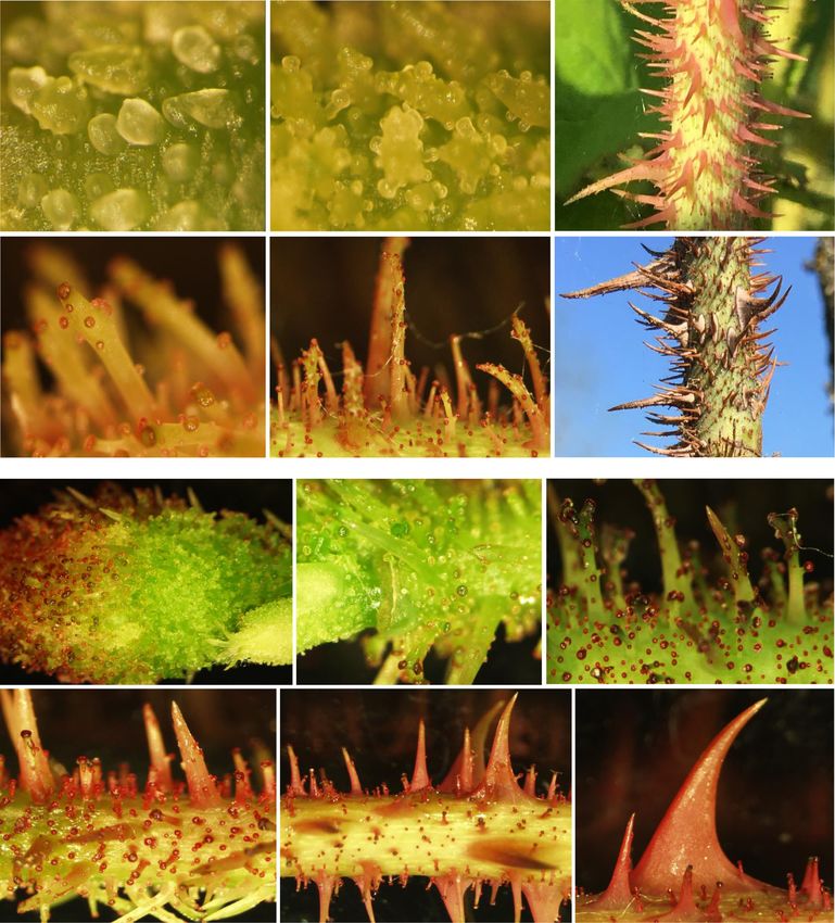
Zhou et al. Horticulture Research (2021)8:221                                                                       Page 2 of 12

their final forms and structures, locations, and func-              trichomes at the molecular level is still a source of debate.
tions10,15. They are mainly divided into nonglandular tri-         A precise histological description of the tissues is
chomes (NGTs) and glandular trichomes (GTs)10. Both                requested for clear conclusions.
types can be unicellular or multicellular and branched or            Another hypothesis is that rose prickles are spines.
unbranched. Presently, the genetic and molecular                   Prickles were proposed to be modified leaves without
mechanisms of NGTs are well understood in Arabidopsis              internal vascular tissues, as the abscission cell layer of
thaliana, and numerous related genes have been identified           prickles resembled the abscission layer of deciduous
(reviewed by Hülskamp16 and Zhou17). These genes                   leaves, with mature prickles easily peeled off9. However,
encode proteins belonging to the MYB, bHLH, WD40,                  no strong evidence was presented to support this
WRKY, and C2H2 zinc finger protein families. A trimeric             hypothesis. Li et al.23 suggested that cells from the prickle
activator complex consisting of MYB (GLABRA1)-bHLH                 abscission region were different from cells of the petiole
(GLABROUS3/ENHANCER OF GL3)-WDR (TRANS-                            abscission zone according to the anatomical structure and
PARENT TESTA GL1) plays a key role in NGT initia-                  chemical composition of tender prickles.
tion16. The genetic pathway for GT initiation is not yet well        Later, Angyalossy et al.13 defined prickles as “sharp
known (reviewed by Huchelmann et al.18 and Chalvin                 outgrowths from the bark, without vascular tissue”, based
et al.19). In Solanum, an HD-ZIP IV transcription factor           on longitudinal sections through the developed prickles of
(WOOLLY) may interact with the B-type cyclin CycB2 and             Polyscias mollis, Piptadenia gonoacantha, and Oplopanax
the C2H2 zinc-finger protein (HAIR) to induce GT                    horridus. However, the “bark” term is unprecise, as it
initiation (reviewed by Chalvin et al.19). In Artemisia            refers to all tissues exterior to the vascular cambium,
annua, an HD-ZIP IV transcription factor (AaHD8) may               including tissues such as the periderm (composed of cork,
interact with a MIXTA-like protein (AaMIXTA1), which               cork cambium, and phelloderm), cortex (comprising
activates AaHD1, leading to GT initiation20.                       ground tissues), phloem and epidermis32,33. In conclusion,
  Prickles are common structures in plants, which are              the origin of prickles in plants is still controversial and
involved in defense against insects and large mammalian            requires further investigation.
herbivores21. The morphogenetic and molecular mechan-                Wild roses belong to the genus Rosa in the family
isms underlying prickle initiation and development remain          Rosaceae. The genus Rosa is composed of ∼200 species
largely unknown. A few reports have described the ana-             and is widely distributed in cold temperate to subtropical
tomical structures of prickles13, especially in roses9,22,23. As   regions34. Rose is a such beautiful flower with wonderful
the analyses were performed in late developmental stages,          fragrant, have always been popular at different periods
conclusions about the tissues from which prickles originate        and in many civilizations since it plays a part in many
are difficult to draw, leading to different and controversial       religions and has come to symbolize romance. Today, rose
hypotheses developed below.                                        is one of the most economically important ornamental
  The mainstream hypothesis is that prickles originate             plants in the world. Most roses have prickles on their
from multiple cellular divisions of the epidermis3,24,25 and       stems. For cut rose production, removing prickles is an
are considered as modified GTs, with lignification leading           essential step before packaging. This process causes
to a hard and sharp appendage2,5,22,26. Nonglandular               wounding on the stem, largely affects transportation tol-
prickles (NGPs) were described as a late stage of glandular        erance and vase life, and reduces the ornamental value.
prickles (GPs)22. Based on this hypothesis, molecular              Furthermore, prickles represent a risk of injury to work-
approaches were developed to test the trichome origin of           ers. Therefore, rose cultivars with many prickles are
prickles in rose and Rubus. A comparison of transcript             generally not accepted for the production of cut roses,
accumulation between rose F1 genotypes with no, low-               even if they have other outstanding ornamental traits. In
density (NGPs) and high-density (GPs and NGPs) prickles            rose, prickles are very diverse, showing different types,
revealed significant differences for some candidate genes,          shapes, sizes, densities, and colors. Furthermore, genetic
such as RcTTG227. Unfortunately, prickle types, NGPs               resources such as several high-density SNP-based genetic
and GPs, were mixed in the previous study. Later, Zhou             maps from rose F1 populations27,35,36 and GWAS col-
et al.28 proposed that prickle and trichome initiation             lections27,37,38 are available. The recent production of two
involve different genetic pathways, as no major difference         high-quality reference genome sequences27,39 allows
was observed during prickle initiation for candidate gene          genomic approaches. Therefore, rose is a good model
homologs of genes known to control trichome initiation             plant with which to study the molecular and genetic bases
in A. thaliana. However, on the basis of a transcriptomic          of prickle initiation and development.
approach, the molecular network controlling prickle                  In this study, our main objectives were to characterize
initiation was proposed to be similar in Rubus29 and in            in detail the initiation and development of prickles in
roses30,31 to the one described for trichome initiation            roses using histological approaches and to investigate
in A. thaliana. The relationship between prickles and              their diversity in terms of form. The major questions are
Zhou et al. Horticulture Research (2021)8:221 Page 3 of 12
as follows: (i) which types of prickles exist in roses? (ii) macroscopic analysis, we previously determined two
Which tissues do prickles originate from? and (iii) How categories of prickles on the stems of the OW progeny: (i)
do the prickles develop? We clearly demonstrated that “nonglandular prickles (NGPs)” and (ii) “glandular
prickles in rose originate from the ground meristem and prickles (GPs)” refer to the prickles without and with
are not modified trichomes. Two major types of prickles glands, respectively28. In addition, “prickless” refers to
are described in rose: glandular and nonglandular prick- stems without prickles. For detailed morphological and
les. These histological analyses are necessary for precise anatomical studies of NGPs and GPs, we selected one
genetic and genomic studies. individual presenting the two types of prickles, OW9106,
and one without prickles, OW9068. According to the
Results specific morphogenetic events during prickle develop-
First, we performed a detailed analysis on individuals of ment, we defined developmental stages for NGPs and GPs
a F1 progeny (macroscopic and microscopic analyses). on the rose stem (as defined in ref. 28).
Then, based on these observations, we performed a survey Stage I corresponds to prickle initiation and to the first
of prickle diversity in the genus Rosa, with more precise outgrowth. Initiation appeared at the early stage of
observations of twelve representative genotypes. internode development (probably simultaneously with the
first internode, under the petiole (Fig. 1a, white dotted
Prickle type determination and anatomical study in the frame)). It appeared just below the formation of leaf pri-
OW population mordium. The first visible sign of an NGP was prolifera-
OW population obtained from the cross between Rosa tion of multiple cells of the ground meristem (Fig. 1d).
chinensis ‘Old Blush’ (OB) and hybrid of Rosa wichurana The rapid division of those cells causes an oblique rise
(W), and both parents present prickles on their stems. leading to a triangular protuberance (100–500 µm), which
Very clear separation of prickle traits (type and density) can be observed on the macroscope (Fig. 1a, d). This
on stems was observed in the F1 hybrids. Based on the process was absent in the prickless OW9068 genotype: no
d e f g h i
GM E
procambium
I
PM
Pro Pro
E
PM
j k l E
200µm 200µm 200µm
a b c
m n o p q r
E (or Pro)
elongation
PM
I
G
Pro
Pro E G
GM PM PG
E
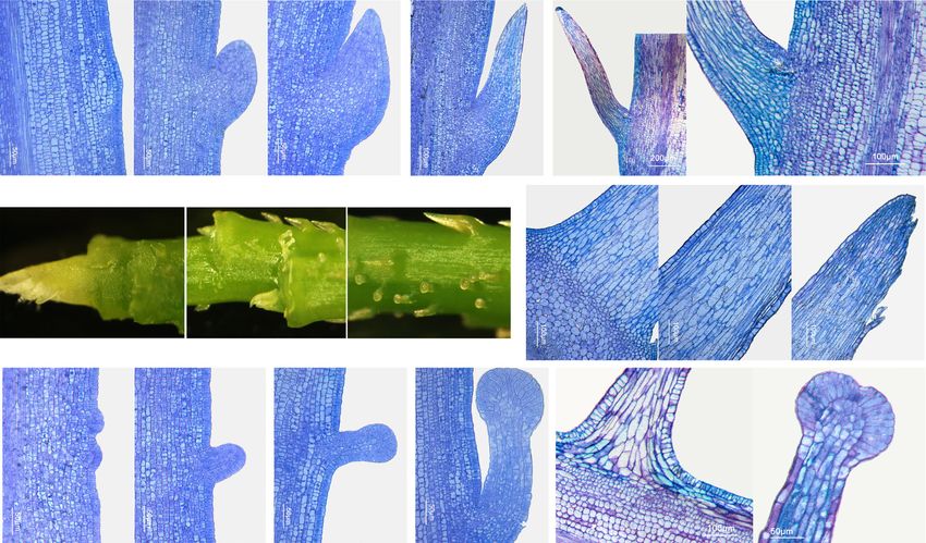
Fig. 1 Anatomy of nonglandular prickles (NGPs) and glandular prickles (GPs) in OW9106. a–c Macroscopic images of the different stages of
GPs and NGPs on the stem (leaves and leaf primordia were removed). Anatomy of stage I (d–f), IIa (g), IIb (h, i) and IIc (j–l) of nonglandular prickles.
Anatomy of stage I (m–o), IIa (p), and III (q, r) of glandular prickles. The white dotted frame represents the first internode. I: prickle initiation; Pro:
protoderm; GM: ground meristem; PM: prickle meristem; PMCL: prickle meristematic cell-like; E: epidermis; PG: precursor gland; G: gland; AL:
abscission layer structure-like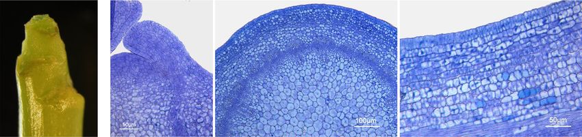
Zhou et al. Horticulture Research (2021)8:221                                                                                         Page 4 of 12

                                     apical                         GM
  a                          b       meristems            c                                            d
                                                              Pro
                                             leaf
                                             primordium
                                                                     procambium                                    GM

                                        procambium
                   500µm
                                                                                                                        procambium

 Fig. 2 Anatomy of prickless stems in OW9068. a Macroscopic image of a prickless shoot tip (leaves and leaf primordia were removed).
 b Longitudinal sections of shoot tips. c, d Cross- and longitudinal sections below the apical meristems, respectively. GM: ground meristem. Pro:
 protoderm. E: epidermis

appendages were observed (Fig. 2). For GPs, 2–4 cells                             downwardly curved hooks28. All the cells gradually stop
located at the first (and/or second) layers of the ground                          dividing and continue to elongate lengthwise (Fig. 1j–l).
meristem first appeared to differentiate and to divide                                The developmental stages of GPs are similar to those of
(Fig. 1m), and they gave rise to a cylindrical bump                               NGPs, except for the development of the gland head
(~50 µm) (Fig. 1n). At this early stage, a difference was                         (Fig. 1p; ref. 28). The gland is usually surrounded by one
observed between GPs and NGPs of OW9106 in the                                    cell layer and occasionally by two cell layers (Fig. 1p). Cell
number of primordial cells (Fig. 1d, m). The GP primordia                         division stops at early stage IIa. Then, the cells only
are smaller than the NGP ones. Then, in GPs and NGPs,                             enlarge, leading to the formation of a glandular head
the rapid cell division of a limited region of the ground                         (Fig. 1p). Their size only slightly increases during GP
meristem gives rise to a new structure on the surface of                          development (100–150 µm).
the stem (Fig. 1e, n). For GPs, the protoderm of this new                            The NGPs and GPs enter stage III when they begin to
structure differentiates into precursor gland cells (Fig. 1o),                    lignify and gradually harden28. An abscission layer
which will give rise to gland cells. This differentiation is                      structure-like is also formed (Fig. 1q). Thus, the prickles
absent in NGPs, where the protoderm (or the epidermis)                            can be easily separated from the stem. At the end of this
only continues to grow by cell division (Fig. 1f).                                stage, the cells are fully enlarged and lignified.
   In Stage II, both NGPs and GPs show continuous                                    Stage IV is defined as the mature stage, in which the
growth, color development, and shape development                                  NGPs and GPs completely harden, lose moisture and
(Supplementary Fig. 1 presented in Zhou et al.28). The                            exhibit gradual cell death28.
difference between GPs and NGPs is that the precursor
gland cells of GPs form a new structure—the glandular                             Discovery of different types of prickles among the rose
head—whereas no such structure is observed at the tip                             resources
of the NGPs (Fig. 1g, p). We have divided stage II into                             To describe the different types of prickles that are
three substages:                                                                  present in the Rosa genus, we conducted a survey of
   For NGPs, epidermal cells maintain normal cell pro-                            prickle types in 110 wild rose species, varieties, and
liferation during prickle development (Fig. 1g–l). In                             ancient hybrids (Supplementary Table 1, Fig. 3). Twelve
stage IIa, prickles continue to grow upwards28 because                            representative individuals (highlighted in pink in Sup-
the young prickle is covered by unopened leaves. Ana-                             plementary Table 1), which represent different sections
tomical analysis showed that the upper cells (from up to                          of the Rosa classification, were selected for detailed
down) of the prickle begin to enlarge, suggesting that the                        morphological analysis. According to macroscopic
cells gradually lose their ability to divide, while the cells                     observations, we classified prickles into two general
of the lower part may still continue to divide (small cells)                      categories, glandular prickles (GPs) and nonglandular
(Fig. 1g). These cell proliferation abilities and cell divi-                      prickles (NGPs), as we previously observed for OW
sion orientations may determine the prickle shape and                             individuals. The majority of roses present NGPs (98 out
the width of the prickle base in the later stages. In stage                       of 110), including 81 roses that presented NGPs only and
IIb, as the leaves open, the prickles grow outwards28. The                        17 that presented NGPs and GPs simultaneously (as
cells of the lower half of the prickle gradually stop pro-                        previously shown for OW9106) (Fig. 3). The NGPs and
liferating and begin to elongate (from top to bottom,                             GPs in these 98 roses were all unbranched. The
Fig. 1h, i). In stage IIc, after the leaves are fully opened,                     unbranched NGPs and GPs of a few roses (7 and 4,
the prickles are almost fully developed and form                                  respectively) are covered with hairs (hairy), whereas the
Zhou et al. Horticulture Research (2021)8:221                                                                                                                                  Page 5 of 12

                                                                             Stem prickles in roses

                    NGP (98)                                                               GP (22)
                                                                                                                                        Prickless (NP) or almost (7)

               Unbranched (98)                                        Unbranched (17)                         Branched (5)              NP or almost (4)               NP (3)
                                                                                                                                                                      R. Fraxinifolia lindl
                                                                                                                                                If have
                                                                                                                                                                      R. multiflora ‘Inermis’
      Naked (91)                 Hair (7)                   Naked (13)                    Hair (4)               Naked (5)                 NGP (4)                    R. wichuraiana ‘Basyes’
  R. hultemia persica        R. minutifolia Engelm       R. stellata                                          R. centifolia ‘Chou’       R. banksiae var.normalis     Thornless’
                                                                                  R. bracteata
  R. praelucens              R. cymose                   R. caninae freya                                     R. centifolia ‘muscosa’    R. banksiae ‘alba plena’     OW9068
                                                                                  R. rugosa scabrosa
  R. roxburghii              R. bracteate                R. horrida                                           R. × damascena ‘Quatre     R. banksiae ‘lutea’
  R. carolina                                            R. damascena             R. marie bugnet
                             R. rugosa scabrosa                                                               Saisons Blanc Mousseux’    R. pimpinellifolia ‘lutea’
  R. stellata                R. iwara                    R. gallica officinalis   R. ‘Grootendorst Supreme’
                                                                                                              R. ‘Général Kléber’
  ……                         R. marie bugnet             R. prattii
                                                                                                              R. ‘Parkzauber’
  R. multiflora              R. ‘Grootendorst Supreme’   R. willmottiae
  R. chinensis 'old blush'                               R. tsinglingensis
  R. wichuraiana                                         R. marmorata
  ……                                                     ……
  OW9137                                                 OW9106
  OW9106

           Unbranched NGPs only (81)                 Unbranched NGPs and GPs (17 )

                                          NGPs (98)
                                                                                  GPs (22)
                                                                                                    5 branched GPs

 Fig. 3 Different types of prickles among the rose resources. The number of roses is presented in the brackets. The “4” in red means that four
 prickless genotypes can sometimes present a few NGPs (R. banksiae var. normalis, R. banksiae ‘alba plena’, R. banksiae ‘lutea’, and R. pimpinellifolia
 ‘lutea’). OW F1 individuals (shown in blue) are not counted in the number of each category indicated in the figure. Ninety-eight roses present NGPs,
 including 81 roses presenting NGPs only and 17 presenting NGPs and GPs simultaneously (as in OW9106)

majority (91 and 13, respectively) did not have hair                                                   stages I and IIa of the prickles (previously defined for
(naked). Five genotypes present only branched and                                                      OW9106) are similar in these species (except the bristle
naked GPs (Fig. 3, Supplementary Table 1). Seven roses                                                 prickles, Fig. 4i–n). The primordial cells gave rise to an
have glabrous stem, but among these roses, four can                                                    oblique triangular structure (100–500 µm) that grows
sometimes be observed with a few NGPs. Here, we                                                        upwards (Fig. 4a, b). A large difference in shape appears
describe the characteristics and developmental process                                                 at the later stages. We also observed slight differences in
of the different types of prickles through detailed analysis                                           prickle initiation in different genotypes. In R. ecae
of examples of rose resources.                                                                         (Supplementary Fig. 2a–e), R. laxa (Supplementary Fig.
                                                                                                       2f–i) and R. omeiensis (large wing-like prickle, Fig. 4a–h),
Unbranched NGPs                                                                                        prickle initiation occurred only at the shoot tip, and the
Naked                                                                                                  same stage of prickles appeared in the same region of
  The unbranched and naked NGPs (91 genotypes in our                                                   the stem. Their development is quite similar to that
rose collection) are the most common type of prickles in                                               described for the OW9137 prickles28. In R. sherardi and
our rose collection. Among the representative genotypes,                                               R. moschata, prickle initiation occurred not only at the
five (R. omeiensis, R. ecae, R. laxa, R. sherardii, and                                                 shoot tip but also later during stem growth (Supple-
R. moschata) present only naked NGPs. The mature                                                       mentary Fig. 3b and i). In these two species, prickle
prickles are highly diverse in terms of shape, color, size,                                            initiation can take place over a longer period, and the
and density (Fig. 4, Supplementary Figs. 2 and 3). In                                                  prickles that initiate later are smaller at maturity (Sup-
particular, there are two very different shapes of NGPs                                                plementary Fig. 3). Thus, the time and place of prickle
present in R. omeiensis, which are needle-like (bristles)                                              initiation are important factors that impact the size of
and wing-like (Fig. 4a). We found that the morphology of                                               prickles on the mature stem.
Zhou et al. Horticulture Research (2021)8:221 Page 6 of 12
a b c d
I
I IIb
I
IIa
I
IIa
100µm 200µm 500µm 500µm
e f g h
IIb
IIb
III
IIb
IIc
1mm 1mm 1mm 1mm
i j k
IIa
I
IIb
100 µm 100 µm 500 µm
l m n 1mm
1mm
200 µm IV
III
IIc
Fig. 4 Nonglandular prickle developmental process in R. omeiensis. The initiation of needle-like prickles (i–n) occurs later than that of wing-like
prickles (a–h). White and yellow alphabetic marks and arrows refer to the development stages of wing-like and needle-like prickles, respectively
Hairy and three hybrids of R. rugosa (R. iwara (Supplementary
Some unbranched NGPs are covered with hairs (tri- Fig. 4h–q), R. “Grootendorst Supreme” (Supplementary
chomes). Only seven genotypes (7 out of 110) in our Fig. 4r–v) and R. ‘Marie bugnet’). In R. rugosa ‘scabrosa’,
collection have this type of prickle (Fig. 3, Supplemen- hairs are present on the stem and on the prickles. On the
tary Table 1): R. minutifolia Engelm, R. cymosa, R. stem, high-density hairs are present all along the shoot.
bracteata, R. rugosa ‘scabrosa’ (Supplementary Fig. 4a–g) Their initiation occurred earlier than that of prickles.Zhou et al. Horticulture Research (2021)8:221 Page 7 of 12
During prickle development, no hairs were visible in Some GPs present only one gland head (Supplementary
stage I (Supplementary Fig. 4a). Later, hairs appeared on Figs. 4 and 5), whereas some have several glands randomly
the lower part of the prickle, and the upper part remains distributed on their surface (Fig. 5). Some genotypes are
naked throughout development. In R. iwara, the hairs not easy to classify in the previous categories. For exam-
appeared later and at a lower density (Supplementary ple, one or several small GPs can develop on a large NGP
Fig. 4k). Prickles and stems have no hairs during (Fig. 5l), as observed in R. ‘General Kleber’.
stages I to IIb (Supplementary Fig. 4h, i and m), and the All prickles go through initiation, development, and
hairs appear clearly at stages IIc and III (Supplementary senescence. Most prickles do not fall from the stem, but a
Fig. 4k and l). few do (Supplementary Fig. 3g). In such cases, only a scar
is visible.
Unbranched GPs
Seventeen roses in our collection present unbranched Discussion
GPs, with 13 presenting naked GPs and 4 presenting hairy Two types of prickles in roses, glandular and nonglandular,
GPs (Supplementary Table 1, Fig. 3). with two distinct gene networks
On the basis of the morphology and anatomy of
Naked prickles, from their initiation to their complete develop-
Concerning the unbranched and naked GPs, their ment, and their distribution in the OW population, we
developmental process and origin were described in the proposed a categorization scheme for the presence or
previous section. We found that these prickles always absence of prickles in roses. We defined two major types
coexist with NGPs in roses (Supplementary Table 1), as in of prickles in roses: nonglandular prickles (NGPs) and
the following species or varieties: R. iwara (Supplemen- glandular prickles (GPs).
tary Fig. 4n and o), R. stellata, R. caninae ‘freya’, R. hor- For the first time, NGP and GP initiation and devel-
rida, R. rubella (Supplementary Fig. 5a–f), R. damascena opment were histologically characterized in detail. At the
(Supplementary Fig. 5g–j), R. gallica officinalis, R. prattii, initiation stage, no essential difference was observed
R. willmottiae, R. tsinglingensis, R. marmorata, R. pimpi- between GPs and NGPs. Both arose from the ground
nellifolia ‘King of the Scots’, R. pimpinellifolia ‘aïcha’ and meristem beneath the protoderm. Later, differences
R. anemoniflora. appeared between NGPs and GPs. GPs rapidly developed
a gland that was absent in NGPs. Our results do not
Hairy support previous research showing that GPs and NGPs
Unbranched GPs are covered with hairs (trichomes). are the early and later stages of the same prickle2,5,22,26.
Only four genotypes have this type of prickle: R. brac- Furthermore, our conclusions are different from those of
teata, R. rugosa ‘scabrosa’, R. ‘Marie Bugnet’ and R. previous studies, which reported that prickles originated
‘Grootendorst Supreme’ (two hybrids of R. rugosa) from the epidermis5,24,25 and were modified from gland-
(Supplementary Fig. 4t and u). They also present hairy ular trichomes22,26, were induced from glandular tri-
NGPs on their stems. chome signals40, or originated from bark tissue13. The
sub-epidermal origin of prickle in rose was confirmed in a
Branched GPs recent study for the cultivar “First Red”, where prickles are
Branched GPs were found in only five roses: R. centifolia proposed to be originated from the cortical parenchyma30.
‘chou’, R. centifolia ‘muscosa’, R. × damascena ‘Quatre We suggest that GPs may be modified from NGPs for
Saisons Blanc Mousseux’, and the two hybrids R. ‘Général the following reasons. NGPs are the most common type of
Kléber’ (Fig. 5a–f) and R. ‘Parkzauber’ (Fig. 5g–l). All the prickles in roses, and GPs (except for a few genotypes with
prickles were naked, and no hairy types were found in this branched GPs) always coexist with NGPs. Furthermore,
subcategory. GPs and NGPs have a common initiation process, and
Interestingly, these roses belong to a particular type of their development differs later (Fig. 1). For GPs, gland cells
roses, the moss roses (see “Discussion”). At stage I, the (a specific structure of GPs) are not produced at prickle
developmental process of branched GPs is more compli- initiation but during prickle development (Fig. 1m–r). A
cated than the one of unbranched GPs. Thus, we divided similar hypothesis was proposed for the trichomes. From
stage I into three substages. In stage Ia, multiple divisions an evolutionary perspective, the earliest glandular tri-
give rise to a nearly round protuberance (Fig. 5a). The chomes (GTs) were proposed to be modified from non-
appearance of branch bumps is a sign of entrance into glandular trichomes (NGTs)41,42. Another possibility is
stage Ib (Fig. 5a). In stage Ic, the bumps continue to grow that GPs and NGPs have different genetic pathways in
and to differentiate into glands and stalks (Fig. 5b). The terms of the fate of the first mother cells. Additional stu-
subsequent stages are similar to those of the unbranched dies are needed to test these different hypotheses.
GPs (Fig. 5c–f). The genetic results support the last hypothesis28.Zhou et al. Horticulture Research (2021)8:221 Page 8 of 12
a b c
Ic
Ib
Ic
III
Ia
100µm 200µm
d 200µm e 500µm
f
IV
IIc
IIb
g h i
IIb
IIa
500um 500um
j 500um
k 1mm
l 1mm
III
IIc
Fig. 5 Branched and naked glandular prickle development in R. ‘General Kleber’ and R. ‘Parkzauber’. Stage Ia, Ib (a), Ic (b), IIb (d), IIc (e), III (c)
and IV (f) of GPs in R. ‘General Kleber’. g Early stage of GPs on bud of R. ‘Parkzauber’. Stage IIa (h), IIb (i), IIc (j), III (k) of GPs in R. ‘Parkzauber’. l A small GP
developed on a large NGP
At the morphological level, we also observed a large hair initiation occurs later than prickle initiation, and
diversity of GPs and NGPs in the Rosa genus. The these hairs cover not only the prickles but also the stem
glandular and nonglandular prickles can be covered by (Supplementary Fig. 4). Therefore, we hypothesize that:
hairs (hairy) or not (naked). GPs can present branches the genetic pathways for hairy and naked NGP initiation
(branched versus unbranched GPs). For hairy prickles, are the same; the genetic mechanism controlling hairZhou et al. Horticulture Research (2021)8:221 Page 9 of 12
initiation is different from that controlling prickle initia- natural conditions. The absence of prickles may be due
tion. Similarly, branches appear during prickle develop- to mutations, and selection may eliminate this trait,
ment, suggesting that the pathways controlling GP similar to selection for recurrent blooming trait44.
initiation are similar between unbranched and branched Therefore, our hypothesis is that glabrous mutants were
GPs, whereas another signaling pathway might control selected and rescued by humans. This could explain the
whether a prickle branches or not. rarity of genotypes without prickles found under natural
conditions. These glabrous roses are interesting materi-
Suggestions for genetic and genomic studies on rose als for genetic and genomic studies aiming to understand
prickles prickle initiation.
As we suggested for the first time that different genetic
pathways are involved in GP and NGP initiation28, these Branched GPs in moss roses
pathways should be studied separately in genetic and Branched GPs are also rare and were present in only 5
molecular studies. In roses, prickles are present on organs of the 110 roses examined here: R. centifolia ‘Chou’
other than the stem, such as petioles, pedicels, and fruits. (Inconnu,Zhou et al. Horticulture Research (2021)8:221 Page 10 of 12
respond through intracellular signaling pathways and buffer at pH 7.2. The solution volume was equal to 50
eventually adopt a specific cell fate, thereby producing times the volume of the sample. Each sample was put
different organs or tissues47. Therefore, for trichomes and under vacuum to remove air, with the vacuum setting
prickles, the tissues they originate from are different paused every 4 min. After 2 h of vacuum, we changed the
(protoderm versus subprotoderm), which may indicate glutaraldehyde solution (4% v/v), stored the tubes at 4 °C
that different gene networks control prickle and trichome 12 h, rinsed the samples twice with phosphate buffer pH
initiation. This hypothesis is supported by molecular 7.2 and stored the samples at 4 °C.
evidence in roses, where no strong link can be found Dehydration at room temperature: Samples were rinsed 3
between the trichome and prickle pathways. Indeed, Zhou times with distilled water and immersed in 50% (v/v)
et al.28 characterized rose gene homologs known in alcohol for 10 min, 70% (v/v) alcohol for 10 min, 90%
A. thaliana to control trichome initiation. These genes (v/v) alcohol for 10 min, and 100% alcohol for 15 min.
were not transcriptionally regulated during prickle Preinfiltration: Samples were transferred to preinfiltra-
initiation, suggesting that the genetic pathway controlling tion solution (100° alcohol/Technovit® 7100 resin
prickle initiation is different from that controlling tri- (Heraeus Kulzer, Wehrheim, Germany) (v/v)) at 4 °C
chome initiation. Therefore, we suggest that different and under vacuum for 2 h, and then the samples were
genetic pathways control the initiation of NGPs and stored for 12 h at 4 °C.
NGTs. This conclusion is different from the current Infiltration: Samples were transferred to infiltration
hypothesis: rose NGPs and A. thaliana unicellular NGTs solution (1 sachet of hardener I dissolved in 100 mL
share the same genetic pathway for their initiation30,31. (Heraeus Kulzer, Wehrheim, Germany) of Technovit®
7100 resin) under vacuum for at least 20 min at 4 °C, and
Materials and methods the tubes were then stored for 12 h at 4 °C.
Plant materials Inclusion: Samples were included using an inclusion
A diploid OW population obtained from a cross solution (1 mL of hardener II® (Heraeus Kulzer,
between Rosa chinensis ‘Old Blush’ (OB) and Rosa × Wehrhrim, Germany) and 15 ml of infiltration solution)
wichurana (RW) was grown in a field and managed by the and stored at 37 °C. Sections were made after a week
Horticulture Experimental Unit (INRAE, Angers, France). at 37 °C.
To obtain more vegetative branches, we selected two The samples were cut into 3 µm sections for anatomical
once-flowering individuals OW9068 and OW9106. These observation using a Leica RM2165 rotary microtome.
genotypes were cut and managed in IRHS greenhouses in After being stained with toluidine blue 1%48, the samples
November 2017. were observed and photographed using an ergonomic
Rosa resources were planted at the Loubert Rose Gar- system microscope (Leica DM1000).
dens (Rosiers sur Loire, France), INRAE (Angers, France)
and Flower Research Institute (FRI, Kunming, China). We Scoring of prickle type among the 110 roses
selected twelve representative genotypes to perform Taxonomical nomenclature followed that described in
detailed analyses of the type and developmental stages of Yu49, Gu and Robertson34 and Masure43. Each species or
prickles: Rosa ecae, Rosa laxa, Rosa sherardi, Rosa hybrid was associated with the types of prickles that were
moschata, Rosa omeiensis, Rosa damascena, Rosa rugosa previously determined in the OW population and in the
‘scabrosa’, Rosa iwara, Rosa ‘Grootendorst Supreme’, Rosa twelve representative roses. The prickle types on each
rubella, Rosa ‘General Kleber’ and Rosa ‘Parkzauber’. For species were characterized based on photographs, which
110 genotypes (Supplementary Table 1), we scored the were mainly taken at the Loubert Rose Gardens (https://
prickle type on the first and second branches, and only www.pepiniere-rosesloubert.com/) and FRI. For some
considered prickles on the stem (prickles on pedicel were species, the conclusions are based on professional
excluded). knowledge and experience and online photographs. All
the roses and their origins are presented in Supplemen-
Macroscopy and stereoscopy tary Table 1.
The experiments were performed at Platform IMAC
(SFR QuaSav, Angers). Fresh rose stems were photo- Acknowledgements
graphed with a Leica M205FA stereomicroscope. We are grateful to the IMAC technical platforms of SFR Quasav for supporting
the histological experiment and especially thank Aurelia Rolland for
participating in the discussion during the histological experiment. We are
Histological study grateful to the Loubert Rose Gardens (Rosiers sur Loire, France) for providing
Sample dissections were performed under a microscope the experimental materials. We would also like to thank Hong-ying Jian and
to remove the leaves. Various steps were performed: Shu-Fa Li (FRI, Kunming, China) for wild rose collection and species
identification. We thank Xue-wu DOU (Angers University, France) for helping
Fixation at 4 °C: Samples were immersed in a 4% (v/v) produce Supplementary Fig. 1. We thank Latifa HAMAMA (IRHS, Agrocampus-
glutaraldehyde solution mixed with 0.2 mol/L phosphate ouest, Angers, France) for communication of tissue anatomical terms.Zhou et al. Horticulture Research (2021)8:221 Page 11 of 12
We thank the PHENOTIC platform and experimental unit Horticulture for 15. Esau, K. Plant Anatomy, Vol. 75. (Wiley, 1953).
managing the plants. This work was supported by funding from the National 16. Hülskamp, M. Plant trichomes: a model for cell differentiation. Nat. Rev. Mol.
Natural Science Foundation of China (31760585) and the China Scholarship Cell Biol. 5, 471–480 (2004).
Council ([2017]3109). 17. Zhou, N. N. Genetics and genomics of prickles on rose stem. PhD thesis
(University of Angers, Angers, France. Doctor dissertation, 2021).
Author details 18. Huchelmann, A., Boutry, M. & Hachez, C. Plant glandular trichomes:
1 natural cell factories of high biotechnological interest. Plant Physiol. 175,
Univ Angers, Institut Agro, INRAE, IRHS, SFR QUASAV, F-49000 Angers, France.
2 6–22 (2017).
National Engineering Research Center for Ornamental Horticulture; Flower
Research Institute (FRI), Yunnan Academy of Agricultural Sciences, Kunming 19. Chalvin, C., Drevensek, S., Dron, M., Bendahmane, A. & Boualem, A. Genetic
650231, China. 3Univ Angers, INRAE, SFR QUASAV, F-49000 Angers, France control of glandular trichome development. Trends Plant Sci. 25, 477–487
(2020).
Author contributions 20. Yan, T. et al. A novel HD-ZIP IV/MIXTA complex promotes glandular trichome
N.N. Zhou, F. Foucher, and L. Hibrand Saint Oyant conceived and designed the initiation and cuticle development in Artemisia annua. N. Phytologist 218,
study. N.N. Zhou performed the experiments, collected and analyzed the data, 567–578 (2018).
and drafted the manuscript. F. Foucher and L. Hibrand-Saint Oyant were 21. Bagella, S. et al. Thorn, spine and prickle patterns in the Italian flora. Plant
responsible for supervising the project and for revising the manuscript. Biosyst. 153, 118–133 (2019).
F. Simonneau provided full technical knowledge for performing the 22. Kellogg, A. A., Branaman, T. J., Jones, N. M., Little, C. Z. & Swanson, J.-D.
histological experiments. T. Thouroude contributed to F1 cutting plant Morphological studies of developing Rubus prickles suggest that they are
management in the greenhouse and to recording planting information on modified glandular trichomes. Botany 89, 217–226 (2011).
wild rose resources. 23. Li, H. et al. Studies on anatomical structure and chemical composition in
prickles of Rosa hybrida. Acta Horticulturae Sin. 39, 1321–1329 (2012).
24. Esau, K. Anatomy of seed plants. Soil Sci. 90, 149 (1960).
Data availability
25. Peitersen, A. K. Blackberries of New England–genetic status of the plants.
All data supporting the results of this study are included in the manuscript and
Vermont Agricultural Experiment Station.(1921).
its additional files.
26. Khadgi, A. & Weber, C. A. Morphological characterization of prickled and
prickle-free Rubus using scanning electron microscopy. HortScience 55,
Conflict of interest 676–683 (2020).
The authors declare no competing interests. 27. Hibrand-Saint Oyant, L. et al. A high-quality genome sequence of Rosa chi-
nensis to elucidate ornamental traits. Nat. Plants 4, 473 (2018).
Supplementary information The online version contains supplementary 28. Zhou, N. N. et al. Genetic determinism of prickles in rose. Theor. Appl. Genet.
material available at https://doi.org/10.1038/s41438-021-00689-7. 133, 3017–3035 (2020).
29. Khadgi, A. & Weber, C. A. RNA-Seq analysis of prickled and prickle-free epi-
dermis provides insight into the genetics of prickle development in red
Received: 28 May 2021 Revised: 7 July 2021 Accepted: 13 July 2021
raspberry (Rubus ideaus l.). Agronomy 10, 1904 (2020).
30. Swarnkar, M. K., Kumar, P., Dogra, V. & Kumar, S. Prickle morphogenesis in rose
is coupled with secondary metabolite accumulation and governed by
canonical MBW transcriptional complex. Plant Direct 5, e00325 (2021).
References 31. Zhong, M. C. et al. Rose without prickle: genomic insights linked to moisture
1. Bell, A. D. Plant form: an illustrated guide to flowering plant morphology. adaptation. Nat. Sci. Rev. nwab092, https://doi.org/10.1093/nsr/nwab092
(Oxford University Press, USA, 1991). (2021).
2. Ma, Z. Y., Wen, J., Ickert-Bond, S. M., Chen, L. Q. & Liu, X. Q. Morphology, 32. Dickison, W. C. Integrative Plant Anatomy, 1st edn. (Academic Press, California,
structure, and ontogeny of trichomes of the Grape genus (Vitis, Vitaceae). USA, 2000).
Front. Plant Sci. 7, 704 (2016). 33. Evert, R. F. & Eichhorn, S. E. Esau’s plant anatomy: meristems, cells, and tissues
3. Canli, F. A. & Skirvin, R. M. Separation of thornless rose chimeras into of the plant body: their structure, function, and development, 3rd edn. (John
their (Rosa sp.) consistent genotypes in vitro. Pak. J. Biol. Sci. 6, Wiley & Sons, 2006).
1644–1648 (2003). 34. Gu, G. Z. & Robertson, K. R. Flora of China - Rosaceae: Rosa, Vol. 9. http://www.
4. Castro, P., Stafne, E. T., Clark, J. R. & Lewers, K. S. Genetic map of the primocane- iplant.cn/info/rosa?t=foc (Science Press, Beijing, 2003).
fruiting and thornless traits of tetraploid blackberry. Theor. Appl. Genet. 126, 35. Bourke, P. M. et al. Partial preferential chromosome pairing is genotype
2521–2532 (2013). dependent in tetraploid rose. Plant J. 90, 330–343 (2017).
5. Coyner, M. A., Skirvin, R. M., Norton, M. A. & Otterbacher, A. G. Thornlessness in 36. Vukosavljev, M. et al. High-density SNP-based genetic maps for the parents of
blackberries. Small Fruits Rev. 4, 83–106 (2005). an outcrossed and a selfed tetraploid garden rose cross, inferred from
6. Hall, H. K., Cohen, D. & Skirvin, R. M. The inheritance of thornlessness from admixed progeny using the 68k rose SNP array. Horticulture Res. 3, 1–8 (2016).
tissue culture-derived ‘thornless evergreen’ blackberry. Euphytica 35, 891–898 37. Nguyen, T. H. N., Schulz, D., Winkelmann, T. & Debener, T. Genetic dissection of
(1986). adventitious shoot regeneration in roses by employing genome-wide asso-
7. Kariyat, R. R., Hardison, S. B., De Moraes, C. M. & Mescher, M. C. Plant spines ciation studies. Plant Cell Rep. 36, 1493–1505 (2017).
deter herbivory by restricting caterpillar movement. Biol. Lett. 13, 20170176 38. Schulz, D. F. et al. Genome-wide association analysis of the anthocyanin and
(2017). carotenoid contents of rose petals. Front. Plant Sci. 7, 1798 (2016).
8. McPheeters, K. & Skirvin, R. M. Histogenic layer manipulation in chimeral 39. Raymond, O. et al. The Rosa genome provides new insights into the
thornless evergreen trailing blackberry. Euphytica 32, 351–360 (1983). domestication of modern roses. Nat. Genet. 50, 772–777 (2018).
9. Asano, G., Kubo, R. & Tanimoto, S. Growth, structure and lignin localization in 40. Pandey, S. et al. Transcriptome analysis provides insight into prickle devel-
rose prickle. Bull. Fac. Agriculture 93, 117–125 (2008). opment and its link to defense and secondary metabolism in Solanum viarum
10. Werker, E. Trichome diversity and development. In Advances in Botanical dunal. Sci. Rep. 8, 1–12 (2018).
Research, Vol. 31, 1–35 (Academic Press, 2000). 41. Krings, M., Kellogg, D. W., Kerp, H. & Taylor, T. N. Trichomes of the seed fern
11. Blaser, H. W. Morphology of the determinate thorn-shoots of Gleditsia. Am. J. Blanzyopteris praedentata: implications for plant-insect interactions in the late
Bot. 43, 22–28 (1956). carboniferous. Botanical J. Linn. Soc. 141, 133–149 (2003).
12. Boke, N. H. Developmental anatomy and the validity of the genus Bartschella. 42. Lange, B. M. The evolution of plant secretory structures and emergence of
Am. J. Bot. 43, 819–827 (1956). terpenoid chemical diversity. Annu. Rev. Plant Biol. 66, 139–159 (2015).
13. Angyalossy, V. et al. Iawa list of microscopic bark features. IAWA J. 37, 585–587 43. Masure, P. Guide Des Rosiers Sauvages: 500 Espèces, Variétés et Hybrides Du
(2016). Monde. (Delachaux et Niestlé, Paris, France, 2013).
14. Delbrouck, C. (1875). Die Pflanzen-Stacheln. Botanische Abhandlungen aus 44. Soufflet-Freslon, V. et al. Diversity and selection of the continuous-flowering
dem Gebiet der Morphologie und Physiologie. Adolph Marcus (1875). gene, RoKSN, in rose. Horticulture Res. 8, 1–11 (2021).Zhou et al. Horticulture Research (2021)8:221 Page 12 of 12
45. Nédelec, P. Y. Roses grandeur nature: la collection des roses loubert. Delachaux 47. Larkin, J. C., Brown, M. L. & Schiefelbein, J. How do cells know what they want
et Niestlé (2018). to be when they grow up? Lessons from epidermal patterning in Arabidopsis.
46. Caissard, J.-C., Bergougnoux, V., Martin, M., Mauriat, M. & Baudino, S. Annu. Rev. Plant Biol. 54, 403–430 (2003).
Chemical and histochemical analysis of ‘quatre saisons blanc mous- 48. O’Brien, T. P., Feder, N. & McCully, M. E. Polychromatic staining of plant cell
seux’, a moss rose of the Rosa× damascena group. Ann. Bot. 97, walls by toluidine blue o. Protoplasma 59, 368–373 (1964).
231–238 (2006). 49. Yu, D. J. Flora of China, Vol. 36 (Science Press, Beijing, 1974).You can also read