Movements of Mycoplasma mobile Gliding Machinery Detected by High-Speed Atomic Force Microscopy
←
→
Page content transcription
If your browser does not render page correctly, please read the page content below
Movements of Mycoplasma mobile Gliding
Machinery Detected by High-Speed Atomic
Force Microscopy

Kohei Kobayashi, Noriyuki Kodera, Taishi Kasai, Yuhei O. Tahara,

Takuma Toyonaga, Masaki Mizutani, Ikuko Fujiwara, Toshio

Ando, Makoto Miyata

   Citation    mBio. 12(3); e00040-21
 Issue Date    2021-06-29
  Published    2021-05-28
    Type       Journal Article
 Textversion   Publisher
               ◆最小細胞である「マイコプラズマ・モービレ」の分子モーターの動きを世界で初めて検出。
 Highlights    ◆マイコプラズマの滑走運動時の動きを捉えた世界初の研究。
               ◆ナノスケールのデバイスや医薬品を開発するための基礎となる。
               研究チームは、最小の細菌である「マイコプラズマ・モービレ」が滑走するための分子モー
               ターの動きを検出することに世界で初めて成功しました。この発見は、ナノスケールのデバ
               イスや医薬品の開発への応用が期待されます。
               【今回の発見】
               マイコプラズマは、菌体の片側に小さな突起“接着器官”を形成し、この突起で宿主組織の
    概要
               表面にはりつき、はりついたまま“滑走運動”を行います。滑走運動時には ATP 合成酵素
               から進化した特殊な分子モーターが細胞内部で力を発生していることが示唆されていまし
               たが、その動きが捉えられたことはありませんでした。今回、最先端技術である高速原子間
               力顕微鏡(高速 AFM)を用いることで、滑走の分子モーターの動きをナノレベルで細胞外
               から検出することに成功しました。
               © 2021 Kobayashi et al. This is an open-access article distributed under the terms of the
   Rights      Creative Commons Attribution 4.0 International license.
               https://creativecommons.org/licenses/by/4.0/
    DOI        10.1128/mBio.00040-21

               Placed on: Osaka City University Repository
Kobayashi K, Kodera N, Kasai T, Tahara YO, Toyonaga T, Mizutani M, Fujiwara I, Ando T, Miyata M.
2021. Movements of Mycoplasma mobile gliding machinery detected by high-speed atomic force
microscopy. mBio 12:e00040-21. https://doi.org/10.1128/mBio.00040-21.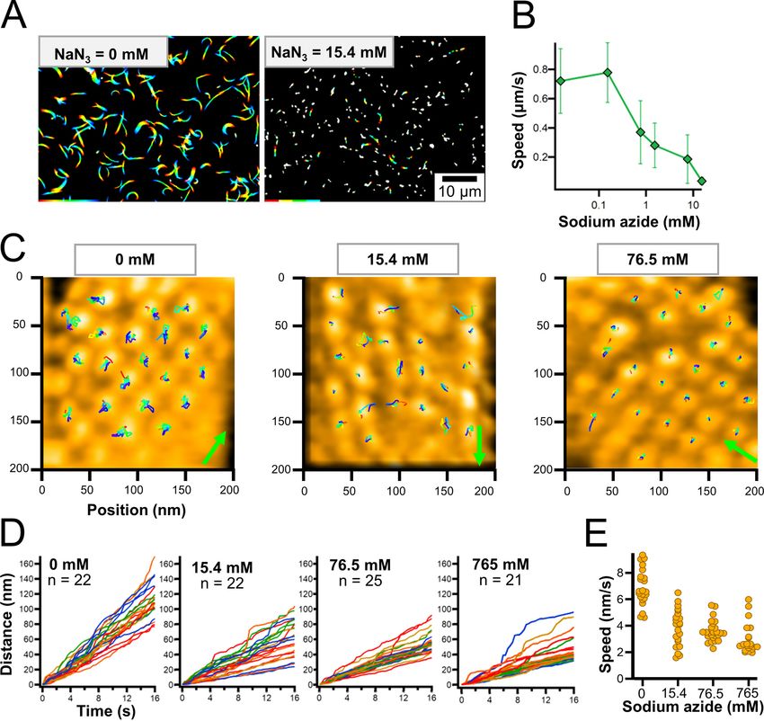
本研究では、マイコプラズマを生きたままガラスに固定し、高速 AFM の細い針で細胞
                の表面を軽くたたきながら探ることにより、分子モーターの構造をリアルタイムにビデオ
                に記録しました。
                  さらに得られたビデオ画像に隠された信号を計算によって抽出することで、分子モータ
                ー粒子が ATP を加水分解して力を発生する際の動きをナノレベルで追跡することに成功
                しました。その結果、鎖状に連なったモーター粒子が、細胞進行方向に向かって右側に約
                9 ナノメートル、細胞内側に 2 ナノメートル、300 ミリ秒以内の時間に動くことを明らか
                にしました。このことは、他に類を見ない構造であるマイコプラズマの分子モーターがど
                のようなメカニズムで滑走運動を行っているか、そしてそのメカニズムが ATP 合成酵素の
                どのような性質から進化してきたかを説き明かす大きな手掛かりとなります。

  Description

                可視化された細胞内部のモーター粒子とその動き

                縦方向に並ぶ白い楕円がモーター粒子。

                緑の矢印は滑走運動の方向を示す。

                それぞれの粒子の重心の 5 秒間の動きが虹色の線で示されている。

                ©大阪市立大学      宮田真人

                  ガラスに固定した細胞から膜を除去したり、単離した分子モーターを使ったりすること
                により、空間的、時間的により高い分解能における解析を目指します。それとは別に単離
                した分子モーター構造を電子顕微鏡で解析することで、原子レベルの解像度で明らかにし
                ます。これらの情報を統合することで、滑走運動メカニズムを原子レベルで理解します。
                  滑走の構造とメカニズムを詳細に明らかにすることで、運動能の起源と動作原理に迫る
                ことができ、ナノスケールのデバイスや医薬品を開発するための基盤になることが期待さ
                れます。

                ‘世界初!マイコプラズマの滑走運動における分子モーターの動きをナノレベルで検出!’.
                大阪市立大学. https://www.osaka-cu.ac.jp/ja/news/2021/210528-2. (参照 2021-05-28.)

Kobayashi K, Kodera N, Kasai T, Tahara YO, Toyonaga T, Mizutani M, Fujiwara I, Ando T, Miyata M.
2021. Movements of Mycoplasma mobile gliding machinery detected by high-speed atomic force
microscopy. mBio 12:e00040-21. https://doi.org/10.1128/mBio.00040-21.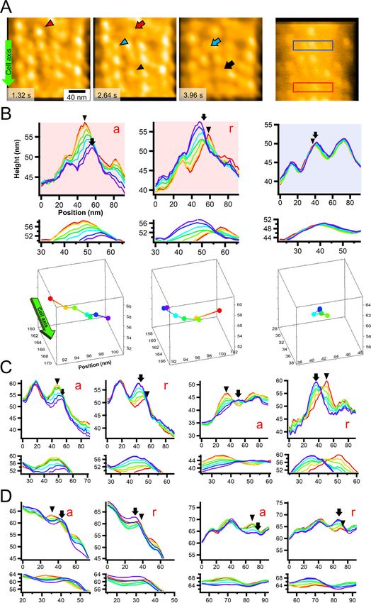
■高速原子間力顕微鏡(高速 AFM)
                  原子間力顕微鏡(Atomic Force Microscopy: AFM)は、探針と試料の間に働く原子
                間力を元に、分子の形状をナノメートル(10-9 m)程度の高い空間分解能で可視化す
    補   足       る顕微鏡。高速 AFM は、金沢大学の安藤敏夫特任教授のグループによって開発され
                た超高速で観察できる AFM で、サブ秒(~0.1 秒)という時間分解能で、水溶液中に
                あるタンパク質などの生体分子や細胞の形状や動態をその周囲の環境を含めて観察す
                ることができる。
                動画 : マイコプラズマ・モービレの滑走の様子
    参   考
                https://www.youtube.com/watch?v=-LRdogB3U8s

Kobayashi K, Kodera N, Kasai T, Tahara YO, Toyonaga T, Mizutani M, Fujiwara I, Ando T, Miyata M.
2021. Movements of Mycoplasma mobile gliding machinery detected by high-speed atomic force
microscopy. mBio 12:e00040-21. https://doi.org/10.1128/mBio.00040-21.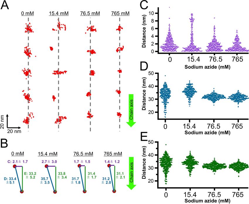
RESEARCH ARTICLE

Movements of Mycoplasma mobile Gliding Machinery Detected
by High-Speed Atomic Force Microscopy
Kohei Kobayashi,a Noriyuki Kodera,b Taishi Kasai,a* Yuhei O. Tahara,a,c Takuma Toyonaga,a                                        Masaki Mizutani,a*
  Ikuko Fujiwara,a,d Toshio Ando,b Makoto Miyataa,c

a   Graduate School of Science, Osaka City University, Sumiyoshi-ku, Osaka, Japan
b   Nano Life Science Institute (WPI-NanoLSI), Kanazawa University, Kanazawa, Ishikawa, Japan
c   The OCU Advanced Research Institute for Natural Science and Technology (OCARINA), Osaka City University, Sumiyoshi-ku, Osaka, Japan
d   Department of Bioengineering, Nagaoka University of Technology, Nagaoka, Niigata, Japan

Kohei Kobayashi and Noriyuki Kodera contributed equally to this work. Kohei Kobayashi performed most experiments and Noriyuki Kodera provided AFM setups and technologies.

ABSTRACT       Mycoplasma mobile, a parasitic bacterium, glides on solid surfaces, such
as animal cells and glass, by a special mechanism. This process is driven by the force
generated through ATP hydrolysis on an internal structure. However, the spatial and
temporal behaviors of the internal structures in living cells are unclear. In this study,
we detected the movements of the internal structure by scanning cells immobilized
on a glass substrate using high-speed atomic force microscopy (HS-AFM). By scan-
ning the surface of a cell, we succeeded in visualizing particles, 2 nm in height and
aligned mostly along the cell axis with a pitch of 31.5 nm, consistent with previously
reported features based on electron microscopy. Movements of individual particles
were then analyzed by HS-AFM. In the presence of sodium azide, the average speed
of particle movements was reduced, suggesting that movement is linked to ATP hy-
drolysis. Partial inhibition of the reaction by sodium azide enabled us to analyze par-
ticle behavior in detail, showing that the particles move 9 nm right, relative to the
gliding direction, and 2 nm into the cell interior in 330 ms and then return to their
original position, based on ATP hydrolysis.
IMPORTANCE The Mycoplasma genus contains bacteria generally parasitic to animals
and plants. Some Mycoplasma species form a protrusion at a pole, bind to solid                                            Citation Kobayashi K, Kodera N, Kasai T, Tahara
surfaces, and glide by a special mechanism linked to their infection and survival. The                                    YO, Toyonaga T, Mizutani M, Fujiwara I, Ando T,
                                                                                                                          Miyata M. 2021. Movements of Mycoplasma
special machinery for gliding can be divided into surface and internal structures that
                                                                                                                          mobile gliding machinery detected by high-
have evolved from rotary motors represented by ATP synthases. This study suc-                                             speed atomic force microscopy. mBio 12:
ceeded in visualizing the real-time movements of the internal structure by scanning                                       e00040-21. https://doi.org/10.1128/mBio
                                                                                                                          .00040-21.
from the outside of the cell using an innovative high-speed atomic force microscope
                                                                                                                          Editor Dominique Soldati-Favre, University of
and then analyzing their behaviors.                                                                                       Geneva
                                                                                                                          Copyright © 2021 Kobayashi et al. This is an
KEYWORDS AFM, probing, pathogenic bacteria, ATPase, class Mollicutes
                                                                                                                          open-access article distributed under the terms
                                                                                                                          of the Creative Commons Attribution 4.0
                                                                                                                          International license.

M       any bacteria translocate to nutrient-rich places and escape from repellent sub-
        stances by manipulating external appendages, such as flagella and pili (1, 2).
However, class Mollicutes, a small group of bacteria, have as many as three of their own
                                                                                                                          Address correspondence to Makoto Miyata,
                                                                                                                          miyata@sci.osaka-cu.ac.jp.
                                                                                                                          * Present address: Taishi Kasai, Department of
                                                                                                                          Life Science, Rikkyo University, Toshima-ku,
motility mechanisms. Class Mollicutes evolved from phylum Firmicutes by losing pepti-
                                                                                                                          Tokyo, Japan; Masaki Mizutani, Bioproduction
doglycan synthesis and flagella swimming to evade host innate immunity in their para-                                      Research Institute, National Institute of
sitic life (1). They have a single-layered cell membrane featured by a high content of                                    Advanced Industrial Science and Technology
                                                                                                                          (AIST), Tsukuba, Ibaraki, Japan.
sterols (25 to 30% of the weight of total membrane lipids) and lipoproteins as periph-
                                                                                                                          Received 27 January 2021
eral structures (3–7). Among Mollicutes, the gliding motility of Mycoplasma mobile, the                                   Accepted 19 April 2021
subject of this study, is suggested to have evolved from a combination of ATP synthase                                    Published 28 May 2021
and cell adhesion (1, 5, 8–12).
                                                                                                                                                        ®
May/June 2021 Volume 12 Issue 3 e00040-21                                                                                                                   mbio.asm.org     1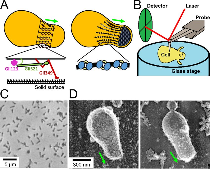
®
Kobayashi et al.

FIG 1 Experimental design and conditions for HS-AFM observation. (A) Schematic illustrations of M.
mobile gliding machinery. The gliding machinery formed as a protrusion can be divided into surface
(left) and internal (right) structures. The surface structure is composed of about 450 units, including
three large proteins—Gli123 (purple), Gli521 (green), and Gli349 (red)—as shown at the bottom.
Gli349 repeatedly catches sialylated oligosaccharides fixed on the solid surface and pulls the cell
forward. The internal structure can be divided into a large mass at the cell front, “bell” and a chain
structure. The chain structure is composed of particles that have been suggested to evolve from F-
type ATPase/synthase. (B) Schematic illustration of an M. mobile cell being scanned by high-speed
atomic force microscopy (HS-AFM). The surface of an immobilized cell on glass stage (blue) is
scanned by an AFM cantilever probe (gray), and the cantilever movement is monitored by a detector
(green). (C) Phase-contrast image of M. mobile cells on a coverslip. Living cells were immobilized onto
a coverslip using poly-L-lysine and glutaraldehyde. (D) Quick-freeze, deep-etch electron microscopic
(EM) image of M. mobile cells on a coverslip. The cell was immobilized on the coverslip by poly-L-
lysine and glutaraldehyde (left) and allowed to glide on the coverslip coated with sialylated
oligosaccharides (right). The cell axis and front are indicated by a green arrow in panels A and D.

    M. mobile, isolated from a freshwater fish, is a flask-shaped bacterium with a length
of 0.8 m m (Fig. 1A). M. mobile glides in the direction of its tapered end on solid surfaces,
such as animal cells, glass, and plastics. Its gliding speed is 2.5 to 4 m m/s, which is 3 to
5 times its own cell length (10, 13). The gliding machinery is divided into surface and
internal structures, both of which are composed of 450 units (Fig. 1A) (5, 8, 10, 14). The
internal structure is characterized by multiple chains. An M. mobile cell has approxi-
mately 28 chains around the base of the protrusion (Fig. 1A). Each chain consists of
uniformly sized particles, which are 13 nm in width and 21 nm in length (5).
Interestingly, the amino acid sequence of component proteins suggests that this chain
structure has evolved from ATP synthase (5, 8, 10, 12, 15). Recently, the isolated inter-
nal structure was shown to hydrolyze ATP through conformational changes, suggest-
ing that the internal structure functions as a motor and generates the force for gliding
(5, 10). The surface structure is composed of three large proteins, Gli349, Gli521, and
Gli123. Gli349 has a binding site for sialylated oligosaccharide at its tip and plays the
role of a “leg” in gliding (9, 16–20). Gli521 and Gli123 have been proposed to act as a
“crank” that transmits force (21–24) and as a “mount” to correctly localize the surface
proteins (19). A working model for the gliding mechanism has been suggested as fol-
lows (5, 10, 13, 25): the force for gliding generated based on ATP-derived energy by
the special motor is transmitted across the membrane to the surface structure, includ-
ing the leg structure. Then, the foot (the tip structure of the leg) repeatedly catches,
pulls, and releases the sialylated oligosaccharides (9, 16), the major structures on host

May/June 2021 Volume 12 Issue 3 e00040-21                                                                 mbio.asm.org   2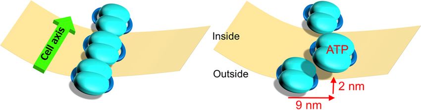
®
Gliding Mechanism of Mycoplasma mobile

animal surfaces (26–28), resulting in cell migration (21, 29–32). This explains the gliding
mechanism at the bacterial surface; however, the spatial and temporal behaviors and
movements of internal motors in living cells have not been examined.
    Atomic force microscopy (AFM) (33) is a powerful method to image the surface struc-
tures and to study the mechanical properties of a biological sample at the submolecular
level (34). In this method, a sample placed on a substrate is scanned with a nanometer-
scale probe under dry and wet conditions. The usefulness of this method has been dem-
onstrated also in the field of microbiology (35, 36). In high-speed AFM (HS-AFM), the scan-
ning speed of AFM has been dramatically improved to ;20 frames per second (fps) while
maintaining minimal invasiveness (37). Then, the dynamic behaviors of biomolecules and
cells can be captured in aqueous solution (37), and their functional mechanisms have
been elucidated (37–40). Notably, HS-AFM has been applied to understand the structures
on the cell wall (41) or below the cell membrane (42).
    In this study, we succeeded in visualizing the internal structure of M. mobile gliding
machinery by scanning the surface of cells immobilized on a glass substrate using HS-
AFM. The particle structure, a component of the internal structure, showed movements
mainly in the right and inward directions relative to the gliding direction of an M. mo-
bile cell.

RESULTS
    Immobilization of living cells on the glass surface. We attempted to visualize the
gliding machinery by scanning the upper side of living cells immobilized on the sub-
strate surface (Fig. 1B), since the gliding machinery is arranged around the base of the
protruded region (Fig. 1A). Cell suspension in a buffer was placed on a glass substrate
reactivated for amino groups and kept for 10 min at 25 to 28°C. Phase-contrast micros-
copy showed that the cells adhered to the glass substrate at a density of 1 cell per
approximately 6 m m2 (Fig. 1C). When the buffer was replaced by growth medium con-
taining sialylated oligosaccharides (scaffolds for gliding), half of the cells recovered to
glide, suggesting that the cells were alive on the glass. Serum included in the medium
contained sialylated oligosaccharides conjugated to fetuin, a serum protein. Fetuin
was likely adsorbed onto the glass and worked as a scaffold for mycoplasma gliding
(26–28, 43).
    To observe the shape of immobilized cells, we adopted quick-freeze, deep-etch
electron microscopy that visualizes cells under aqueous conditions with nanometer
spatial resolution (44, 45). The morphology of immobilized cells (Fig. 1D, left) was
not significantly different from that of the gliding cell visualized without any chemi-
cal fixation (Fig. 1D right).
    Visualization of immobilized cells by HS-AFM. Next, the cells immobilized on the
glass surface were scanned by HS-AFM (Movie S1 and Fig. 2A). A typical M. mobile cell
with a flask shape was found at a density of a single cell per approximately 100 m m2.
As can be seen by comparing cell appearance in optical and electron microscopy, the
cell images obtained here suggest that cells are characterized by rigidity in the front
region (Fig. 1C and D), consistent with previous observations showing an internal rigid
“bell” structure (5, 12). The average size of a cell was 0.93 6 0.33 m m in length and
0.33 6 0.08 m m in width (n = 20, Fig. 2A). We also measured the height along the long
axis of the cell. Two peaks were found; one was near the front end, and the other was
near the tail end of the cell, consistent with previously reported characteristics of M.
mobile cells (49, 51).
    The Young’s modulus of the M. mobile cells was roughly estimated to be ;20 kPa
based on the Hertz model of the spherical tip (46), by assuming that the Poisson’s ratio
of the cell and the nominal radius of the tip are 0.5 and ;5 nm, respectively. This value
is comparable to that of live animal cells (10 to 100 kPa) (47), whose architecture of the
cell membrane is a single lipid bilayer like M. mobile cells, but much smaller than that
of live Escherichia coli cells (;8 MPa) (48), which have the outer membrane and the
peptidoglycan layer.
    To visualize the gliding machinery, the cell surface was scanned by HS-AFM at a

May/June 2021 Volume 12 Issue 3 e00040-21                                                     mbio.asm.org   3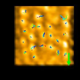
®
Kobayashi et al.
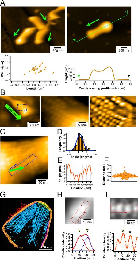
FIG 2 Chain imaging by HS-AFM. (A) (Left) Cluster of cells immobilized on a glass surface (upper) and
distribution of cell dimensions (n = 20) (lower). (Right) The height profile along the broken line (upper)
(Continued on next page)
May/June 2021 Volume 12 Issue 3 e00040-21 mbio.asm.org 4®
Gliding Mechanism of Mycoplasma mobile
scanning rate of 300 ms per frame in an area of 300 nm2. Interestingly, we found parti-
cle structures aligned mostly along the cell axis at the front side of the cells (Fig. 2B).
The particle structures appeared when the average tapping force exceeded ;40 pN
(piconewtons) (see Materials and Methods). They were aligned at an angle of approxi-
mately 4.6° relative to the cell axis (Fig. 2C and D, n = 99 chains from 20 cells). The parti-
cle height was approximately 2 nm (Fig. 2E), and the pitches were distributed as
31.5 6 4.9 nm (Fig. 2F, n = 98), in good agreement with a previous number, 31 nm,
measured by electron cryotomography (Fig. 2G) (5). To measure the dimensions of the
particles in detail, we collected 19 particle images and averaged them (Fig. 2H). The
averaged image showed an elliptical structure, 27.2 nm long and 14.2 nm wide, with
two height peaks. The distance between the two peaks of a particle was 10.0 nm.
These features were consistent with the results from electron cryotomography (Fig. 2I)
(5), showing that the particle structure observed in HS-AFM is identical to the internal
structure observed by electron cryotomography.
The internal structure of M. mobile is detected by HS-AFM from the surface. An
M. mobile cell has three huge proteins, Gli521, Gli349, and Gli123, on its surface (Fig. 1A,
left). To confirm that the particle structures visualized with HS-AFM are not the surface
structures, the cell surface was treated with proteinase K, a serine protease with broad
specificity, and scanned by HS-AFM. First, we confirmed that M. mobile cells gliding on
the glass surface were stopped 1 min after the addition of 0.2 mg/ml proteinase K
(Fig. S1A), suggesting that the surface proteins involved in the gliding machinery are
sensitive to proteinase K. Then, we observed the cell surface by HS-AFM after the immo-
bilized cells were treated with proteinase K for 20 min. The particle structures were
observed on the surface of the cell even after proteinase K treatment. The particle
pitches of cells with and without proteinase K treatment were 31.2 6 3.2 (n = 31) and
28.9 6 3.6 nm (n = 33), respectively (Fig. S1B), showing a significant difference between
them (P = 0.00651 by Student’s t test). Based on these observations, we concluded that
the particle structure detected by HS-AFM was inside the structure but influenced by the
surface treatment with proteinase K, consistent with a previous observation (12).
During the observation of intact cells immobilized on glass surfaces, we observed
the removal of the cell membrane by chance, resulting in the exposure of the inside
structure. The exposed inside structure showed features similar to the internal jellyfish-
like structure of M. mobile (5, 12) (Movie S2, Fig. S1C). We compared the features of par-
ticle structures before and after the removal of the cell membrane (Fig. S1D). After re-
moval, the height of the particle relative to the background increased, resulting in a
clearer appearance than before removal. The particle pitches were 30.3 6 4.1 and
31.8 6 7.3 nm before and after removal, respectively, without a statistically significant
difference (P = 0.277 by Student’s t test). The average heights of particles observed
before and after removal of the cell membrane were 257 and 18 nm, respectively, from
the lowest position of the image. The difference between them was 239 nm, compara-
FIG 2 Legend (Continued)
is plotted along the green arrow (lower). The cell axis and front are shown by an arrow. (B) Detailed
structure of a cell. (Left) Whole-cell image. The cell axis and front are indicated by a green arrow.
(Middle) Magnified image of the boxed area of the left panel. (Right) The middle panel image was
processed with a bandpass filter. (C to F) Image analyses of particles. (C) Cell image featuring a
representative chain structure. The cell axis and front are indicated by a green arrow. (D) Distribution
of chain angle relative to the cell axis fitted by a Gaussian curve (n = 99 chains from 20 cells). (E) Image
profile of the boxed area along the direction of blue arrow in panel C. (F) Scatter dot plot for distances
between peak positions of the chain profile. The average was 31.5 6 4.9 nm (n = 98). (G) Three-
dimensional rendered image for a 146-nm-thick slice of permeabilized cell reconstructed by electron
cryotomography, modified from a previous study (5). The surface filamentous structures, cell
membrane, undercoating at the front and side membranes, and internal chain are colored red, orange,
yellow, and purple, respectively. (H) Averaged image of 19 particle structures from HS-AFM (upper) and
image profile of boxed area (lower). The profile (orange squares) was fitted by the sum (purple solid
line) of two Gaussian curves (red and blue). Yellow triangles show peaks of the Gaussian curves. (I)
Averaged images of chain structure (blue part in panel G) from electron cryotomography (upper) (5)
and image profile of the boxed area along the chain axis (lower). Yellow triangles show peaks of
Gaussian curves. In all HS-AFM imaging, the surface was scanned left to right for line and lower to
upper for image.
May/June 2021 Volume 12 Issue 3 e00040-21 mbio.asm.org 5®
Kobayashi et al.
ble to the height of M. mobile cells (Fig. 2A and Fig. S1D). Therefore, the particles
detected before and after cell membrane removal were proposed to be the structure
beneath the upper cell membrane and the one on the lower cell membrane facing the
glass substrate, respectively. This occasional observation is likely related to the charac-
ter of M. mobile surface structure, that is a soft single-layered membrane (5). However,
we could not remove the cell membrane intentionally. Then, we focused on analyzing
the internal structure beneath the upper cell membrane.
Behavior of particle structure detected by HS-AFM. The surface protrusion of M.
mobile cells was scanned with a scanning rate of 200 or 330 ms per frame with a scan
area of 200 by 200 nm2. Projected images were processed using a bandpass filter to
improve the image contrast, by drift correction, and by averaging three sequential
images for better signal/noise ratio (Movie S4). In most cases, the particles were diffi-
cult to trace over time because of image discontinuity, even when particle images
were clear. This is probably due to the stability of the cell immobilized onto the glass
surface and damage to the scanning probe. However, we succeeded in tracing the
behaviors of individual particles in some videos and used them for further analyses.
Sodium azide suppressed particle movement. To discuss the behaviors of internal
particles, we needed to confirm that the particle movements are caused by ATP hydroly-
sis on the internal structure. In a previous study, the ATPase activity of the internal struc-
ture of M. mobile was inhibited by sodium azide (5). The binding activity and gliding
speed of “gliding heads,” the gliding machinery isolated from the cell protrusion, were
also inhibited by sodium azide (5). In the present study, we examined the effect of sodium
azide on the gliding speed of intact M. mobile cells. The averaged gliding speed of intact
M. mobile cells was decreased from 0.77 6 0.17 to 0.04 6 0.02 m m/s by the addition of
15.4 mM sodium azide (Fig. 3A and B), suggesting that sodium azide affected the ATPase
activity of the internal structure and the force generation for gliding.
We then scanned the cell surfaces by HS-AFM in the presence and absence of so-
dium azide (Movie S4 to S7). The tracking of the mass center every 200 ms (no azide)
or 330 ms (with azide) for 16.2 s showed that most particles were moving independ-
ently (Fig. 3C). These movements were significantly reduced by the addition of sodium
azide. We calculated the accumulated moving distances and estimated the speeds for
the particle movements from a linear fitting of the accumulated moving distance
(Fig. 3D and E). At concentrations of 0, 15.4, 76.5, and 765 mM sodium azide, the
speeds calculated from accumulated moving distances were 6.9 6 1.4, 3.9 6 1.4,
3.6 6 0.8, and 3.0 6 1.1 nm/s, respectively, suggesting that the movement of particle
structures is linked to ATP hydrolysis. Interestingly, in 15.4 mM sodium azide, the par-
ticles can be classified as either active or static, and the different types tend to form an
adjacent pair in chains (Fig. 3C).
Particle displacements traced as an image profile. Not all particles moved in the
same direction at the same time (Fig. 3C to E), and this feature was more obvious in
15.4 mM sodium azide (Movie S5, Fig. 4A), indicating that the movements were linked
to ATP hydrolysis, not caused by artificial drift in the measurements. The addition of so-
dium azide may allow easier detection of individual movements by reducing some of
the movements. Analysis of 27 particles in a 200 by 200 nm2 field in the presence of
15.4 mM sodium azide for 23.1 s showed that 19 particles moved distances longer than
6 nm, distinct from other movements. The frequency of such long movements in the
whole field was 1.17 events/s (Fig. 4A). Next, we focused on particle movements. Since
the particles appeared to move mainly perpendicular to the particle chain in the cell
surface plane, the height profile of a box perpendicular to the particle chain was traced
over time (Fig. 4B, upper graphs). Six particles did not move (static particle), while 15
active particles showed remarkable movements, and a returning path for some par-
ticles was observed. As shown in the “a” panels of Fig. 4B and C, the movements of the
particles showed a tendency moving 9.1 6 2.5 nm (n = 15) in the left direction perpen-
dicular to the chain axis and 2.3 6 3.0 nm (n = 8) on the cytoplasmic side in the Z direc-
tion. These behaviors can be traced three-dimensionally as shown for the representa-
tive particle movements (Fig. 4B, lowermost graphs). The profile continued to change
May/June 2021 Volume 12 Issue 3 e00040-21 mbio.asm.org 6® Gliding Mechanism of Mycoplasma mobile FIG 3 Effects of sodium azide on particle displacements. (A) Rainbow traces of gliding cells for 5 s with and without sodium azide from phase-contrast microscopy. Video frames were overlaid with different colors from red to blue. (B) Gliding speed under various concentrations of sodium azide. Speeds of 2.5 to 20 s were averaged for 140 to 223 cells. (C) HS-AFM images with continuous traces of individual particles for 13.2 s. HS-AFM images were processed by bandpass filter, drift correction, and sequential averaging. Particles were traced every 200 ms for no sodium azide, and 330 ms in the presence of sodium azide, as presented by the color change from red to blue. The cell axis and front are indicated by a green arrow. The surface was scanned left to right for line and lower to upper for imaging. Movies are shown as supplemental data as Movies S4, S5, S6, and S7 for imaging in 0, 15.4, 76.5, 765 mM sodium azide, respectively. (D) Time course of accumulated moving distances of individual particles under various concentrations of sodium azide. (E) Scatter dot plot of particle speed under various concentrations of sodium azide. Speeds were estimated from a linear fitting of accumulated moving distance. for approximately five frames of 330 ms. However, the movement was likely completed in a single 330-ms frame, because the image was profiled after averaging three consec- utive video images every 330 ms to reduce image noise. Eleven particles showed returning movements in the video, with speeds similar to those of their advancing movements, as shown in the panels marked “r” in Fig. 4B and C. In conclusion, the active particles moved to 9 nm left and 2 nm lower relative to the cell axis in 330 ms and came back to the original position in another 330 ms. May/June 2021 Volume 12 Issue 3 e00040-21 mbio.asm.org 7
®
Kobayashi et al.

FIG 4 Movements of individual particles. (A) Video frames of particle chains under 15.4 mM sodium azide (Movie S5).
The green arrow on the left shows the cell axis and front. The left three panels show consecutive video frames showing
                                                                                           (Continued on next page)

May/June 2021 Volume 12 Issue 3 e00040-21                                                                                mbio.asm.org   8
®
Gliding Mechanism of Mycoplasma mobile
Next, particle movements perpendicular to the cell axis were searched in the ab-
sence of sodium azide. Observation of 21 particles for 16.6 s showed that movements
longer than 6 nm appeared at a frequency of 2.17 events/s (Movie S4 and Fig. 4D). The
distance moved was 8.0 6 1.9 nm (n = 24) in the left direction perpendicular to the axis
of the chain alignment within 200 ms and 2.0 6 1.9 nm (n = 18) on the cytoplasmic side
in the Z direction (Fig. 4D).
Particle displacements traced as a positional distribution. To study the direction
of movements of the particles on the membrane surface statistically, the distributions
of the particles as the mass center were analyzed every 200 and 330 ms for observa-
tions in the absence and presence of sodium azide, respectively (Fig. 5A and Movies S5
to S7). In this analysis, we determined the particle positions for the x and y axes
(Fig. 5A), while only the x and z axes were shown in panels C and D of Fig. 4. Instead,
we did not trace the particle positions with time in Fig. 5. The faster scan speed for the
observation in the absence of sodium azide was applied, as we assumed that the par-
ticles moved faster under these conditions. However, this difference in the scanning
speed should not affect the conclusion, because no difference was found, even when
the analysis was performed using 400-ms intervals for the measurements without so-
dium azide (Fig. S2). Analysis showed that the distributions were larger in the presence
of 15.4 mM and smaller at 76.5 and 765 mM than in the absence of sodium azide
(Fig. 5A). Next, we measured the distributions of three distances (Fig. 5B) as follows:
the particle position to the chain axis (Fig. 5C), the distance to the adjacent particle
(Fig. 5D), and the distance to the adjacent particle projected to the chain axis (Fig. 5E).
These results are schematically summarized (Fig. 5B), suggesting that movements per-
pendicular to the chain axis of the particles (presented as distance “c” in Fig. 5) should
be present but not easy to detect in the absence of sodium azide; they were observed
more clearly when the frequency of movements was reduced by sodium azide, and
they were inhibited under high concentrations of sodium azide.
DISCUSSION
Internal structure was traced from the outside surface. The particle features
traced by HS-AFM in this study were consistent with those of the internal structure
reported in previous studies (Fig. 2) (5, 12), suggesting that HS-AFM visualized the in-
ternal structure. The large surface proteins Gli521, Gli349, and Gli123 exist on the cell
surface of M. mobile as components of the gliding machinery (7, 14, 17–20, 22, 24, 49).
A group of surface proteins, Mvsps, which are responsible for antigenic variations, also
exist on the cell surface (3, 50). These surface proteins may interfere with probing the
internal structure from the surface. However, the chain structures observed by HS-AFM
did not show obvious differences before and after protease treatment of the cells
(Fig. S1B). Furthermore, similar structures were observed before and after mechanical
removal of the cell membrane (Fig. S1C and D). These results showed that the particles
traced by HS-AFM were not on the surface structure, but were inside the cell. The sur-
face structure, composed of mainly large filamentous proteins, may be too thin and/or
mobile to be detected by the current scanning performance of HS-AFM on the cell
FIG 4 Legend (Continued)
remarkable particle movements. The particles with movements are marked before (triangles) and after (arrows) the
movements with coordinated colors. Particles moved to the left relative to the gliding direction. The rightmost panel
shows a raw image of the video frame showing areas profiled for active (red) and static (blue) particles shown in panel
B. (B) Consecutive image profile of representative active and static particles. (Upper six graphs) Image profiles of active
(red background) and static (blue background) particles every 330 ms for 1.98 s. (Lowermost graphs) Three-dimensional
positions of peaks of particles tracing from red to purple. Y positions are shown only in these graphs in Fig. 4. The
green arrow on the left shows the cell axis and front. (C) Consecutive image profiles showing particle movements every
330 ms for 1.98 s in 15.4 mM sodium azide. (D) Consecutive image profiles showing particle movements every 200 ms for
1.2 s without sodium azide (Movie S4). (B to D) Consecutive profiles of each frame from red to purple. Advancing (a) and
returning (r) movements are presented. Peak positions of focusing particles are marked by a triangle and an arrow,
respectively, for the initial and the end time points. Distances between peaks before and after movement were manually
measured for statistical analysis of particle movements. The profile of heights and positions is presented with a common
X- Y- scale in the lower panel for each data set.
May/June 2021 Volume 12 Issue 3 e00040-21 mbio.asm.org 9®
Kobayashi et al.
FIG 5 Analyses of particle distribution. (A) Distribution of particles in a chain. The particle positions and the axis of the particle positions are indicated by
red dots and gray dashed lines, respectively. The particle positions were detected every 200 and 330 ms, respectively, without and with sodium azide at 82,
66, 70, and 66 points under 0, 15.4, 76.5, and 765 mM sodium azide, respectively. The axis of particle positions was determined by a linear approximation
of the average position of each particle. (B) Schematic illustration of three distances with average and standard deviation (SD) values in nm. (C to E) The
particle position to the chain axis (C, purple), the distance to the adjacent particle (D, blue), and the distance to the adjacent particle projected to the
chain axis (E, green) are shown. Bar lengths are not to scale. Movies S4 to S7 were analyzed. The chain axis is indicated by a green arrow pointing mostly
to the cell front in panels A and B.
membrane (16–18, 22). The lack of a peptidoglycan layer should be advantageous for
visualizing the inside structure, due to the lack of stiffness (44, 45, 51). Moreover, the
internal structure should be sufficiently stiff and positioned beneath the cell mem-
brane, reminiscent of cortical actin in animal cells (42).
Effects of sodium azide. Sodium azide inhibits many ATPases by blocking ADP
release (52). In M. mobile gliding, the reagent inhibited cell gliding (Fig. 3A and B) and
the isolated gliding machinery (5). Particle behaviors became more visible in the pres-
ence of 15.4 mM sodium azide. Under this condition, cell gliding was reduced to 20
times slower than the original, suggesting that ATP hydrolysis occurred 20 times less
frequently. If the particles move in a rapid and independent manner, it may be difficult
to trace the movements of individual particles. However, if the reaction was partially
inhibited by 15.4 mM sodium azide, most particles may be in their home position,
while some particles move to another position. In this case, the movements could be
traced easily. This assumption is supported by the observation that the particle
May/June 2021 Volume 12 Issue 3 e00040-21 mbio.asm.org 10®
Gliding Mechanism of Mycoplasma mobile
FIG 6 Schematic illustration of particle movement in M. mobile visualized by HS-AFM. The internal
chain of the gliding machinery and cell membrane are indicated by blue objects and a beige plate,
respectively. Here, we focus on the particle chain lining the lower side of cell membrane, while we
scanned mostly the particle chain beneath the upper side of the cell membrane in this study. The left
and right panels show the particles before and after the advancing movement, respectively. The
central particle moves as an ATP- or ADP/Pi-bound form to the right and inner sides for a distance of
9 and 2 nm, respectively.
distances between neighboring particles are 1.7 to 2.5 nm shorter under high concen-
trations of sodium azide than those without the reagent (Fig. 5D). A previous study
based on electron microscopy showed that the particle distances in the ADP and
unbound forms were approximately 2 nm shorter than those in the AMPPNP, ADP-Vi,
and ADP-AlFx states (5). As sodium azide is thought to inhibit the release of ADP (52),
the changes in particle distance observed in the present study are consistent with the
results of electron microscopy (Fig. 5B) (5).
Particle behavior in the gliding mechanism. The particles moved approximately
9 nm to the right of the gliding direction and 2 nm to the cytoplasmic side within
330 ms (Fig. 6). This movement may be coupled with the transition from ADP or
unbound form to ATP or the ADP/Pi form (5). Considering the fact that the particles are
structurally linked to the surface structures of the gliding machinery (5), the move-
ments observed in the present study are likely involved in the gliding mechanism.
There are two possibilities for the relation between the particle movements and
gliding motility. As M. mobile gliding is caused probably by repetitive leg strokes (21,
29, 32), the particle movements may be coupled to the stroke. Previous studies have
reported that the step size of M. mobile is approximately 70 nm under no load and ad-
justable to various loads (21, 29, 32, 53). The moving distances of particles, approxi-
mately 10 nm is much shorter than the step size. However, this difference can be
explained because the surface structure contains two large proteins with dimensions
comparable to the step size; that is, the Gli349 “leg” that catches the scaffold and the
Gli521 “crank” that transmits force for gliding are 100 and 120 nm long, respectively
(Fig. 1A) (16, 17, 22). Therefore, the movements occurring in the internal structure can
be amplified through the huge protein molecules on the surface or through an
unknown structure that connects the internal and surface structures (Fig. 1A and 2G).
This assumption can explain the previous observation that the single leg exerts a force
of 1.5 pN, a few times smaller than motor proteins (21), assuming elastic components
are equipped in the large surface complex. Another possibility is that the movements
observed here are caused as a “reaction” of cooperating many particles in a large com-
plex, besides a direct transmission linking the particle and the surface structures. In
this scenario, the movements of the transmission were too small to be detected by HS-
AFM, for example rotation of rod proteins.
In a previous study, M. mobile gliding showed a leftward directional change of
about 8.5° with 1-m m cell progress (31). This gliding property may be related to the ob-
servation that the particle movements are pointed to the right relative to the gliding
direction (Fig. 6). Otherwise, the tilting of the chain axis about 4.6° from the cell axis
may cause a directional change in gliding (Fig. 2D).
To elucidate the mechanism of M. mobile gliding, we need to further visualize the
behaviors and structures of the machinery in detail, including those of both internal
and surface structures. The combination of electron microscopy and HS-AFM may pro-
vide better insights in the near future.
May/June 2021 Volume 12 Issue 3 e00040-21 mbio.asm.org 11®
Kobayashi et al.
MATERIALS AND METHODS
Cell preparation. A mutant strain (gli521[P476R]) of M. mobile 163K (ATCC 43663) activated for bind-
ing (21, 23, 54) was grown in Aluotto medium at 25 to 28°C, as previously described (8, 49). Cultured
cells were collected by centrifugation at 12,000 g for 4 min at 25 to 28°C and suspended in phosphate-
buffered saline with glucose (PBS/G) consisting of 75 mM sodium phosphate (pH 7.3), 68 mM NaCl, and
10 mM glucose (21, 26, 30, 31). This process was repeated twice, and finally the cells were resuspended
in PBS/G to a 20-fold density of the original culture.
Gliding analyses. A tunnel chamber assembled as previously described (3-mm interior width, 22-
mm length, 40-m m wall thickness) was treated with Aluotto medium for 15 min at 25 to 28°C (21, 30),
and then the medium was replaced by PBS/G. The cell suspension was inserted into the tunnel chamber
with video recording. PBS/G was replaced with PBS/G containing 0.2 mg/ml proteinase K (Qiagen N. V.,
Hilden, Germany) or various concentrations of sodium azide, as necessary.
Cell immobilization on the glass surface. A glass slide was treated with saturated KOH-ethanol so-
lution for 15 min and washed 10 times with water. For analyses with an imaging rate of 1,000 and
330 ms per frame, the glass was treated with 0.1% poly-L-lysine for 5 min. After the solution was
removed, the glass was washed with water and dried. Then, the glass was treated with 0.1% glutaralde-
hyde for 5 min, washed with water, and covered with PBS/G. For analyses with an imaging rate of
200 ms per frame, the glass was treated with sandpaper, saturated with KOH-ethanol solution for
15 min, washed 10 times with water, and then dried. The washed glass was treated with 1,000-fold
diluted 3-aminopropyldiethoxymethylsilane for 5 min at 25 to 28°C, washed, and treated with glutaral-
dehyde as described above. Finally, the cell suspension was placed onto the glass substrate and left for
10 min at 25 to 28°C.
Microscopy. To examine the immobilizing conditions using phase-contrast microscopy, the glass
slide was assembled into a tunnel chamber (19). The cell suspension was loaded into the tunnel, kept
for 10 min at 25 to 28°C, washed with PBS/G, and observed by phase-contrast microscopy IX71
(Olympus, Tokyo, Japan) (21, 27, 31). To analyze the immobilizing conditions, quick-freeze deep-etch
electron microscopy, fixation, and washing were performed on the coverslip. When the cells were frozen
without immobilization, we followed the procedure for the electron microscopy method described pre-
viously (44, 45). Briefly, the cells on the glass were pressed against a copper block cooled with liquid he-
lium and frozen. Then, the frozen sample was fractured and etched to expose it. Subsequently, the
exposed surface was shadowed with platinum to create a replica membrane, which was observed under
a JEM-1010 transmission electron microscope (JEOL, Tokyo, Japan) at 80 kV, equipped with a FastScan-
F214 (T) charge-coupled device (CCD) camera (TVIPS, Gauting, Germany).
Observation by HS-AFM. Imaging was performed with a laboratory-built HS-AFM in tapping mode
(55, 56). Small cantilevers (BLAC10DS-A2; Olympus) with a resonant frequency of ;0.5 MHz in water, a
quality factor (Qc) of ;1.5 in water, and a spring constant (kc) of ;0.08 N/m were used. The cantilever’s
free oscillation amplitude (A0) and set-point amplitude (Asp) were set at ;2.5 nm and ;0.8 A0, respec-
tively. Under these conditions, the average tapping force ,F. can be approximated as ;40 pN using
the following equation:
kc qffiffiffiffiffiffiffiffiffiffiffiffiffiffiffi
ffi
F¼ A20 2A2sp
2Qc
For searching cells, the sample was scanned at an imaging rate of 1,000 ms per frame in an area of
3,000 by 3,000 nm2 with 150 by 150 pixels. To observe the particle structure, the cell surface was scanned
with an imaging rate of 330 or 200 ms per frame in an area of 200 by 200 nm2 with 100 by 100 pixels.
Video analyses. To trace particles in the XY plane, videos were processed by three methods (Movies
S3 to S7). (i) The image contrast was improved by a bandpass filter. (ii) Image drifts were corrected by a
plugin, “align slices in stack” (57), equipped with ImageJ. (iii) Image noises were removed by averaging
three consecutive slices. Then, each particle image was cropped, binarized, and traced for the mass cen-
ter. Here, the threshold for binarization was determined independently for each particle of interest. The
cell axes in Fig. 2 were determined by fitting a cell image as an ellipse. All analyses were performed with
ImageJ version 1.52A. Image averaging of particles was performed using EMAN, version 2.3.
SUPPLEMENTAL MATERIAL
Supplemental material is available online only.
MOVIE S1, AVI file, 0.2 MB.
MOVIE S2, AVI file, 2.7 MB.
MOVIE S3, AVI file, 2.6 MB.
MOVIE S4, AVI file, 0.8 MB.
MOVIE S5, AVI file, 0.6 MB.
MOVIE S6, AVI file, 0.7 MB.
MOVIE S7, AVI file, 0.6 MB.
FIG S1, PDF file, 0.3 MB.
FIG S2, PDF file, 0.2 MB.
May/June 2021 Volume 12 Issue 3 e00040-21 mbio.asm.org 12®
Gliding Mechanism of Mycoplasma mobile
ACKNOWLEDGMENTS
We appreciate Yuya Sasajima at Osaka City University for helpful discussions.
This work was supported by Grants-in-Aid for Scientific Research (A) (MEXT KAKENHI,
grant number JP17H01544), JST CREST (grant number JPMJCR19S5), Osaka City
University (OCU) Strategic Research Grant 2018 for top priority research, and by a Grant-
in-Aid for the Fugaku Trust for Medicinal Research to M. Miyata.
REFERENCES
1. Miyata M, Robinson RC, Uyeda TQP, Fukumori Y, Fukushima SI, Haruta S, 18. Metsugi S, Uenoyama A, Adan-Kubo J, Miyata M, Yura K, Kono H, Go N.
Homma M, Inaba K, Ito M, Kaito C, Kato K, Kenri T, Kinosita Y, Kojima S, 2005. Sequence analysis of the gliding protein Gli349 in Mycoplasma mobile.
Minamino T, Mori H, Nakamura S, Nakane D, Nakayama K, Nishiyama M, Biophysics (Nagoya-Shi) 1:33–43. https://doi.org/10.2142/biophysics.1.33.
Shibata S, Shimabukuro K, Tamakoshi M, Taoka A, Tashiro Y, Tulum I, 19. Uenoyama A, Kusumoto A, Miyata M. 2004. Identification of a 349-kilodal-
Wada H, Wakabayashi KI. 2020. Tree of motility: a proposed history of mo- ton protein (Gli349) responsible for cytadherence and glass binding dur-
tility systems in the tree of life. Genes Cells 25:6–21. https://doi.org/10 ing gliding of Mycoplasma mobile. J Bacteriol 186:1537–1545. https://doi
.1111/gtc.12737. .org/10.1128/jb.186.5.1537-1545.2004.
2. Nakamura S, Minamino T. 2019. Flagella-driven motility of bacteria. Bio- 20. Kusumoto A, Seto S, Jaffe JD, Miyata M. 2004. Cell surface differentiation
molecules 9:279. https://doi.org/10.3390/biom9070279. of Mycoplasma mobile visualized by surface protein localization. Microbi-
3. Adan-Kubo J, Yoshii SH, Kono H, Miyata M. 2012. Molecular structure of iso- ology (Reading) 150:4001–4008. https://doi.org/10.1099/mic.0.27436-0.
lated MvspI, a variable surface protein of the fish pathogen Mycoplasma mo- 21. Mizutani M, Tulum I, Kinosita Y, Nishizaka T, Miyata M. 2018. Detailed
bile. J Bacteriol 194:3050–3057. https://doi.org/10.1128/JB.00208-12. analyses of stall force generation in Mycoplasma mobile gliding. Biophys J
4. Kawamoto A, Matsuo L, Kato T, Yamamoto H, Namba K, Miyata M. 2016. Pe- 114:1411–1419. https://doi.org/10.1016/j.bpj.2018.01.029.
riodicity in attachment organelle revealed by electron cryotomography sug- 22. Nonaka T, Adan-Kubo J, Miyata M. 2010. Triskelion structure of the Gli521
gests conformational changes in gliding mechanism of Mycoplasma pneu- protein, involved in the gliding mechanism of Mycoplasma mobile. J Bac-
moniae. mBio 7:e00243-16–e00216. https://doi.org/10.1128/mBio.00243-16. teriol 192:636–642. https://doi.org/10.1128/JB.01143-09.
5. Nishikawa M, Nakane D, Toyonaga T, Kawamoto A, Kato T, Namba K, 23. Uenoyama A, Seto S, Nakane D, Miyata M. 2009. Regions on Gli349 and
Miyata M. 2019. Refined mechanism of Mycoplasma mobile gliding based Gli521 protein molecules directly involved in movements of Mycoplasma
on structure, ATPase activity, and sialic acid binding of machinery. mBio mobile gliding machinery, suggested by use of inhibitory antibodies and
10:e02846-19. https://doi.org/10.1128/mBio.02846-19. mutants. J Bacteriol 191:1982–1985. https://doi.org/10.1128/JB.01012-08.
6. Rottem S. 1980. Membrane lipids of mycoplasmas. Biochim Biophys Acta 24. Seto S, Uenoyama A, Miyata M. 2005. Identification of a 521-kilodalton
604:65–90. https://doi.org/10.1016/0304-4157(80)90004-0. protein (Gli521) involved in force generation or force transmission for
7. Wu HN, Miyata M. 2012. Whole surface image of Mycoplasma mobile, sug- Mycoplasma mobile gliding. J Bacteriol 187:3502–3510. https://doi.org/10
gested by protein identification and immunofluorescence microscopy. J .1128/JB.187.10.3502-3510.2005.
Bacteriol 194:5848–5855. https://doi.org/10.1128/JB.00976-12. 25. Chen J, Neu J, Miyata M, Oster G. 2009. Motor-substrate interactions in
8. Tulum I, Kimura K, Miyata M. 2020. Identification and sequence analyses Mycoplasma motility explains non-Arrhenius temperature dependence.
of the gliding machinery proteins from Mycoplasma mobile. Sci Rep Biophys J 97:2930–2938. https://doi.org/10.1016/j.bpj.2009.09.020.
10:3792. https://doi.org/10.1038/s41598-020-60535-z. 26. Kasai T, Hamaguchi T, Miyata M. 2015. Gliding motility of Mycoplasma mo-
9. Hamaguchi T, Kawakami M, Furukawa H, Miyata M. 2019. Identification of bile on uniform oligosaccharides. J Bacteriol 197:2952–2957. https://doi
novel protein domain for sialyloligosaccharide binding essential to Myco- .org/10.1128/JB.00335-15.
plasma mobile gliding. FEMS Microbiol Lett 366:fnz016. https://doi.org/10 27. Kasai T, Nakane D, Ishida H, Ando H, Kiso M, Miyata M. 2013. Role of bind-
.1093/femsle/fnz016. ing in Mycoplasma mobile and Mycoplasma pneumoniae gliding analyzed
10. Miyata M, Hamaguchi T. 2016. Prospects for the gliding mechanism of through inhibition by synthesized sialylated compounds. J Bacteriol
Mycoplasma mobile. Curr Opin Microbiol 29:15–21. https://doi.org/10 195:429–435. https://doi.org/10.1128/JB.01141-12.
.1016/j.mib.2015.08.010. 28. Nagai R, Miyata M. 2006. Gliding motility of Mycoplasma mobile can
11. Tulum I, Yabe M, Uenoyama A, Miyata M. 2014. Localization of P42 and occur by repeated binding to N-acetylneuraminyllactose (sialyllactose)
F1-ATPase alpha-subunit homolog of the gliding machinery in Myco- fixed on solid surfaces. J Bacteriol 188:6469–6475. https://doi.org/10
plasma mobile revealed by newly developed gene manipulation and .1128/JB.00754-06.
fluorescent protein tagging. J Bacteriol 196:1815–1824. https://doi.org/ 29. Kinosita Y, Miyata M, Nishizaka T. 2018. Linear motor driven-rotary motion
10.1128/JB.01418-13. of a membrane-permeabilized ghost in Mycoplasma mobile. Sci Rep
12. Nakane D, Miyata M. 2007. Cytoskeletal “jellyfish” structure of Myco- 8:11513. https://doi.org/10.1038/s41598-018-29875-9.
plasma mobile. Proc Natl Acad Sci U S A 104:19518–19523. https://doi 30. Tanaka A, Nakane D, Mizutani M, Nishizaka T, Miyata M. 2016. Directed
.org/10.1073/pnas.0704280104. binding of gliding bacterium, Mycoplasma mobile, shown by detachment
13. Miyata M. 2010. Unique centipede mechanism of Mycoplasma gliding. force and bond lifetime. mBio 7:e00455-16. https://doi.org/10.1128/mBio
Annu Rev Microbiol 64:519–537. https://doi.org/10.1146/annurev.micro .00455-16.
.112408.134116. 31. Morio H, Kasai T, Miyata M. 2016. Gliding direction of Mycoplasma mobile.
14. Uenoyama A, Miyata M. 2005. Identification of a 123-kilodalton protein J Bacteriol 198:283–290. https://doi.org/10.1128/JB.00499-15.
(Gli123) involved in machinery for gliding motility of Mycoplasma mobile. J 32. Kinosita Y, Nakane D, Sugawa M, Masaike T, Mizutani K, Miyata M,
Bacteriol 187:5578–5584. https://doi.org/10.1128/JB.187.16.5578-5584.2005. Nishizaka T. 2014. Unitary step of gliding machinery in Mycoplasma mo-
15. Béven L, Charenton C, Dautant A, Bouyssou G, Labroussaa F, Skollermo A, bile. Proc Natl Acad Sci U S A 111:8601–8606. https://doi.org/10.1073/
Persson A, Blanchard A, Sirand-Pugnet P. 2012. Specific evolution of F1- pnas.1310355111.
like ATPases in mycoplasmas. PLoS One 7:e38793. https://doi.org/10 33. Binnig G, Quate CF, Gerber C. 1986. Atomic force microscope. Phys Rev
.1371/journal.pone.0038793. Lett 56:930–933. https://doi.org/10.1103/PhysRevLett.56.930.
16. Lesoil C, Nonaka T, Sekiguchi H, Osada T, Miyata M, Afrin R, Ikai A. 2010. 34. Muller DJ, Dufrene YF. 2008. Atomic force microscopy as a multifunctional
Molecular shape and binding force of Mycoplasma mobile's leg protein molecular toolbox in nanobiotechnology. Nat Nanotechnol 3:261–269.
Gli349 revealed by an AFM study. Biochem Biophys Res Commun doi:https://pubmed.ncbi.nlm.nih.gov/18654521/.
391:1312–1317. https://doi.org/10.1016/j.bbrc.2009.12.023. 35. Dufrene YF, Viljoen A, Mignolet J, Mathelie-Guinlet M. 2021. AFM in cellu-
17. Adan-Kubo J, Uenoyama A, Arata T, Miyata M. 2006. Morphology of iso- lar and molecular microbiology. Cell Microbiol 12:e13324. https://doi.org/
lated Gli349, a leg protein responsible for Mycoplasma mobile gliding via 10.1111/cmi.13324:e13324.
glass binding, revealed by rotary shadowing electron microscopy. J Bac- 36. Pasquina-Lemonche L, Burns J, Turner RD, Kumar S, Tank R, Mullin N,
teriol 188:2821–2828. https://doi.org/10.1128/JB.188.8.2821-2828.2006. Wilson JS, Chakrabarti B, Bullough PA, Foster SJ, Hobbs JK. 2020. The
May/June 2021 Volume 12 Issue 3 e00040-21 mbio.asm.org 13®
Kobayashi et al.
architecture of the Gram-positive bacterial cell wall. Nature 582:294–297. rheology on the nanoscale. Soft Matter 11:4584–4591. https://doi.org/10
https://doi.org/10.1038/s41586-020-2236-6. .1039/c4sm02718c.
37. Ando T. 2019. High-speed atomic force microscopy. Curr Opin Chem Biol 48. Mathelie-Guinlet M, Asmar AT, Collet JF, Dufrene YF. 2020. Lipoprotein
51:105–112. https://doi.org/10.1016/j.cbpa.2019.05.010. Lpp regulates the mechanical properties of the E. coli cell envelope. Nat
38. Heath GR, Scheuring S. 2019. Advances in high-speed atomic force mi- Commun 11:1789. https://doi.org/10.1038/s41467-020-15489-1.
croscopy (HS-AFM) reveal dynamics of transmembrane channels and 49. Miyata M, Yamamoto H, Shimizu T, Uenoyama A, Citti C, Rosengarten R.
transporters. Curr Opin Struct Biol 57:93–102. https://doi.org/10.1016/j 2000. Gliding mutants of Mycoplasma mobile: relationships between motil-
.sbi.2019.02.008. ity and cell morphology, cell adhesion and microcolony formation. Microbi-
39. Lyubchenko YL, Shlyakhtenko LS. 2016. Imaging of DNA and protein-DNA ology 146:1311–1320. https://doi.org/10.1099/00221287-146-6-1311.
complexes with Atomic Force Microscopy. Crit Rev Eukaryot Gene Expr 50. Wu HN, Kawaguchi C, Nakane D, Miyata M. 2012. “Mycoplasmal antigen
26:63–96. https://doi.org/10.1615/CritRevEukaryotGeneExpr.v26.i1.70. modulation,” a novel surface variation suggested for a lipoprotein specifi-
40. Rajendran A, Endo M, Sugiyama H. 2012. Structural and functional analy- cally localized on Mycoplasma mobile. Curr Microbiol 64:433–440. https://
sis of proteins by high-speed atomic force microscopy. Adv Protein Chem doi.org/10.1007/s00284-012-0090-y.
Struct Biol 87:5–55. https://doi.org/10.1016/B978-0-12-398312-1.00002-0. 51. Nakane D, Miyata M. 2012. Mycoplasma mobile cells elongated by deter-
41. Yamashita H, Taoka A, Uchihashi T, Asano T, Ando T, Fukumori Y. 2012. gent and their pivoting movements in gliding. J Bacteriol 194:122–130.
Single-molecule imaging on living bacterial cell surface by high-speed
https://doi.org/10.1128/JB.05857-11.
AFM. J Mol Biol 422:300–309. https://doi.org/10.1016/j.jmb.2012.05.018.
52. Bowler MW, Montgomery MG, Leslie AG, Walker JE. 2006. How azide
42. Zhang Y, Yoshida A, Sakai N, Uekusa Y, Kumeta M, Yoshimura SH. 2017. In
inhibits ATP hydrolysis by the F-ATPases. Proc Natl Acad Sci U S A
vivo dynamics of the cortical actin network revealed by fast-scanning
103:8646–8649. https://doi.org/10.1073/pnas.0602915103.
atomic force microscopy. Microscopy (Oxf) 66:272–282. https://doi.org/10
53. Miyata M, Ryu WS, Berg HC. 2002. Force and velocity of Mycoplasma mo-
.1093/jmicro/dfx015.
bile gliding. J Bacteriol 184:1827–1831. https://doi.org/10.1128/jb.184.7
43. Jaffe JD, Miyata M, Berg HC. 2004. Energetics of gliding motility in Myco-
.1827-1831.2002.
plasma mobile. J Bacteriol 186:4254–4261. https://doi.org/10.1128/JB.186
54. Uenoyama A, Miyata M. 2005. Gliding ghosts of Mycoplasma mobile.
.13.4254-4261.2004.
44. Tulum I, Tahara Y, Miyata M. 2019. Peptidoglycan layer and disruption Proc Natl Acad Sci U S A 102:12754–12758. https://doi.org/10.1073/
processes in Bacillus subtilis cells visualized using quick-freeze, deep-etch pnas.0506114102.
electron microscopy. Microscopy (Oxf) 68:441–449. https://doi.org/10 55. Uchihashi T, Kodera N, Ando T. 2012. Guide to video recording of struc-
.1093/jmicro/dfz033. ture dynamics and dynamic processes of proteins by high-speed atomic
45. Miyata M, Petersen JD. 2004. Spike structure at the interface between force microscopy. Nat Protoc 7:1193–1206. https://doi.org/10.1038/nprot
gliding Mycoplasma mobile cells and glass surfaces visualized by rapid- .2012.047.
freeze-and-fracture electron microscopy. J Bacteriol 186:4382–4386. 56. Ando T, Kodera N, Takai E, Maruyama D, Saito K, Toda A. 2001. A high-
https://doi.org/10.1128/JB.186.13.4382-4386.2004. speed atomic force microscope for studying biological macromolecules.
46. Lin DC, Dimitriadis EK, Horkay F. 2007. Robust strategies for automated Proc Natl Acad Sci U S A 98:12468–12472. https://doi.org/10.1073/pnas
AFM force curve analysis–I. Non-adhesive indentation of soft, inhomoge- .211400898.
neous materials. J Biomech Eng 129:430–440. https://doi.org/10.1115/1 57. Tseng Q, Duchemin-Pelletier E, Deshiere A, Balland M, Guillou H, Filhol O,
.2720924. Thery M. 2012. Spatial organization of the extracellular matrix regulates
47. Hecht FM, Rheinlaender J, Schierbaum N, Goldmann WH, Fabry B, Schaffer cell-cell junction positioning. Proc Natl Acad Sci U S A 109:1506–1511.
TE. 2015. Imaging viscoelastic properties of live cells by AFM: power-law https://doi.org/10.1073/pnas.1106377109.
May/June 2021 Volume 12 Issue 3 e00040-21 mbio.asm.org 14You can also read