Influence of embedding media on the accuracy of working length determination by means of apex locator: an ex vivo study
←
→
Page content transcription
If your browser does not render page correctly, please read the page content below
www.nature.com/scientificreports
OPEN Influence of embedding media
on the accuracy of working length
determination by means of apex
locator: an ex vivo study
Thomas Gerhard Wolf1,2*, Anna Krauß‑Mironjuk2, Richard Johannes Wierichs1 &
Benjamín Briseño‑Marroquín1,2
The aim of this research was to determine ex vivo the influence on accuracy of five different
embedding media, for investigative and educational purposes, and one electronic apex locator. 110
human extracted mature roots of permanent single-rooted human teeth were used. The roots were
embedded in alginate, stick sponge, 2% agar–agar and 6% and 12% gelatin. The actual working length
to the physiological foramen was determined under a stereo-microscope (16 ×) and the electronic
working lengths with the Elements Diagnostic Unit and a K-file ISO 10. The accuracy ranges of the
accumulated measurements, when allowing a ± 0.5 mm tolerance, went from 98.2% (6% and 12%
gelatin), 93.7% (alginate), 92.8% (2% agar–agar) to 91.7% (sponge). The exact measurements at
the physiological foramen ranged from 80.0% (6% gelatin), 76.5% (2% agar–agar), 71.8% (12%
gelatin), 68.2% (alginate) to 64.5% (sponge). Although relatively seldom (n = 24), measurements
with deviations of more than ± 0.5 mm were also observed; thus, the accuracy of the working length
determination results per se can be considered as clinically acceptable. The results of this research
allow a recommendation of the investigated embedding media for electronic working length
determination models for educational and research purposes in endodontics.
Assessment of the working length can be considered to be an imperative procedure during root canal preparation
procedure and its accurate determination to be of the outmost importance for successful endodontic treatment1,2.
The working length can be defined as the distance between the most coronal or incisal edge or cusp tip and an
apical reference point given by the physiological f oramen3. If the working length is underestimated, tissue residues
and/or bacteria will remain in the non-instrumented areas of the root canal system. On the other hand, if the
working length is determined beyond the apical boundaries, vital and/or infected material will be transported
into the periapical tissues. An erroneously determined working length will most probably compromise the out-
come of an endodontic treatment, as it will lead to shaping and filling procedures that are inaccurate. This can lead
to periapical tissue inflammation and/or infection1,2. All possible measures should be undertaken to constrain
mechanical procedures as well as chemicals and possible toxins from irritating materials within the root canal
system, but not beyond the physiological foramen limits in order to minimize the risk of bacterial contamination
and/or mechanical or chemical irritation of the peri-radicular tissues due to irrigating solutions, filling materi-
als and over-instrumentation4. These precautions will enhance the success rate of an endodontic t reatment4.
It has been reported5,6, in contrast with a report from Switzerland 25 years a go7, that in the last decade, a
majority of operators surveyed determine the working length by means of an apex locator. However, although
it has been reported in several in vitro studies8–13 that a solely electronic working length determination under
different clinical conditions leads to clinically acceptable results, the actual guidelines of professional endodontic
societies14,15 suggest that the working length should be determined electronically and subsequently substantiated
by means of an X-ray image. Despite this fact, different research groups report5,6 that only approximately 50% of
the surveyed operators routinely combine the electronic and radiological working length determination methods.
This electronic and radiological combination method rationale is based in the remaining possible limitations that
electronic apex locators still h ave8. In an ex vivo investigation, no statistical differences were reported between
1
Department of Restorative, Preventive and Pediatric Dentistry, School of Dental Medicine, University of Bern,
Freiburgstrasse 7, 3010 Bern, Switzerland. 2Department of Periodontology and Operative Dentistry, University
Medical Center of the Johannes-Gutenberg-University Mainz, Mainz, Germany. *email: thomas.wolf@
zmk.unibe.ch
Scientific Reports | (2021) 11:3340 | https://doi.org/10.1038/s41598-021-82942-6 1
Vol.:(0123456789)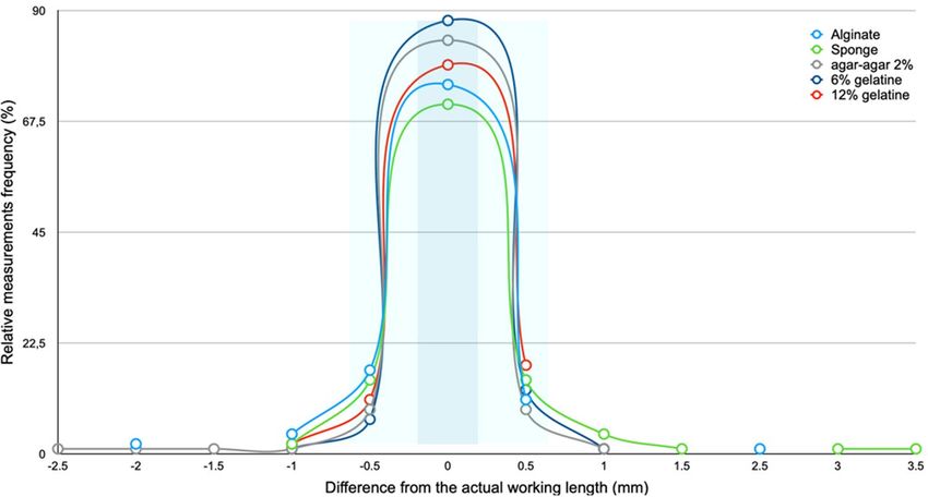
www.nature.com/scientificreports/
the radiologic working length determination (included as gold standard) and an electronic device16; however, it
would be clinically advisable to keep in mind that the working length determination by means of a radiograph
alone could lead to overestimation and unintentional over-enlargement of the physiological foramen17. If the
ALARA principle (“as low [radiation] as reasonably achievable”) is routinely taken into consideration, a combined
clinical strategic combination employment of an electronic device and radiograph during the working length
determination will enhance the working length determination accuracy and, concomitantly, patient radiation
exposure reduction16,18.
Regardless of the working length determination method employed, an accurate preparation boundary is of
great significance to ensure endodontic success4. The accuracy of electronic apex locators has been evaluated with
in vivo or in vitro research methods, whereas the precision of the working length determination depends on the
device and/or type of irrigation employed rather than the pulp tissue s tatus19. Moreover, most ex vivo or in vitro
studies8–10,12,13,16,20–25 usually compare the accuracy of specific devices under different clinical conditions with
different embedding media. Especially for the implementation of electronic devices to determine the working
length in a university teaching scenario25–27, the question of which embedding media is most suitable not only
for teaching purposes but also for research arises and, according to the actual specific literature, this matter is
not completely elucidated. Thus, the aim of this study was to compare five different embedding media with an
ex vivo research model and to establish if all the investigated embedding media provide similar accuracy results
when determining the working length—a result which would consequently enhance educational and investigative
confidence. The null hypothesis stated that the electronic working lengths measured with the embedding media
investigated would result within a tolerance range of ± 0.5 mm. To reject this hypothesis, an ex vivo study was
designed and carried out to assess if one or more of the embedding materials investigated would not be suitable
conductive media for apex locator working length determination.
Materials and methods
A total of 110 single-rooted human permanent teeth with mature apices were collected from an oral surgery
department of a German university dental school for reasons (usually for periodontal, endodontic, orthodontic
and traumatic reasons) unrelated to this investigation and included in this study. This research material can be
considered as so-called excess material, and hence fulfills the legal regulations of the University Medical Center of
the Johannes Gutenberg University of Mainz, Germany (Contract General Terms [AVB], §14 Organ explantation/
further use of body material, Status: 1. April 2017) and may be used for medical research without any additional
approval of the local ethics committee. This regulation is supported and approved by the ethics committee of
the Medical State Association of Rhineland-Palatinate, Germany, for scientific purposes. Informed tooth extrac-
tion and further investigative purposes with the excess material consent was obtained from each individual. All
methods were performed in accordance with the guidelines and regulations and experimental protocols at the
University Medical Center of the Johannes Gutenberg University of Mainz, Germany.
Selection criteria were: complete root development, no signs of root fracture or resorption, no radicular
or coronal caries, no partially or completely obliterated root canals and no previous endodontic treatment. In
order to dissolve any superficial remaining tissue, the teeth were stored in a 1% sodium hypochlorite (Apotheke
der Universitätsmedizin Mainz, Mainz, Germany) solution for 14 days. The teeth surfaces were thoroughly
cleansed from tissue and calculus residue with an ultrasonic device (Piezon 150; EMS, Nyon, Switzerland). The
teeth crowns were then separated at the enamel-cement interface with a 2-N feed force and a grain-size D64
diamond-coated cutting belt (EXAKT 300 CL; Exakt Advanced Technologies, Norderstedt, Germany) transverse
to the tooth longitudinal axis; thus, defining a leveled and reproducible reference landmark. The length between
this landmark and the corresponding physiological foramen was defined as the actual working length. Root
canal patency was confirmed with a K-file 06 (Flexofile; Dentsply, Ballaigues, Switzerland) and the teeth fixed in
Nalgene tubes (Nalgene; Rochester, NY, USA) with plaster enabling a direct contact between the root(s) and the
embedding media. After root canal patency verification, no further irrigating or preparation procedures were
undertaken, thus the electronic measurements were made under a relative low humidity in the root canals. The
root canal entries were blocked with wax plates (Pinnacle Modellierwachs Standard; Dentsply De Trey, Konstanz,
Germany) to prevent plaster from flowing into the root canals. The tubes were numbered consecutively.
Five embedding media were investigated: alginate (Blueprint; Dentsply De Trey, Konstanz, Germany), stick
sponge (Steckschaum; Blume 2000, Norderstedt, Germany), 2% agar–agar (Becton Dickinson, Sparks, MD,
USA), bovine skin 6% gelatin and 12% gelatin (Bovine gelatin; SIGMA, Steinheim, Germany). A 20 ml syringe
(ECOJECT; Dispomed Witt, Gelnhausen, Germany) was then filled with the alginate, using a cement spatula.
The Nalgene tubes were then injected with moderate pressure and a vibrator, taking care to prevent air inclu-
sions and allowing a homogenous alginate filling of the tubes. Directly afterwards, the tube lids with the fixed
roots were screwed on tightly, allowing maximum surface contact between the roots and the embedding media.
The alginate was allowed to set for 2.5 min at 23 °C. A 0.5 cm length and 0.5 cm diameter stick sponge was cut
off from a sponge stick, inserted into the Nalgene tubes and trimmed according to the respective tube length.
The sponge was then moistened using a 10 ml syringe (ECOJECT; Dispomed Witt, Gelnhausen, Germany) and
0.9% saline solution (Bacto Agar; Becton Dickinson, Sparks, MD, USA) until the entire sponge was soaked. The
agar–agar solution was prepared from 2 g agar–agar powder (Bacto Agar; Becton Dickinson, Sparks, MD, USA),
0.9 g sodium chloride, 0.095 g disodium hydrogen phosphate dehydrate, 0.018 g potassium dihydrogen phos-
phate (Optipur; Merck, Darmstadt, Germany) and 100 ml distilled water (Aqua B. Braun; B. Braun Melsungen,
Melsungen, Germany). The mixture was heated to 150 °C under constant stirring and boiled until a homogene-
ous agar–agar suspension was formed. The Nalgene tubes were completely filled with the agar–agar solution by
means of a disposable 10 ml pipette (Eppendorf Research Plus 10–100 µl; Eppendorf, Hamburg, Germany), the
lids with the fixed roots were screwed on tightly and the solution, of gel-like consistency, was allowed to cool.
Scientific Reports | (2021) 11:3340 | https://doi.org/10.1038/s41598-021-82942-6 2
Vol:.(1234567890)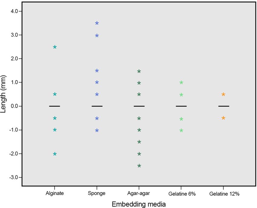
www.nature.com/scientificreports/
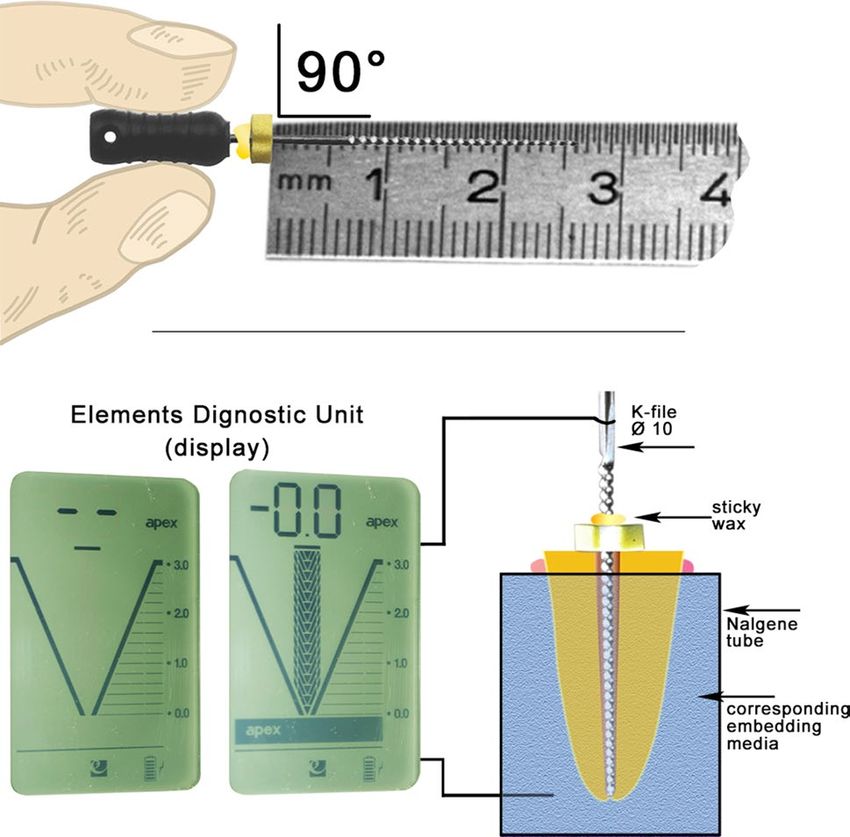
Figure 1. Diagrammatic visualization of the experimental setup of the electronic working length measurements
(below) and precautions taken during the transmission of the corresponding measurements made (above). The
Elements Diagnostic Unit apex locator measurement was determined to be correct as the display scale reached
the “0.0” level, the “apex” sign below it appeared and a corresponding acoustic signal was heard.
The 6% and 12% gelatin solutions were prepared with 6 and 12 g gelatin, respectively, and a 100 ml 0.9% saline
solution. The solutions were heated at a slowly increasing temperature rate up to 150 °C for 15 min and stirred
continuously; they were subsequently allowed to cool and solidify.
All electronic measurements were carried out with the Elements Diagnostic Unit apex locator (Kerr, Brea,
CA, USA) immediately after the corresponding preparation procedures were completed and within a time period
of at most 15 min; otherwise, a new embedding media would have to be prepared. In accordance with the unit
operating instructions, the measurements were carried out when the unit battery was sufficiently charged and
after checking proper functioning of all cables and plug connections. A series of ex vivo measurements with
teeth not included in this investigation were made under magnification (16 × ; Stemi DRC; Carl Zeiss Jena, Jena,
Germany) until the working length with the Elements Diagnostic Unit apex locator at the physiological foramen
was determined to be accurate. The electronic measurements were determined to be correct as the device display
scale reached the “0.0” level, the “apex” sign below it appeared and a corresponding acoustic signal was heard
(Fig. 1). The loop-shaped electrode, which is usually placed in the corner of the patient’s mouth and establishes
contact with the cheek, was replaced by a 1.7 mm Ø, 30 mm length stainless-steel wire (Stainless steel hard
rods—316–1.70 mm/0.0393 inch; Sadevinox; Seynod, France) during the research procedures. The wire fitted
tightly into the device connection socket and into a circular perforation made on the corresponding Nalgene
tube. This perforation was made in the lower third of each Nalgene tube with a red round diamond bur (016;
Premium Diamantschleifer; Busch & CO., Engelskirchen, Germany). The stainless-steel wire was glued (Supergel
Sekundenkleber; UHU, Bühl, Germany) to the Nalgene tube in order to firmly affix the wire and to prevent the
embedding media from flowing out through the perforation (Fig. 1).
All electronic measurements were carried out with K-files ISO 10 (Flexofile; Dentsply, Ballaigues, Switzer-
land), whereby the file was gently advanced towards the root tip until the apex locator showed a stable reading
at the physiological foramen for five seconds. After having reached the working length, the silicone stopper was
placed flat on the coronal root reference landmark, fixed on the K-file (Sticky Wax; Kerr, Brea, CA, USA) and
the K-file was removed from the root canal carefully to ensure that the stopper position was not modified. Sub-
sequently, the measured working length was determined with a 15 cm stainless-steel ruler (Rumold, Stuttgart,
Germany) with half millimeter marks (± 0.2 mm) by an experienced and calibrated single operator (Fig. 1).
Care was taken to ensure that the root surfaces were clean from any embedding media prior to any new work-
ing length measurement. After the working lengths were determined with the five different embedding media,
the actual working lengths were determined as the tip of the measuring instrument reached the physiological
foramina under direct view with a stereo microscope (16 ×) by one previously calibrated operator (A.M.) with an
experience of over 100 physiological foramina localizations and according to a previously reported physiological
foramina morphological d escription3.
The actual working length was established as a reference measurement for the purpose of comparison and to
investigate the accuracy of the results obtained with the different embedding media. The working length of all
root canals was determined with each embedding media (n = 550); thus, a total of 660 measurements (includ-
ing the actual working length measurements) were made. The statistical evaluation was carried out with SPSS
Scientific Reports | (2021) 11:3340 | https://doi.org/10.1038/s41598-021-82942-6 3
Vol.:(0123456789)www.nature.com/scientificreports/
2% agar–
Actual Alginate Sponge agar 6% gelatin 12% gelatin
WL n % n % n % n % n % n %
9.5 1 0.9
10.0 1 0.9 2 1.8 1 0.9 3 2.7 2 1.8
10.5 6 5.5 12 10.9 7 6.4 3 2.7 6 5.5 6 5.5
11.0 6 5.5 6 5.5 10 9.1 9 8.1 9 8.2 9 8.2
11.5 13 11.8 7 6.4 10 9.1 14 12.7 10 9.1 8 7.3
12.0 9 8.2 9 8.2 5 4.5 5 4.5 7 6.4 10 9.1
12.5 10 9.1 9 8.2 13 11.8 9 8.2 10 9.1 8 7.3
13.0 14 12.7 13 11.8 12 10.9 15 13.6 17 15.5 14 12.7
13.5 16 14.5 15 13.6 12 10.9 14 12.7 15 13.6 15 13.6
14.0 11 10.0 14 12.7 12 10.9 11 10.0 13 11.8 14 12.7
14.5 10 9.1 9 8.2 9 8.2 11 10.0 10 9.1 10 9.1
15.0 6 5.5 6 5.5 6 5.5 6 5.5 5 4.5 5 4.5
15.5 4 3.6 5 4.5 6 5.5 4 3.6 4 3.6 6 5.5
16.0 1 0.9 1 0.9 1 0.9 2 1.8 1 0.9
16.5 2 1.8 1 0.9 4 3.6 2 1.8 2 1.8 1 0.9
17.0 1 0.9 1 0.9 2 1.8 1 0.9 1 0.9 2 1.8
Total 110 100.0 110 100.0 110 100.0 110 100.0 110 100.0 110 100.0
Table 1. Actual working length and different embedding media working lengths determination (mm)
distributed according to the corresponding root canal working lengths measured (WL). Significant higher
differences between the results obtained with alginate and the actual working lengths are noticeable. However,
it should be taken into consideration that the clinically acceptable tolerance of ± 0.5 mm accepted in this
investigation, is not being taken into account in this table (n = 110 per research group).
n Min Max Mean MED 95% CI SD
Alginate 110 10.0 17.0 12.98 13.0 12.68–13.28 1.59
Sponge 110 10.0 17.0 13.15 13.0 12.83–13.47 1.68
2% agar–agar 110 9.5 17.0 13.05 13.0 12.74–13.35 1.60
6% gelatin 110 10.5 17.0 13.09 13.0 12.81–13.37 1.48
12% gelatin 110 10.0 17.0 13.07 13.0 12.78–13.37 1.55
Table 2. Statistical evaluation of the working length determination with the Elements Diagnostic Unit apex
locator and five different embedding media (mm; MED = median, 95% CI = 95% confidence interval of the
difference; SD = standard deviation; n = 110 per research group.
15 Statistics Software (IBM; Armonk, NY, USA) and the Institute for Medical Biometry, Epidemiology and
Informatics (IMBEI) at the University Medical Center facilities in Mainz, Germany. The results of the different
embedding media were compared with the reference measurements of the actual working length obtained under
the microscope. A measurement difference between the electronic and actual working lengths of ± 0.5 mm was
defined as clinically acceptable. The absolute and relative frequencies of the results, measurements within the
acceptable and non-acceptable tolerance ranges and with a significant difference from the reference measure-
ments were calculated for each embedding media and graphically displayed using histograms and box plots. The
Wilcoxon test for paired samples (α = 0.050) was bilaterally calculated and therefore considered as exploratory.
Results
The working length results of 110 root canals measured with the Elements Diagnostic Unit apex locator and five
different embedding media (alginate, sponge, 2% agar–agar, 6% and 12% gelatin) can be considered as normally
distributed; thus, the results are described with mean values and confidence intervals. The absolute and relative
results of the actual working length and different embedding media are given in Table 1. Only one significant
difference between the actual and alginate root canal working lengths, when not taking into consideration
the ± 0.5 mm clinical tolerance, could be observed (p = 0.035). The results obtained with all embedding media
delivered homogeneous results. The descriptive statistic of the results showed that the mean values of all meas-
urements ranged between 12.98 mm (alginate) and 13.15 mm (sponge) with the exception of 2% agar–agar and
6% gelatin, where a minimum of 9.5 mm and 10.5 mm, respectively, was measured, whereas all other embedding
media showed a minimum of 10.0 mm. A maximum of 17.0 mm was obtained with all embedding media. The
standard deviation ranged from 1.48 for 6% gelatin and 1.68 for sponge (Table 2).
Scientific Reports | (2021) 11:3340 | https://doi.org/10.1038/s41598-021-82942-6 4
Vol:.(1234567890)www.nature.com/scientificreports/
Figure 2. Diagram depicting the relative frequency (%) differences of the working length measurements
obtained with the different embedding media and the actual working length of the root canals investigated.
A ± 0.5 mm difference was considered in this investigation as clinically acceptable (the exact and clinically
acceptable measurements are highlighted with dark and light blue, respectively; n = 110 per research group).
Only few shorter and longer measurements within the non-clinically acceptable working length were
observed. Overall, 10 measurements 1.0 mm shorter than the working length (4 = alginate, 2 = sponge and 12%
gelatin and 1 = 2% agar–agar and 6% gelatin) and five measurements 1.5 mm to 2.5 mm shorter than the working
length were observed (2 = alginate and 3 2% agar–agar). Six measurements 1 mm longer than the working length
(4 = sponge and 1 = 2% agar–agar and 6% gelatin) and five measurements 1.5 mm to 3.5 mm shorter than the
working length were observed (1 = alginate and 4 sponge). The high accuracy of measurements obtained at the
physiological foramen and within the ± 0.5 mm clinical tolerance can be observed in Fig. 2. The results range and
individual boxes show overall a relative uniformity of the working length measurements made with the different
embedding media. It could be observed that the largest measurement at 17.0 mm and the median position at
13.0 mm are very similar for all embedding media (Fig. 3). The differences between the electronically determined
working lengths in the respective embedding media and the actual working lengths are shown in Fig. 4.
Discussion
The working length results of 110 root canals at the physiological foramen level determined by using the Elements
Diagnostic Unit apex locator and alginate, sponge, 2% agar–agar, 6% and 12% gelatin used as embedding media
do not allow to reject the null hypothesis. Moreover, in accordance with several reports22,28 a clinically acceptable
tolerance range for working length determination accuracy at ± 0.5 mm was also allowed in this investigation.
Whereas a relatively high accuracy of working length determination with the radiographic method has been
reported with an ex vivo research model16, it should be taken into account that, within this type of research set-
up, the bone structures are not depicted, which could clinically represent a burdensome clinical situation when
detecting meticulous endodontic areas, particularly for operators with little experience. Thus, it seems reasonable
to permit a working length determination accuracy tolerance range due to the fact that an exact m orphological3
or radiological29–31 determination of the physiological foramen is not possible in daily practice. When considering
the clinical implication of these two parameters (working length determination method and clinical tolerance),
a tolerance allowance can also be supported based on a radiological i nvestigation1 in which the authors report
a 95% success rate for endodontic treatments.
The morphological landmarks of the apical region illustrate a particular terminology problem in scientific
research. Theoretically, the terminus of a root canal is where the pulp tissue or dentin root canal comes apically
to an end. This landmark has been recommended as the terminus of the root canal shaping and obturation
procedures4,32. Consequently, this landmark is where the working length should be determined by means of either
a radiograph or an electronic device or a combination of both m ethods14,15,33. Typically, the root canal narrows
consistently from coronal expanding apically to form the physiological foramen (apical constriction)3,34. The
major (apical) foramen is considered to be located at the root surface, whereas the physiological foramen (minor
foramen; apical constriction) is considered to be the narrowest (minor) diameter of the root canal located at the
cementodentinal junction approximately 0.5 to 1 mm away from the radiological apex34. However, it has been
reported that an apical constriction was observed in less than 50% of the teeth i nvestigated35 and that the cement
root canal area has not only tapered walls but also parallel w alls36. Furthermore, it has also been reported that
the physiological foramen cannot always be clearly ex vivo delimited under magnification (40 ×)3; yet, it can be
delimited when being investigated under micro-CT37.
In morphological and working length investigations, the physiological foramen has been termed as “api-
cal constriction”17,19,21,23,27,30–32,38–41 “anatomic apex”42,43, “1 mm from the anatomical foramen”44, “minor
Scientific Reports | (2021) 11:3340 | https://doi.org/10.1038/s41598-021-82942-6 5
Vol.:(0123456789)www.nature.com/scientificreports/
Figure 3. 100% box-and-whisker plot of the working length determination measurement results with the
different embedding media investigated (mm; n = 110 per research group).
Figure 4. Box plot showing the differences distribution between the actual working length (0.0) and the ones
obtained with the different embedding media. The lines at “0.0” represent the 25th quartile, median and 75th
quartile. Consequently, a characteristic box cannot be seen in the present figure. A slight tendency of too long
and too short measurements for sponge and agar–agar, respectively, can be observed. For alginate and gelatin in
both concentrations the scattering is similar (mm; n = 110 per research group).
foramen”38–40,45, “flush with the apex”46, “histological foramen”34, among others. The term physiological fora-
men has been proposed to define this morphological l andmark3 since this landmark is invariably located at the
junction between the pulpal connective and interstitial loose connective tissues of the periodontal ligament
independently if an apical constriction is assuredly present or if its shape is conical or parallel. The accuracy
range of electronic apex locators has been reported to be c ontrasting21,38–40. This wide accuracy range might
also be explained due to the apical morphological landmarks terminology consistency lack within the reported
investigations. In this investigation the positions of the physiologic foramina were determined through ex vivo
observations under magnification (16 ×) made by one operator and a corresponding “0.0” reading of the elec-
tronic device employed. Although in the pertaining Elements Diagnostic Unit apex locator user’s m anual47 the
Scientific Reports | (2021) 11:3340 | https://doi.org/10.1038/s41598-021-82942-6 6
Vol:.(1234567890)www.nature.com/scientificreports/
physiological foramen is termed as “apical foramen”, the highly accurate measurements results obtained in this
investigation strongly suggest that the term apical foramen corresponds with physiological foramen.
This ex vivo investigation examined the accuracy influence of different embedding media on endodontic
working length determination and an electronic device (Elements Diagnostic Unit) routinely used in clinical
practice. In order to reproduce a clinical situation as closely as possible, an embedding media with electrical
conductivity similar to that of the human periodontal tissue is indispensable in ex vivo investigations. A 2%
phosphate-buffered agar–agar solution to mimic the periodontium conditions was p roposed48. The advantage
of this medium is that an electrical resistance of 6.5 kΩ with a 7.3 pH corresponds with the values of human
periodontal tissue. Other reports10 have used the same agar–agar solution in their experiments, or with a 1%
concentration49. The use of a 0.9% isotonic saline solution41 and gelatin50 as embedding media for ex vivo elec-
tronic working length determination experiments has also been also reported. Two different gelatin concentra-
tions were included in this research in an effort to prove if possible different fluidities could have an impact on
the electronic working length measurements, specifically in an educational scenario where the time required to
make a measurement could be inherently extensive. Measurements using sponges or gels soaked in isotonic saline
solution have been suggested as a further alternative method51. The working length accuracy results obtained in
this research range between 91.7% (sponge) and 98.2% (gelatin 6% and 12%) and are similar to the ones reported
by other investigation groups with research on different embedding media9,21,49. The use of sponges soaked in
isotonic saline solution and gelatin has also been i nvestigated52 and, similar to the results of this investigation,
no statistically significant differences were reported. Several studies8,9,16,21,23,27,53 have compared the accuracy
of different apex locators in vitro and alginate as embedding media and report accuracies ranging from 31 to
100%. However, a direct comparison of these results is cumbersome, mainly as a consequence of the different
individual parameters investigated. In a systematic review and literature meta-analysis, the a uthors19 conclude
that the precision of electrical length measurement thus depends on both the device and the type of irrigation.
Different research g roups53–55 have dealt with the influence of different embedding media on the measurement
accuracy of electronic apex locators. Successive measurements in 1% agar–agar, alginate, gelatin and 0.9% saline
and flower sponge as embedding media and the Root ZX apex locator were investigated. These a uthors54 report
that flower sponge was the only embedding media in which the working length was determined beyond the api-
cal limit in 20% of cases; however, no statistically significant differences between the embedding materials were
observed. In the present study, the longest measurements, which were up to 3.5 mm beyond the actual working
length and thus clearly far beyond the clinically acceptable tolerance, were also obtained with sponge sticks. A
possible explanation for these long measurements could be that sponge is easily deformable and therefore an
unstable contact between the root apical region and embedding media is a given.
Alginate could be considered as a widespread embedding media in ex vivo endodontic apex locator
research16,22,23,53–55. In contrast with this investigation, in which the only statistically significant difference
working length accuracy measurement was determined for alginate (6 = shorter and 1 = longer), in a different
investigation54 it has been reported that the most accurate measurements obtained were with alginate; however,
this was when the file tip was at most 1 mm away from the “apical foramen”. The significant differences obtained
in this investigation could be explained by arguing that the after-setting time of alginate could influence the
contact between sample and embedding media. Although it could be postulated that the lower accuracy of the
alginate media has no clinical significance, the possibility that a defective contact between the embedding media
and sample could happen should be taken into consideration within an educational scenario in which time
plays a decisive role. A different research g roup55, using a Root ZX II device, report that alginate has a higher
electronic root canal length determination accuracy when compared with saline, and floral foam and gauze
both soaked in 0.9% saline. In another similar investigation, the a uthors53 using the Raypex 5 and Dentaport
ZX, report that alginate at a 0.5 mm and 1.0 mm tolerance showed the highest accuracy when compared with
sugar-free gelatin and a 0.9% sodium hypochlorite solution; however, the authors do not report any statistically
significant differences. This is in contrast to this investigation in which the results showed exact measurements
in 64.5% = sponge, 68.2% = alginate, 71.8% = 12% gelatin, 76.4% = 2% agar–agar and 80% = 6% gelatin. In addition
when a clinically acceptable tolerance (± 0.5 mm) was assumed, the range of correct measurements obtained rose
to 91.7% = sponge, 92.8% = 2% agar–agar, 93.7% = alginate and 98.2% = 6% and 12% gelatin. These differences
could be explained through the employment of different root canal irrigation research parameters and through
the different clinical accuracy tolerances allowed in both investigations.
25 years ago, electronic apex locators were seldom routinely e mployed7 in daily practice, most probably
due a lack of trust in the electronic devices on the side of the operator and to high interference susceptibility in
older generation devices. However, nowadays the use of electronic apex locators in daily practice has markedly
increased5,6 since actual working length determination devices have proven to be highly reliable in moist root
canals9,19,22, in root resorption13, root fracture11 and during endodontic re-treatment cases12. Yet, the possibility of
an incorrect measurement with an electronic working length determination device, even if these occur relatively
seldom, should always be kept in mind. Therefore, a combined electronic and radiographic working length deter-
mination is to be preferred. The implementation of cone-beam computed tomography (CBCT) to determine the
working length has been also d iscussed56; however, at least for the time being, CBCT cannot be recommended
as a primary working length determination method. Under the presumption that a combined, educated clinical
deployment strategy of an electronic device and invasive methods during the working length determination
will enhance the working length determination accuracy, the ALARA principle (“as low [radiation] as reason-
ably achievable”) should always be taken into consideration16,18. The accuracy of different electronic devices has
been extensively discussed. In fact, nowadays the use of an electronic apex locator and a radiograph is actually
recommended by different professional endodontic s ocieties14,15,33 as a routine clinical procedure to determine
working length. The authors completely agree with this clinical guideline. Further discussion of this clinical
aspect is beyond the scope of this research, namely the accuracy of different embedding media in endodontic
Scientific Reports | (2021) 11:3340 | https://doi.org/10.1038/s41598-021-82942-6 7
Vol.:(0123456789)www.nature.com/scientificreports/
working length determination for investigative and educational purposes. Although the results obtained in this
research cannot be considered to refute the null hypothesis and the accuracy of alginate as an embedding media
proved to be high, it should be kept in mind that this type of embedding media could be negatively influenced
by the material manipulation time, especially within an educational scenario.
Conclusions
According to the results obtained and within the limitations of this study, it can be concluded that all investigated
embedding media are suitable for ex vivo endodontic investigative and educational purposes.
Received: 17 June 2020; Accepted: 27 January 2021
References
1. Sjögren, U., Hagglund, B., Sundqvist, G. & Wing, K. Factors affecting the long-term results of endodontic treatment. J. Endod. 16,
498–504. https://doi.org/10.1016/S0099-2399(07)80180-4 (1990).
2. Farzaneh, M., Abitbol, S., Lawrence, H. P. & Friedman, S. Treatment outcome in endodontics-the Toronto Study. Phase II: Initial
treatment. J. Endod. 30, 302–309. https://doi.org/10.1097/00004770-200405000-00002 (2004).
3. Briseño Marroquín, B., El-Sayed, M. A. A. & Willershausen-Zönnchen, B. Morphology of the physiological foramen: I. Maxillary
and mandibular molars. J. Endod. 30, 321–328. https://doi.org/10.1097/00004770-200405000-00005 (2004).
4. Ricucci, D. Apical limit of root canal instrumentation and obturation, Part 1. Literature review. Int. Endod. J. 31, 384–393. https
://doi.org/10.1046/j.1365-2591.1998.00184.x (1998).
5. Savani, G. M., Sabbah, W., Sedgley, C. M. & Whitten, B. Current trends in endodontic treatment by general dental practitioners:
Report of a United States national survey. J. Endod. 40, 618–624. https://doi.org/10.1016/j.joen.2014.01.029 (2014).
6. Zaugg, B., Stassinakis, A. & Hotz, P. Influence of magnification tools on the recognition of simulated preparation and filling errors.
Schweiz. Monatsschr. Zahnmed. 114, 890–896 (2004).
7. Barbakow, F., Lutz, F. & Tóth, L. Root canal treatment in Switzerland. The quantitative aspects—A determination of its status.
Schweiz. Monatsschr. Zahnmed. 105, 1412–1417 (1995).
8. Briseño Marroquín, B., Frajlich, S., Goldberg, F. & Willershausen, B. Influence of instrument size on the accuracy of different apex
locators: An in vitro study. J. Endod. 34, 698–702. https://doi.org/10.1016/j.joen.2008.02.019 (2008).
9. Cunha D’Assunção, F. L., Santana de Albuquerque, D. & Correia de Queiroz Ferreira, L. The ability of two apex locators to locate
the apical foramen: An in vitro study. J. Endod. 32, 560–562. https://doi.org/10.1016/j.joen.2005.11.011 (2006).
10. Carneiro, E. et al. Accuracy of root length determination using Tri Auto ZX and ProTaper instruments: An in vitro study. J. Endod.
32, 142–144. https://doi.org/10.1016/j.joen.2005.10.030 (2006).
11. Goldberg, F., Frajlich, S., Kuttler, S., Manzur, E. & Briseño Marroquín, B. The evaluation of four electronic apex locators in teeth
with simulated horizontal oblique root fractures. J. Endod. 34, 1497–1499. https://doi.org/10.1016/j.joen.2008.09.002 (2008).
12. Goldberg, F., Briseño Marroquín, B., Frajlich, S. & Dreyer, C. In vitro evaluation of the ability of three apex locators to determine
the working length during retreatment. J. Endod. 31, 676–678. https://doi.org/10.1097/01.don.0000155226.03483.ff (2005).
13. Goldberg, F., De Silvio, A. C., Manfré, S. & Nastri, N. In vitro measurement accuracy of an electronic apex locator in teeth with
simulated apical root resorption. J. Endod. 28, 461–463. https://doi.org/10.1097/00004770-200206000-00011 (2002).
14. Gluskin, A. H. et al. The standard of practice in contemporary endodontics. Endodontics. Colleagues for Excellence, 2014 1–11.
Reprint at: https://www.aae.org/specialty/wp-content/uploads/sites/2/2017/06/ecfefall2014standardofpractice.pdf.
15. European Society of Endodontology. Quality guidelines for endodontic treatment: Consensus report of the European Society of
Endodontology. Int. Endod. J. 39, 921–930. https://doi.org/10.1111/j.1365-2591.2006.01180.x (2006).
16. Wolgin, M., Grundmann, M. J., Tchorz, J. P., Frank, W. & Kielbassa, A. M. ex vivo investigation on the postoperative integrity of
the apical constriction after the sole use of electronic working length determination. J. Dent. 64, 52–57. https://doi.org/10.1016/j.
jdent.2017.06.005 (2017).
17. Vieyra, J. P. & Acosta, J. Comparison of working length determination with radiographs and four electronic apex locators. Int.
Endod. J. 44, 510–518. https://doi.org/10.1111/j.1365-2591.2011.01855.x (2011).
18. Council of the European Union. Council Directive 2013/59/Euratom of 5 December 2013 laying down basic safety standards for
protection against the dangers arising from exposure to ionising radiation, and repealing Directives 89/618/Euratom, 90/641/
Euratom, 96/29/Euratom, 97/43/Euratom and 2003/122/.Euratom. Official Journal of the European Union L 13–1–L 13–73 (2014).
Reprint at https://eur-lex.europa.eu/legal-content/EN/TXT/PDF/?uri=CELEX:32013L0059&from=EN.
19. Tsesis, I. et al. The precision of electronic apex locators in working length determination: A systematic review and meta-analysis
of the literature. J. Endod. 41, 1818–1823. https://doi.org/10.1016/j.joen.2015.08.012 (2015).
20. ElAyouti, A., Weiger, R. & Löst, C. The ability of Root ZX apex locator to reduce the frequency of overestimated radiographic
working length. J. Endod. 28, 116–119. https://doi.org/10.1097/00004770-200202000-00017 (2002).
21. Plotino, G., Grande, N. M., Brigante, L., Lesti, B. & Somma, F. Ex vivo accuracy of three electronic apex locators: Root ZX, Ele-
ments Diagnostic Unit and Apex Locator and ProPex. Int. Endod. J. 39, 408–414. https://doi.org/10.1111/j.1365-2591.2006.01095
.x (2006).
22. Cunha D’Assunção, F. L., Santana de Albuquerque, D., Salazar-Silva, J. R., Correia de Queiroz Ferreira, L. & Medeiros Bezerra, P.
The accuracy of root canal measurements using the Mini Apex Locator and Root ZX-II: An evaluation in vitro. Oral Surg. Oral
Med. Oral Pathol. Oral Radiol. Endod. 104, e50–e53. https://doi.org/10.1016/j.tripleo.2007.03.025 (2007).
23. Bernardes, R. A. et al. Evaluation of precision of length determination with 3 electronic apex locators: Root ZX, Elements Diag-
nostic Unit and Apex Locator, and RomiAPEX D-30. Oral Surg. Oral Med. Oral Pathol. Oral Radiol. Endod. 104, 91–94. https://
doi.org/10.1016/j.tripleo.2007.05.016 (2007).
24. Briseño Marroquín, B., Fernández, C., Schmidtmann, I., Willershausen, B. & Goldberg, F. Accuracy of electronic apex locators to
detect root canal perforations with inserted metallic posts: An ex vivo study. Head Face Med. 10, 57. https://doi.org/10.1186/s1300
5-014-0057-2 (2014).
25. Tchorz, J. P., Hellwig, E. & Altenburger, M. J. An improved model for teaching use of electronic apex locators. Int. Endod. J. 45,
307–310. https://doi.org/10.1111/j.1365-2591.2011.01975.x (2012).
26. Tchorz, J. P., Hellwig, E. & Altenburger, M. J. Teaching model for artificial teeth and endodontic apex locators. J. Dent. Educ. 77,
626–629. https://doi.org/10.1002/j.0022-0337.2013.77.5.tb05512.x (2013).
27. Wolgin, M., Wiedemann, P., Frank, W., Wrbas, K.-T. & Kielbassa, A. M. Development and evaluation of an endodontic simulation
model for dental students. J. Dent. Educ. 79, 1363–1372. https://doi.org/10.1016/j.jdent.2017.06.005 (2015).
28. Ricard, O., Roux, D., Bourdeau, L. & Woda, A. Clinical evaluation of the accuracy of the Evident RCM Mark II Apex Locator. J.
Endod. 17, 567–569. https://doi.org/10.1016/S0099-2399(06)81723-1 (1991).
29. Chunn, C. B., Zardiackas, L. D. & Menke, R. A. In vivo root canal length determination using the Forameter. J. Endod. 7, 515–520.
https://doi.org/10.1016/S0099-2399(81)80114-8 (1981).
Scientific Reports | (2021) 11:3340 | https://doi.org/10.1038/s41598-021-82942-6 8
Vol:.(1234567890)www.nature.com/scientificreports/
30. Hassanien, E. E., Hashem, A. & Chalfin, H. Histomorphometric study of the root apex of mandibular premolar teeth: An attempt
to correlate working length measured with electronic and radiograph methods to various anatomic positions in the apical portion
of the canal. J. Endod. 34, 408–412. https://doi.org/10.1016/j.joen.2007.12.013 (2008).
31. Pratten, D. H. & McDonald, N. J. Comparison of radiographic and electronic working lengths. J. Endod. 22, 173–176. https://doi.
org/10.1016/S0099-2399(96)80095-1 (1996).
32. Ricucci, D. & Langeland, K. Apical limit of root canal instrumentation and obturation, part 2. A histological study. Int. Endod. J.
31, 394–409. https://doi.org/10.1046/j.1365-2591.1998.00183.x (1998).
33. Swiss Society for Endodontology. Quality guidelines of the Swiss Society of Endodontology. Swiss Dent. J. 124, 1143–1152 (2014).
34. Kuttler, Y. Microscopic investigation of root apexes. J. Am. Dent. Assoc. 50, 544–552. https://doi.org/10.14219/jada.archi
ve.1955.0099 (1955).
35. Wu, M. K., Wesselink, P. R. & Walton, R. E. Apical terminus location of root canal treatment procedures. Oral. Surg. Oral. Med.
Oral. Pathol. Oral. Radiol. Endod. 89, 99–103. https://doi.org/10.1016/s1079-2104(00)80023-2 (2000).
36. Dummer, P. M., McGinn, J. H. & Rees, D. G. The position and topography of the apical canal constriction and apical foramen. Int.
Endod. J. 17, 192–198. https://doi.org/10.1111/j.1365-2591.1984.tb00404.x (1984).
37. Wolf, T. G., Paqué, F., Patyna, M. S., Willershausen, B. & BriseñoMarroquín, B. Three-dimensional analysis of the physiological
foramen geometry of maxillary and mandibular molars by means of micro-CT. Int. J. Oral. Sci. 9, 151–157. https: //doi.org/10.1038/
ijos.2017.29 (2017).
38. Hoer, D. & Attin, T. The accuracy of electronic working length determination. Int. Endod. J. 37, 125–131. https://doi.org/10.111
1/j.0143-2885.2004.00764.x (2004).
39. Hör, D., Krusy, S. & Attin, T. Ex vivo comparison of two electronic apex locators with different scales and frequencies. Int. Endod.
J. 38, 855–859. https://doi.org/10.1111/j.1365-2591.2005.01004.x (2005).
40. Wrbas, K.-T., Ziegler, A. A., Altenburger, M. J. & Schirrmeister, J. F. In vivo comparison of working length determination with two
electronic apex locators. Int. Endod. J. 40, 133–138. https://doi.org/10.1111/j.1365-2591.2006.01199.x (2007).
41. Ushiyama, J. New principle and method for measuring the root canal length. J. Endod. 9, 97–104. https://doi.org/10.1016/S0099
-2399(83)80105-8 (1983).
42. Khandewal, D., Ballal, N. V. & Saraswathi, M. V. Comparative evaluation of accuracy of 2 electronic Apex locators with conventional
radiography: An ex vivo sudy. J. Endod. 41, 201–204. https://doi.org/10.1016/j.joen.2014.10.011 (2015).
43. Keleş, A. & Keskin, C. Deviations of mesial root canals of mandibular first molar teeth at the apical third: A micro-computed
tomographic study. J. Endod. 44, 1030–1032. https://doi.org/10.1016/j.joen.2018.02.028 (2018).
44. Luiten, D. J., Morgan, L. A., Baugartner, J. C. & Marshall, J. G. A comparison of four instrumentation techniques on apical canal
transportation. J. Endod. 21, 26–32. https://doi.org/10.1016/s0099-2399(06)80553-4 (1995).
45. Ivica, A., Deari, S., Patcas, R., Weber, F. E. & Zehnder, M. Transforming growth factor beta 1 distribution and content in the root
dentin of young mature and immature human premolars. J. Endod. 46, 641–647. https: //doi.org/10.1016/j.joen.2020.01.016 (2020).
46. Lally, T., Geist, J. R., Yu, Q., Himel, V. T. & Sabey, K. Evaluation of 4 commercial viewing devices for radiographic perceptibility
and working length determination. J. Endod. 41, 1120–1124. https://doi.org/10.1016/j.joen.2015.02.027 (2015).
47. SybronEndo. Elements Diagnostic Unit. Instruction Guidelines 1–7 (SybronEndo, Glendora, CA, 2020).
48. Aurelio, J. A., Nahmias, Y. & Gerstein, H. A model for demonstrating an electronic canal length measuring device. J. Endod. 9,
568–569. https://doi.org/10.1016/S0099-2399(83)80063-6 (1983).
49. Ebrahim, A. K., Wadachi, R. & Suda, H. Ex vivo evaluation of the ability of four different electronic apex locators to determine the
working length in teeth with various foramen diameters. Aust. Dent. J. 51, 258–262. https://doi.org/10.1111/j.1834-7819.2006.
tb00439.x (2006).
50. Donnelly, J. C. A simplified model to demonstrate the operation of electronic root canal measuring devices. J. Endod. 19, 579–580.
https://doi.org/10.1016/S0099-2399(06)81292-6 (1993).
51. Ounsi, H. F. & Naaman, A. In vitro evaluation of the reliability of the Root ZX electronic apex locator. Int. Endod. J. 32, 120–123.
https://doi.org/10.1046/j.1365-2591.1999.00202.x (1999).
52. De Moor, R. J., Hommez, G. M., Martens, L. C. & De Boever, J. G. Accuracy of four electronic apex locators: An in vitro evaluation.
Endod. Dent. Traumatol. 15, 77–82. https://doi.org/10.1111/j.1600-9657.1999.tb00758.x (1999).
53. Chen, E. et al. An ex vivo comparison of electronic apex locator teaching models. J. Endod. 37, 1147–1151. https: //doi.org/10.1016/j.
joen.2011.03.032 (2011).
54. Baldi, J. V. et al. Influence of embedding media on the assessment of electronic apex locators. J. Endod. 33, 476–479. https://doi.
org/10.1016/j.joen.2006.12.024 (2007).
55. Guerreiro-Tanomaru, J. M., Croti, H. R., Silva, F., Faria, G. & Tanomaru-Filho, M. Tooth embedding medium influences the
accuracy of electronic apex locator. Acta Odontol. Latinoam. 25, 214–217 (2012).
56. Wolf, T. G., Fischer, F. & Schulze, R. K. W. Correlation of objective image quality and working length measurements in different
CBCT machines: An ex vivo study. Sci. Rep. 10, 19414. https://doi.org/10.1038/s41598-020-76424-4 (2020).
Acknowledgements
The authors deny any conflicts of interest related to this study. We appreciate the kind support of the Institute of
Medical Biometry, Epidemiology and Informatics (IMBEI) of the University of Mainz (Germany) and Dr. Kathy
Saranpa (University of Eastern Finland) for her kind support in the preparation of the manuscript.
Author contributions
T.G.W. and B.B.M. designed and planned the study. A.K.M. was in charge of the experimental and R.J.W. of the
statistical conduction of the investigation. The manuscript was prepared by T.G.W. and B.B.M. All authors read
and approved the final manuscript.
Competing interests
The authors declare no competing interests.
Additional information
Correspondence and requests for materials should be addressed to T.G.W.
Reprints and permissions information is available at www.nature.com/reprints.
Publisher’s note Springer Nature remains neutral with regard to jurisdictional claims in published maps and
institutional affiliations.
Scientific Reports | (2021) 11:3340 | https://doi.org/10.1038/s41598-021-82942-6 9
Vol.:(0123456789)www.nature.com/scientificreports/
Open Access This article is licensed under a Creative Commons Attribution 4.0 International
License, which permits use, sharing, adaptation, distribution and reproduction in any medium or
format, as long as you give appropriate credit to the original author(s) and the source, provide a link to the
Creative Commons licence, and indicate if changes were made. The images or other third party material in this
article are included in the article’s Creative Commons licence, unless indicated otherwise in a credit line to the
material. If material is not included in the article’s Creative Commons licence and your intended use is not
permitted by statutory regulation or exceeds the permitted use, you will need to obtain permission directly from
the copyright holder. To view a copy of this licence, visit http://creativecommons.org/licenses/by/4.0/.
© The Author(s) 2021
Scientific Reports | (2021) 11:3340 | https://doi.org/10.1038/s41598-021-82942-6 10
Vol:.(1234567890)You can also read