Progress in Neuropsychopharmacology & Biological Psychiatry
←
→
Page content transcription
If your browser does not render page correctly, please read the page content below
Progress in Neuropsychopharmacology & Biological Psychiatry 104 (2021) 110028

                                                                       Contents lists available at ScienceDirect

                                                    Progress in Neuropsychopharmacology
                                                           & Biological Psychiatry
                                                            journal homepage: www.elsevier.com/locate/pnp

Psychiatric autoimmune conditions in children and adolescents: Is catatonia                                                                                                  T
a severity marker?
                                    ⁎
Vladimir Ferrafiata,b, , Elise Riquinc, Elena Frerid, Tiziana Granatad, Nardo Nardoccid,
François Medjkanee, Claire Corfiottie, Alessandra Tozzod, Huges Pellerina, Xavier Benarousa,
Julien Harochef, Zahir Amouraf, Philippe Duvergerc, Renaud Jardrie, Priscille Gerardinb,
David Cohena,g, Angèle Consolia,h, Marie Raffina,h
a
  Department of Child and Adolescent Psychiatry, Sorbonne Université, Hôpital Pitié-Salpêtrière, AP-HP, 47-83 Boulevard de l’Hôpital, 75013 Paris, France
b
  Department of Child and Adolescent Psychiatry, Université de Rouen, Hôpital Charles Nicolle, 1 rue de Germont, 76000 Rouen, France
c
  Department of Child and Adolescent Psychiatry, Hôpital Universitaire d’Angers, 4 Rue Larrey, 49100 Angers, France
d
  Department of Pediatric Neuroscience, Fondazione IRCCS Istituto Neurologico Carlo Besta, Via Celoria 11, 20133 Milan, Italy
e
  Department of Child and Adolescent Psychiatry, Université Lille Nord de France, CHRU de Lille, F-59037 Lille Cedex, France
f
  French National Reference Center for Rare Systemic AutoImmune Disorders, E3M Institute, Université Pierre et Marie Curie, Hôpital Pitié-Salpêtrière, AP-HP, 47-83
Boulevard de l’Hôpital, 75013 Paris, France
g
  CNRS UMR 7222, Institut des Systèmes Intelligents et Robotiques, Université Pierre et Marie Curie, Hôpital Pitié-Salpêtrière, AP-HP, 47-83 Boulevard de l’Hôpital, 75013
Paris, France
h
  GRC 15 PSYDEV. Troubles psychiatriques et développement. Sorbonne Université, Paris, France

A R T I C LE I N FO                                           A B S T R A C T

Keywords:                                                     Objectives: Patients with autoimmune encephalitis (AE) are likely to exhibit an acute onset of severe psychiatric
Autoimmune condition                                          features, including psychosis and/or catatonia. Based on the high prevalence of catatonia in AE and our clinical
Autoimmune encephalitis                                       experience, we hypothesized that catatonia might be a marker of severity requiring more aggressive treatment
Immunosuppressive treatment                                   approaches.
Catatonia
                                                              Methods: To reach a sufficient number of cases with brain-autoimmune conditions, we pooled two samples
                                                              (N = 58): the first from the French National Network of Rare Psychiatric diseases and the second from the
                                                              largest Italian neuro-pediatrics center for encephalopathies. Autoimmune conditions were diagnosed using a
                                                              multidisciplinary approach and numerous paraclinical investigations. We retrospectively compared patients
                                                              with and without catatonia for psychiatric and non-psychiatric clinical features, biological and imaging as-
                                                              sessments, type of immunotherapy used and outcomes.
                                                              Results: The sample included 25 patients (43%) with catatonia and 33 (57%) without catatonia. Forty-two
                                                              patients (72.4%) had a definite AE (including 27 anti-NMDA receptor encephalitis) and 16 (27.6%) suspected
                                                              autoimmune encephalitis. Patients with catatonia showed significantly more psychotic features [18 (72%) vs 9
                                                              (27.3%), p < 0.001)] and more movement disorders [25 (100%) vs 20 (60.6%), p < 0.001] than patients
                                                              without catatonia. First line (corticoids, immunoglobulin and plasma exchanges) and second line (e.g., ritux-
                                                              imab) therapies were more effective in patients with catatonia, with 24 (96%) vs 22 (66.7%) (p = 0.006) and 17
                                                              (68%) vs 9 (27.3%) (p = 0.002), respectively. However, those with catatonia received more combinations of
                                                              first and second line treatments and had more relapses during outcomes.
                                                              Conclusion: Despite its exploratory design, the study supports the idea that autoimmune catatonia may be a
                                                              marker of severity and morbidity in terms of initial presentation and relapses, requiring the need for early and
                                                              aggressive treatment.

  Abbreviations: ASD, autism spectrum disorders; AE, autoimmune encephalitis; CAUS, causality assessment score; CNV, copy number variation; CSF, cerebral spinal
fluid; CNS, central nervous system; ECT, electro-convulsive therapy; EEG, electroencephalogram; EOS, early onset schizophrenia; FDG PET, 18F-Flurodeoxyglucose
positron emission tomography; HE, Hashimoto encephalopathy; TPO, thyroperoxidase; TG, thyroglobulin; ID, intellectual disability; IgG, immunoglobulin G; MRI,
magnetic Resonance Imaging; OCD, obsessive compulsive disorder; PANS, Pediatric Acute-onset Neuropsychiatric Syndrome; PANDAS, pediatric autoimmune
neuropsychiatric disorders associated with streptococcus infections; PCRS, Pediatric Catatonia Rating Scale; PE, plasma exchange; SLE, systemic lupus erythematosus
  ⁎
    Corresponding author at: Department of Child and Adolescent Psychiatry, Sorbonne Université, Hôpital Pitié-Salpêtrière, AP-HP, 47-83, boulevard de l’Hôpital,
75013, Paris, France.
    E-mail address: vferrafiat@gmail.com (V. Ferrafiat).

https://doi.org/10.1016/j.pnpbp.2020.110028
Received 21 March 2020; Received in revised form 11 June 2020; Accepted 21 June 2020
Available online 01 July 2020
0278-5846/ © 2020 Elsevier Inc. All rights reserved.
V. Ferrafiat, et al.                                                                        Progress in Neuropsychopharmacology & Biological Psychiatry 104 (2021) 110028

1. Introduction                                                                    autoimmune conditions (Ferrafiat et al., 2018).
                                                                                       Here, we aimed to explore whether pediatric patients with auto-
    Pediatric catatonia is a rare and severe psychomotor syndrome as-              immune encephalitis exhibiting catatonia differed from patients with
sociated with a large variety of psychiatric (early onset schizophrenia            autoimmune encephalitis that did not. To do so, we retrospectively
(EOS); autism spectrum disorder (ASD) and intellectual disability (ID))            compared the following factors among a large series of patients with
(Cohen et al., 2005; Cohen, 2006; Dhossche, 2014; Withane and                      autoimmune encephalitis: 1) psychiatric and non-psychiatric clinical
Dhossche, 2019; Ghaziuddin et al., 2015; Fink et al., 2006) and non-               features and paraclinical characteristics; and 2) the clinical outcomes in
psychiatric conditions and with high rates of mortality and morbidity              terms of response to treatment, relapses, sequelae and death between
(Cornic et al., 2009). The prevalence of pediatric catatonia in the hos-           patients with and without catatonia. Based on the high prevalence of
pital setting is estimated at between 0.6% and 17.7%. Symptomatic                  catatonia in autoimmune disorders, our clinical experience with auto-
treatment consists initially of high dosage benzodiazepines (e.g.; lor-            immune catatonia (Consoli et al., 2012; Ferrafiat et al., 2016; Ferrafiat
azepam) (Raffin et al., 2015). In the case of resistance or life-threa-              et al., 2018; Lahutte et al., 2008; Marra et al., 2008), and on a study
tening conditions; electro-convulsive therapy (ECT) is effective and safe           suggesting a predominant catatonic subtype of anti-NMDA receptor
in youth (Dhossche, 2014; Raffin et al., 2015; Puffer et al., 2016;                   encephalitis with poorer treatment response (DeSena et al., 2014), we
Consoli et al., 2010). Approximately 20% of catatonias are secondary to            hypothesized that catatonia might be a marker of severity in terms of
an underlying medical condition, including genetic, neurological, in-              initial clinical presentation and outcomes, with a higher risk of re-
fectious and autoimmune disorders (Consoli et al., 2012; Raffin et al.,              sistance to treatment for autoimmune conditions.
2018). Therefore, specific etiological treatments can lead to catatonia
improvement (Consoli et al., 2012; Ferrafiat et al., 2016; Ferrafiat et al.,         2. Methods
2018; Lahutte et al., 2008; Marra et al., 2008) and have crucial impact
on the prognosis (Ferrafiat et al., 2018; Byrne et al., 2015; Finke et al.,         2.1. Participants
2012; Florance et al., 2009; Dale et al., 2017; Hacohen et al., 2013).
Autoimmune disorders, such as anti-NMDA receptor encephalitis                          This publication involves current clinical practice and is based on a
(Florance et al., 2009; Titulaer et al., 2013), Systemic Lupus Er-                 clinical study in compliance with the Ethics of the University Centers.
ythematosus (SLE) (Tucker et al., 2008; Hoffman et al., 2009;                       The study was conducted and approved according to the hospital ethics
Livingston et al., 2011), Hashimoto Encephalopathy (HE) (Montagna                  committee’s regulations. The French sample included every child and
et al., 2016; Mahmud et al., 2003; Carlone et al., 2013) and Pediatric             adolescent inpatient admitted for a severe acute psychiatric presenta-
Autoimmune Disorders Associated with Streptococcus infections                      tion (catatonia, acute psychosis episode, cognitive regression, halluci-
(PANDAS)/Pediatric Acute-onset Neuropsychiatric Syndrome (PANS)                    nations, and mood disorders) and a diagnosis of autoimmune conditions
(Swedo et al., 2015; Elia et al., 2005; Schlansky et al., 2019), are likely        from 4 departments of Child and Adolescent Psychiatry belonging to a
to exhibit an acute onset of severe psychiatric features at an early stage,        National network for rare psychiatric diseases: i) University Hospital La
including catatonia. The association between catatonia and severe                  Pitié-Salpêtrière, Paris, France between 1993 and 2017; ii) University
psychiatric features (hallucinations, delusions, mood disorders and                Hospital Charles Nicolle, Rouen, France between 2013 and 2017; iii)
cognitive regression) remains rare and atypical in youths and is to be             University Hospital of Lille, France between 2015 and 2017; iv)
considered as a red flag for possible underlying autoimmune disorders.              University Hospital of Angers, France between 2015 and 2017. The
The identification of well characterized antibodies in plasma or cerebral           Italian sample included every child and adolescent inpatient admitted
spinal fluid (CSF) can be challenging, as a substantial proportion of               for suspicion of acute encephalopathy to the Department of Pediatric
youth with suspected autoimmune encephalitis is seronegative (Dale                 Neuroscience at the Foundation IRCCS Neurological Institute “Carlo
et al., 2017; Hacohen et al., 2013; Lee et al., 2016). Those conditions            Besta”, Milan, Italy between 2010 and 2017.
require the early introduction of aggressive immunosuppressive treat-
ment to limit neurological and cognitive sequelae (Byrne et al., 2015;             2.2. Inclusion criteria
Florance et al., 2009; Titulaer et al., 2013). Moreover, autoimmune
catatonia seems to respond well to the early introduction of plasma                   Patients had to fulfill two main criteria:
exchange (Ferrafiat et al., 2016; Ferrafiat et al., 2018; Marra et al.,
2008). Despite potential adverse effects (e.g., infections, malignancies,           1. Showing an autoimmune condition that included: autoimmune
infertility) (Titulaer et al., 2013; Kashyape et al., 2012), the risk–benefit          systemic disorders (SLE, Hashimoto encephalopathy, PANDAS) and
ratio for using immunosuppressive treatment in youth remains favor-                   autoimmune encephalitis. Autoimmune encephalitis (AE) was di-
able due to high mortality and morbidity rates (Byrne et al., 2015;                   vided into two types: definite AE with the existence of well-known
Titulaer et al., 2013; Zekeridou et al., 2015).                                       CSF antibodies (anti-NMDA, anti-GAD, anti-Hu) and suspected AE.
    To date, there are few data regarding catatonia in autoimmune                     The suspected AE criteria included the previously described “pos-
conditions. Most of the studies about autoimmune systemic disorders                   sible AE” and “probable AE” according to the criteria proposed by
and autoimmune encephalitis have focused on neurological aspects,                     Graus et al (Graus et al., 2016). However, a substantial proportion of
such as epilepsy and movement disorders and autonomic dysfunction,                    children with suspected AE was seronegative with negative MRI and
even though catatonia is considered as a movement disorder by some                    normal CSF testing and therefore did not fulfill the current criteria
authors (Granata et al., 2018; Graus et al., 2016). Psychiatric features in           for possible or probable AE proposed by Graus et al. (Dale et al.,
autoimmune conditions that are common have been broadly assessed.                     2017; Hacohen et al., 2013). Hence, we proposed that patients with
Assessments have mainly detailed psychotic and mood symptoms in the                   suspected AE must fulfill the following criteria: i) rapid progression
adult population. However, catatonia has often been reported in pa-                   (less than 3 months) of working memory deficits, altered mental
tients with autoimmune conditions, in particular in anti-NMDA en-                     status, or severe psychiatric symptoms; ii) at least one of the fol-
cephalitis and lupus. One study proposed a subtype of catatonic anti-                 lowing: new focal central nervous system (CNS) findings, seizures
NMDA encephalitis (DeSena et al., 2014). Our previous study under-                    not explained by previous seizure disorder, CSF pleiocytosis, and
lined the key role of catatonia as a red flag for different types of                    MRI or 18F-Flurodeoxyglucose positron emission tomography (FDG

                                                                               2
V. Ferrafiat, et al.                                                                      Progress in Neuropsychopharmacology & Biological Psychiatry 104 (2021) 110028

   PET) features suggestive of encephalitis. Giving recent data sup-             41% MRI abnormalities) (Turpin et al., 2019) and adults (61 patients
   porting that FDG PET could be more sensitive than conventional                with AE, 85% PET abnormalities vs 68% MRI abnormalities) (Probasco
   imaging for the detection of AE (Morbelli et al., 2016; Probasco              et al., 2017). In our study, PET was considered abnormal based on local
   et al., 2017; Turpin et al., 2019), we purposely added PET scan               expert's interpretation. From the most recent literature data in adult
   features to the previous possible AE criteria from Graus et al.; iii)         and pediatric population, they usually consider the presence or absence
   therapeutic     challenge    with     immunosuppressive      or    im-        of metabolism abnormalities mainly in cerebral cortex (Probasco et al.,
   munomodulatory treatments was positive; iv) reasonable exclusion              2017; Turpin et al., 2019) and basal ganglia (Turpin et al., 2019) in-
   of alternative causes. To help define when a medical condition could           cluding: i) hypometabolisms in frontal and/or occipital lobes; ii) hy-
   be causal in pediatric catatonia (Consoli et al., 2012), we also ret-         permetabolisms involving every five lobes; and iii) basal ganglia hy-
   rospectively used a causality assessment score (CAUS). In auto-               permetabolisms. All paraclinical data were retrospectively assessed by
   immune and pediatric setting, CAUS helps diagnose autoimmune                  each center.
   conditions even in the absence of formal identification of auto-                    Due to the absence of treatment guidelines, each center had its own
   antibodies with a validated threshold score ≥ 5, allowing early and           treatment algorithm. Etiological treatments were reviewed and sys-
   aggressive use of immunosuppressive treatment (Ferrafiat et al.,               tematically retrospectively classified into three groups: i) tumor re-
   2018).                                                                        moval; ii) first line treatments, including corticoids, intravenous im-
2. Patients should present acute severe neuropsychiatric symptoms,               munoglobulins (IgG IV) and plasma exchanges (PE); and iii) second line
   including a systematic assessment of catatonia. In addition to cata-          treatments, including cyclophosphamide, mycophenolate mofetil, aza-
   tonia, the main psychiatric presentations included: an acute psy-             thioprine, and rituximab. Despite differences in treatment algorithm, all
   chotic episode; mood disorders, such as manic, mixed or depressive            centers ensured an early and timely introduction of both lines.
   episodes with or without psychotic features; severe cognitive re-                  The response to treatment was clinically and retrospectively eval-
   gression (global or specific) and isolated hallucinations (visual, au-         uated with a three-point scale based on the psychiatric and neurological
   ditory). In the French network, the diagnosis of catatonia was made           clinical improvement evaluation by senior clinicians of each center: 0
   when patients presented at least two motor symptoms or one motor              for “no improvement,” when no improvement of either psychiatric or
   and one non-motor symptom (Mutism, Negativism, Echolalia,                     neurological symptoms was found; 1 for “partial improvement,” if at
   Verbigeration,      Withdrawal,        Incontinence,     Schizophasia,        least one category of symptoms was improved; and 2 for “major im-
   Acrocyanosis, Autonomic abnormality) indicative of severe beha-               provement,” when both psychiatric and neurological symptoms were
   vioral and emotional impairment. The catatonic symptom list was               improved. In addition to treatment response, we also reported possible
   based on a validated modified version of the Bush and Francis scale:           poor outcomes, such as relapse, sequelae and death. The outcome as-
   the Pediatric Catatonia Rating Scale (PCRS) (Benarous et al., 2016).          sessments were performed knowing if the patient was catatonic or not.
   In the Italian center, catatonia was systematically searched using            Relapse was considered when the patient presented a similar or more
   DSM5 criteria. They included the presence of three symptoms from              severe clinical (psychiatric and neurological) presentation within the
   the following list of twelve signs: stupor, catalepsy, waxy flexibility,       coming year following discharge. Sequelae had to occur after treatment
   mutism, negativism, posturing, mannerisms, stereotypy, agitation,             response and included: i) persistent seizures and/or movement dis-
   grimacing, echolalia, and echopraxia.                                         orders; ii) cognitive impairment, such as memory loss, language deficit,
                                                                                 attention deficit, or persistence of the initial cognitive regression; and
2.3. Patient assessment                                                          iii) persistent chronic psychiatric features requiring the long-term use of
                                                                                 standard psychiatric treatment (e.g., antipsychotic, mood stabilizer,
    For each patient, each center systematically and retrospectively             and antidepressant). The follow up period available for all patients was
assessed: i) socio-demographic data (sex, age); ii) autoimmune and               at least one year after treatment introduction.
psychiatric history; iii) the type of autoimmune condition: autoimmune
systemic disorders, definite AE and suspected AE; iv) the presence and
type of catatonia, the presence and type of psychotic features (hallu-           2.4. Statistical analysis
cinations, delusions), the presence of mood disorders (manic, depres-
sive symptoms), ASD features, obsessive compulsive disorder (OCD),                   This is a retrospective analysis of pooled data from 5 centers. We
anxiety features, sleep disorders, and cognitive regression. Regarding           compared patients with catatonia versus patients without catatonia for
the neurological signs, we systematically reported any movement dis-             categorical variables: sex; autoimmune conditions; subtypes of cata-
orders and seizures. All possible systemic localizations, such as cuta-          tonia (stuporous and/or agitated); psychiatric features; neurological
neous, digestive, or rheumatologic expression, were also systematically          symptoms with movement disorders and seizures; systemic localization;
searched.                                                                        plasma biological arguments; CSF biological arguments; abnormal EEG;
    To maximize the accuracy of medical diagnoses, we used previously            abnormal MRI; abnormal PET scan; type of treatment used with tumor
proposed guidelines for clinical and paraclinical investigations to help         removal, first line and second line treatments; treatment response to
determine the medical conditions associated with catatonia and/or                first and/or second line treatments with the categories of “partial” and
acute psychotic episodes (Cornic et al., 2009; Ferrafiat et al., 2018;            “major” improvement combined as “positive response” for binary per-
Lahutte et al., 2008; Sedel et al., 2007; Benarous et al., 2018). Neuro-         centages of improvement ; relapses; sequelae and death. The quanti-
logical and global examinations were performed to identify medical               tative variables compared between the two groups included age and
conditions. Paraclinical investigations were performed accordingly to            CAUS score.
our previous published extensive panel when AE is suspected (Ferrafiat                Quantitative variables were described using the means and standard
et al., 2018). Additional cerebral PET scanning was performed when the           deviation. Categorical variables were described using the numbers and
clinical presentation included fever and/or neurological signs and/or            percentages of occurrences. Quantitative variables were compared
resistance to standard treatment and/or negative MRI. The use of PET             using either Welch's t-test or Wilcoxon rank-sum test depending on the
scan in AE remains as a research topic and is not available in all pe-           graphically assessed normality assumption. Categorical variables were
diatric settings (Morbelli et al., 2016). Some authors argue that FDG-           compared using either Chi-squared test or Fisher’s exact test if at least
PET imaging has low specificity regarding the cause of the disorder               one expected count under the null hypothesis was less than five.
(Graus and Dalmau, 2016). However both Turpin et al. and Probasco                Analyses were run using R software 3.4.0. A p-value less than 0.05 were
et al. found a higher sensibility for PET compared to MRI, respectively          considered significant. Given the sample size and lack of statistical
in pediatric population (34 patients with AE, 94% PET abnormalities vs           correction, this study is only exploratory.

                                                                             3
V. Ferrafiat, et al. Progress in Neuropsychopharmacology & Biological Psychiatry 104 (2021) 110028
3. Results corticoids and/or PE and/or IgG IV). Three patients (5%) underwent
tumor removal besides immunomodulatory treatments. The partici-
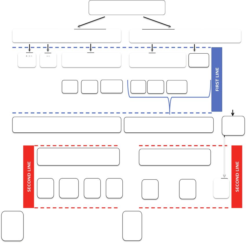
3.1. Demographic and clinical characteristics pants’ treatments are shown in Fig. 2
The French series included 23 patients: 14 patients had catatonia, 3.2. Clinical outcomes
and 9 did not. The Italian series comprised 35 patients: 11 patients had
catatonia, and 24 did not. Fig. 1 summarizes the diagram flow of the First line therapies were effective (major or partial improvement)
study. In total, the sample included 58 patients with autoimmune for 46 (79%) patients and not effective without any improvement for 12
neuropsychiatric conditions, 25 (43%) with catatonia and 33 (57%) (21%) patients. Second line therapies were effective, with major or
without catatonia. The mean age was 13.86 ( ± 4.41) years. The ma- partial improvement for 26 (87%), and 4 (13%) patients had no im-
jority of patients was females (66%). Participants’ characteristics are provement. Seven patients (12%) relapsed. Fifteen (26%) patients ex-
given in Table 1. Among the catatonic group (N = 25), 15 (60%) ex- hibited sequelae within the one-year follow up, including: persistent
hibited a stuporous form, 6 (24%) an agitated form and 4 (16%) a seizures (N = 4), movement disorders (N = 0), cognitive impairment
mixed form. Forty-two patients (72.4%) were diagnosed with definite (N = 5, memory loss and language deficit), and chronic psychiatric
AE, including 27 anti-NMDA receptor encephalitis (46.55%), 5 SLE features (N = 6, anxiety and mood disorders). Outcome data were not
(8.62%), 2 anti-GAD encephalitis (3.45%), 2 anti-Hu encephalitis available for two patients. Only one catatonic patient (2%) died from
(3.45%), 2 Hashimoto encephalopathy (3.45%), 1 anti-VGKC/anti-LGI1 complications of severe autonomic dysfunction due to anti-NMDA en-
encephalitis (1.7%), 2 PANDAS/PANS (3.4%), and 1 SLE with anti- cephalitis.
phospholipid-related chorea (1.7%). Suspected AE was identified for 16 Interestingly most patients with suspected AE exhibited catatonia
patients (27.6%). The distribution of autoimmune conditions per site is (N = 10, 67%). Most of them fully responded to first lines including
listed in Table S1. plasma exchange (N = 13, 81%). Only 3 of them (19%) required both
Regarding first line treatment, 42 patients received an initial ther- first and second line. Half of them (N = 8) exhibited sequelae including
apeutic challenge with high dosage corticoid pulses, 35 patients (60%) cognitive impairment (N = 3) and chronic psychiatric features (N = 5,
received IgG IV, 11 (19%) had plasma exchanges (PE), 36 patients anxiety and mood disorders). Only one (6%) patient relapsed.
(62%) received at least two different first lines, and only one patient
(1.7%) directly received second line treatment (rituximab) without 3.3. Patients with and without catatonia
previous first lines. The other 15 (26%) patients without high dosage
corticoids received another first line: 10 had IgG IV alone, and 5 had PE Comparisons are given in Table 1. There were no differences be-
alone. Second line treatments, including cyclophosphamide (8.6%), tween the two groups regarding sociodemographic data. A few differ-
mycophenolate mofetil (5.2%), azathioprine (8.6%), and rituximab ences emerged regarding clinical characteristics. Catatonic patients
(29.2%), were indicated for 30 patients (52%) to maintain improve- showed significantly more psychotic features than patients without
ment after a positive response to first line treatment (high dosage catatonia (18 (72%) vs 9 (27.3%), p < 0.001), including more
Fig. 1. Flow chart of the study. N: number, AE: autoimmune encephalitis.
4V. Ferrafiat, et al. Progress in Neuropsychopharmacology & Biological Psychiatry 104 (2021) 110028
Table 1
Socio-demographics and clinical characteristics of the patients with neuropsychiatric autoimmune conditions with and without catatonia.
No catatonia (N = 33) Catatonia (N = 25) Total (N = 58) P
Socio-demographics
Sex: N (%) females 20 (60.6%) 18 (72%) 38 (66%) 0.366WILCOXON
Age: mean (SD) 13.39 (4.58) 14.48 (4.17) 13.86 (4.41) 0.243WILCOXON
Etiological classification
CAUS score 7.42 (1.7) 7.56 (1.61) 7.47 (1.65) 0.731
Autoimmune systemic disorders: N (%) 3 (9.1%) 5 (20%) 8 (13.8%) 0.951
Definite AE: N (%) 21 (63.6%) 13 (52%) 34 (58.6%) 0.951
Suspected AE: N (%) 9 (27.3%) 7 (28%) 16 (27.6%) 0.951
Clinical characteristics
Stuporous catatonia: N (%) 0 19 (76%) 19 (33%) < 0.001
Agitated catatonia: N (%) 0 10 (40%) 10 (17%) < 0.001FISHER
Hallucinations: N (%) 7 (21%) 17 (68%) 24 (41%) < 0.001
Delusions: N (%) 6 (18.2%) 14 (56%) 20 (34%) 0.003
Psychotic symptoms: N (%) 9 (27.3%) 18 (72%) 27 (47%) 0.001
Mood disorders: N (%) 17 (51.5%) 19 (76%) 36 (62%) 0.057
ASD features: N (%) 0 4 (16%) 4 (7%) 0.03FISHER
OCD: N (%) 2 (6.1%) 0 2 (3%) 0.501FISHER
Anxiety features: N (%) 33 (100%) 20 (80%) 53 (91%) 0.012FISHER
Sleep disorders: N (%) 19 (57.6%) 18 (72%) 37 (64%) 0.258
Cognitive regression: N (%) 19 (57.6%) 14 (56%) 33 (57%) 0.904
Movement disorders 20 (60.6%) 25 (100%) 45 (78%) < 0.001
Seizures: N (%) 21 (63.6%) 12 (48%) 33 (57%) 0.234
Systemic localization: N (%) 7 (21.2%) 5 (20%) 12 (21%) 0.91
Paraclinical characteristics
Plasma biological arguments: N (%) 7 (21.2%) 6 (24%) 13 (22%) 0.801
CSF biological arguments: N (%) 24 (72.7%) 15 (60%) 39 (67%) 0.306
EEG arguments: N (%) 16 (48.5%) 15 (60%) 31 (53%) 0.384
MRI arguments: N (%) 15 (45.5%) 8 (33%) 23 (40%) 0.357
PET scan arguments: N (%) 1 (50%) 2 (66.7%) 3 (60%) 1FISHER
Treatment
Tumor removal: N (%) 1 (3%) 2 (8%) 3 (5%) 0.572FISHER
Corticoids: N (%) 25 (75.8%) 17 (68%) 42 (72%) 0.513
IgG IV: N (%) 23 (69.7%) 12 (48%) 35 (60%) 0.094
Plasma exchange: N (%) 4 (12 0.1%) 7 (28%) 11 (19%) 0.179FISHER
Cyclophosphamide: N (%) 0 5 (20%) 5 (9%) 0.013FISHER
Mycophemolate mofetil: N (%) 0 3 (12%) 3 (5%) 0.075FISHER
Aziothropine: N (%) 2 (6.1%) 3 (12%) 5 (9%) 0.643FISHER
Ritubimax: N (%) 7 (21.2%) 10 (40%) 17 (30%) 0.066
Outcomes
First line treatment efficacy: N (%) 22 (66.7%) 24 (96%) 46 (79%) 0.006
Second line treatment efficacy: N (%) 9 (27.3%) 17 (68%) 26 (45%) 0.002
Relapses: N (%)* 0 7 (29.2%) 7 (12%) 0.001FISHER
Sequels: N (%) 8 (24%) 7 (28%) 15 (26%) 0.746
Death: N (%) 0 1 (4%) 1 (2%) 0.431FISHER
AE: autoimmune encephalitis, ASD: autism spectrum disorder, OCD: obsessive–compulsive disorder, CSF: cerebro-spinal fluid; *N = 56; In most cases we used Chi2
tests for comparisons except when indicated.
delusional ideas and hallucinations. In addition, they had significantly 4. Discussion
more movement disorders (dystonia, choreiform or other non-specific
abnormal movements): 25 (100%) vs 20 (60.6%), respectively, Few data exist regarding pediatric catatonia related to autoimmune
p < 0.001. In contrast, patients without catatonia had significantly conditions (Ferrafiat et al., 2016; Ferrafiat et al., 2016; Benarous et al.,
more anxiety manifestations than catatonic ones, with 33 (100%) vs 5 2018; Mooneyham et al., 2018; Tanguturi et al., 2019). This study
(20%), respectively, p = 0.012. gathers one of the largest samples over several university hospitals,
Regarding the response to immunomodulatory or im- allowing a multi-disciplinary diagnosis and therapeutic approach
munosuppressive treatment, first line (high dosage corticoids and/or PE through various pediatric departments. First, our study underscores
and/or IgG IV) and second line (including cyclophosphamide, myco- that autoimmune catatonia is significantly more associated with severe
phenolate mofetil, and azathioprine, rituximab) therapies were found psychotic features compared to autoimmune conditions without cata-
to be more effective for catatonic patients, with 24 (96%) versus 22 tonia, suggesting a more severe initial psychiatric presentation. Second,
(66.7%) (p = 0.006) and 17 (68%) versus 9 (27.3%) (p = 0.002), our results stress that patients with catatonia experienced more relapses
respectively, showing a major or partial clinical response. However, that patients without catatonia. These results emphasize that auto-
they had more relapses than patients without catatonia, at 7 (29.2%) immune catatonia could imply a more severe form of autoimmune
versus 0 (0%) (p = 0.001), respectively. No significant differences re- condition, supporting our hypothesis. However, we did not find a
garding sequelae or death were found between the two groups. higher risk of poorer outcomes in terms of treatment resistance, se-
Catatonic patients received all high dose benzodiazepines, and none quelae or death among catatonic patients. Given our results, we discuss
received ECT. four main points: i) catatonia in immune-related conditions; ii) treat-
ment; iii) anti-NMDA receptor encephalitis, as we observed 27 cases;
and iv) the limitations of our study.
5V. Ferrafiat, et al. Progress in Neuropsychopharmacology & Biological Psychiatry 104 (2021) 110028
Fig. 2. Participants’ first and second line treatments according to catatonia. IgG IV: intravenous immunoglobulins; PE: plasma exchange; CYC: cyclophosphamide;
MYP: mycophenolate mofetil; AZP: azathioprine; RTX: rituximab.
Our results support the idea that catatonia is associated with more therapies. Hypothetically, catatonia as a clinical marker of risk of re-
severe forms of autoimmune disorders, but we can generalize this lapse would require earlier (even presumptively) and more aggressive
finding outside the context of anti-NMDA receptor encephalitis, as half immunosuppressive treatment to better prevent relapses and neuro-
of our cases were secondary to another causal encephalitis. Catatonia cognitive sequelae. In contrast to DeSena et al., who hypothesized that
was associated with more psychotic features, emphasizing the fact that anti-NMDA encephalitis with catatonia showed a poor response to im-
autoimmune catatonia is likely to be associated with other severe munosuppressive treatments (DeSena et al., 2014), our catatonic pa-
psychiatric features, such as hallucinations, psychosis, cognitive re- tients had a better response to either first or second line treatment than
gression, and fluctuation of symptoms, leading to an atypical psychia- patients without catatonia. These results might be explained by two
tric presentation in youths (Consoli et al., 2012; Ferrafiat et al., 2018; factors: i) our catatonic patients were clinically more severe and
Bonnot et al., 2015). Regarding the significant difference in anxiety therefore had faster access to immunotherapy and more combinations
features between the two groups, the non-catatonic patients were more of first and second line treatments (only 4 catatonic patients did not
likely to express their anxiety and to be diagnosed, whereas catatonic receive second line treatments compared to 24 without catatonia); ii)
patients presented difficulties in identifying and therefore expressing the lack of standardized international treatment guidelines makes it
their inner subjective feelings and experiences during the acute phase difficult to interpret our treatment data, as patients’ treatment plans
(Cohen, 2006). Furthermore, catatonic patients had more relapses. and access to immunosuppressive treatment were heterogeneous and
Even if the prognosis of neurocognitive sequelae is related to the timing widely differed from one center to another. To illustrate this hetero-
of the introduction of immunosuppressive treatment (Byrne et al., geneity, we found that immunoglobulin was often used in our Italian
2015; Florance et al., 2009; Titulaer et al., 2013; Armangue et al., 2012; sample. Plasma exchanges were only available in 3 centers due to
Luca et al., 2011; Chapman and Vause, 2011; Nosadini et al., 2015), it is limited access to this procedure. Cyclophosphamide was mainly used in
notable that catatonic patients were more severe with more relapses one center according to a published multidisciplinary treatment algo-
regardless of their initial positive response to first or second line rithm (Ferrafiat et al., 2018). The different treatments used in each
6V. Ferrafiat, et al. Progress in Neuropsychopharmacology & Biological Psychiatry 104 (2021) 110028
center are detailed in Fig. 2. consider catatonia as a marker of possible underlying autoimmune
Regarding published data on treatment options, available studies conditions, as many children and adolescents with suspected auto-
suggest that the use of high dosage corticoids via pulses initially gives immune encephalitis are seronegative (Dale et al., 2017). It also un-
good response rates in pediatric populations (Armangue et al., 2012; derscores the idea that catatonia can be a marker of severity and
Luca et al., 2011; Brunner et al., 2008; Levy and Kamphuis, 2012). morbidity in terms of associated psychiatric symptoms and outcomes.
However, plasma exchanges and their peripheral action represent an Finally, the response of autoimmune catatonia to treatment, along with
interesting option in SLE and anti-NMDA receptor encephalitis (Boers a higher relapse rate further supports the existing literature for both
and Colebatch, 2001; DeSena et al., 2015; Hussain et al., 2005; Prytuła early and aggressive immunosuppressive treatments.
et al., 2015; Suppiej et al., 2016) and provide an effective treatment for
catatonia (Ferrafiat et al., 2016; Marra et al., 2008; Elia et al., 2005). In Funding Source
the case of resistance to first line treatments (Armangue et al., 2012) or
relapses, second line therapies are to be considered (Byrne et al., 2015; This study was performed without any specific support.
Florance et al., 2009; Dale et al., 2017; Titulaer et al., 2013; Stingl et al.,
2018). The early introduction of second lines in youths ensure better Ethical Statement
outcomes (Luca et al., 2011; Stingl et al., 2018; Ishiura et al., 2008). For
example, despite rituximab's significant risk of infectious complications, This publication involves current clinical practice and is based on a
a recent study supports its early off-label use in youths with significant clinical study in compliance with the Ethics of the University Centers.
morbidity secondary to autoimmune disorders (Dale et al., 2014). All the parents were informed of the possible further use of clinical,
Regarding anti-NMDA receptor encephalitis, most studies are re- paraclinical and treatment data regarding their child and gave their
cent, and it is the most frequent form of AE. This was also the case in consent for use of those data in the context of research.
our sample, at nearly half of the cases, and 49% of those cases had
catatonia. Movement disorders, such as stereotyped movements, dys- CRediT authorship contribution statement
tonia, chorea and catatonia, are frequent and are considered to be key
symptoms exhibited by patients (Granata et al., 2018; Dash et al., 2019; Vladimir Ferrafiat: Conceptualization, Methodology,
Duan et al., 2016; Dalmau et al., 2008). Graus et al. included rigidity Investigation, Data curation, Writing - original draft, Visualization.
and abnormal postures (that belong to catatonic features) among the Elise Riquin: Methodology, Investigation, Data curation, Writing -
movement disorders in new diagnosis criteria for anti-NMDA receptor original draft. Elena Freri: Methodology, Investigation, Data curation,
encephalitis (Graus et al., 2016). Interestingly, two studies focusing on Writing - original draft. Tiziana Granata: Writing - review & editing.
catatonia found that patients who had hypokinetic disorders, such as Nardo Nardocci: Writing - review & editing. François Medjkane:
catatonia, took significantly longer to improve or did not fully recover Writing - review & editing. Claire Corfiotti: Writing - review & editing.
compared to other forms of movement disorders (Granata et al., 2018; Alessandra Tozzo: . Huges Pellerin: Formal analysis, Writing - ori-
Dash et al., 2019). DeSena et al. proposed distinguishing 3 phenotypes ginal draft. Xavier Benarous: Writing - review & editing. Julien
of anti-NMDA receptor antibody encephalitis in children (DeSena et al., Haroche: Writing - review & editing. Zahir Amoura: Writing - review
2014): type 1 “classic anti-NMDA receptor antibody encephalitis with & editing. Philippe Duverger: Writing - review & editing. Renaud
predominant movement disorder and epilepsy”; type 2 “psychiatric Jardri: Writing - review & editing. Priscille Gerardin: Writing - review
predominant NMDA receptor encephalitis subtype”; and type 3 “cata- & editing. David Cohen: Conceptualization, Methodology,
tonia/stupor predominant anti-NMDA receptor encephalitis sub phe- Investigation, Data curation, Writing - review & editing, Visualization.
notype.” Type 3 patients were often very severe and showed the poorest Angèle Consoli: Writing - review & editing. Marie Raffin:
response even to aggressive immunotherapies (DeSena et al., 2014). Conceptualization, Methodology, Investigation, Data curation, Writing
Anti-NMDA encephalitis emphasizes that our results cross paths with - review & editing, Supervision.
those previous available data in terms of severity, whether the severity
is defined by the initial clinical presentation, treatment response, fre- Declaration of Competing Interest
quency of relapses or sequelae. Hence, our results extend the concept of
catatonia as a marker of severity to autoimmune conditions in general. The authors declare that they have no known competing financial
However, the results should be interpreted in the context of several interests or personal relationships that could have appeared to influ-
limitations. First, the number of subjects recruited was low, despite the ence the work reported in this paper.
multicentric recruitment. This could be explained by the very low
prevalence of catatonia in youths compared to other psychiatric dis- Appendix A. Supplementary data
orders (Cohen et al., 2005) and the low prevalence of autoimmune
conditions among the pediatric population (Dale et al., 2017; Stingl Supplementary data to this article can be found online at https://
et al., 2018). The statistical analysis should be regarded as exploratory. doi.org/10.1016/j.pnpbp.2020.110028.
Second, to increase the number of patients with probable, suspected
and definite AE, we grouped different samples that had recruitment References
biases, leading to heterogeneity. Indeed, 60% of the cases came from
the Italian autoimmune encephalitis specialized center, with a lower Cohen, D., Nicolas, J.-D., Flament, M.F., et al., 2005. Clinical relevance of chronic cata-
proportion of catatonia compared to the French network specialized in tonic schizophrenia in children and adolescents: evidence from a prospective natur-
pediatric catatonia. Additionally, the two centers had a different first- alistic study. Schizophr. Res. 76 (2–3), 301–308. https://doi.org/10.1016/j.schres.
2005.01.014.
step inclusion criterion, highlighting center-based biases. As a con- Cohen, D., 2006. Towards a valid nosography and psychopathology of catatonia in
sequence, generalization may not be valid. Third, we used a multicenter children and adolescents. Int. Rev. Neurobiol. 72, 131–147. https://doi.org/10.1016/
clinical sample of acutely ill patients recruited in university teaching S0074-7742(05)72008-0.
Dhossche, D.M., 2014. Decalogue of catatonia in autism spectrum disorders. Front.
hospitals that may have been particularly enriched for subjects with Psychiatry 5, 157. https://doi.org/10.3389/fpsyt.2014.00157.
more severe forms of neuropsychiatric autoimmune conditions. Withane, N., Dhossche, D.M., 2019. Electroconvulsive treatment for catatonia in autism
spectrum disorders. Child Adolesc Psychiatr Clin N Am. 28 (1), 101–110. https://doi.
org/10.1016/j.chc.2018.07.006.
5. Conclusion Ghaziuddin, N., Nassiri, A., Miles, J.H., 2015. Catatonia in Down syndrome; a treatable
cause of regression. Neuropsychiatr. Dis. Treat. 11, 941–949. https://doi.org/10.
Despite its exploratory nature, this study enhances the need to 2147/NDT.S77307.
7V. Ferrafiat, et al. Progress in Neuropsychopharmacology & Biological Psychiatry 104 (2021) 110028
Fink, M., Taylor, M.A., Ghaziuddin, N., 2006. Catatonia in autistic spectrum disorders: a Lee, W.-J., Lee, S.-T., Moon, J., et al., 2016. Tocilizumab in autoimmune encephalitis
medical treatment algorithm. Int. Rev. Neurobiol. 72, 233–244. https://doi.org/10. refractory to rituximab: an institutional cohort study. Neurother J Am Soc Exp
1016/S0074-7742(05)72014-6. Neurother. 13 (4), 824–832. https://doi.org/10.1007/s13311-016-0442-6.
Cornic, F., Consoli, A., Tanguy, M.-L., et al., 2009. Association of adolescent catatonia Kashyape, P., Taylor, E., Ng, J., Krishnakumar, D., Kirkham, F., Whitney, A., 2012.
with increased mortality and morbidity: evidence from a prospective follow-up study. Successful treatment of two paediatric cases of anti-NMDA receptor encephalitis with
Schizophr. Res. 113 (2–3), 233–240. https://doi.org/10.1016/j.schres.2009.04.021. cyclophosphamide: the need for early aggressive immunotherapy in tumour negative
Raffin, M., Zugaj-Bensaou, L., Bodeau, N., et al., 2015. Treatment use in a prospective paediatric patients. Eur. J. Paediatr. Neurol. EJPN Off. J. Eur. Paediatr. Neurol. Soc.
naturalistic cohort of children and adolescents with catatonia. Eur. Child Adolesc. 16 (1), 74–78. https://doi.org/10.1016/j.ejpn.2011.07.005.
Psychiatry 24 (4), 441–449. https://doi.org/10.1007/s00787-014-0595-y. Zekeridou, A., Karantoni, E., Viaccoz, A., et al., 2015. Treatment and outcome of children
Puffer, C.C., Wall, C.A., Huxsahl, J.E., Frye, M.A., 2016. A 20 year practice review of and adolescents with N-methyl-D-aspartate receptor encephalitis. J. Neurol. 262 (8),
electroconvulsive therapy for adolescents. J. Child Adolesc. Psychopharmacol. 26 (7), 1859–1866. https://doi.org/10.1007/s00415-015-7781-9.
632–636. https://doi.org/10.1089/cap.2015.0139. Granata, T., Matricardi, S., Ragona, F., et al., 2018. Pediatric NMDAR encephalitis: A
Consoli, A., Benmiloud, M., Wachtel, L., Dhossche, D., Cohen, D., Bonnot, O., 2010. single center observation study with a closer look at movement disorders. Eur. J.
Electroconvulsive therapy in adolescents with the catatonia syndrome: efficacy and Paediatr. Neurol. EJPN Off. J. Eur. Paediatr. Neurol. Soc. 22 (2), 301–307. https://
ethics. J ECT 26 (4), 259–265. https://doi.org/10.1097/YCT.0b013e3181fb3924. doi.org/10.1016/j.ejpn.2018.01.012.
Consoli, A., Raffin, M., Laurent, C., et al., 2012. Medical and developmental risk factors of Graus, F., Titulaer, M.J., Balu, R., et al., 2016. A clinical approach to diagnosis of auto-
catatonia in children and adolescents: a prospective case-control study. Schizophr. immune encephalitis. Lancet Neurol. 15 (4), 391–404. https://doi.org/10.1016/
Res. 137 (1–3), 151–158. https://doi.org/10.1016/j.schres.2012.02.012. S1474-4422(15)00401-9.
Raffin, M., Consoli, A., Giannitelli, M., et al., 2018. Catatonia in children and adolescents: DeSena, A.D., Greenberg, B.M., Graves, D., 2014. Three phenotypes of anti-N-methyl-D-
a high rate of genetic conditions. J. Am. Acad. Child Adolesc. Psychiatry 57 (7), aspartate receptor antibody encephalitis in children: prevalence of symptoms and
518–525.e1. https://doi.org/10.1016/j.jaac.2018.03.020. prognosis. Pediatr. Neurol. 51 (4), 542–549. https://doi.org/10.1016/j.
Ferrafiat V, Raffin M, Deiva K, et al. Catatonia and Autoimmune Conditions in Children pediatrneurol.2014.04.030.
and Adolescents: Should We Consider a Therapeutic Challenge? J Child Adolesc Morbelli, S., Djekidel, M., Hesse, S., et al., 2016. Role of (18)F-FDG-PET imaging in the
Psychopharmacol. Published online April 19, 2016. doi:10.1089/cap.2015.0086. diagnosis of autoimmune encephalitis. Lancet Neurol. 15 (10), 1009–1010. https://
Ferrafiat, V., Raffin, M., Freri, E., et al., 2018. A causality algorithm to guide diagnosis doi.org/10.1016/S1474-4422(16)30140-5.
and treatment of catatonia due to autoimmune conditions in children and adoles- Probasco, J.C., Solnes, L., Nalluri, A., et al., 2017. Abnormal brain metabolism on FDG-
cents. Schizophr. Res. 200, 68–76. https://doi.org/10.1016/j.schres.2017.06.036. PET/CT is a common early finding in autoimmune encephalitis. Neurol.
Lahutte, B., Cornic, F., Bonnot, O., et al., 2008. Multidisciplinary approach of organic Neuroimmunol. Neuroinflamm. 4 (4), e352. https://doi.org/10.1212/NXI.
catatonia in children and adolescents may improve treatment decision making. Prog. 0000000000000352.
Neuro-Psychopharmacol. Biol. Psychiatry 32 (6), 1393–1398. https://doi.org/10. Turpin, S., Martineau, P., Levasseur, M.-A., et al., 2019. 18F-Flurodeoxyglucose positron
1016/j.pnpbp.2008.02.015. emission tomography with computed tomography (FDG PET/CT) findings in children
Marra, D., Amoura, Z., Soussan, N., et al., 2008. Plasma exchange in patients with stu- with encephalitis and comparison to conventional imaging. Eur. J. Nucl. Med. Mol.
porous catatonia and systemic lupus erythematosus. Psychother. Psychosom. 77 (3), Imaging 46 (6), 1309–1324. https://doi.org/10.1007/s00259-019-04302-x.
195–196. https://doi.org/10.1159/000120280. Benarous X, Consoli A, Raffin M, et al. Validation of the Pediatric Catatonia Rating Scale
Byrne, S., Walsh, C., Hacohen, Y., et al., 2015. Earlier treatment of NMDAR antibody (PCRS). Schizophr Res. Published online July 1, 2016. doi:10.1016/j.schres.2016.06.
encephalitis in children results in a better outcome. Neurol. Neuroimmunol. 020.
Neuroinflamm. 2 (4), e130. https://doi.org/10.1212/NXI.0000000000000130. Sedel, F., Baumann, N., Turpin, J.-C., Lyon-Caen, O., Saudubray, J.-M., Cohen, D., 2007.
Finke, C., Kopp, U.A., Prüss, H., Dalmau, J., Wandinger, K.-P., Ploner, C.J., 2012. Psychiatric manifestations revealing inborn errors of metabolism in adolescents and
Cognitive deficits following anti-NMDA receptor encephalitis. J. Neurol. Neurosurg. adults. J. Inherit. Metab. Dis. 30 (5), 631–641. https://doi.org/10.1007/s10545-007-
Psychiatry 83 (2), 195–198. https://doi.org/10.1136/jnnp-2011-300411. 0661-4.
Florance, N.R., Davis, R.L., Lam, C., et al., 2009. Anti-N-methyl-D-aspartate receptor Benarous, X., Raffin, M., Ferrafiat, V., Consoli, A., Cohen, D., 2018. Catatonia in children
(NMDAR) encephalitis in children and adolescents. Ann. Neurol. 66 (1), 11–18. and adolescents: new perspectives. Schizophr. Res. 200, 56–67. https://doi.org/10.
https://doi.org/10.1002/ana.21756. 1016/j.schres.2017.07.028.
Dale, R.C., Gorman, M.P., Lim, M., 2017. Autoimmune encephalitis in children: clinical Graus, F., Dalmau, J., 2016. Role of (18)F-FDG-PET imaging in the diagnosis of auto-
phenomenology, therapeutics, and emerging challenges. Curr. Opin. Neurol. 30 (3), immune encephalitis – Authors’ reply. Lancet Neurol. 15 (10), 1010. https://doi.org/
334–344. https://doi.org/10.1097/WCO.0000000000000443. 10.1016/S1474-4422(16)30130-2.
Hacohen, Y., Wright, S., Waters, P., et al., 2013. Paediatric autoimmune en- Mooneyham, G.C., Gallentine, W., Van Mater, H., 2018. Evaluation and management of
cephalopathies: clinical features, laboratory investigations and outcomes in patients autoimmune encephalitis: a clinical overview for the practicing child psychiatrist.
with or without antibodies to known central nervous system autoantigens. J. Neurol. Child Adolesc. Psychiatr. Clin. N. Am. 27 (1), 37–52.
Neurosurg. Psychiatry 84 (7), 748–755. https://doi.org/10.1136/jnnp-2012-303807. Tanguturi, Y.C., Cundiff, A.W., Fuchs, C., 2019. Anti-N-methyl d-aspartate receptor en-
Titulaer, M.J., McCracken, L., Gabilondo, I., et al., 2013. Treatment and prognostic factors cephalitis and electroconvulsive therapy: literature review and future directions.
for long-term outcome in patients with anti-NMDA receptor encephalitis: an ob- Child. Adolesc. Psychiatr. Clin. N. Am. 28 (1), 79–89. https://doi.org/10.1016/j.chc.
servational cohort study. Lancet Neurol. 12 (2), 157–165. https://doi.org/10.1016/ 2018.07.005.
S1474-4422(12)70310-1. Bonnot, O., Herrera, P.M., Tordjman, S., Walterfang, M., 2015. Secondary psychosis in-
Tucker, L.B., Uribe, A.G., Fernández, M., et al., 2008. Adolescent onset of lupus results in duced by metabolic disorders. Front. Neurosci. 9, 177. https://doi.org/10.3389/
more aggressive disease and worse outcomes: results of a nested matched case-con- fnins.2015.00177.
trol study within LUMINA, a multiethnic US cohort (LUMINA LVII). Lupus 17 (4), Armangue, T., Petit-Pedrol, M., Dalmau, J., 2012. Autoimmune encephalitis in children.
314–322. https://doi.org/10.1177/0961203307087875. J. Child Neurol. 27 (11), 1460–1469.
Hoffman, I.E.A., Lauwerys, B.R., De Keyser, F., et al., 2009. Juvenile-onset systemic lupus Luca, N., Daengsuwan, T., Dalmau, J., et al., 2011. Anti-N-methyl-D-aspartate receptor
erythematosus: different clinical and serological pattern than adult-onset systemic encephalitis: a newly recognized inflammatory brain disease in children. Arthritis
lupus erythematosus. Ann. Rheum. Dis. 68 (3), 412–415. https://doi.org/10.1136/ Rheum. 63 (8), 2516–2522. https://doi.org/10.1002/art.30437.
ard.2008.094813. Chapman, M.R., Vause, H.E., 2011. Anti-NMDA receptor encephalitis: diagnosis, psy-
Livingston, B., Bonner, A., Pope, J., 2011. Differences in clinical manifestations between chiatric presentation, and treatment. Am. J. Psychiatry 168 (3), 245–251. https://doi.
childhood-onset lupus and adult-onset lupus: a meta-analysis. Lupus 20 (13), org/10.1176/appi.ajp.2010.10020181.
1345–1355. https://doi.org/10.1177/0961203311416694. Nosadini, M., Mohammad, S.S., Ramanathan, S., Brilot, F., Dale, R.C., 2015. Immune
Montagna, G., Imperiali, M., Agazzi, P., et al., 2016. Hashimoto’s encephalopathy: a rare therapy in autoimmune encephalitis: a systematic review. Expert Rev. Neurother. 15
proteiform disorder. Autoimmun. Rev. 15 (5), 466–476. https://doi.org/10.1016/j. (12), 1391–1419. https://doi.org/10.1586/14737175.2015.1115720.
autrev.2016.01.014. Brunner, H.I., Gladman, D.D., Ibañez, D., Urowitz, M.D., Silverman, E.D., 2008.
Mahmud, F.H., Lteif, A.N., Renaud, D.L., Reed, A.M., Brands, C.K., 2003. Steroid-re- Difference in disease features between childhood-onset and adult-onset systemic
sponsive encephalopathy associated with Hashimoto’s thyroiditis in an adolescent lupus erythematosus. Arthritis Rheum. 58 (2), 556–562. https://doi.org/10.1002/
with chronic hallucinations and depression: case report and review. Pediatrics 112 (3 art.23204.
Pt 1), 686–690. Levy, D.M., Kamphuis, S., 2012. Systemic lupus erythematosus in children and adoles-
Carlone, C., Todini, L., Marini, I., et al., 2013. Acute psychiatric presentation of steroid- cents. Pediatr. Clin. N. Am. 59 (2), 345–364. https://doi.org/10.1016/j.pcl.2012.03.
responsive encephalopathy: the under recognized side of autoimmune thyroiditis. 007.
Riv. Psichiatr. 48 (2), 169–173. https://doi.org/10.1708/1272.14042. Boers, P.M., Colebatch, J.G., 2001. Hashimoto’s encephalopathy responding to plasma-
Swedo, S.E., Seidlitz, J., Kovacevic, M., et al., 2015. Clinical presentation of pediatric pheresis. J. Neurol. Neurosurg. Psychiatry 70 (1), 132.
autoimmune neuropsychiatric disorders associated with streptococcal infections in DeSena, A.D., Noland, D.K., Matevosyan, K., et al., 2015. Intravenous methylprednisolone
research and community settings. J. Child Adolesc. Psychopharmacol. 25 (1), 26–30. versus therapeutic plasma exchange for treatment of anti-N-methyl-D-aspartate re-
https://doi.org/10.1089/cap.2014.0073. ceptor antibody encephalitis: a retrospective review. J. Clin. Apheresis 30 (4),
Elia, J., Dell, M.L., Friedman, D.F., et al., 2005. PANDAS with catatonia: a case report. 212–216. https://doi.org/10.1002/jca.21363.
Therapeutic response to lorazepam and plasmapheresis. J. Am. Acad. Child Adolesc. Hussain, N.S., Rumbaugh, J., Kerr, D., Nath, A., Hillis, A.E., 2005. Effects of prednisone
Psychiatry 44 (11), 1145–1150. https://doi.org/10.1097/01.chi.0000179056. and plasma exchange on cognitive impairment in Hashimoto encephalopathy.
54419.5e. Neurology 64 (1), 165–166. https://doi.org/10.1212/01.WNL.0000148580.
Schlansky K, Facer B, Tanguturi YC, Cundiff AW, Fuchs DC. Pediatric Acute-Onset 98997.C5.
Neuropsychiatric Syndrome and Catatonia: A Case Report. Psychosomatics. Prytuła, A., Vande Walle, J., Verhelst, H., et al., 2015. Therapeutic plasma exchange in
Published online April 3, 2019. doi:10.1016/j.psym.2019.03.007. children with acute autoimmune central nervous system disorders. Int. J. Artif.
8V. Ferrafiat, et al. Progress in Neuropsychopharmacology & Biological Psychiatry 104 (2021) 110028
Organs 38 (9), 494–500. https://doi.org/10.5301/ijao.5000435. org/10.1212/WNL.0000000000000570.
Suppiej A, Nosadini M, Zuliani L, et al. Plasma exchange in pediatric anti-NMDAR en- Dash, D., Ihtisham, K., Tripathi, M., Tripathi, M., 2019. Proportion and spectrum of
cephalitis: A systematic review. Brain Dev. Published online February 26, 2016. movement disorders in adolescent and adult patients of autoimmune encephalitis of
doi:10.1016/j.braindev.2016.01.009. non-neoplastic aetiology. J. Clin. Neurosci. Off. J. Neurosurg. Soc. Australas 59,
Stingl, C., Cardinale, K., Van Mater, H., 2018. An update on the treatment of pediatric 185–189. https://doi.org/10.1016/j.jocn.2018.10.076.
autoimmune encephalitis. Curr. Treat Options Rheumatol. 4 (1), 14–28. https://doi. Duan, B.-C., Weng, W.-C., Lin, K.-L., et al., 2016. Variations of movement disorders in
org/10.1007/s40674-018-0089-z. anti-N-methyl-D-aspartate receptor encephalitis: a nationwide study in Taiwan.
Ishiura, H., Matsuda, S., Higashihara, M., et al., 2008. Response of anti-NMDA receptor Medicine (Baltimore) 95 (37), e4365. https://doi.org/10.1097/MD.
encephalitis without tumor to immunotherapy including rituximab. Neurology 71 0000000000004365.
(23), 1921–1923. https://doi.org/10.1212/01.wnl.0000336648.43562.59. Dalmau, J., Gleichman, A.J., Hughes, E.G., et al., 2008. Anti-NMDA-receptor encephalitis:
Dale, R.C., Brilot, F., Duffy, L.V., et al., 2014. Utility and safety of rituximab in pediatric case series and analysis of the effects of antibodies. Lancet Neurol. 7 (12),
autoimmune and inflammatory CNS disease. Neurology 83 (2), 142–150. https://doi. 1091–1098. https://doi.org/10.1016/S1474-4422(08)70224-2.
9You can also read