Metagenomic analysis of gut microbiota in non treated plaque psoriasis patients stratified by disease severity: development of a new ...
←
→
Page content transcription
If your browser does not render page correctly, please read the page content below
www.nature.com/scientificreports
OPEN Metagenomic analysis of gut
microbiota in non‑treated plaque
psoriasis patients stratified
by disease severity: development
of a new Psoriasis‑Microbiome
Index
Ignacio Dei‑Cas1,2,3, Florencia Giliberto4,5, Leonela Luce4,5, Hernán Dopazo6 &
Alberto Penas‑Steinhardt7,8*
Psoriasis is an immune-mediated skin disorder. Imbalance of gut microbial populations has been
implicated in many diseases. We aimed to investigate whether there were differences in gut
microbiota in psoriasis patients vs non-psoriasis controls and between psoriasis severity groups. 55
psoriasis patients and 27 controls were included. V3–V4 regions of the 16S rRNA gene of fecal samples
were analyzed using Illumina MiSeq. Bioinformatic analysis was performed. We found changes
in gut microbiome composition depending on their psoriasis status as determined by weighted
unifrac (p < 0.05), in particular an increase in Firmicutes and depletion of Bacteroidetes in psoriasis
patients. Additionally, the Faecalibacterium and Blautia genus were higher in psoriasis patients while
Bacteroides and Paraprevotella in non-psoriasis controls (p < 0.05, LDA score > 2). Moderate-to-severe
psoriasis patients had lower biodiversity than mild psoriatic patients (p = 0.049). No differences for
beta-diversity were found. We developed a Psoriasis-Microbiota Index (PMI), which discriminated
among psoriasis patients and controls with sensitivity: 0.78 and specificity: 0.79. Furthermore, we
performed a meta-analysis with published data to validate this index. We demonstrated gut dysbiosis
in psoriasis patients, suggesting a role in psoriasis pathophysiology. Furthermore, we developed
a PMI with the potential to discriminate between psoriasis patients and controls across different
populations, which could be used as a biomarker in the clinical practice.
Psoriasis is a chronic, immune-mediated inflammatory skin disease. It ranges in severity from a few scattered red,
scaly plaques to involvement of almost the entire body s urface1. Psoriasis is estimated to affect about 2–4% of the
population in western countries, causes considerable psychosocial disability and has a major impact on patients’
quality of life2,3. Skin lesions are characterized by angiogenesis, an inflammatory reaction with recruitment of T
cells into the skin, hyperproliferation of keratinocytes and altered epidermal differentiation4. Genetic and envi-
nderstood5,6.
ronmental factors are implicated in psoriasis, although, the exact etiology of the disease is not fully u
1
Hospital Interzonal General de Agudos Presidente Perón, Servicio de Dermatología, Avellaneda,
Argentina. 2Psoriasis BsAs, Buenos Aires, Argentina. 3Facultad de Medicina, Universidad de Buenos Aires,
Buenos Aires, Argentina. 4Facultad de Farmacia y Bioquímica, Departamento de Microbiología, Inmunología,
Biotecnología y Genética, Cátedra de Genética, Laboratorio de Distrofinopatías, Universidad de Buenos Aires,
Buenos Aires, Argentina. 5Instituto de Inmunología, Genética y Metabolismo (INIGEM), CONICET - Universidad de
Buenos Aires, Buenos Aires, Argentina. 6CONICET, Biocódices, Buenos Aires, Argentina. 7Laboratorio de Genómica
Computacional, Departamento de Ciencias Básicas, Universidad Nacional de Luján, Luján, Argentina. 8Instituto
Universitario de Ciencias de la Salud Fundación H A Barceló, Buenos Aires, Argentina. *email: pufetin@gmail.com
Scientific Reports | (2020) 10:12754 | https://doi.org/10.1038/s41598-020-69537-3 1
Vol.:(0123456789)www.nature.com/scientificreports/
Psoriasis patients Non-psoriasis controls p
n: 55 n: 27
Age (years), mean ± SD 44.8 (16.9) 48.7 (18.8) NS
Female (%) 49.1 57.7 NS
Male (%) 50.9 42.3 NS
Age of Psoriasis symptom onset (years), mean ± SD 30.5 (17.5) NA
Type 1 Psoriasis (%) 69.1 NA
Last outbreak of Psoriasis symptoms (months), mean ± SD 4.2 (2.0) NA
Duration of Psoriasis (years), mean ± SD 14.3 (12.0) NA
Moderate-to-severe Psoriasis (%) 49.1 NA
Hypertension (%) 29.1 NA
Diabetes (%) 16.4 NA
Weight, mean ± SD 81.8 (19.9) 75 (15.1) NS
Heigh, mean ± SD 1.66 (0.1) 1.63 (0.1) NS
BMI, mean ± SD 29.6 (5.5) 28.1 (5.2) NS
Metabolic syndrome (%) 21.8 NA
Overweight (%) 29.1 42.3 NS
Obesity (%) 45.5 30.7 NS
PASI, mean ± SD 9.9 (7.2) NA
BSA, mean ± SD 14.5 (18.5) NA
Table 1. Characteristics of the sample.
The Human Microbiome Project (HMP) was initiated to fill a gap between our current understanding derived
from Human Genome Project and actual physiological phenomenon. The HMP created a new view of ourselves
as ‘super-organisms’ consisting of a human host and thousands of microbial symbionts7.
Imbalance of gut microbial populations or dysbiosis has important functional consequences and has been
implicated in many digestive diseases, diabetes, obesity, metabolic syndrome, psoriatic arthritis, celiac disease,
psychiatric disorders and o thers8–13.
There is a well-known relationship between psoriasis and other inflammatory diseases (obesity, inflammatory
bowel disease, psoriatic arthritis, etc.)14. More importantly, bowel mucosa of active psoriasis patients without
bowel symptoms show microscopic lesions, even when mucosa appeared macroscopically normal, with immune
cellular infiltrates capable of producing pro-inflammatory c ytokines15. Bacterial DNA translocation from the
intestinal lumen has been described in patients with psoriasis suggesting that the gut microbiome may potentially
act in skin d iseases16,17,18.
Recent investigations point to the IL-23/Th17 axis as playing a major role in psoriasis p athogenesis19. The
adhesion of specific members of gut microbiome to intestinal epithelial cells is found to be essential for the induc-
tion of Th17 c ells20–22. Mice exposed to antibiotics showed inhibition of psoriasis induction by a dysregulation
of gut and skin m icrobiota23–25.
There have been only limited studies of microbiota in psoriasis patients using molecular methods, which
showed contradicting results regarding the most abundant taxa in the disease. These studies involved relatively
small numbers of subjects, skin and gut microbiota and unmatched study d esigns6,17,26–38. Furthermore, none of
existing reports evaluated changes in the gut microbiota among disease severity groups.
In the present study, we aimed to investigate whether the microbiota composition of non-treated chronic
plaque psoriasis patients, as a group and divided according to disease severity, differs from non-psoriasis controls.
We used strict inclusion and exclusion criteria. We included only patients with chronic plaque psoriasis and
excluded patients with PsA and IBD (psoriasis comorbidities that are related to changes in the gut microbiota)
and those patients under active systemic treatment, since there is evidence that methotrexate and biologic drugs
induce compositional changes in the gut m icrobiota39–41. In addition, controls should not have family history of
icrobiota42. Granted that there is abundant
psoriasis in first degree relatives as genetics could also shape the gut m
evidence that overweight or obese subjects have changes in their gut microbiota in relation to controls and that
obesity and metabolic syndrome are comorbidities of psoriasis, we matched patients by sex, age and BMI43,44.
Furthermore, we designed a Psoriasis-Microbiota Index (PMI) to discriminate patients against controls and
performed a meta-analysis with previously published data to validate this index.
Results
Background of study cohort. This study included 55 untreated chronic plaque psoriasis patients and 27
unrelated non-psoriasis controls. The background of patients and controls are shown in Table 1. The patient
group included 28 with mild disease and 27 with moderate-to-severe psoriasis. Table 2 represents the demo-
graphic data between mild and moderate-to-severe psoriasis groups, in which patients were comparable except
for disease duration (longer in moderate-to-severe patients) and time since last relapse (longer for mild psoria-
sis).
Scientific Reports | (2020) 10:12754 | https://doi.org/10.1038/s41598-020-69537-3 2
Vol:.(1234567890)www.nature.com/scientificreports/
Mild psoriasis patients Moderate-to-severe psoriasis patients p
n: 28 n: 27
Age, mean ± SD 41 ± 14.2 48.6 ± 18.9 NS
Female n: 27 (%) 42.9 55.6 NS
Male n: 28 (%) 57.1 44.4 NS
Age of Psoriasis symptom onset (years), mean ± SD 31 ± 15 29.9 ± 19.8 NS
Type 1 Psoriasis n: 38 (%) 47.4 52.6 NS
Last outbreak of Psoriasis symptoms (months), mean ± SD 4.7 ± 2.1 3.6 ± 1.7 0.04
Years with Psoriasis, mean ± SD 9.9 ± 8.7 18.6 ± 13.4 0.008
Hypertension n: 16 (%) 28.6 29.6 NS
Diabetes n: 9 (%) 17.9 14.9 NS
Weight, mean ± SD 85 ± 19.5 79.1 ± 20.2 NS
Heigh, mean ± SD 1.68 ± 0.1 1.64 ± 0.1 NS
BMI, mean ± SD 29.9 ± 5.8 29.5 ± 5.3 NS
Metabolic syndrome n: 12 (%) 21.4 22.2 NS
Overweight n: 16 (%) 28.6 29.6 NS
Obesity n: 25 (%) 46.4 44.4 NS
PASI, mean ± SD 3.7 ± 1.1 16.3 ± 4.8 0.000001
BSA, mean ± SD 2 ± 1.2 27.5 ± 19.2 0.000001
Table 2. Demographic data in mild and moderate-to-severe psoriasis patients.
Psoriasis vs non‑psoriasis controls. Sequence analysis and comparison of microbial communities. The
hypervariable region V3-V4 of bacterial 16S gene was sequenced using MiSeq-Illumina system, obtaining
152,939.46 ± 18,320.34 sequences per sample. Rarefaction plots reached an asymptotic state, indicating that the
sequence depth was sufficient to represent the bacterial community richness and diversity (data not shown).
Therefore, when we compared species richness (Chao1 index), there were no significant differences between
psoriasis patients and controls. For beta-diversity as determined by Unifrac, we found significant differences be-
tween both groups, p = 0.034 for weighted UniFrac (Fig. 1) but not for unweighted UniFrac p = 0.255 (ADONIS).
Psoriasis patients differ from controls in the observed community structure. The dominant phyla in psoriasis
patients were Bacteroidetes 47.1%, Firmicutes 44.6%, Proteobacteria 5.4%, Actinobacteria 0.8% and Fusobacteria
0.7%, while the principal phyla found in controls were Bacteroidetes 59.9%, Firmicutes 33.0%, Proteobacteria
4.2%, Verrucomicrobia 1.4% and Actinobacteria 0.8% (Fig. 2).
Phyla-level differences were detected between the two groups (control vs Psoriasis patients) including differ-
ences in Bacteroidetes and Firmicutes, with a Firmicutes to Bacteroidetes ratio of 0.63 ± 0.32 in non-psoriasis con-
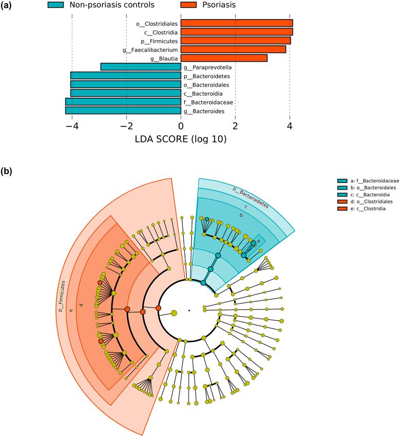
trols and 1.29 ± 0.81 in psoriasis patients (p = 0.0002). LefSe analysis revealed that these differences were mainly
driven by changes in the Bacteroides and Paraprevotella genus which were more abundant in non-psoriasis con-
trols while Faecalibacterium and Blautia in psoriasis patients (logarithmic LDA scores threshold was 2.0) (Fig. 3).
We did not observe significant differences in gut microbiota associated with changes in age, weight and BMI.
Mild vs moderate‑to‑severe psoriasis. Species richness in moderate-to-severe psoriasis patients was
lower comparing with mild psoriasis patients (p = 0.049). Comparing the principal phyla detected, we did not
find differences between both psoriasis groups (Supplementary Fig. S1).
We did not find differences in beta-diversity in mild vs moderate-to-severe psoriasis patients Supplementary
Fig. S2. We did not observe significant differences for age, gender, age at psoriasis onset, years with psoriasis,
hypertension, diabetes, weight, BMI, PASI and BSA. Only significant differences were found for metabolic syn-
drome (p = 0.002) in unweighted analysis.
Psoriasis‑Microbial index. Considering the results of relative abundance of different taxa, we generated
the PMI to discriminate between psoriasis and non-psoriasis controls (Fig. 4a). We evaluated its applicability by
ROC analysis. The Area Under the Curve (AUC) for the classification of Psoriasis (training dataset) was 0.797
(Fig. 4b), determining an optimal cut-off value of PMI = − 1.00 (sensitivity = 0.78 and specificity = 0.79) (Fig. 4c).
When we applied the PMI according to psoriasis severity, the AUC was 0.849 and 0.743 for mild and moderate-
to-severe psoriasis respectively.
Meta‑analysis. We validated this PMI using datasets from previously reported literature on PubMed. We
identified 7 related 16S datasets6,17,26,27,29,34,35. Only the study of Hidalgo-Cantabrana et al.34 fulfilled the inclusion
criteria.
When we applied the PMI to the downloaded sequence data from Hidalgo-Cantabrana et al.(test dataset)34
(Fig. 4a), the AUC was 0.953, Sensitivity = 0.89 and Specificity = 0.90, using cut-off value obtained with our
dataset (PMI = − 1.00);(Fig. 4b). Sensitivity vs Specificity curves of both datasets were plotted in Fig. 4c showing
Scientific Reports | (2020) 10:12754 | https://doi.org/10.1038/s41598-020-69537-3 3
Vol.:(0123456789)www.nature.com/scientificreports/
Figure 1. PCoA of beta-diversity values (Weighted Unifrac distances). Comparison of the gut microbiota from
psoriasis patients and non-psoriasis controls. Ellipses show 95% confidence intervals.
Figure 2. Bar plot showing the relative abundance of phyla distribution of each operational taxonomic unit
(OTU) within samples.
Scientific Reports | (2020) 10:12754 | https://doi.org/10.1038/s41598-020-69537-3 4
Vol:.(1234567890)www.nature.com/scientificreports/
Figure 3. Plot from LEfSe analysis indicating enriched bacterial genus associated either with psoriasis patients
(red) or non-psoriasis controls (blue). The length of the bar column represents the LDA score (a). Cladogram
plotted from LEfSe analysis showing the differences in relative abundance of taxa at five levels between psoriasis
patients vs non-psoriasis controls. (P < .05; LDA score 2) (b).
concordant results, indicating that PMI would be a powerful tool capable of discriminating between patients
with psoriasis and controls from different populations.
Discussion
Intestinal dysbiosis is a possible actor in chronic inflammation, even in distant tissue sites, such as the skin. Imbal-
ance in gut microbiota induces epithelial changes resulting in increased intestinal inflammation and altered gut
permeability, which in susceptible individuals may trigger the development of different chronic disease states
Scientific Reports | (2020) 10:12754 | https://doi.org/10.1038/s41598-020-69537-3 5
Vol.:(0123456789)www.nature.com/scientificreports/
Figure 4. PMI distinguishes non psoriasis controls from psoriasis patients. Violin plot showing PMI in control
and psoriasis fecal samples (a). Performance of cross-city prediction using each city-specific AD diagnosis
model, as assessed via the area under the ROC curve (AUROC). The ROC curve of tenfold cross-validation was
marked as blue lines and the ROC curve of the prediction as red lines. Performance of PMI, assessed via the area
under the ROC curve (AUROC) (b). Sensitivity and specificity vs. PMI (Cutoff) plot in both populations (c).
such as IBD, obesity, diabetes, multiple sclerosis, atopic dermatitis and cancer, among others45–50. However, until
now, only a few studies have addressed this question in p soriasis6,17,26,27,29,34–36,33.
Our work demonstrates that there are differences in gut microbiota between psoriasis patients and non-
psoriasis controls. We evaluated 55 untreated chronic plaque psoriasis patients (27 with moderate-to-severe
psoriasis and 28 with mild disease), being according to our knowledge the study with the highest number of
psoriasis patients and the first which evaluates changes in gut microbiota according to psoriasis severity based
on well-defined strict criteria. We made a comparison of our study design with all the available publications on
gut microbiota and psoriasis up to March 31, 2020 (Table 3).
Alpha-diversity has been observed to be decreased in a dysbiotic gut51. A lower microbial diversity has been
found in some psoriasis studies6,29,34 but not by other investigators17,26,27,35. Our study does not show a lower
alpha-diversity.
We found that Bacteroidetes and Firmicutes were the most prevalent phyla in patients and controls. How-
ever, there were significant differences between both phyla in psoriasis patients. The Firmicutes to Bacteroidetes
ratio was 1.29 ± 0.81 in psoriasis patients and 0.63 ± 0.32 in non-psoriasis controls. In line with our results, other
investigations showed a high Firmicutes:Bacteroidetes ratio6,27,29,34,35.
Short Chain Fatty Acid (SCFA) like acetate, propionate and butyrate, are known to regulate not only gut
specific but also distant inflammatory responses through the induction of immune cells52. An increase in
Firmicutes:Bacteroidetes ratio has been implicated in a higher acetate and lower butyrate production. Butyrate
is the preferred fuel for the colonic epithelial cells and the major regulator of cell proliferation and differentiation,
and has important anti-inflammatory, antioxidant and anti-carcinogenic functions53. Low levels of butyrate may
affect the integrity of the mucous layer compromising the gut epithelial barrier and enhance chronic colonic
and systemic i nflammation54.
Beta-diversity showed that genus Faecalibacterium and Blautia (both belong to the phylum Firmicutes, class
Clostridia and order Clostridiales) were the most relevant genus in psoriasis patients that discriminated against
non-psoriasis controls. Faecalibacterium prausnitzii (F. prausnitzii) can regulate T helper 17 cell (Th17)/regula-
tory T cell (Treg) differentiation and has been consistently reported as one of the main butyrate producers found
Scientific Reports | (2020) 10:12754 | https://doi.org/10.1038/s41598-020-69537-3 6
Vol:.(1234567890)www.nature.com/scientificreports/
Hidalgo-
Our study Codoñer et al.20 Tan et al.29 Cantabrana et al.37 Chen et al.30 Huang et al.6 Scher et al.32 Shapiro et al.38
Publication year 2018 2018 2019 2018 2018 2015 2019
Caucasian/Argen-
Population Caucasian/Spain Asian/China Caucasian/Spain Asian/China Asian/China Caucasian/US Caucasian/Israel
tine
Psoriasis patients (n) 55 52 14 19 35 32 15 Ps / 16 PsA 24
Non-Psoriasis
27 300 (from HMP) 14 20 27 64 17 22
controls (n)
Plaque psoriasis
yes yes yes yes yes no NA NA
exclusive
Age, sex & comor-
Matchead by Age, sex & BMI No No Age Age, sex & BMI No Age & sex
bidities
Active systemic
No No NA No Yes Yes No Yes
treatment
Stratified by severity Yes No No No NA Yes No No
Concomitant PsA no NA NA NA yes no yes NA
16S region analyzed V3–V4 V3–V4 V4 V2–V3 V3–V4 V4–V5 V1–V2 V4
Platform Illumina Illumina Illumina Ion Chef Illumina Illumina Illumina Illumina
Average reads 152,939 ~ 85,000 ~ 30,000 233,113 ~ 85,000 NA NA ~ 50,000
Avaiable upon
Raw data available at PRJNA574485 request to the cor- NA PRJNA517056 PRJNA379878 NA NA NA
responding author
Table 3. Study design of the available publications included in the meta-analysis (up to March 31, 2020).
in the intestine53,55. The role of F. prausnitzii in maintaining immune and physiological functions promoted this
bacterium as a next generation p robiotic56.
In psoriasis, a decrease in relative abundance of F. Prausnitzii has been reported in some s tudies30,34, but not
by other investigators17,27,35. In our study the genus Faecalibacterium showed higher values in psoriasis patients.
Lopez-Siles et al. determined that F. prausnitzii includes two phylogroups and recent studies suggest that other
Faecalibacterium genus and species could not be ruled out53,57,58. The relative abundance, as well as which phylum
and species of Faecalibacterium population are disbalanced in different diseases, makes it difficult to establish
the use of a single bacteria as a general biomarker for all diseases. The use of F. prausnitzii as a gold standard of
a healthy gut microbiota is limited53.
The genus Blautia includes obligate anaerobic intestinal commensal bacteria that belong to the family Lach-
nospiraceae and includes more than 100 different s pecies59,60. Blautia are important members of the healthy
human gut m icrobiota61. Jenq et al. found a lower mortality due to a graft versus-host disease after allogeneic
blood/marrow transplantation among patients with high abundance of Blautia and Bajaj et al.found that Blautia
was one of the bacteria associated with improved outcomes in patients with liver cirrhosis62,63. Genus Blautia
has been also related to cancer. Chen et al.reported a detrimental association between lower concentrations of
Blautia in the gut and colorectal c ancer64. On the contrary, Luu et al. found that higher levels of Blautia were
associated with poor prognosis in patients with early-stage breast c ancer65. Considering the limited data avail-
able on Blautia and the huge number of species reported, we can not explain the reasons why genus Blautia was
increased in our work. Additional data are required to determine their true role in human diseases.
The fact that most relevant genus in psoriasis patients that discriminated against non-psoriasis controls were
Faecalibacterium and Blautia, taxa producing high levels of butyrate, contradicts the traditional association of
butyrate producers observed in diseases such as IBD66,67. Therefore, results highlight the need for additional
research given the observational nature and limits of 16S used in this study.
In our control group, the predominant genus were Bacteroides and Paraprevotella. These bacteria differ only in
family (Bacteroideaceae for Bacteroides and Prevotellaceae for Paraprevotella)68,69. Increasing evidence proposes
that Bacteroides harness complex recalcitrant g lycans70. SCFAs are the major metabolic products of anaerobic
fermentation of glycans by gut bacteria and have been shown to impact on the host p hysiology71. The beneficial
effect of Bacteroides is consistent with our findings, where this genus was increased in controls and depleted in
psoriasis patients.
There is evidence that age, diet, geographical location, genetics and antibiotics, among other factors, influence
gut microbiota72,73. We selected unrelated controls matched by sex, age and BMI to moderate-to-severe psoriasis
patients, living in the same area and with a similar diet in order to reduce those confounding factors. We did not
find differences in beta-diversity according to personal features, so we postulate that changes in gut microbiota
would then be dependent on psoriasis and not on other covariates.
We found that patients with moderate-to-severe psoriasis had a lower diversity (species richness) than patients
with mild disease, although this difference was subtle. Only Huang et al.also studied whether the composition
of the intestinal microbiota differed depending on the severity of the disease and they found that the genus
Bacteroides was increased in patients with psoriasis and that it was characteristic of the subgroup with severe
disease6. In our study, the genus Bacteroides was found to be diminished in patients with psoriasis but no dif-
ferences were found between mild and moderate-to-severe psoriasis patients. For example, these distinctions
could be due to different inclusion criteria.
Scientific Reports | (2020) 10:12754 | https://doi.org/10.1038/s41598-020-69537-3 7
Vol.:(0123456789)www.nature.com/scientificreports/
When we compared whether the microbiota of patients with mild psoriasis vs patients with moderate-to-
severe psoriasis was affected by age, sex, age at onset of the disease, years of illness and comorbidities such as
hypertension or diabetes, we could not establish differences between both severity groups. These results could
also explain that changes of the gut microbiota in psoriasis would be dependent on the presence of the disease
and would not be affected by its severity.
Codoñer et al., Shapiro et al.and Hidalgo-Cantabrana et al.reported similar results to our study regarding
the bacteria genus increased in psoriasis and controls17,34,35. This concordance suggests that there is probably
a core gut microbiota in psoriasis patients. Unfortunately, not all the studies met the inclusion criteria for the
meta-analysis. Codoñer et al. did not use a control group from the same geographic location as they used pub-
licly available data from The Human Microbiome Project and the raw data from Shapiro et al.were not available.
This serves as another example of the importance of unrestricted access to raw sequencing data, which has been
already recognized by the scientific c ommunity74. Despite variations among Hidalgo-Cantabrana et al.and our
study, a psoriasis model can be applied across populations from different geographical locations. The proposed
PMI proved to be able to discriminate between psoriasis and controls across cities and continents with an optimal
cut-off value of PMI = − 1.00.
Given that general dermatologists are able to make a diagnosis of psoriasis with a simple physical exam,
the diagnostic applicability of the test will have to await further clinical experience. The PMI represents, a step
forward as a combined practical, ready to use, clinical and research tool. The index will allow us to gain more
knowledge on the microbial component of psoriasis and provide the possibility of increasing our understanding
of the role played by the microbiome in the disease process. Moreover, as PMI was only tested in 2 cohorts of
non-treated patients, we cannot exclude its role as a biomarker for evaluating treatment response. Further studies
of metagenome shotgun sequencing at the species/strain levels might be useful for the update and improvement
of the developed PMI.
In summary, our findings demonstrate variations in gut microbiota profiles between non-treated plaque
psoriasis patients and non-psoriasis controls. This results suggest that it is likely that altered gut microbiota
plays a pathophysiological role in psoriasis. However, whether modulation of gut microbiota could modify the
course of the disease remains to be explored. This study is unique in being the first to propose a PMI with the
ability to discriminate between psoriasis patients and age-sex-and BMI matched controls and between samples
from communities of different continents. Further studies are needed to better interpret the role of the PMI as a
potential biomarker test in psoriasis, and to test this index in larger and diverse populations to confirm its validity.
Methods
Study participants. This cross-sectional study recruited unrelated individuals, including consecutive
chronic plaque psoriasis patients and non-psoriasis controls. Controls were matched to moderate-to-severe pso-
riasis patients according to sex, age (± 2 years) and Body Mass Index (BMI; ± 1). Participants were caucasian,
above 18 years old and from the same geographical location. Samples were collected between October 2017 and
April 2018.
Psoriasis patients were subdivided based on their severity in mild and moderate-to-severe psoriasis. Mild
psoriasis was defined as actual Body Surface Area covered by psoriasis (BSA) < 10%, Psoriasis Area and Sever-
ity Index (PASI) < 10, Investigator Global Assessment (IGA) < 3 and absence of episodes of moderate-to-severe
psoriasis in the past. Moderate-to-severe psoriasis was defined as BSA ≥ 10%, PASI ≥ 10 and IGA ≥ 3.
Two visits were conducted over a period of 4 weeks to take a detailed assessment of psoriasis, medical history,
and a complete physical exam, including PASI, IGA and BSA involvement. Type 1 psoriasis was defined if the
symptoms began on or before age 40 years; a BMI ≥ 25 was considered as excessive weight and BMI ≥ 30 as obesity.
Key exclusion criteria for psoriasis patients included concomitant diagnosis of psoriatic arthritis accord-
ing to CASPAR criteria, inflammatory bowel disease (IBD), current topical treatment, systemic treatment for
psoriasis (including phototherapy) 3 months previous to sample collection, assuming that immunosuppression
could modify gut microbiota.
The exclusion criteria for controls were the presence of other dermatosis, family history of psoriasis in first
degree relatives, immunological disorders, hypertension, fatty liver disease, diabetes mellitus, malignancy, any
other serious internal disease, smoking and alcohol abuse.
Exclusion criteria applied to all groups were: antibiotic therapy 3 months previous to sample collection,
extreme diet, consumption of probiotics, positive HIV test or any gastrointestinal tract surgery leaving perma-
nent residua.
Sample collection and DNA extraction. All participants were apprised for the stool sampling collection
method by receiving a standardized protocol for the collection of approximately 5 g of stool in a sterile bacterio-
static buffer t ube75. Participants were asked to collect samples 24 h before the second visit. DNA extraction was
performed from 200 mg of feces using QIAamp-PowerFecal DNA-Kit.
Comparison of microbial communities and sequence analysis. Hypervariable regions V3–V4 of
the 16S rRNA gene were amplified with primers 337F/805R and sequenced in paired-end mode using a MiSeq
sequencer (IlluminaⓇ), warranting an average of 152,939 sequences per sample.
De-multiplexed reads were quality trimmed using Trimmomatic(V0.36)76. Sequences generated were
analyzed using Quantitative Insights Into Microbial Ecology (QIIME) version 1.9.1 software package77. For
this purpose, the sequences obtained were compared with those from Greengenes 13_8 database78. Chimeric
sequences were filtered using VSEARCH79. Operative Taxonomic Units (OTUs) were assigned to each read with
an open_reference OTU picking process. SortMeRNA (v2.1)80 was used for the reference OTU picking steps
Scientific Reports | (2020) 10:12754 | https://doi.org/10.1038/s41598-020-69537-3 8
Vol:.(1234567890)www.nature.com/scientificreports/
(with sortmerna_coverage = 0.8) and sumaclust (v1.0.20)81 for the de novo OTU picking steps (with 10% of the
failures subsampled). Low-confidence OTUs called by < 0.1% of the reads were removed using the script remove_
low_confidence_otus.py82. An average of 29,872.93 ± 6,452.75 mapping high-quality sequences were obtained,
leading to 455.91 ± 126.99 unique OTUs per sample. For multiple comparisons, p-values were adjusted by Bonfer-
roni correction83. To compare microbial communities in different sample groups, we used Unifrac a lgorithm84.
Differences on beta-diversity were assessed using ADONIS. In order to compare the relative abundance of the
different taxa between groups, we performed Linear Discriminant Analysis (LDA) effect implemented in L EfSe85 .
Psoriasis‑Microbiome Index development. PMI was defined as the logarithm of total abundance of
organisms increased in psoriasis over total abundance of organisms decreased in psoriasis for all samples (at
genus level) using the compute_taxonomy_ratio.py s cript86. Then, we evaluated how these PMI performed for
classification subjects by psoriasis status through Receiver Operating Characteristic (ROC) analysis (training
dataset). ROC analysis was performed using ROCR package (RStudio version 1.1.453)87. Cut-off value was
selected as the point where the sensitivity and specificity functions intersect each other, i.e. jointly maximizing
the sensitivity and specificity of PMI.
Meta‑analysis. We performed a systematic literature search of PubMed databases up to March 31, 2020
using the following terms: “Psoriasis” and “gut microbiota” or “gut microbiome”. The study inclusion criteria
were: Case–control studies with publicly available raw 16S data and metadata, indicating case/control status for
each sample. Studies including patients with other clinical forms different from plaque psoriasis and patients
under systemic treatment (DMARDS and biologics) were excluded.
Data accession. Raw sequences of 16S rRNA gene reported in this article have been deposited in NCBI
Short Read Archive (SRA) and are accessible under the accession number PRJNA574485.
Ethical statement. This study received approval by the Ethics Committee of Hospital Español, Buenos
Aires Argentina according to local regulations and Helsinki declaration. Written informed consent was obtained
from all study participants.
Received: 24 January 2020; Accepted: 7 July 2020
References
1. Griffiths, C. E. M. & Barker, J. N. Pathogenesis and clinical features of psoriasis. The Lancet 370, 263–271 (2007).
2. Kurd, S. K. & Gelfand, J. M. The prevalence of previously diagnosed and undiagnosed psoriasis in US adults: results from NHANES
2003–2004. J. Am. Acad. Dermatol. 60, 218–224 (2009).
3. Rapp, S. R., Feldman, S. R., Exum, M. L., Fleischer, A. B. Jr. & Reboussin, D. M. Psoriasis causes as much disability as other major
medical diseases. J. Am. Acad. Dermatol. 41, 401–407 (1999).
4. Baliwag, J., Barnes, D. H. & Johnston, A. Cytokines in psoriasis. Cytokine 73, 342–350 (2015).
5. Singh, S. et al. Genomic alterations driving psoriasis pathogenesis. Gene 683, 61–71 (2019).
6. Huang, L. et al. Dysbiosis of gut microbiota was closely associated with psoriasis. Sci. China Life Sci. https://doi.org/10.1007/s1142
7-018-9376-6 (2018).
7. The Human Microbiome Project Consortium. Structure, function and diversity of the healthy human microbiome. Nature 486,
207–214 (2012).
8. Landman, C. & Quévrain, E. Gut microbiota: description, role and pathophysiologic implications. Rev. Med. Int. 37, 418–423
(2016).
9. Carrera-Quintanar, L. et al. The human microbiota and obesity: a literature systematic review of in vivo models and technical
approaches. Int. J. Mol. Sci. 19, 3827 (2018).
10. Chen, X. & Devaraj, S. Gut Microbiome in Obesity, Metabolic Syndrome, and Diabetes. Curr. Diab. Rep. 18, 129 (2018).
11. Gulas, E., Wysiadecki, G., Strzelecki, D., Gawlik-Kotelnicka, O. & Polguj, M. Can microbiology affect psychiatry? A link between
gut microbiota and psychiatric disorders. Psychiatr. Pol. 52, 1023–1039 (2018).
12. Gilis, E. et al. The role of the microbiome in gut and joint inflammation in psoriatic arthritis and spondyloarthritis. J. Rheumatol.
Suppl. 94, 36–39 (2018).
13. Cenit, M. C., Codoñer-Franch, P. & Sanz, Y. Gut microbiota and risk of developing celiac disease. J. Clin. Gastroenterol. 50(Suppl
2), S148–S152 (2016).
14. Takeshita, J. et al. Psoriasis and comorbid diseases: Epidemiology. J. Am. Acad. Dermatol. 76, 377–390 (2017).
15. Scarpa, R. et al. Microscopic inflammatory changes in colon of patients with both active psoriasis and psoriatic arthritis without
bowel symptoms. J. Rheumatol. 27, 1241–1246 (2000).
16. Ramírez-Boscá, A. et al. Identification of bacterial DNA in the peripheral blood of patients with active psoriasis. JAMA Dermatol.
151, 670–671 (2015).
17. Codoñer, F. M. et al. Gut microbial composition in patients with psoriasis. Sci. Rep. 8, 3812 (2018).
18. Visser, M. J. E., Kell, D. B. & Pretorius, E. Bacterial dysbiosis and translocation in psoriasis vulgaris. Front. Cell. Infect. Microbiol.
9, 7 (2019).
19. Blauvelt, A. & Chiricozzi, A. The immunologic role of IL-17 in psoriasis and psoriatic arthritis pathogenesis. Clin. Rev. Allergy
Immunol. 55, 379–390 (2018).
20. Atarashi, K. et al. Th17 cell induction by adhesion of microbes to intestinal epithelial cells. Cell 163, 367–380 (2015).
21. Wilck, N. et al. Salt-responsive gut commensal modulates TH17 axis and disease. Nature https://doi.org/10.1038/nature24628
(2017).
22. Flannigan, K. L. & Denning, T. L. Segmented filamentous bacteria-induced immune responses: a balancing act between host
protection and autoimmunity. Immunology https://doi.org/10.1111/imm.12950 (2018).
Scientific Reports | (2020) 10:12754 | https://doi.org/10.1038/s41598-020-69537-3 9
Vol.:(0123456789)www.nature.com/scientificreports/
23. Zákostelská, Z. et al. Intestinal microbiota promotes psoriasis-like skin inflammation by enhancing Th17 response. PLoS ONE 11,
e0159539 (2016).
24. Zanvit, P. et al. Antibiotics in neonatal life increase murine susceptibility to experimental psoriasis. Nat. Commun. 6, 8424 (2015).
25. Stehlikova, Z. et al. Crucial role of microbiota in experimental psoriasis revealed by a gnotobiotic mouse model. Front. Microbiol.
10, 236 (2019).
26. Tan, L. et al. The Akkermansia muciniphila is a gut microbiota signature in psoriasis. Exp. Dermatol. 27, 144–149 (2018).
27. Chen, Y.-J. et al. Intestinal microbiota profiling and predicted metabolic dysregulation in psoriasis patients. Exp. Dermatol. 27,
1336–1343 (2018).
28. Chang, H.-W. et al. Alteration of the cutaneous microbiome in psoriasis and potential role in Th17 polarization. Microbiome 6,
154 (2018).
29. Scher, J. U. et al. Decreased bacterial diversity characterizes the altered gut microbiota in patients with psoriatic arthritis, resembling
dysbiosis in inflammatory bowel disease. Arthritis Rheumatol 67, 128–139 (2015).
30. Eppinga, H. et al. Similar depletion of protective Faecalibacterium prausnitziiin psoriasis and inflammatory bowel disease, but not
in hidradenitis suppurativa. J. Crohn’s Colitis 10, 1067–1075 (2016).
31. Langan, E. A. et al. The role of the microbiome in psoriasis: moving from disease description to treatment prediction?. Br. J. Der-
matol. 178, e360–e360 (2018).
32. Tett, A. et al. Unexplored diversity and strain-level structure of the skin microbiome associated with psoriasis. NPJ Biofilms Micro-
biomes 3, 14 (2017).
33. Nakajima, S., Harrison, O., Merrill, E., Linehan, J. & Belkaid, Y. 648 Candida albicans colonization exacerbates skin inflammation
in a murine model of psoriasis. J. Invest. Dermatol. 137, S112 (2017).
34. Hidalgo-Cantabrana, C. et al. Gut microbiota dysbiosis in a cohort of patients with psoriasis. Br. J. Dermatol. https://doi.
org/10.1111/bjd.17931(2019).
35. Shapiro, J. et al. Psoriatic patients have a distinct structural and functional fecal microbiota compared with controls. J. Dermatol.
46, 595–603 (2019).
36. Quan, C. et al. Psoriatic lesions are characterized by higher bacterial load and imbalance between Cutibacterium and Corynebac-
terium. J. Am. Acad. Dermatol. https://doi.org/10.1016/j.jaad.2019.06.024 (2019).
37. Langan, E. A. et al. Combined culture and metagenomic analyses reveal significant shifts in the composition of the cutaneous
microbiome in psoriasis. Br. J. Dermatol. https://doi.org/10.1111/bjd.17989 (2019).
38. Stehlikova, Z. et al. Dysbiosis of skin microbiota in psoriatic patients: co-occurrence of fungal and bacterial communities. Front.
Microbiol. 10, 438 (2019).
39. Sayers, E., MacGregor, A. & Carding, S. R. Drug-microbiota interactions and treatment response: relevance to rheumatoid arthritis.
AIMS Microbiol 4, 642–654 (2018).
40. Aden, K. et al. Metabolic functions of gut microbes associate with efficacy of tumor necrosis factor antagonists in patients with
inflammatory bowel diseases. Gastroenterology https://doi.org/10.1053/j.gastro.2019.07.025 (2019).
41. Doherty, M. K. et al. Fecal microbiota signatures are associated with response to Ustekinumab therapy among Crohn’s disease
patients. mBio https://doi.org/10.1128/mBio.02120-17 (2018).
42. Goodrich, J. K. et al. Human genetics shape the gut microbiome. Cell 159, 789–799 (2014).
43. Turnbaugh, P. J. et al. A core gut microbiome in obese and lean twins. Nature 457, 480–484 (2009).
44. Takeshita, J. et al. Psoriasis and comorbid diseases: implications for management. J. Am. Acad. Dermatol. 76, 393–403 (2017).
45. Ni, J., Wu, G. D., Albenberg, L. & Tomov, V. T. Gut microbiota and IBD: causation or correlation?. Nature Reviews Gastroenterology
& Hepatology 14, 573–584 (2017).
46. Maruvada, P., Leone, V., Kaplan, L. M. & Chang, E. B. The human microbiome and obesity: moving beyond associations. Cell Host
Microbe 22, 589–599 (2017).
47. Patterson, E. et al. Gut microbiota, obesity and diabetes. Postgrad. Med. J. 92, 286–300 (2016).
48. Tremlett, H., Bauer, K. C., Appel-Cresswell, S., Finlay, B. B. & Waubant, E. The gut microbiome in human neurological disease: a
review. Ann. Neurol. 81, 369–382 (2017).
49. Song, H., Yoo, Y., Hwang, J., Na, Y.-C. & Kim, H. S. Faecalibacterium prausnitzii subspecies-level dysbiosis in the human gut
microbiome underlying atopic dermatitis. J. Allergy Clin. Immunol. 137, 852–860 (2016).
50. Saha, A. & Robertson, E. S. Microbiome and human malignancies. Microbiome Cancer https://doi.org/10.1007/978-3-030-4155-
7_1 (2019).
51. Chatelier, E. L. et al. Richness of human gut microbiome correlates with metabolic markers. Nature 500, 541–546 (2013).
52. Maslowski, K. M. & Mackay, C. R. Diet, gut microbiota and immune responses. Nat. Immunol. 12, 5–9 (2011).
53. Lopez-Siles, M., Duncan, S. H., Garcia-Gil, L. J. & Martinez-Medina, M. Faecalibacterium prausnitzii: from microbiology to
diagnostics and prognostics. ISME J. 11, 841–852 (2017).
54. Bach Knudsen, K. E. et al. Impact of diet-modulated butyrate production on intestinal barrier function and inflammation. Nutrients
10, 1499 (2018).
55. Zhou, L. et al. Faecalibacterium prausnitzii produces butyrate to maintain Th17/Treg balance and to ameliorate colorectal colitis
by inhibiting histone deacetylase 1. Inflamm. Bowel Dis. https://doi.org/10.1093/ibd/izy182 (2018).
56. Martín, R., Bermúdez-Humarán, L. G. & Langella, P. Searching for the bacterial effector: the example of the multi-skilled com-
mensal bacterium. Front. Microbiol. 9, 346 (2018).
57. Fitzgerald, C. B. et al. Comparative analysis of Faecalibacterium prausnitzii genomes shows a high level of genome plasticity and
warrants separation into new species-level taxa. BMC Genom. 19, 931 (2018).
58. Lopez-Siles, M. et al. Cultured representatives of two major phylogroups of human colonic Faecalibacterium prausnitzii can utilize
pectin, uronic acids, and host-derived substrates for growth. Appl. Environ. Microbiol. 78, 420–428 (2012).
59. Durand, G. A. et al. Blautia massiliensis sp. nov., isolated from a fresh human fecal sample and emended description of the genus
Blautia. Anaerobe 43, 47–55 (2017).
60. Website. https://www.ncbi.nlm.nih.gov/Taxonomy/Browser/wwwtax.cgi?mode=Undef&id=572511&lvl=3&keep=1&srchm
ode=1&unlock [accessed on 24 August 2019].
61. Touyama, M., Jin, J. S., Kibe, R., Hayashi, H. & Benno, Y. Quantification of Blautia wexlerae and Blautia luti in human faeces by
real-time PCR using specific primers. Benef. Microbes 6, 583–590 (2015).
62. Jenq, R. R. et al. Intestinal blautia is associated with reduced death from graft-versus-host disease. Biol. Blood Marrow Transplant.
21, 1373–1383 (2015).
63. Bajaj, J. S. et al. Colonic mucosal microbiome differs from stool microbiome in cirrhosis and hepatic encephalopathy and is linked
to cognition and inflammation. Am. J. Physiol. Gastrointest. Liver Physiol. 303, G675–G685 (2012).
64. Chen, W., Liu, F., Ling, Z., Tong, X. & Xiang, C. Human intestinal lumen and mucosa-associated microbiota in patients with
colorectal cancer. PLoS ONE 7, e39743 (2012).
65. Luu, T. H. et al. Intestinal proportion of Blautia sp. is associated with clinical stage and histoprognostic grade in patients with
early-stage breast cancer. Nutr. Cancer 69, 267–275 (2017).
66. Lavelle, A. & Sokol, H. Gut microbiota-derived metabolites as key actors in inflammatory bowel disease. Nat. Rev. Gastroenterol.
Hepatol. 17, 223–237 (2020).
67. Lloyd-Price, J. et al. Multi-omics of the gut microbial ecosystem in inflammatory bowel diseases. Nature 569, 655–662 (2019).
Scientific Reports | (2020) 10:12754 | https://doi.org/10.1038/s41598-020-69537-3 10
Vol:.(1234567890)www.nature.com/scientificreports/
68. Taxonomy. Taxonomy browser (Bacteroides). https: //www.ncbi.nlm.nih.gov/Taxono my/Browse r/wwwtax .cgi?mode=Info&id=816.
69. Website. Taxonomy browser (Paraprevotella) [WWW Document]. URL https: //www.ncbi.nlm.nih.gov/Taxono my/Browse r/wwwta
x.cgi?mode=Info&id=577309&lvl=3&lin=f&keep=1&srchmode=1&unlock [accessed on 24 August 2019].
70. Luis, A. S. et al. Dietary pectic glycans are degraded by coordinated enzyme pathways in human colonic Bacteroides. Nat Microbiol
3, 210–219 (2018).
71. Singh, R. P. Glycan utilisation system in Bacteroides and Bifidobacteria and their roles in gut stability and health. Appl. Microbiol.
Biotechnol. https://doi.org/10.1007/s00253-019-10012-z (2019).
72. Lagier, J.-C., Million, M., Hugon, P., Armougom, F. & Raoult, D. Human gut microbiota: repertoire and variations. Front. Cell.
Infect. Microbiol. 2, 136 (2012).
73. Belforte, F. S. et al. Getting to know the gut microbial diversity of metropolitan Buenos Aires inhabitants. Front. Microbiol. 10, 965
(2019).
74. Langille, M. G. I., Ravel, J. & Florian Fricke, W. ‘Available upon request’: not good enough for microbiome data!. Microbiome https
://doi.org/10.1186/s40168-017-0394-z (2018).
75. Gray, M. A., Pratte, Z. A. & Kellogg, C. A. Comparison of DNA preservation methods for environmental bacterial community
samples. FEMS Microbiol. Ecol. 83, 468–477 (2013).
76. Bolger, A. M., Lohse, M. & Usadel, B. Trimmomatic: a flexible trimmer for Illumina sequence data. Bioinformatics 30, 2114–2120
(2014).
77. Caporaso, J. G. et al. QIIME allows analysis of high-throughput community sequencing data. Nat. Methods 7, 335–336 (2010).
78. DeSantis, T. Z. et al. Greengenes, a chimera-checked 16S rRNA gene database and workbench compatible with ARB. Appl. Environ.
Microbiol. 72, 5069–5072 (2006).
79. Rognes, T., Flouri, T., Nichols, B., Quince, C. & Mahé, F. VSEARCH: a versatile open source tool for metagenomics. PeerJ 4, e2584
(2016).
80. Kopylova, E., Noé, L. & Touzet, H. SortMeRNA: fast and accurate filtering of ribosomal RNAs in metatranscriptomic data. Bioin-
formatics 28, 3211–3217 (2012).
81. Mercier, C., Boyer, F., Bonin, A. & Coissac, E. SUMATRA and SUMACLUST: fast and exact comparison and clustering of sequences.
GitLab https://git.metabarcoding.org/obitools/sumaclust/wikis/home (2013).
82. Comeau, A. M., Douglas, G. M. & Langille, M. G. I. Microbiome Helper: a custom and streamlined workflow for microbiome
Research. mSystems https://doi.org/10.1128/mSystems.00127-16 (2017).
83. Benjamini, Y. & Hochberg, Y. Controlling the false discovery rate: a practical and powerful approach to multiple testing. J. R. Stat.
Soc. B 57, 289–300 (1995).
84. Lozupone, C. A. & Knight, R. The UniFrac significance test is sensitive to tree topology. BMC Bioinform. 16, 211 (2015).
85. Segata, N. et al. Metagenomic biomarker discovery and explanation. Genome Biol. 12, R60 (2011).
86. Gevers, D. et al. The treatment-naive microbiome in new-onset Crohn’s disease. Cell Host Microbe 15, 382–392 (2014).
87. Sing, T., Sander, O., Beerenwinkel, N. & Lengauer, T. ROCR: visualizing classifier performance in R. Bioinformatics 21, 3940–3941
(2005).
Acknowledgements
This research was supported by Novartis Pharma, CAIN457AAR02T. The authors thank Dr. Federico Rey for his
constructive comments and suggestions and the “Centro de Investigación, Docencia y Extensión en Tecnologías
de la Información y las Comunicaciones” (CIDETIC, http:/cidetic.unlu.edu.ar/), Universidad Nacional de Luján,
Luján, Argentina for human and computational resources. We are grateful to Fundación H.A. Barceló, Instituto
Universitario de Ciencias de la Salud, CABA, Buenos Aires, for excellent technical support.
Author contributions
I.D., F.G., H.D. and A.P.S. designed the study. I.D. performed the recruitment of the volunteers. I.D., F.G. and
L.L. collected the stool samples and the extraction of fecal bacterial DNA. A.P.S. processed the raw sequences
and performed the bioinformatic and statistical analysis. I.D., F.G. and A.P.S. analyzed the results. All authors
wrote and reviewed the manuscript.
Competing interests
Dr Dei-Cas has received compensation as a speaker, consultant, and investigator for Novartis, Eli Lilly and
Janssen. Dr Penas-Steinhardt has received compensation as a speaker for Novartis. Dr Florecina Giliberto, Dr.
Leonela Luce and Dr. Hernán Dopazo declare no competing interest.
Additional information
Supplementary information is available for this paper at https://doi.org/10.1038/s41598-020-69537-3.
Correspondence and requests for materials should be addressed to A.P.-S.
Reprints and permissions information is available at www.nature.com/reprints.
Publisher’s note Springer Nature remains neutral with regard to jurisdictional claims in published maps and
institutional affiliations.
Open Access This article is licensed under a Creative Commons Attribution 4.0 International
License, which permits use, sharing, adaptation, distribution and reproduction in any medium or
format, as long as you give appropriate credit to the original author(s) and the source, provide a link to the
Creative Commons license, and indicate if changes were made. The images or other third party material in this
article are included in the article’s Creative Commons license, unless indicated otherwise in a credit line to the
material. If material is not included in the article’s Creative Commons license and your intended use is not
permitted by statutory regulation or exceeds the permitted use, you will need to obtain permission directly from
the copyright holder. To view a copy of this license, visit http://creativecommons.org/licenses/by/4.0/.
© The Author(s) 2020
Scientific Reports | (2020) 10:12754 | https://doi.org/10.1038/s41598-020-69537-3 11
Vol.:(0123456789)You can also read