PLASMA NFL LEVELS AND LONGITUDINAL CHANGE RATES IN C9ORF72 AND GRN-ASSOCIATED DISEASES: FROM TAILORED REFERENCES TO CLINICAL APPLICATIONS
←
→
Page content transcription
If your browser does not render page correctly, please read the page content below
Neurogenetics
J Neurol Neurosurg Psychiatry: first published as 10.1136/jnnp-2021-326914 on 4 August 2021. Downloaded from http://jnnp.bmj.com/ on September 17, 2021 by guest. Protected by
Original research
Plasma NfL levels and longitudinal change rates in
C9orf72 and GRN-associated diseases: from tailored
references to clinical applications
Dario Saracino ,1,2,3 Karim Dorgham ,4 Agnès Camuzat,1,5 Daisy Rinaldi,1,2
Armelle Rametti-Lacroux,6 Marion Houot,1,2,7 Fabienne Clot,8 Philippe Martin-Hardy,1
Ludmila Jornea,1 Carole Azuar,1,2,6 Raffaella Migliaccio ,1,2,6 Florence Pasquier,9
Philippe Couratier,10 Sophie Auriacombe,11 Mathilde Sauvée,12
Claire Boutoleau-Bretonnière,13 Jérémie Pariente,14,15 Mira Didic,16,17
Didier Hannequin,18 David Wallon,18 the French Research Network on FTD/FTD-ALS,
the PREV-DEMALS and Predict-PGRN study groups, Olivier Colliot,1,3 Bruno Dubois,1,2,6
Alexis Brice,1 Richard Levy,1,2,6 Sylvie Forlani,1 Isabelle Le Ber 1,2,6
►► Additional supplemental ABSTRACT INTRODUCTION
material is published online Objective Neurofilament light chain (NfL) is a GRN and C9orf72 gene mutations are the main
only. To view, please visit
the journal online (http://dx. promising biomarker in genetic frontotemporal dementia genetic causes of frontotemporal dementia (FTD)
doi.org/10.1136/jnnp-2021- (FTD) and amyotrophic lateral sclerosis (ALS). We and/or amyotrophic lateral sclerosis (ALS).1–4
326914). evaluated plasma neurofilament light chain (pNfL) levels GRN-associated phenotypes are dominated by the
in controls, and their longitudinal trajectories in C9orf72 behavioural variant of FTD (bvFTD),5 whereas
For numbered affiliations see
end of article.
and GRN cohorts from presymptomatic to clinical stages. C9orf72 expansions lead to bvFTD, ALS or a
Methods We analysed pNfL using Single Molecule combination of both.3 5 Less typical C9orf72-
Array (SiMoA) in 668 samples (352 baseline and 316 related phenotypes are characterised by psychiatric
copyright.
Correspondence to
Dr Isabelle Le Ber, Sorbonne follow-up) of C9orf72 and GRN patients, presymptomatic disorders6 or by a very slowly progressive disease in
Université, Paris Brain Institute– carriers (PS) and controls aged between 21 and 83. a subset of carriers.7 8
Institut du Cerveau (ICM), They were longitudinally evaluated over a period of A new era is emerging in genetic FTD and
Inserm U1127, CNRS UMR
7225; Reference Centre for >2 years, during which four PS became prodromal/ ALS, with the development of GRN and C9orf72
Rare or Early Dementias, IM2A, symptomatic. Associations between pNfL and clinical– disease-modifying therapies. The presymptomatic
Département de Neurologie, genetic variables, and longitudinal NfL changes, were or prodromal phases appear to be the ideal time
Hopital Universitaire Pitie investigated using generalised and linear mixed-effects to deliver preventive treatments, before emer-
Salpetriere, Paris, France;
models. Optimal cut-offs were determined using the gence of overt clinical manifestations. In this fast-
isabelle.leber@upmc.fr
Youden Index. moving context, detecting progression since disease
Received 19 April 2021 Results pNfL levels increased with age in controls, beginning, at the biological level, up to full-blown
Accepted 13 July 2021 from ~5 to~18 pg/mL (p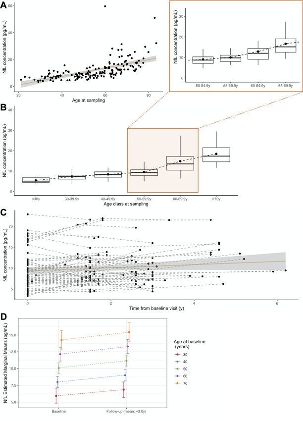
Neurogenetics
J Neurol Neurosurg Psychiatry: first published as 10.1136/jnnp-2021-326914 on 4 August 2021. Downloaded from http://jnnp.bmj.com/ on September 17, 2021 by guest. Protected by
Table 1 Descriptive data of the studied population
Patients PS
Controls Overall C9orf72 GRN Overall C9orf72 GRN
N 165 102 54 48 85 48 37
Gender (F/M) 96/69 46/56 24/30 22/26 52/33 30/18 22/15
Disease phenotype –
FTD (N) – 75 27* 48 – –
ALS (N) 6 6† –
FTD/ALS (N) 10 10‡ –
Psychiatric (N) 11 11§ –
Age at disease onset – 58.0 (53.0–64.8) 58.0 (50.3–67.0) 58.0 (54.8–63.0) – – –
(years)
Age at baseline sampling 56.5 (45.9–66.3) 62.9 (58.3–69.6) 64.4 (58.0–71.5) 62.1 (58.5–66.2) 41.2 (34.2–47.5) 42.0 (34.4–47.4) 40.9 (33.2–48.8)
(years)
Age at baseline, range 21.1–83.5 35.5–79.9 39.8–79.9 35.5–76.2 20.4–79.4 24.0–79.4 20.4–68.8
(years)
Disease duration at – 3.5 (2.3–5.9) 5.1 (2.9–9.0)¶ 2.9 (2.2–3.5)¶ – – –
sampling (years)
pNfL at baseline (pg/mL) 9.88 (7.42–14.36)** 66.25 (33.74–98.86)** 39.49 (23.89–74.42)†† 86.21 (58.17–118.13)†† 8.08 (6.08–10.10)** 8.48 (6.71–11.52) 7.70 (5.59–9.23)
Mean (±SD) pNfL at 12.08 (±7.57)** 81.21 (±75.99)** 64.52 (±63.92)†† 99.99 (±84.40)†† 8.79 (±4.02)** 9.76 (±4.69)‡‡ 7.52 (±2.44)‡‡
baseline
Individuals with follow- 65 44 29 15 66 43 23
up (N)
Mean (±SD) follow-up 2.96 (±1.16) 2.00 (±1.21) 1.95 (±1.26) 2.08 (±1.13) 2.99 (±1.30) 2.83 (±0.65) 3.29 (±2.01)
duration (years)
Mean ARC (%) +3.9** +26.7** +24.7†† +29.3†† +3.2** +3.2 +3.3
Values are indicated as median and IQR, except where differently specified. There were no statistically significant differences between the groups, apart from specific occurrences, as follows.
*3/27 patients with FTD had SP course.
†2/6 patients with ALS had SP course.
‡3/10 patients with FTD/ALS had SP course.
§4/11 patients with psychiatric presentations had SP course.
¶Different disease duration at baseline between C9orf72 and GRN patients (p=0.0001).
**Higher values in patients compared with controls (p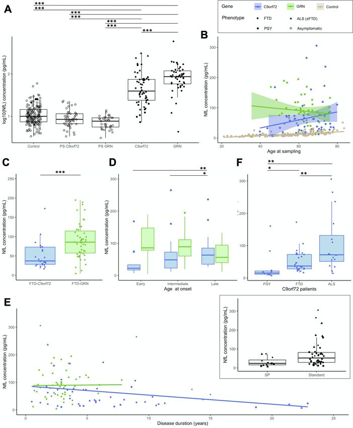
Neurogenetics
J Neurol Neurosurg Psychiatry: first published as 10.1136/jnnp-2021-326914 on 4 August 2021. Downloaded from http://jnnp.bmj.com/ on September 17, 2021 by guest. Protected by
the follow-up as they developed subtle cognitive/behavioural To perform unbiased longitudinal analyses in patients and in
and/or motor symptoms, and reached CDR+NACC-FTLD=0.5 PS, we selected separate subgroups of controls based on demo-
(online supplemental table A1). graphic features and follow-up duration (online supplemental
For a subgroup analysis in C9orf72 patients, we included five tables A2 and A3).
patients with primary psychiatric disorders whose demographic All groups were split to separately analyse 10-year discrete age
data were comparable to the former. classes, from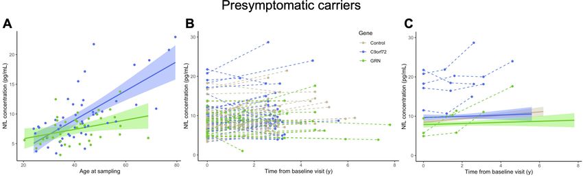
Neurogenetics

                                                                                                                                                                       J Neurol Neurosurg Psychiatry: first published as 10.1136/jnnp-2021-326914 on 4 August 2021. Downloaded from http://jnnp.bmj.com/ on September 17, 2021 by guest. Protected by
                                                                                                                                                                                                                                                         copyright.

Figure 1 pNfL levels in controls. (A) Association of pNfL levels with the age at sampling (r=0.766, p
Neurogenetics
J Neurol Neurosurg Psychiatry: first published as 10.1136/jnnp-2021-326914 on 4 August 2021. Downloaded from http://jnnp.bmj.com/ on September 17, 2021 by guest. Protected by
Genotype-specific and age-specific cut-offs
Table 2 Plasma neurofilament light chain levels in each of the age
We determined cut- off values discriminating patients from
classes in controls
controls (table 3 and online supplemental figure A2). Given
Median the distinct gene-specific trajectories, we separately determined
Age class (50th
thresholds for C9orf72 and for GRN patients. A cut- off at
(years) N 5th P 25th P percentile) 75th P 95th P
19.00 pg/mL yielded the best sensitivity/specificity trade-off toNeurogenetics
J Neurol Neurosurg Psychiatry: first published as 10.1136/jnnp-2021-326914 on 4 August 2021. Downloaded from http://jnnp.bmj.com/ on September 17, 2021 by guest. Protected by
copyright.
Figure 2 Baseline pNfL levels in patients. (A) pNfL levels in C9orf72 and GRN patients compared with presymptomatic carriers and controls. (B) pNfL
levels according to the age at sampling in C9orf72 (r=0.284, p=0.037) and in GRN (r=−0.123, p=0.406) patients, with controls displayed for comparison.
(C) Comparison of pNfL levels between C9orf72 and GRN patients, restricting the analysis to those with FTD phenotype only. (D) Comparison of pNfL levels
according to the age at onset, classified as early (before 50 years), intermediate (between 50 and 65 years) and late (after 65 years). Levels significantly
differed in C9orf72 patients, but not in GRN patients. (E) pNfL levels according to disease duration, evidencing a negative correlation in C9orf72 patients
(r=−0.311, p=0.021) but not in GRN patients (r=0.088, p=0.552). In the insert, C9orf72 carriers with atypical, SP disease course are compared with
patients with standard disease duration. (F) Comparison of pNfL levels according to clinical phenotype in C9orf72 patients; patients with ALS were
considered as a unique group, regardless of the presence of associated FTD. Asterisks indicate the significance of post hoc comparisons between the
groups: *pNeurogenetics
J Neurol Neurosurg Psychiatry: first published as 10.1136/jnnp-2021-326914 on 4 August 2021. Downloaded from http://jnnp.bmj.com/ on September 17, 2021 by guest. Protected by
Figure 3 Longitudinal pNfL changes in patients and controls. (A) Mean baseline and follow-up pNfL levels in 44 patients and 36 controls with comparable
demographic variables undergoing longitudinal sampling (mean follow-up: 2 years). There was greater increase in C9orf72 and GRN patients compared with
controls (pNeurogenetics
J Neurol Neurosurg Psychiatry: first published as 10.1136/jnnp-2021-326914 on 4 August 2021. Downloaded from http://jnnp.bmj.com/ on September 17, 2021 by guest. Protected by
Figure 4 Baseline pNfL levels and longitudinal changes in presymptomatic carriers. (A) pNfL levels at baseline according to the age at sampling in
C9orf72 (r=0.651, pNeurogenetics
J Neurol Neurosurg Psychiatry: first published as 10.1136/jnnp-2021-326914 on 4 August 2021. Downloaded from http://jnnp.bmj.com/ on September 17, 2021 by guest. Protected by
copyright.
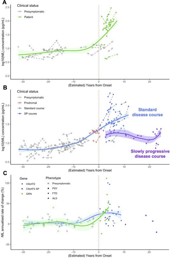
Figure 5 Modelisation of pNfL trajectories and progression rates over the entire disease course, from presymptomatic phase to clinical phase, in GRN and
C9orf72 carriers. (A,B) pNfL levels at baseline and at follow-up visits in presymptomatic and symptomatic carriers of GRN (A) and C9orf72 (B) mutations,
at individual and group levels, according to their clinical status and their (estimated) distance to/from disease onset. (C) pNfL annualised rates of change
(%) in presymptomatic and symptomatic GRN and C9orf72 carriers according to their (estimated) distance to/from disease onset. Patients are classified
according to their phenotype. Among C9orf72 patients, those with SP disease course are presented in a different colour. On the x axis, the disease duration
from onset is given for patients, and the estimated years to clinical onset is given for presymptomatic carriers. Estimated years to onset were calculated for
each individual, taking into account the mean age of disease onset in his/her family. For prodromal C9orf72 carriers, the age at their first subtle cognitive/
behavioural and/or motor symptoms was considered. ALS, amyotrophic lateral sclerosis; FTD, frontotemporal dementia; NfL, neurofilament light chain; pNfL,
plasma neurofilament light chain; PSY, psychiatric presentations; SP, slowly progressive.
Saracino D, et al. J Neurol Neurosurg Psychiatry 2021;0:1–11. doi:10.1136/jnnp-2021-326914 9Neurogenetics
J Neurol Neurosurg Psychiatry: first published as 10.1136/jnnp-2021-326914 on 4 August 2021. Downloaded from http://jnnp.bmj.com/ on September 17, 2021 by guest. Protected by
ALS and the proposed thresholds should be used to predict clin- Dubois (Hôpital Pitié-Salpêtrière, Paris), Charles Duyckaerts (Hôpital Pitié-Salpêtrière,
ical evolution in presymptomatic carriers only when the muta- Paris), Frédérique Etcharry-Bouyx (CHU Angers), Maïté Formaglio (CHU Lyon),
Véronique Golfier (CHU Rennes), Didier Hannequin (CHU Charles Nicolle, Rouen),
tion status is known. They are not intended to be used in sporadic Lucette Lacomblez (Hôpital Pitié-Salpêtrière, Paris), Isabelle Le Ber (Hôpital Pitié-
forms, or when other diseases are in the differential diagnosis. Salpêtrière, Paris), Bernard-François Michel (CH Sainte-Marguerite, Marseille),
In the modelisation of pNfL trajectories, the estimation of the Jérémie Pariente (CHU Rangueil, Toulouse), Florence Pasquier (CHU Lille), Daisy
years to disease onset in presymptomatic carriers was performed Rinaldi (CHU Pitié-Salpêtrière, Paris), Mathilde Sauvée (CHU Grenoble Alpes),
taking into account the mean age at onset in their families, which François Sellal (CH Colmar), Christel Thauvin-Robinet (CHU Dijon), Catherine Thomas-
Anterion (CH Plein-Ciel, Lyon), and Martine Vercelletto (CHU Laennec, Nantes). PREV-
is known to show an imperfect correlation with the individu- DEMALS & Predict-PGRN study groups: Elisabeth Auffray-Calvier (CHU Nantes), Eric
al’s actual age at onset.5 Lastly, our findings should be replicated Bardinet (ICM, Paris), Eve Benchetrit (Hôpital Pitié-Salpêtrière, Paris), Isabelle Berry
in other control populations, as well as in independent genetic (CHU Toulouse), Hugo Bertin (Hôpital Pitié-Salpêtrière, Paris), Anne Bertrand (Hôpital
cohorts, before employing references and thresholds in clinical Pitié-Salpêtrière, Paris), Anne Bissery (Hôpital Pitié-Salpêtrière, Paris), Stéphanie
Bombois (CHU Roger Salengro, Lille), Marie-Paule Boncoeur (CHU Limoges), Alexis
practice. A standardised system for pNfL measurement would be
Brice (ICM, Paris), Claire Boutoleau-Bretonnière (CHU Laennec, Nantes), Agnès
highly recommended to reduce the variability across centres and Camuzat (ICM, Paris), Valérie Causse-Lemercier (Hôpital Pitié-Salpêtrière, Paris),
harmonise the interpretation of the results. Mathieu Chastan (CHU Charles Nicolle, Rouen), Yaohua Chen (CHU Roger Salengro,
Our study provides valuable information on pNfL dynamics Lille), Marie Chupin (ICM, Paris), Olivier Colliot (ICM, Paris), Philippe Couratier (CHU
under physiological conditions, and in C9orf72 and GRN Limoges), Xavier Delbeuck (CHU Roger Salengro, Lille), Christine Delmaire (CHU
Roger Salengro, Lille), Vincent Deramecourt (CHU Roger Salengro, Lille), Mira Didic
diseases, improving their interpretability as biomarkers in future (CHU La Timone, Marseille), Aurélie Funkiewiez (Hôpital Pitié-Salpêtrière, Paris),
studies and as potential prognostic indexes in clinical practice. In Emmanuel Gerardin (CHU Charles Nicolle, Rouen), Nadine Girard (CHU La Timone,
particular, the impact of age in the healthy and the specific pNfL Marseille), Eric Guedj (CHU Marseille), Marie-Odile Habert (Hôpital Pitié-Salpêtrière,
trajectories in the two different genetic cohorts led us to propose Paris), Didier Hannequin (CHU Charles Nicolle, Rouen), Aurélie Kas (Hôpital Pitié-
age-specific and gene-specific thresholds and change rates. They Salpêtrière, Paris), Gregory Kuchinski (CHU Lille), Géraldine Lautrette (CHU Limoges),
Isabelle Le Ber (Hôpital Pitié-Salpêtrière, Paris), Benjamin Le Toullec (ICM, Paris),
allow partial filling of the gaps of knowledge currently existing Marie-Anne Mackowiak (CHU Roger Salengro, Lille), Olivier Martinaud (CHU Charles
in pNfL dynamics and may prove their usefulness to spot unusual Nicolle, Rouen), Merry Masmanian (Hôpital Pitié-Salpêtrière, Paris), Jacques Monteil
values in at-risk subjects. (CHU Limoges), Assi-Hervé Oya (Hôpital Pitié-Salpêtrière, Paris), Amandine Pallardy
(CHU Nantes), Jérémie Pariente (CHU Rangueil, Toulouse), Florence Pasquier (CHU
Author affiliations Roger Salengro, Lille), Grégory Petyt (CHU Roger Salengro, Lille), Pierre Payoux (CHU
1
Sorbonne Université, Paris Brain Institute - Institut du Cerveau - ICM, Inserm U1127, Toulouse), Daisy Rinaldi (Hôpital Pitié-Salpêtrière, Paris), Adeline Rollin-Sillaire (CHU
CNRS UMR 7225, AP-HP - Hôpital Pitié-Salpêtrière, Paris, France Roger Salengro, Lille), Sabrina Sayah (Hôpital Pitié-Salpêtrière, Paris), and David
2
Reference Centre for Rare or Early Dementias, IM2A, Départment de Neurologie, Wallon (CHU Charles Nicolle Rouen).
AP-HP - Hôpital Pitié-Salpêtrière, Paris, France Contributors Full access to all the data in the study and responsibility for the
3
Aramis Project Team, Inria Paris Research Centre, Paris, France integrity of the data and the accuracy of the data analysis: DS. Study concepts
copyright.
4
Sorbonne Université, INSERM, Centre d’Immunologie et des Maladies Infectieuses- and study design, literature research: DS and ILB. Laboratory analysis: DS and KD.
Paris (CIMI-Paris), Paris, France Statistical analysis: DS and MH. Obtainment of funding and study supervision: ILB.
5
EPHE, PSL Research University, Paris, France Administrative, technical or material support: SF and ILB. Acquisition, analysis or
6
Inserm U 1127, CNRS UMR 7225, Sorbonne Université, Paris Brain Institute–Institut interpretation of data; manuscript drafting or manuscript revision for important
du Cerveau (ICM), FRONTlab, Paris, France intellectual content; approval of final version of submitted manuscript: all authors.
7
Centre of Excellence of Neurodegenerative Disease (CoEN), ICM, CIC Neurosciences,
Funding The research leading to these results received funding from the
Département de Neurologie, AP-HP, Hôpital Pitié-Salpêtrière, Sorbonne Université,
’Investissements d’avenir’ ANR-11-INBS-0011. This work was partially funded by
Paris, France
8 the Programme Hospitalier de Recherche Clinique (PHRC) Predict-PGRN (to ILB,
UF de Neurogénétique Moléculaire et Cellulaire, Département de Génétique, AP-HP,
promotion by Assistance Publique–Hôpitaux de Paris), the PHRC FTLD-exome
Hôpitaux Universitaires La Pitié Salpêtrière-Charles Foix, Paris, France
9 (to ILB, promotion by Assistance Publique–Hôpitaux de Paris), by the ANR-PRTS
Univ Lille, Inserm U1171, CHU Lille, DistAlz, LiCEND, CNR-MAJ, Lille, France
10 PREV-DEMALS project (to ILB, grant number ANR-14-CE15-0016-07, promotion by
CMRR Service de Neurologie, CHU de Limoges, Limoges, France
11 Assistance Publique–Hôpitaux de Paris) and the Fondation Vaincre Alzheimer (to ILB,
CMRR Nouvelle Aquitaine, Institut des Maladies Neurodégénératives Clinique
grant number FR-17035).
(IMNc), CHU de Bordeaux Hôpital Pellegrin, Bordeaux, France
12
CMRR de l’Arc Alpin, POLE PRéNeLE, CHU Grenoble Alpes, Grenoble, France Disclaimer The sponsors had no role in study design, data analysis or
13
CHU Nantes, Inserm CIC04, Department of Neurology, Centre Mémoire de interpretation, writing or decision to submit the report for publication.
Ressources et Recherche, Nantes, France
14 Competing interests Disclosure of interests unrelated to the present article: ILB
Department of Neurology, Toulouse University Hospital, Toulouse, France
15 served as a member of advisory board for Prevail Therapeutics and of the steering
Toulouse NeuroImaging Centre (ToNIC), Inserm, UPS, University of Toulouse,
committee for Alector, and received research grants from ANR, DGOS, PHRC, ARSla
Toulouse, France
16 Association, Fondation Plan Alzheimer outside of the present work.
APHM, Timone, Service de Neurologie et Neuropsychologie, Hôpital Timone Adultes,
Marseille, France Patient consent for publication Not required.
17
Institut de Neurosciences des Systèmes (INS), Aix-Marseille University, Inserm, Ethics approval These clinical–genetic studies were approved by the Paris-Necker
Marseille, France Hospital/AP-HP Ile-de-France VI ethics committees (CPP 68–15, ID RCB 2015-
18
Department of Neurology and CNR-MAJ, Normandy Center for Genomic and A00856-43). All participants or legal representatives gave informed consent.
Personalized Medicine, Normandie University, UNIROUEN, Inserm U1245 and Rouen
University Hospital, Rouen, France Provenance and peer review Not commissioned; externally peer reviewed.
Data availability statement Data are available upon reasonable request. Data
Acknowledgements We are grateful to the DNA and cell bank of the Institut du supporting the findings of this study are available from the corresponding author
Cerveau (ICM) for the technical assistance (DNA and cell bank, ICM, Pitié-Salpêtrière upon reasonable request.
hospital, Paris, France) and to Dr Foudil Lamari and Benoit Rucheton (DMU Biogem- Supplemental material This content has been supplied by the author(s).
Metabolic Biochemistry Department, Neurometabolic and Neurodegenerative Unit, It has not been vetted by BMJ Publishing Group Limited (BMJ) and may not
APHP Sorbonne, Pitié-Salpêtrière hospital, Paris, France). The study was partially have been peer-reviewed. Any opinions or recommendations discussed are
conducted with the support of the Centre d’Investigation Clinique Neuroscience (CIC solely those of the author(s) and are not endorsed by BMJ. BMJ disclaims all
1422), Pitié-Salpêtrière Hospital, Paris. liability and responsibility arising from any reliance placed on the content.
Collaborators The French Research Network on FTD/FTD-ALS: Sophie Auriacombe Where the content includes any translated material, BMJ does not warrant the
(CHU Pellegrin, Bordeaux), Serge Belliard (CHU Rennes), Frédéric Blanc (Hôpitaux accuracy and reliability of the translations (including but not limited to local
Civils, Strasbourg), Claire Boutoleau-Bretonnière (CHU Laennec, Nantes), Alexis regulations, clinical guidelines, terminology, drug names and drug dosages), and
Brice (Hôpital Pitié-Salpêtrière, Paris), Mathieu Ceccaldi (CHU La Timone, Marseille), is not responsible for any error and/or omissions arising from translation and
Philippe Couratier (CHU Limoges), Mira Didic (CHU La Timone, Marseille), Bruno adaptation or otherwise.
10 Saracino D, et al. J Neurol Neurosurg Psychiatry 2021;0:1–11. doi:10.1136/jnnp-2021-326914Neurogenetics
J Neurol Neurosurg Psychiatry: first published as 10.1136/jnnp-2021-326914 on 4 August 2021. Downloaded from http://jnnp.bmj.com/ on September 17, 2021 by guest. Protected by
Open access This is an open access article distributed in accordance with the 21 Rojas JC, Wang P, Staffaroni AM, et al. Plasma neurofilament light for prediction
Creative Commons Attribution Non Commercial (CC BY-NC 4.0) license, which of disease progression in familial frontotemporal lobar degeneration. Neurology
permits others to distribute, remix, adapt, build upon this work non-commercially, 2021;96:e2296–312.
and license their derivative works on different terms, provided the original work is 22 Le Ber I, Guedj E, Gabelle A, et al. Demographic, neurological and behavioural
properly cited, appropriate credit is given, any changes made indicated, and the use characteristics and brain perfusion SPECT in frontal variant of frontotemporal
is non-commercial. See: http://creativecommons.org/licenses/by-nc/4.0/. dementia. Brain 2006;129:3051–65.
23 Caroppo P, Habert M-O, Durrleman S, et al. Lateral temporal lobe: an early imaging
ORCID iDs marker of the presymptomatic GRN disease? J Alzheimers Dis 2015;47:751–9.
Dario Saracino http://orcid.org/0000-0002-4 299-9743 24 Bertrand A, Wen J, Rinaldi D, et al. Early Cognitive, Structural, and Microstructural
Karim Dorgham http://orcid.org/0 000-0001-9539-3203 Changes in Presymptomatic C9orf72 Carriers Younger Than 40 Years. JAMA Neurol
Raffaella Migliaccio http://orcid.org/0000-0002-6960-8474 2018;75:236.
Isabelle Le Ber http://orcid.org/0000-0002-2 508-5181 25 Kmetzsch V, Anquetil V, Saracino D, et al. Plasma microRNA signature in
presymptomatic and symptomatic subjects with C9orf72-associated frontotemporal
dementia and amyotrophic lateral sclerosis. J Neurol Neurosurg Psychiatry
REFERENCES
2021;92:485–93.
1 Baker M, Mackenzie IR, Pickering-Brown SM, et al. Mutations in progranulin
26 Devenney EM, Ahmed RM, Halliday G, et al. Psychiatric disorders in C9orf72 kindreds:
cause tau-negative frontotemporal dementia linked to chromosome 17. Nature
study of 1,414 family members. Neurology 2018;91:e1498–507.
2006;442:916–9.
2 Cruts M, Gijselinck I, van der Zee J, et al. Null mutations in progranulin cause 27 Millecamps S, Boillée S, Le Ber I, et al. Phenotype difference between ALS patients
ubiquitin-positive frontotemporal dementia linked to chromosome 17q21. Nature with expanded repeats in C9ORF72 and patients with mutations in other ALS-related
2006;442:920–4. genes. J Med Genet 2012;49:258–63.
3 DeJesus-Hernandez M, Mackenzie IR, Boeve BF, et al. Expanded GGGGCC 28 Cammack AJ, Atassi N, Hyman T, et al. Prospective natural history study of
hexanucleotide repeat in noncoding region of C9orf72 causes chromosome 9p-linked C9orf72 ALS clinical characteristics and biomarkers. Neurology 2019;93:10.1212/
FTD and ALS. Neuron 2011;72:245–56. WNL.0000000000008359.
4 Renton AE, Majounie E, Waite A, et al. A hexanucleotide repeat expansion in C9orf72 29 Preische O, Schultz SA, Apel A, et al. Serum neurofilament dynamics predicts
is the cause of chromosome 9p21-linked ALS-FTD. Neuron 2011;72:257–68. neurodegeneration and clinical progression in presymptomatic Alzheimer’s disease.
5 Moore KM, Nicholas J, Grossman M, et al. Age at symptom onset and death and Nat Med 2019;25:277–83.
disease duration in genetic frontotemporal dementia: an international retrospective 30 Weydt P, Oeckl P, Huss A, et al. Neurofilament levels as biomarkers in asymptomatic
cohort study. Lancet Neurol 2020;19:145–56. and symptomatic familial amyotrophic lateral sclerosis. Ann Neurol 2016;79:152–8.
6 Snowden JS, Rollinson S, Thompson JC, et al. Distinct clinical and pathological 31 Mattsson N, Andreasson U, Zetterberg H, et al. Association of plasma neurofilament
characteristics of frontotemporal dementia associated with C9orf72 mutations. Brain light with neurodegeneration in patients with Alzheimer disease. JAMA Neurol
2012;135:693–708. 2017;74:557.
7 Khan BK, Yokoyama JS, Takada LT, et al. Atypical, slowly progressive behavioural 32 Lleó A, Alcolea D, Martínez-Lage P, et al. Longitudinal cerebrospinal fluid biomarker
variant frontotemporal dementia associated with C9ORF72 hexanucleotide expansion. trajectories along the Alzheimer’s disease continuum in the BIOMARKAPD study.
J Neurol Neurosurg Psychiatry 2012;83:358–64. Alzheimers Dement 2019;15:742–53.
8 Valente ES, Caramelli P, Gambogi LB, et al. Phenocopy syndrome of behavioral variant 33 Khalil M, Pirpamer L, Hofer E, et al. Serum neurofilament light levels in normal aging
frontotemporal dementia: a systematic review. Alzheimers Res Ther 2019;11:30. and their association with morphologic brain changes. Nat Commun 2020;11:812.
9 Khalil M, Teunissen CE, Otto M, et al. Neurofilaments as biomarkers in neurological 34 Gray E, Oeckl P, Amador MDM, et al. A multi-center study of neurofilament assay
copyright.
disorders. Nat Rev Neurol 2018;14:577–89. reliability and inter-laboratory variability. Amyotroph Lateral Scler Frontotemporal
10 Gaetani L, Blennow K, Calabresi P, et al. Neurofilament light chain as a biomarker in Degener 2020;21:1–7.
neurological disorders. J Neurol Neurosurg Psychiatry 2019;90:870–81. 35 Mattsson N, Cullen NC, Andreasson U, et al. Association between longitudinal plasma
11 Bridel C, van Wieringen WN, Zetterberg H, et al. Diagnostic value of cerebrospinal neurofilament light and neurodegeneration in patients with Alzheimer disease. JAMA
fluid neurofilament light protein in neurology: a systematic review and meta-analysis. Neurol 2019;76:791.
JAMA Neurol 2019;76:1035. 36 Caroppo P, Le Ber I, Camuzat A, et al. Extensive white matter involvement in
12 Meeter LH, Dopper EG, Jiskoot LC, et al. Neurofilament light chain: a biomarker for patients with frontotemporal lobar degeneration: think progranulin. JAMA Neurol
genetic frontotemporal dementia. Ann Clin Transl Neurol 2016;3:623–36. 2014;71:1562.
13 Steinacker P, Anderl-Straub S, Diehl-Schmid J, et al. Serum neurofilament light chain in 37 Sudre CH, Bocchetta M, Heller C, et al. White matter hyperintensities in progranulin-
behavioral variant frontotemporal dementia. Neurology 2018;91:e1390–401. associated frontotemporal dementia: a longitudinal GENFI study. Neuroimage
14 van der Ende EL, Meeter LH, Poos JM, et al. Serum neurofilament light chain in 2019;24:102077.
genetic frontotemporal dementia: a longitudinal, multicentre cohort study. Lancet 38 Feneberg E, Oeckl P, Steinacker P, et al. Multicenter evaluation of neurofilaments in
Neurol 2019;18:1103–11. early symptom onset amyotrophic lateral sclerosis. Neurology 2018;90:e22–30.
15 Benussi A, Karikari TK, Ashton N, et al. Diagnostic and prognostic value of serum NfL 39 Al Shweiki MHDR, Steinacker P, Oeckl P, et al. Neurofilament light chain as a
and p-Tau181 in frontotemporal lobar degeneration. J Neurol Neurosurg Psychiatry blood biomarker to differentiate psychiatric disorders from behavioural variant
2020;91:960–7. frontotemporal dementia. J Psychiatr Res 2019;113:137–40.
16 Cajanus A, Katisko K, Kontkanen A, et al. Serum neurofilament light chain in FTLD: 40 Fourier A, Formaglio M, Kaczorowski F, et al. A combination of total tau and
association with C9orf72, clinical phenotype, and prognosis. Ann Clin Transl Neurol neurofilaments discriminates between neurodegenerative and primary psychiatric
2020;7:903–10. disorders. Eur J Neurol 2020;27:1164–9.
17 Steinacker P, Huss A, Mayer B, et al. Diagnostic and prognostic significance of 41 Panman JL, Venkatraghavan V, van der Ende EL, et al. Modelling the cascade of
neurofilament light chain NF-L, but not progranulin and S100B, in the course of biomarker changes in GRN -related frontotemporal dementia. J Neurol Neurosurg
amyotrophic lateral sclerosis: data from the German MND-net. Amyotroph Lateral Psychiatry 2021;92:494–501.
Scler Frontotemporal Degener 2017;18:112–9. 42 Rohrer JD, Nicholas JM, Cash DM, et al. Presymptomatic cognitive and
18 Verde F, Steinacker P, Weishaupt JH, et al. Neurofilament light chain in serum neuroanatomical changes in genetic frontotemporal dementia in the genetic
for the diagnosis of amyotrophic lateral sclerosis. J Neurol Neurosurg Psychiatry frontotemporal dementia initiative (GENFI) study: a cross-sectional analysis. Lancet
2019;90:157–64. Neurol 2015;14:253–62.
19 Benatar M, Zhang L, Wang L, et al. Validation of serum neurofilaments as prognostic 43 Lee SE, Sias AC, Mandelli ML, et al. Network degeneration and dysfunction in
and potential pharmacodynamic biomarkers for ALS. Neurology 2020;95:e59–69. presymptomatic C9ORF72 expansion carriers. Neuroimage 2017;14:286–97.
20 Benatar M, Wuu J, Lombardi V, et al. Neurofilaments in pre-symptomatic ALS 44 Le Blanc G, Jetté Pomerleau V, McCarthy J, et al. Faster Cortical Thinning and Surface
and the impact of genotype. Amyotroph Lateral Scler Frontotemporal Degener Area Loss in Presymptomatic and Symptomatic C9orf72 Repeat Expansion Adult
2019;20:538–48. Carriers. Ann Neurol 2020;88:113–22.
Saracino D, et al. J Neurol Neurosurg Psychiatry 2021;0:1–11. doi:10.1136/jnnp-2021-326914 11You can also read