Increased expression of secreted frizzled related protein 1 (SFRP1) predicts ampullary adenocarcinoma recurrence - Nature
←
→
Page content transcription
If your browser does not render page correctly, please read the page content below
www.nature.com/scientificreports
OPEN Increased expression of secreted
frizzled related protein 1
(SFRP1) predicts ampullary
adenocarcinoma recurrence
Li‑Chin Cheng1, Ying‑ Jui Chao2,3, Michael J. Overman4, Chih ‑Yang Wang5,6,7,8,
Nam Nhut Phan9, Yi‑Ling Chen10, Tzu‑Wen Wang2, Hui‑Ping Hsu2,11*, Yan‑Shen Shan2,3* &
Ming‑ Derg Lai5,6,12
Ampullary adenocarcinoma is a rare gastrointestinal cancer in which WNT signalling dysregulation
has been previously reported. Secreted frizzled related protein 1 (SFRP1) is one of the extracellular
ligands of WNT signalling. We performed bioinformatics analyses of SFRP1 expression in human
cancer. Microarray analysis of SFRP1 in periampullary adenocarcinoma was obtained from the
Gene Expression Omnibus GSE39409 dataset. SFRP1 expression in ampullary adenocarcinoma was
detected by immunohistochemistry staining and correlated with patients’ clinical outcomes. Our
results showed that SFRP1 expression had different clinical applications in all types of human cancer.
No detected alteration of SFPR1 gene and SFRP1 expression in ampullary adenocarcinoma was lower
than that in other periampullary adenocarcinomas. However, high expression levels of SFRP1 protein
were correlated with cancer recurrence, peritoneal carcinomatosis and poor patient prognosis. Gene
set enrichment analysis showed downregulation of multiple WNT-related genes in primary culture
cells from ampullary adenocarcinoma, but SFRP1 expression was increased. We found an interaction
between WNT, bone morphogenetic protein and hedgehog signalling with SFRP1. Furthermore, a high
expression of SFRP1 predicted poor prognosis for ampullary adenocarcinoma patients. Because it is a
multifunctional protein, SFRP1 targeting serves as a potential therapy for ampullary adenocarcinoma
patients.
The incidence of periampullary adenocarcinoma is low. Ampullary adenocarcinoma, a subset of periampul-
lary adenocarcinoma, has a high rate of r esectability1. The ampulla of Vater is located in second portion of the
duodenum at the orifice of the common channel, the conjunction of pancreatic duct and common bile duct. The
mucosa of the ampulla of Vater is continuously exposed to an alkaline environment, contacted with enzymes
from pancreatic juice, toxins from bile juice, and microbiota in duodenal juice. This complex environment could
account for the development of ampullary cancer2. Adenocarcinoma is the most common histological type of
1
Division of Colorectal Surgery, Department of Surgery, Chi-Mei Medical Center, Tainan, Taiwan. 2Department
of Surgery, National Cheng Kung University Hospital, College of Medicine, National Cheng Kung University,
No. 138, Sheng‑Li Rd., Tainan 70403, Taiwan. 3Institute of Clinical Medicine, College of Medicine, National
Cheng Kung University, Tainan, Taiwan. 4Department of Gastrointestinal Medical Oncology, The University of
Texas MD Anderson Cancer Center, Houston, TX, USA. 5Department of Biochemistry and Molecular Biology,
College of Medicine, National Cheng Kung University, Tainan, Taiwan. 6Institute of Basic Medical Sciences,
College of Medicine, National Cheng Kung University, Tainan, Taiwan. 7Ph.D. Program for Cancer Molecular
Biology and Drug Discovery, College of Medical Science and Technology, Taipei Medical University, Taipei,
Taiwan. 8Graduate Institute of Cancer Biology and Drug Discovery, College of Medical Science and Technology,
Taipei Medical University, Taipei, Taiwan. 9NTT Institute of Hi‑Technology, Nguyen Tat Thanh University, Ho
Chi Minh City, Vietnam. 10Senior Citizen Service Management, Chia-Nan University of Pharmacy and Science,
Tainan, Taiwan. 11Department of Biostatistics, Vanderbilt University Medical Center, Nashville, TN, USA. 12Center
for Infectious Diseases and Signaling Research, College of Medicine, National Cheng Kung University, Tainan,
Taiwan. *email: hphsu@mail.ncku.edu.tw; ysshan@mail.ncku.edu.tw
Scientific Reports | (2020) 10:13255 | https://doi.org/10.1038/s41598-020-69899-8 1
Vol.:(0123456789)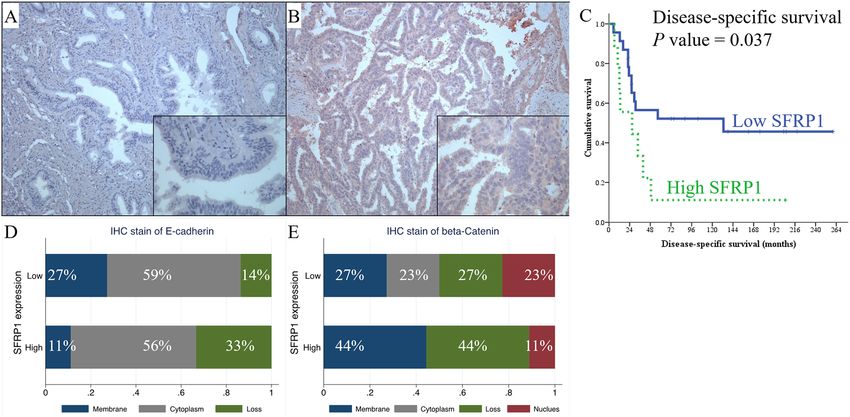
www.nature.com/scientificreports/
ampullary cancer3. Ampullary adenocarcinoma can be subcategorised into pancreaticobiliary and intestinal
subtypes4. Patients with pancreaticobiliary subtypes have a worse prognosis than those with intestinal subtypes.
Furthermore, the carcinogenesis of ampullary cancer involves aberrant activation of WNT s ignalling5. The
canonical WNT-β-catenin pathway begins with the extracellular ligands, the WNT proteins. The binding of
WNT proteins to membrane receptors activates the phosphorylation cascade of downstream molecules, includ-
ing frizzled receptors, which are transmembranous G protein-coupled receptors, and β-catenin, the intracellular
representative protein of the WNT pathway.
The canonical pathway of WNT activation in cancer cells is the accumulation of intracellular and nuclear
β-catenin due to a loss of chaperon p roteins6. The movement of β-catenin into the nucleus results in the tran-
scription of a series of genes involved in cell cycle p rogression7. β-catenin accumulation has been reported in
ampullary adenocarcinoma, particularly in the intestinal subtypes8. Genetic mutation of WNT signalling has
been observed in ampullary and periampullary cancer, including APC (adenomatosis polyposis coli), AXIN1,
SOX9, and FBXW75,9,10. CTNNB1 mutation (catenin beta 1, β-catenin protein) induces the nuclear accumulation
of β-catenin in ampullary cancer11. In contrast, the loss of β-catenin has been correlated with poor prognosis
for ampullary cancer p atients12,13. Other mutations have been reported in gastrointestinal and colorectal can-
cer, such as mutation of APC, CTNNB1, RNF43 (ring finger protein 43), WNT1 (WNT family member 1), and
CDH1 (cadherin 1, E-cadherin)14–16. Thus, the aberrant or non-canonical regulation of WNT signalling possibly
contributes to ampullary cancer development.
Secreted frizzled related protein 1 (SFRP1) belongs to the SFRP family of proteins with a cysteine-rich domain.
The cysteine-rich domain mimics the WNT-binding site of frizzled receptor17. SFRP family proteins act as modu-
lators of WNT signalling. SFRP1 inhibits WNT-dependent transcription and decreases the intracellular level of
β-catenin18. The majority of studies have reported that SFRP1 loss is correlated with poor prognosis in breast
cancer, lung cancer, cholangiocarcinoma, and hepatocellular carcinoma19–22. The methylation of SFRP1 promoter
induces gene silencing and has been detected in 29 cancer types23,24. However, some studies contradict these
findings. In metastatic renal cell carcinoma, SFRP1 hypomethylation increases SFRP1 expression and activates
histone modifications25. Overexpression of SFRP1 protein is related to cell line i nvasiveness25. Furthermore, it is
correlated with lymph node metastasis, cell proliferation, epithelial–mesenchymal transition (EMT), invasion,
and poor survival of gastric cancer patients26. The complex function of SFRP1 in human cancer requires further
investigation. Ampullary cancer originates from complex environment and the mechanism of carcinogenesis is
obscure. In the present study, we utilised bioinformatics analysis to investigate SFRP1 function in multiple types
of cancers. Then, we studied protein and mRNA expression in clinical samples from patients with ampullary
adenocarcinoma to verify the function of SFRP1 in ampullary adenocarcinoma.
Results
SFRP1 expression in different cancer types. The expression level of SFRP1 mRNA was diverse
(Fig. 1A), suggesting that function of SFRP1 may be distinct in different types of cancer. A microarray study
of 182 extrahepatic cholangiocarcinomas detected SFRP1 expression (Fig. 1B). Because ampullary adenocarci-
noma is rare, there was no data available in The Human Protein Atlas. To verify the difference of SFRP-associated
phenotypes, we analysed a pan-cancer panel of Kaplan–Meier Plotter based on 21 cancer types. The probability
of survival was analysed according to expression level of SFRP1 mRNA. The correlation between high SFRP1
expression with patients’ survival was diverse and was correlated with poor prognoses in bladder carcinoma,
kidney renal papillary cell carcinoma, lung squamous cell carcinoma, ovarian cancer, and rectal and stomach
adenocarcinoma (Fig. 2). However, a high expression level of SFRP1 was associated with better prognosis in
patients with breast carcinoma, esophageal adenocarcinoma, head and neck squamous cell carcinoma, kidney
renal clear cell carcinoma, and pancreatic ductal adenocarcinoma (Fig. 3). In 10 other cancer types, the cor-
relation between SFRP1 expression and patient survival was weak, with a wide 95% confidence interval (CI)
(Supplementary Fig. S1).
There are several microarray panels available with variable detection sensitivity. The PrognoScan database
is a collection of multiple array types in an abundant number of cancers. The expression of SFRP1 was divided
into high or low level of mRNA. The Cox proportional hazard ratio (HR) was calculated to obtain a P-value. We
selected datasets where the P-value was < 0.05. The expression of SFRP1 and cancer prognosis are presented in
Supplementary Table S1 and Supplementary Table S2. The results from datasets of lung adenocarcinoma were
controversial. Low SFRP1 was correlated with poor prognosis of patients in one of the datasets (GSE31210),
and high SFRP1 was associated with poor prognosis in a different dataset (jacob-00182-CANDF). Moreover,
the result from PrognoScan in patients with head and neck squamous cell carcinoma (GSE2837) was completely
different from the overall survival in Kaplan–Meier Plotter (Fig. 3). The results of the former dataset showed
that high SFRP1 expression was associated with a poor prognosis, and the latter software concluded that high
SFRP1 expression was correlated with a better prognosis. All these controversial results from the bioinformatics
databases required additional research for clarity.
Transcription regulation involves DNA methylation and histone deacetylation. Information about promoter
methylation of SFRP1 was acquired from the DNA Methylation Interactive Visualisation Database (DNMIVD).
We were unable to find any ampullary cancer datasets from Kaplan–Meier Plotter, PrognoScan, and DNMIVD.
We selected two gastrointestinal adenocarcinoma (gastric and colon adenocarcinoma) and two periampullary
adenocarcinoma (pancreatic adenocarcinoma and cholangiocarcinoma) cases to simulate ampullary adeno-
carcinoma (Fig. 4). SFRP1 expression and differential methylation of the promoter region were compared. An
inverse ratio of gene expression (Fig. 4A,F,K,P) and promoter methylation (Fig. 4B,G,L,Q) was detected in all
four types of adenocarcinoma. Pearson’s and Spearman’s correlation coefficients were calculated, and a nega-
tive correlation was confirmed in gastric, colon and pancreatic adenocarcinoma. High methylation of SFRP1
Scientific Reports | (2020) 10:13255 | https://doi.org/10.1038/s41598-020-69899-8 2
Vol:.(1234567890)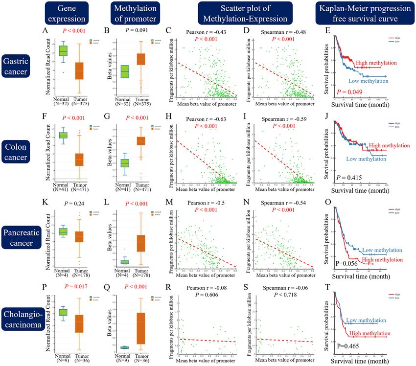
www.nature.com/scientificreports/
Figure 1. Expression of SFRP1 mRNA in different datasets. (A) SFRP1 expression in pan-cancer atlas of The
Cancer Genome Atlas (TCGA), extracted from The Human Protein Atlas programme. (B) Expression of SFRP1
in extrahepatic cholangiocarcinoma from the GSE132305 dataset. Expression level of SFRP1 is different in
various types of cancer. SFRP1, secreted frizzled related protein 1.
represented decreased gene expression (Fig. 4C,D,H,I,M,N). The limited number of patients or wide variation of
gene expression and promoter methylation in cholangiocarcinoma resulted in failure to establish the correlation
(Fig. 4R,S). The impact of DNA methylation was correlated with patients’ survival. For the patients with gastric
adenocarcinoma, low methylation of SFRP1 (high expression levels of SFRP1) was correlated with poor patient
prognosis (Fig. 4E). Low methylation of SFRP1 (high expression of SFRP1) showed a trend of correlation with
better prognosis in patients with pancreatic adenocarcinoma and cholangiocarcinoma (Fig. 4O,T). The results of
gastric and pancreatic adenocarcinoma in DNMIVD were similar with the survival correlation in Kaplan–Meier
Plotter (Figs. 2F and 3E).
Expression of WNT‑related genes in different kinds of cancer. To study other WNT-related genes
in four types of adenocarcinoma, we collected gene expression level from DNMIVD and mutation data from the
cBioPortal platform. APC expression gene was decreased in gastric adenocarcinoma. The expression of WNT1,
APC was decreased in colon adenocarcinoma, and that of CTNNB1 was decreased in cholangiocarcinoma. The
level of RNF43 was increased in gastric, colon and pancreatic adenocarcinoma. The expression of CDH1 gene
was controversial (Supplementary Fig. S2). The differences in gene expression between tumor and normal tissue
were relatively small, but that of SFRP1 gene expression was larger (Fig. 4).
The mutation status of these cancers and ampullary adenocarcinoma are shown in Supplementary Fig. S3
and Supplementary Table S3. Whole exome sequencing data of 98 ampullary adenocarcinomas using Illumina
multiplexing was o btained5. In ampullary adenocarcinoma, there was no alteration of SFRP1 in DNA sequenc-
ing or in copy number. In other cancers, the incidence of SFRP1 alteration was also low (0.4%–3.0% in these 4
kinds of cancer). The most frequent gene alteration in ampullary cancer was found in APC (23%), followed by
CTNNB1 (9%), RNF43 (8%), CDH1 (1.9%), and WNT1 (1.3%). We observe thee co-occurrence of WNT1-RNF43
alteration in two patients, APC-RNF43 in two patients, APC-CDH1 in three patients, and APC-CTNNB1 in one
patient (Supplementary Fig. S3A). The co-occurrence tendency was evaluated by calculating the odds ratio of the
number of patients with genetic alterations (Supplementary Table S3). The co-occurrence of SFRP1 with these
five genes was confirmed in pancreatic cancer; however, the correlation was not established in other cancers due
to the relatively low alteration rate in the SFRP1 gene.
SFRP1 expression in ampullary adenocarcinoma. We employed immunohistochemistry (IHC) stain-
ing to detect the expression of SFRP1 protein in cancer samples to verify the impact of SFRP1 on the survival
of ampullary adenocarcinoma patients (Fig. 5A,B). Of the 32 samples analysed, a low level of SFRP1 expression
was detected in 23 (72%) and a high level of expression in 9 (28%) patients. The patient cohort was too small
for a complete analysis (Table 1). However, the patients with a high expression of SFRP1 tended to have higher
risk of early recurrence within 12 months of operation, and increased proportion of peritoneal carcinomatosis
(Table 2). The disease-specific survival rate of patients with high SFRP1 expression levels was worse than those
with low SFRP1 expression levels (Fig. 5C). The IHC results indicated that the influence of SFRP1 expression in
Scientific Reports | (2020) 10:13255 | https://doi.org/10.1038/s41598-020-69899-8 3
Vol.:(0123456789)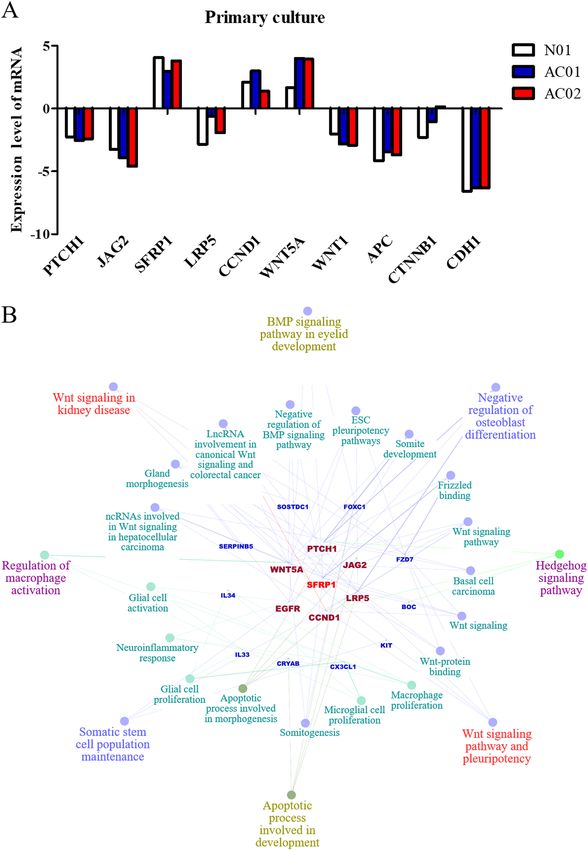
www.nature.com/scientificreports/
Figure 2. Assessment of the effect of SFRP1 mRNA expression by Kaplan–Meier Plotter. High expression of
SFRP1 was correlated with poor prognosis in patients with (A) bladder carcinoma, (B) kidney renal papillary
cell carcinoma, (C) lung squamous cell carcinoma, (D) ovarian cancer, (E) rectal adenocarcinoma, and (F)
stomach adenocarcinoma. SFRP1, secreted frizzled related protein 1; HR, hazard ratio.
survival of ampullary adenocarcinoma patients was similar to rectal and gastric adenocarcinomas (Figs. 2E,F,
and 4E). The result was contrary to the survival analyses determined for pancreatic adenocarcinoma or cholan-
giocarcinoma (Figs. 3E, 4O,T). Thus, the characteristics of ampullary adenocarcinoma was seemingly prone to
certain types of gastrointestinal adenocarcinoma.
In our previous study, the loss of β-catenin was correlated with a poor prognosis for patients with ampullary
adenocarcinoma12. The original dataset correlates with the present study. A high expression of SFRP1 was cor-
related with the loss of E-cadherin, and a low expression of SFRP1 was correlated with a trend of membranous
staining of E-cadherin (P = 0.362, Fig. 5D). The high expression of SFRP1 was associated with membranous
staining of β-catenin, and low expression of SFRP1 was slightly associated with nuclear staining of β-catenin
(P = 0.301, Fig. 5E). The patient cohort was too small to determine the statistical significance; however, our data
indicated that SFRP1expression was associated with the dysregulation of WNT signalling.
SFRP1 expression in periampullary adenocarcinoma. Because ampullary adenocarcinoma is a rare
cancer, there is minimal bioinformatic information available in public databases. The GSE39409 dataset from the
National Center for Biotechnology Information contains data of 14 ampullary adenocarcinoma, two cholangio-
carcinoma, eight duodenal adenocarcinoma, and eight pancreatic adenocarcinoma c ases27. The mRNA expres-
sion of SFRP1 was detected using the Affymetrix U133 Plus 2.0 genome array. The l og2 ratio of SFRP1 expression
in cancer vs. normal tissue was calculated. The patients with ampullary adenocarcinoma had median log2 ratio
of SFRP1 of 7.22 (range, 7.02–8.55). Pancreatic adenocarcinoma patients had a median log2 ratio of SFRP1 of
7.68 (range, 7.03–8.72). SFRP1 expression was relatively lower in ampullary adenocarcinoma than in pancreatic
adenocarcinoma (Fig. 6A). The expression level of SFRP1 mRNA was similar in patients with a recurrence of
ampullary adenocarcinoma (median log2 ratio of SFRP1, 7.24; range, 7.02–8.55) and in patients without recur-
rence (median log2 ratio of SFRP1, 7.21; range, 7.02–8.55) (Fig. 6B). SFRP1 expression was not correlated with
a recurrent status in patients with intestinal subtype (Fig. 6C). For patients with pancreaticobiliary subtypes,
the recurrent patients had a higher level of SFRP1 (Fig. 6D). Expression of WNT1, APC, RNF43, and CDH1
were similar between patients with or without recurrence (Supplementary Figure S4). However, the patients
Scientific Reports | (2020) 10:13255 | https://doi.org/10.1038/s41598-020-69899-8 4
Vol:.(1234567890)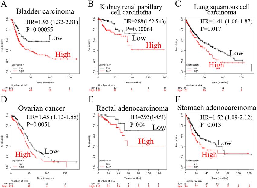
www.nature.com/scientificreports/
Figure 3. Assessment of the effect of SFRP1 mRNA expression by Kaplan–Meier Plotter. Low expression
of SFRP1 was correlated with poor prognosis in patients with (A) breast carcinoma, (B) esophageal
adenocarcinoma, (C) head and neck squamous cell carcinoma, (D) kidney renal clear cell carcinoma, and (E)
pancreatic ductal adenocarcinoma. SFRP1, secreted frizzled related protein 1; HR, hazard ratio.
with cancer recurrence showed a higher level of CTNNB1 gene expression than those without recurrence (Sup-
plementary Figure S4D). However, it must be remembered that the number of patients in our study was limited.
WNT‑associated signalling in ampullary adenocarcinoma. SFRP1 is one of the extracellular ligands
of frizzled receptors. SFRP1 binds to Wnt ligands, which interacts with frizzled receptors in canonical WNT
signalling17. WNT signalling activation induces nuclear accumulation of β-catenin and switches on the tran-
scription of downstream genes6. In our previous study, the loss of β-catenin was correlated with poor prog-
nosis of patients with ampullary adenocarcinoma12. Thus, the dysregulation of WNT signalling in ampullary
adenocarcinoma was a possibility. Therefore, WNT-associated signalling required consideration to explain the
function of SFRP1 in ampullary adenocarcinoma. We collected fresh tissue to establish primary cultured cells
and studied the genes that interacted with SFRP1. One clone from adjacent duodenal mucosa (N01) and two
clones from ampullary adenocarcinomas (AC01 and AC02) were established and analysed with a cDNA micro-
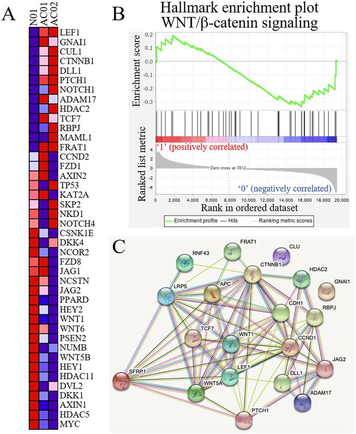
array. The WNT-related genes were selected to generate heatmap (Fig. 7A). Highly differentiated genes that
were upregulated in cancer cells and downregulated in normal cells were identified. LEF1 (lymphoid enhancer-
binding factor 1) was identified as the gene with largest difference and ranked 126th in all 41,000 genes. Gene
set enrichment analysis (GSEA) was performed to analyse the WNT signalling (Fig. 7B). The enrichment score
was − 0.34 and most WNT-related genes were downregulated in AC01 and AC02 cells. The potential networks of
protein–protein interaction of WNT-related genes were drawn using Search Tool for the Retrieval of Interacting
Genes (STRING) software (Fig. 7C), which showed that SFRP1 was linked with multiple genes.
We chose SFRP1-related genes and performed an analysis on these choices using primary culture cells
(Fig. 8A). The expression of SFRP1 was increased in normal and in adenocarcinoma cells. Decreased expression
of PTCH1 (protein: protein patched homolog 1), JAG2 (protein: jagged canonical notch ligand 2), LRP5 (protein:
low-density lipoprotein receptor-related protein 5), WNT1 (protein: WNT family member 1), APC, CTNNB1,
and CDH1 mRNA was detected. Increased expression of CCND1 (protein: cyclin D1) and WNT5A (protein:
WNT family member 5A) was showed. The relationship of these genes is shown in Fig. 8B with predicted clinical
Scientific Reports | (2020) 10:13255 | https://doi.org/10.1038/s41598-020-69899-8 5
Vol.:(0123456789)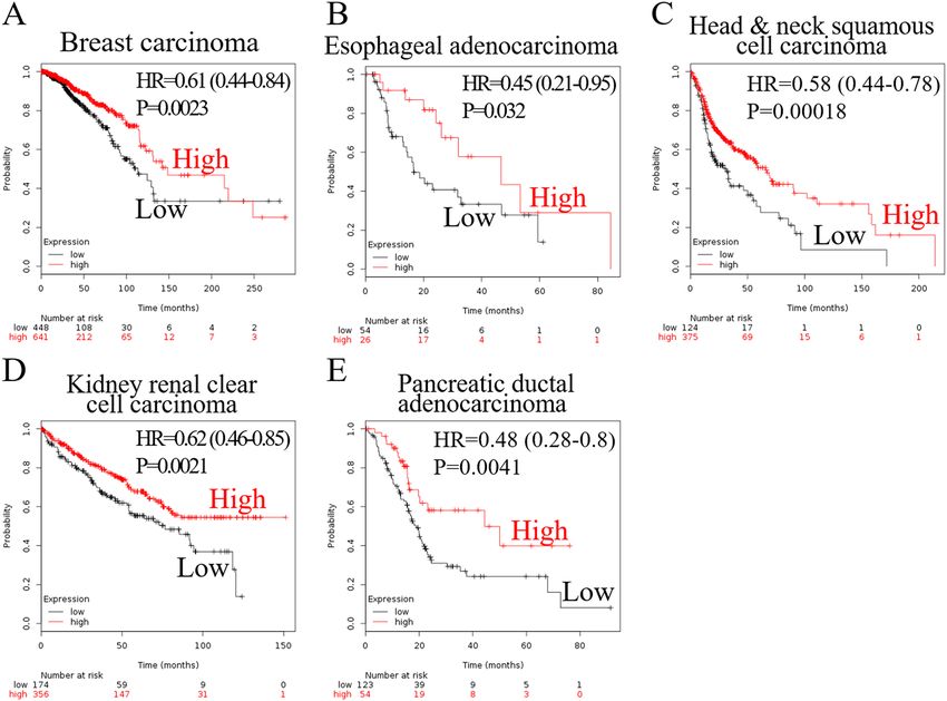
www.nature.com/scientificreports/

                                      Figure 4.  Analysis of SFRP1 in DNA methylation interactive visualisation database. Five different profiles
                                      of information was shown in four kinds of cancer. (A,F,K,P) Gene expression of SFRP1 in RNA-sequencing
                                      read count. (B,G,L,Q) Differential methylation of the promoter region of SFRP1. (C,H,M,R) Scatter plots of
                                      promoter methylation and gene expression, analysed by Pearson’s method. (D,I,N,S) Spearman’s correlation
                                      analysis of promoter methylation and gene expression. (E,J,O,T) Kaplan–Meier progression-free survival curve
                                      by the degree of methylation of SFRP1. (A–E), gastric cancer; (F–J), colon cancer. (K–O), pancreatic cancer;
                                      (P–T), cholangiocarcinoma. SFRP1, secreted frizzled related protein 1.

                                      phenotypes. Crosstalk between WNT, bone morphogenic protein (BMP), and hedgehog signalling was been
                                      implicated.

                                      Discussion
                                      Ampullary adenocarcinoma is a rare malignancy from periampullary region. Because of its characteristics, it
                                      is unreasonable to treat ampullary adenocarcinoma patients with identical modalities as other gastrointestinal
                                      cancers and personalised therapy should be established. Dysregulation of oncogenic WNT signalling has been
                                      reported in ampullary adenocarcinoma. The present study focused on SFRP1, one of the extracellular ligands
                                      in the WNT pathway. SFRP1 mRNA expression was detected in 22 types of cancer. The correlation between
                                      SFRP1 expression and cancer patients’ survival was discordant according to the bioinformatics analyses with
                                      Kaplan–Meier Plotter and PrognoScan. Increased methylation of the promoter region of SFRP1 was consistent
                                      with low expression of SFRP1. There was no alteration of SFRP1 found on microarray analysis of ampullary
                                      adenocarcinoma. We employed IHC staining to study SFRP1 expression in ampullary adenocarcinoma. High
                                      SFRP1expression was weakly associated with decreased membranous staining of E-cadherin. High SFRP1 expres-
                                      sion was correlated with poor prognosis, early recurrence and peritoneal carcinomatosis of ampullary adenocar-
                                      cinoma patients. SFRP1 mRNA expression in ampullary adenocarcinoma was lower than in other periampullary

          Scientific Reports |   (2020) 10:13255 |             https://doi.org/10.1038/s41598-020-69899-8                                            6

Vol:.(1234567890)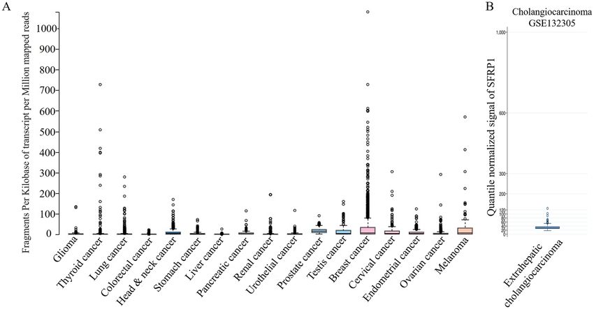
www.nature.com/scientificreports/
Figure 5. Immunohistochemistry staining of SFRP1 in ampullary adenocarcinoma. (A) Negative expression
of SFRP1. (B) High expression of SFRP1. (C) Kaplan–Meier analysis of the impact of SFRP1 expression on
disease-specific survival in ampullary adenocarcinoma patients (P = 0.037). (D) Correlation between localisation
of E-cadherin staining and expression of SFRP1 in using immunohistochemistry (IHC). High expression
of SFRP1 is slightly correlated with the loss of E-cadherin, and low expression of SFRP1 is correlated with
membranous staining of E-cadherin (P = 0.362). (E) Correlation between the localisation of β-catenin staining
and expression of SFRP1 using IHC. High expression of SFRP1 is slightly associated with membranous staining
of β-catenin, and low expression of SFRP1 is associated with nuclear staining of β-catenin (P = 0.301). IHC,
immunohistochemistry; SFRP1, secreted frizzled related protein 1.
adenocarcinomas; however, there was a trend of higher SFRP1 mRNA in ampullary adenocarcinoma patients
with recurrence. We performed a cDNA microarray study of primary culture cells from ampullary adenocar-
cinoma. Increased expression of SFRP1, CCND1 and WNT 5A was related to the downregulation of PTCH1,
JAG2, LRP5, WNT1, APC, CTNNB1, and CDH1. Interaction between these genes may lead to the non-canonical
signalling of SFRP1 and result in cancer dissemination.
The ampulla of Vater is the confluent orifice of the pancreatic duct and common bile duct. The growth of
ampullary adenocarcinoma derives primarily from the mucosa of the common channel of both ductal systems.
The carcinogenesis of ampullary adenocarcinoma is under study, and the activation of WNT signalling is one of
the possible mechanisms. The mutation of tumor suppressors (e.g., APC, GSK-3, and AXIN) in the WNT pathway
was found to induce nuclear accumulation of β-catenin9. The membranous loss of E-cadherin and β-catenin
was observed in ampullary a denocarcinoma8,12. Aberrant nonmembranous β-catenin expression was observed
in intestinal subtype of ampullary c ancer11. SFRP family proteins are extracellular ligands of frizzled receptors,
and the functions of SFRP family proteins differ. Promoter hypermethylation of SFRP1, SFRP2, SFRP4, and
SFRP5 is associated with gene suppression and higher cancer risk28. However, there are several databases that
have shown equivocal correlation between gene expression of SFRP1 and survival of cancer patients. Molecular
subtypes of cancer may alter the regulatory system of SFRP1. Intrinsic subtypes of breast cancer are grouped by
gene expression patterns and clinical relevance. Smid et al. performed microarray analysis of 344 primary breast
tumors. High expression levels of SFRP1 was reported in basal subtype and brain relapse patients with a poor
prognosis; however, low expression levels of SFRP1 was revealed in luminal B and bone relapse patients with a
rognosis29. SFRP1 overexpression of is detected in human gastric cancer and correlated with
relatively better p
lymph node metastasis and poor patient p rognosis26. Upregulation of SFRP1 is detected in metastatic renal cell
carcinoma cell lines and the knockdown of SFRP1 by small interfering RNA inhibits the invasive p otential25.
In the canonical pathway, SFRP1 interacts with WNT protein and restrains the binding of WNT with frizzled
protein, which is the key step of WNT signalling. SFRP1 acts as a WNT inhibitor in canonical pathway. Never-
theless, there are several conflicting reports. The cysteine-rich domain of SFRP1 interacts with frizzled receptors
in prostate cancer cells, and the SFRP1/frizzled complexes activate a signal that may lead to the repression of
downstream signalling30. Treatment with SFRP1 decreases signalling through the Wnt/β-catenin pathway and
the increases cell proliferation of prostate epithelial cells31. Co-activation of the transforming growth factor-
beta (TGF-β) signalling pathway and SFRP1 induces cell proliferation and lung metastasis in mouse model
of gastric cancer26. SFRP1 activates glycogen synthase kinase 3β (GSK3β) and Rac family small GTPase 1 to
interact with TGF-β in promoting cell proliferation, migration, and invasion32. Collaboration of TGF-β and
BMP, WNT-β-catenin, and hedgehog pathways are critical in the EMT of c ancer33. Secretion of extracellular
SFRP1 establishes crosstalk with adhesion molecules in acute myeloid leukaemia cells and contributes to drug
Scientific Reports | (2020) 10:13255 | https://doi.org/10.1038/s41598-020-69899-8 7
Vol.:(0123456789)www.nature.com/scientificreports/
SFRP1 expression
Low High P
No. (%) 23 (72%) 9 (28%)
Age, years, median (range) 66 (36–90) 65 (32–80) NS
Sex NS
Female 12 (75%) 4 (25%)
Male 11 (69%) 5 (31%)
Total bilirubin, mg/dL, median (range) 2.7 (0.5–10.8) 2.3 (0.2–14.4) NS
Direct bilirubin, mg/dL, median (range) 1.7 (0–9.4) 2.4 (0.1–12.4) NS
CEA, units/ml, median (range) 2.5 (0.1–21.7) 2.0 (0.9–11.1) NS
CA125, units/ml, median (range) 13.6 (2.7–44.0) 18.7 (8.3–66.7) NS
CA199, units/ml, median (range) 44.2 (1.4–1,860) 137.7 (5–2,120) NS
Tumor type NS
Polypoid 9 (64%) 5 (46%)
Infiltrative 7 (78%) 2 (22%)
Mixed 6 (75%) 2 (25%)
Tumor size, cm, median (range) 3 (0.8–6.5) 2.5 (1.0–8.0) NS
Differentiation NS
Well 8 (67%) 4 (33%)
Moderate 12 (80%) 3 (20%)
Poor 2 (67%) 1 (33%)
Tumor stage NS
T1 2 (67%) 1 (33%)
T2 5 (63%) 3 (37%)
T3 12 (80%) 3 (20%)
T4 4 (67%) 2 (33%)
Lymph node status NS
Negative 14 (78%) 4 (22%)
Positive 9 (75%) 3 (25%)
Pancreatic invasion NS
Negative 7 (70%) 3 (30%)
Positive 16 (76%) 5 (24%)
AJCC TNM stage NS
Stage I 6 (60%) 4 (40%)
Stage II 12 (86%) 2 (14%)
Stage III 4 (67%) 2 (33%)
Stage IV 1 (50%) 1 (50%)
Table 1. Correlation of histopathological factors and SFRP1 expression detected by IHC staining in
patients with ampullary adenocarcinoma. SFRP1, secreted frizzled related protein 1; NS, not significant;
CEA, carcinoembryonic antigen; CA125, cancer antigen 125; CA199, cancer antigen 199; AJCC TNM stage,
American Joint Committee on Cancer’s tumor, node, and metastases staging system.
r esistance34. The hedgehog signalling pathway is upstream of SFRP1, and hedgehog activation increases the tran-
scription of SFRP135. SFRP1 suppresses WNT signalling; concomitantly, hedgehog signalling promotes stemness
of cancer cells36. SFRP1 is the hinge of WNT- or hedgehog-predominant pathway in cancer, and collaboration
of SFRP1 with other signalling pathways is possible. In the present study, we collected data from The Human
Protein Atlas, Kaplan–Meier Plotter, PrognoScan and DNMIVD. Survival analysis was equivocally associated
with SFRP1 expression. We also used microarray analysis to study SFRP1 and potential interacting molecules.
The fresh samples of periampullary adenocarcinoma from the University of Texas MD Anderson Cancer Center
(UTMDACC) were analysed by Affymetrix U133 Plus 2.0 Array, and the differentially expressed genes were
studied (Fig. 6). The primary culture cells from patients with ampullary neoplasms were established in National
Cheng Kung University Hospital (NCKUH) and investigated using the Agilent Human Whole Genome Oligo
4 × 44 K Microarray chip (Figs. 7 and 8). The different origin of mRNA and dissimilar detection platform of
the microarrays resulted in direct comparisons between the two datasets being impossible. The expression of
SFRP1 mRNA in the UTMDACC dataset was higher in ampullary adenocarcinoma patients with recurrence,
especially in those with pancreaticobiliary subtypes. Most WNT-associated genes were downregulated in the
GSEA analysis. However, the SFRP1 mRNA in the NCKUH dataset was increased in normal and in cancerous
primary culture cells. Additionally, we performed IHC staining to verify expression of SFRP1 protein in ampul-
lary adenocarcinoma. The patients with high SFRP1 staining had a poor prognosis and increased proportion
Scientific Reports | (2020) 10:13255 | https://doi.org/10.1038/s41598-020-69899-8 8
Vol:.(1234567890)www.nature.com/scientificreports/
Expression of SFRP1
Low High P
No recurrence, n (%)a 10 (50%) 1 (13%) NS
Recurrence, n (%)a 10 (50%) 7 (87%)
Delayed recurrence (after postoperative 12 months) 4 2 NS
Early recurrence (within postoperative 12 months) 6 5 0.056
Patterns of recurrencea,b
Liver metastasis 3 3 NS
Local recurrence 5 5 0.061
Peritoneal carcinomatosis 1 3 0.015
Bone metastasis 5 0 NS
Other metastasisc 4 0 NS
Table 2. Correlation between disease recurrence and SFRP1 expression detected by IHC staining in patients
with ampullary adenocarcinoma after curative resection. SFRP1, secreted frizzled related protein 1; NS,
not significant. a Excluding two patients who died due to surgical complications and two patients who lost
follow-up in NCKUH. b Some patients developed more than one kind of metastases. c Including brain, lung, and
ovary metastases.
Figure 6. Expression of SFRP1 in microarrays. (A) Gene expression level of SFRP1 mRNA in the GEO
GSE39409 dataset. Expression of SFRP1 in ampullary adenocarcinoma was lower than other periampullary
adenocarcinoma. (B) Correlation of SFRP1 mRNA with recurrent status of ampullary adenocarcinoma patients
in the GSE39409 dataset. (C) Correlation of SFRP1 mRNA with recurrent status of patients with intestinal
subtypes. (D) Correlation of SFRP1 mRNA with recurrent status of patients with pancreaticobiliary subtypes.
The patients with recurrence had a trend of higher ratio of SFRP1 expression. SFRP1, secreted frizzled related
protein 1.
of peritoneal carcinomatosis. The upregulation of SFRP1 and other interacting genes established a network to
connect WNT, BMP, and hedgehog pathways. The present study provided a possible explanation regarding the
interaction with different oncogenic pathways in ampullary adenocarcinoma.
Ampullary adenocarcinoma is a heterogeneous cancer with intestinal and pancreaticobiliary subtypes4.
Molecular signatures are utilised to group patients. Patients with pancreaticobiliary subtypes have a worse prog-
nosis than those with intestinal subtypes3,4. The pathologist at NCKUH did not recognise pancreaticobiliary
Scientific Reports | (2020) 10:13255 | https://doi.org/10.1038/s41598-020-69899-8 9
Vol.:(0123456789)www.nature.com/scientificreports/
Figure 7. Expression of WNT-associated genes by microarray of primary culture cells. (A) Primary culture
cells of ampullary adenocarcinoma were analysed by microarray of oligo-chips. A heatmep was generated
by comparison with normal duodenal cells (N01) with early and advanced ampullary cancer. Red represents
upregulated genes, and blue represents downregulated genes. (B) Gene set enrichment analysis (GSEA), using
the MSigDB hallmark gene set collection database in primary culture cells, associated with WNT-β-catenin
signalling. Enrichment score is shown in the upper third of the graph. Each bar over the middle third represents
one gene located in the ranking. Red indicates a positive correlation and blue as a negative correlation. The
rank distribution along with the gene list is shown as the grey part in the lower-third of each graph. (C)
Protein–protein interaction of WNT-associated genes was predicted by STRING software. SFRP1 was linked
with multiple genes. N01, primary culture cells from normal duodenum. AC01, primary culture cells from
well-differentiated ampullary adenocarcinoma, T1N0, stage IA. AC02, primary culture cells from moderately-
differentiated ampullary adenocarcinoma, T2N1, stage IIB. GSEA, gene set enrichment analysis; SFRP1, secreted
frizzled related protein 1; STRING, search tool for the retrieval of interacting genes.
Scientific Reports | (2020) 10:13255 | https://doi.org/10.1038/s41598-020-69899-8 10
Vol:.(1234567890)www.nature.com/scientificreports/
or intestinal subtypes of ampullary adenocarcinoma before 2014, and the present study was executed from
September 1994 to August, 2007, before the usage of the classification system at NCKUH. We collected samples
from all subtypes of ampullary adenocarcinoma and elucidated the function of SFRP1. The weak point of the
present study was the lack of clear classification of subtypes in ampullary adenocarcinoma and minimal amounts
of samples. Our results included real world heterogeneity. Moreover, the mRNA microarray from UTMDACC
had similar results to our findings, thus the results of the present study could represent the general condition of
ampullary adenocarcinoma.
Even though there are plenty of studies regarding the somatic mutation of ampullary adenocarcinoma, the
mechanism of carcinogenesis and dissemination is not well understood. The survival of patients with ampul-
lary cancer ranges from 30 to 50%2,3,37. Adjuvant chemotherapy for high-risk patients is not associated with
improved survival38. 5-Fluorouracil-based chemotherapy has some benefit to improve survival, but the results
are unsatisfactory39. Studying oncogenic pathways will help to identify new therapeutic agents. Dysregulation of
the WNT pathway is reported in ampullary cancer5,7–13. SFRP family proteins bind to Wnt ligands and frizzled
receptors to suppress downstream s ignalling15. The expression of SFRP1 predicts a better prognosis in patients
with several kinds of c ancer19–24,28. However, high expression of SFRP1 is correlated with poor prognosis of
patients with ampullary adenocarcinoma in our present study. SFRP1 is a protein with multiple functions.
Understanding the network interaction surrounding SFRP1 will help to select candidates for target therapy.
Solute SFRP1 proteins should be designed for cancers with SFRP1 as a good predictor, including pancreatic and
esophageal adenocarcinoma. Monoclonal anti-SFRP1 antibody is a potential drug for cancers with malignant
SFRP1 networks, including gastric and ampullary adenocarcinoma.
Conclusion
SFRP1 is one of the extracellular ligands of the WNT pathway. SFRP1 expression is detected in various types of
human cancer, and its clinical impact differs based on the cancer type. High expression levels of SFRP1 predicts
poor prognosis in patients with gastric adenocarcinoma but represents better survival of patients with pancre-
atic adenocarcinoma. High methylation levels of SFRP1 promoter suppresses gene expression. In patients with
ampullary adenocarcinoma, the expression level of SFRP1 is lower than other periampullary adenocarcinomas.
However, a high expression level of SFRP1 is correlated with cancer recurrence, peritoneal carcinomatosis and
poor prognosis for patients. Understanding SFRP1 expression phenotype in ampullary adenocarcinoma will
help in the development of new therapeutic agents.
Materials and methods
Bioinformatics analysis. All human proteins are mapped in The Human Protein Atlas (https://proteinatl
as.org)40. Additionally, cancer information from The Cancer Genome Atlas (TCGA) provides the analysis of 17
primary cancers from ≥ 8,000 patients. RNA expression was calculated and expressed as fragments per kilobase
of transcript per million mapped reads. The Kaplan–Meier Plotter is a publically available database of microar-
ray results in the Gene Expression Omnibus (GEO) and clinical outcomes from 21 cancer types (https://kmplo
t.com/analysis/index.php/)41. The expression level of target gene in microarray was calculated. The cutoff point
of high or low expression was the median level of gene expression in obtained from RNA sequencing of a par-
ticular cancer. The univariate Cox regression analysis and Kaplan–Meier survival analysis were performed to
validate the prognostic significance. Overall survival was chosen as the default setting. The survival curve was
expressed as an HR and P-value. The PrognoScan database collects many publicly available microarray datasets
of cancer (https://www.prognoscan.org/)42. The patients included in the survival analysis were grouped with
the optimal cutoff point in gene expression and chosen with a minimum P-value. The datasets were selected if
the P-value was < 0.05 and the 95% CI of HR was not across 0. DNMIVD provided prognostic models based on
DNA methylation and gene expression data from TCGA (https://www.unimd.org/dnmivd/)43. The correlation
between the methylation of gene promoters and patient survival was also available online.
Datasets of cholangiocarcinoma from GEO were searched, and the selection criteria were set as cholangio-
carcinoma, Homo species and extrahepatic cancer. The GSE132305 dataset was the newest one, with microarray
data from 182 extrahepatic c holangiocarcinomas44. The raw data were downloaded and analysed for the expres-
sion of SFRP1 mRNA.
Fresh samples of periampullary adenocarcinoma were obtained from the UTMDACC and analysed using
the Affymetrix U133 Plus 2.0 Array. The data were deposited online in GEO (accession number GSE39409)
(https://www.ncbi.nlm.nih.gov/geo/query/acc.cgi?acc=GSE39409)27. Clinical information and disease status
were collected by one of our authors (M.J.O.).
The ClueGo and CluePedia database were operated via Cytoscape software version 3.5.145,46. Selection options
of GO pathways, organic layout, and colors of nodes were kept according to default setting. The nodes were drawn
according to enrichment of genes in GO pathways. The correlation between genes was calculated using Pearson’s
correlation, Spearman’s rank correlation coefficient, distance correlation and maximal information coefficient,
according to default settings. The related genes and pathways were linked with lines to build interacting networks.
Multidimensional cancer genomics and datasets were collected in the cBioPortal for Caner G enomics47,48.
One dataset of ampullary adenocarcinoma, seven datasets of gastric adenocarcinoma, ten datasets of colorectal
adenocarcinoma or cancer, five datasets of pancreatic adenocarcinoma, and seven datasets of cholangiocarci-
noma were selected. Mutation, deletion, insertion, truncation, fusion and copy number alteration of SFRP1 and
other genes were explored.
GSEA software was applied for enrichment analysis of gene sets that had a common biological function,
chromosome location or signal pathways49. The primary culture cells from one normal duodenum and two
ampullary adenocarcinomas were analysed. The MSigDB hallmark gene set collection was applied for matching
Scientific Reports | (2020) 10:13255 | https://doi.org/10.1038/s41598-020-69899-8 11
Vol.:(0123456789)www.nature.com/scientificreports/
Figure 8. Expression of SFRP1-associated genes by microarray of primary culture cells. (A) Primary culture ▸
cells of ampullary adenocarcinoma was analysed by oligochip microarray. Most WNT-associated genes were
downregulated in these cells. Upregulation of SFRP1 and oncogenes, CCND1 and WNT5A, were detected. (B)
ClueGo and CluePedia were used to create the network with gene GO terms. SFRP1 was shown in the centre
(red), and the other genes were located in the inner track of concentric circles (dark red). Downstream genes
were placed in the middle track (blue), and the phenotypes were placed in the outer two rings. The first inner
ring included PTCH1, JAG2, LRP5, CCND1, EGFR and WNT5A. Interaction between SFRP1 and the first-ring
genes was deduced to genes in the middle circle (blue). The correlated phenotypes were presented in the outer
two circles. The node size reflected the enrichment of genes in each of the GO terms. The connecting lines
showed the correlation between genes and/or pathways. N01, primary culture cells from normal duodenum.
AC01, primary culture cells from well-differentiated ampullary adenocarcinoma, T1N0, stage IA. AC02,
primary culture cells from moderately-differentiated ampullary adenocarcinoma, T2N1, stage IIB. APC,
adenomatosis polyposis coli; BMP, bone morphogenetic protein; BOC, BOC cell adhesion associated, oncogene
regulated; CCND1, cyclin D1; CDH1, cadherin 1; CRYAB, crystallin alpha B; CTNNB1, catenin beta 1; CX3CL1,
C-X3-C motif chemokine ligand 1; EGFR, epidermal growth factor receptor; ESC, embryonic stem cell; FOXC1,
forkhead box C1; FZD7, frizzled class receptor 7; IL33, interleukin 33; IL34, interleukin 34; JAG2, jagged
canonical notch ligand 2; KIT, KIT proto-oncogene, receptor tyrosine kinase; LRP5, low-density lipoprotein
receptor-related protein 5; PTCH1, protein patched 1; SERPINB5, serpin family b member 5; SFRP1, secreted
frizzled related protein 1; SOSTDC1, sclerostin domain containing 1; WNT1, Wnt family member 1; WNT5A,
WNT family member 5A.
molecular signatures50. We used STRING v 11.0 software to analyse the protein–protein interactions based on
the WNT-related genes. This software contains protein–protein interaction networks from 5,090 organisms,
24.6 million proteins, and over 2,000 million interactions51. The k-means clustering algorithm was selected to
classify proteins into different groups.
Patient population. Patients diagnosed with resectable ampullary adenocarcinoma from September, 1994,
to August, 2007, were enrolled from a total of 98 patients that had received curative resection at NCKUH, Tai-
wan, during the time period. The demographics and pathological reports were retrospectively reviewed from
the medical records. The pathological stage was classified according to the guidelines defined by the American
Joint Committee on Cancer Staging Manual, 7th e dition52. Ampullary adenocarcinoma was diagnosed by gross
anatomical location and/or transition of normal–dysplasia–carcinoma from the ampulla of Vater and was made
by the pathologist on duty. All the patients underwent regular follow-ups and annual examinations that included
physical examinations, laboratory tests, abdominal sonography and/or computed tomography. The follow-up
planning was scheduled according to the physician’s specialty and routine practice in Taiwan.
Only patients that had agreed to donate specimens to the Human Biobank in NCKUH were included. Some
patients were excluded due to a small tumor size and an inadequate sample amount. All participants gave written
informed consent, and the samples were preserved in the Human Biobank. Formalin-fixed paraffin-embedded
(FFPE) tumor sections, fresh cancer samples or adjacent normal duodenum samples were obtained and de-
identified for use in our study.
Recurrence was defined as metastasis to a different organ (liver, bone, brain, lung or ovary), locoregional
recurrence, and peritoneal carcinomatosis. Disease recurrence was clarified as biopsy-proven disease or radio-
logical evidence of recurrence. The disease-specific survival rate was limited to those who died due to ampullary
adenocarcinoma, and the time period was recorded from the operation date to date of death.
IHC staining. The FFPE samples obtained from ampullary adenocarcinoma of 32 patients were in the form
of 5-μm thick FFPE sections mounted on slides. The slides were de-paraffinised and rehydrated, and the epitopes
were reexposed using a heat retrieval method with an autoclave. The nonspecific activity of peroxidase was
blocked, and the slides were treated with monoclonal anti-SFRP1 antibody (GeneTex, Irvine, CA, USA) at 4 °C
overnight. The secondary antibody was conjugated with a horseradish peroxidase (HRP)-labelled polymer. We
used 3-amino-9-ethylcarbazole (Zymed Laboraotries, San Fancisco, MA, USA) to detect the expression of the
HRP substrates. All slides were counterstained with hematoxylin and reviewed by a single researcher (H.P.H.).
The intensity of SFRP1 immunoreactivity was defined as negative, weak, moderate or strong. SFRP1 was detected
in the cytoplasm of adenocarcinoma cells. A negative control for IHC staining was used a confirmation; this was
accomplished by the omitting the primary or secondary antibody. A low level of SFRP1 expression was indicated
by a negative or weak expression in IHC staining, and a high level of SFRP1 was indicated by a moderate or
strong expression.
cDNA microarray analysis of primary culture cells. Fresh samples from two ampullary adenocarci-
noma patients were collected immediately following curative resection with pancreaticoduodenectomy. A sam-
ple of the adjacent mucosa of normal duodenum was collected at least 2 cm apart from the ampulla of Vater in
one patient. All the tissues were finely dissected into pieces < 0.2 mm, immersed in Dulbecco’s modified Eagle’s
(DMEM) with 10% fetal bovine serum and placed in an incubator at 37 °C with 95% humidity and 5% C O2. The
media was changed every 2–3 days. After attaining 80% confluence, the cells were trypsinised (0.05% trypsin).
Unmovalbe cells were defined as fibroblasts and the floating cells considered as immune cells. After subculture
for ≥ 3 passages, the cells from the primary culture were collected for cDNA microarray.
Scientific Reports | (2020) 10:13255 | https://doi.org/10.1038/s41598-020-69899-8 12
Vol:.(1234567890)www.nature.com/scientificreports/
Scientific Reports | (2020) 10:13255 | https://doi.org/10.1038/s41598-020-69899-8 13
Vol.:(0123456789)www.nature.com/scientificreports/
Total RNAs were extracted using TRIzol reagent (Invitrogen, Carlsbad, CA, USA). The RNA integrity of
each sample was evaluated on an Agilent 2,100 Bioanalyzer (Agilent, Santa Clara, CA, USA). The absorbance
was measured at 260, 230, and 280 nm, and the absorbance (A) ratios (A260/A230 and A 260/A280) of RNA were
determined using a MaestroNano spectrophotometer (Maestrogen, Las Vegas, NV, USA), and the target range
was set as 1.8–2.1. During the in vitro transcription process, the RNA was labelled with Cy3 dye (PerkinElmer,
Waltham, MA, USA). The Agilent Human Whole Genome Oligo 4 × 44 K Microarray chip (Agilent) was hybrid-
ised with Cy-labelled complementary RNA. The scanning wavelength of microarray was set at 535 nm for Cy3.
Lowe’s method was used for normalisation of the scanned image. Data analysis was performed using Agilent
GeneSpring software (Agilent, Santa Clara, CA, USA).
Statistical analysis. All statistical analyses were performed using SPSS, v17.0 (SPSS, Chicago, IL, USA).
Univariate analysis of categorical variables was performed using Pearson’s chi-squared test. The nonparametric
Kruskal–Wallis test was utilised for continuous variables. The disease-specific survival rate was calculated by the
Kaplan–Meier method and the log-rank test. The level of statistical significance was set as a P-value < 0.5.
Data availability
Correspondence and requests for materials should be addressed to H.P.H.
Received: 26 March 2020; Accepted: 20 July 2020
References
1. Jiang, Z. Q. et al. A retrospective study of ampullary adenocarcinomas: Overall survival and responsiveness to fluoropyrimidine-
based chemotherapy. Ann. Oncol. 24, 2349–2353 (2013).
2. Rostain, F. et al. Trends in incidence and management of cancer of the ampulla of Vater. World J. Gastroenterol. 20, 10144–10150
(2014).
3. Albores-Saavedra, J., Schwartz, A. M., Batich, K. & Henson, D. E. Cancers of the ampulla of vater: Demographics, morphology,
and survival based on 5,625 cases from the SEER program. J. Surg. Oncol. 100, 598–605 (2009).
4. Overman, M. J. et al. Performance and prognostic utility of the 92-gene assay in the molecular subclassification of ampullary
adenocarcinoma. BMC Cancer 16, 668. https://doi.org/10.1186/s12885-016-2677-3 (2016).
5. Gingras, M. C. et al. Ampullary cancers harbor ELF3 tumor suppressor gene mutations and exhibit frequent WNT dysregulation.
Cell Rep. 14, 907–919 (2016).
6. Taciak, B., Pruszynska, I., Kiraga, L., Bialasek, M. & Krol, M. Wnt signaling pathway in development and cancer. J. Physiol. Phar-
macol. 69, 185–196 (2018).
7. Makena, M. R. et al. WNT/β-catenin signaling: The culprit in pancreatic carcinogenesis and therapeutic resistance. Int. J. Mol. Sci.
20, E4242. https://doi.org/10.3390/ijms20174242 (2019).
8. Sung, R. et al. Differential expression of E-cadherin, β-catenin, and S100A4 in intestinal type and nonintestinal type ampulla of
Vater cancers. Gut Liver 8, 94–101 (2014).
9. Jayaramayya, K., Balachandar, V. & Santhy, K. S. Ampullary carcinoma—A genetic perspective. Mutat. Res. 776, 10–22 (2018).
10. Mafficini, A. et al. Sequencing analysis identifies TP53 status as a novel independent prognostic factor and potentially actionable
ERBB, PI3K, and WNT pathways gene mutations. Ann. Surg. 267, 149–156 (2018).
11. Kawakami, M. et al. beta-Catenin alteration in cancer of the ampulla of Vater. J. Exp. Clin. Cancer Res. 21, 23–27 (2002).
12. Hsu, H. P., Shan, Y. S., Jin, Y. T., Lai, M. D. & Lin, P. W. Loss of E-cadherin and β-catenin is correlated with poor prognosis of
ampullary neoplasms. J. Surg. Oncol. 101, 356–362 (2010).
13. Park, S., Kim, S. W., Lee, B. L., Jung, E. J. & Kim, W. H. Expression of E-cadherin and beta-catenin in the adenoma-carcinoma
sequence of ampulla of Vater cancer. Hepatogastroenterology 53, 28–32 (2006).
14. Peng, C. et al. Comprehensive bioinformatic analysis of Wnt1 and Wnt1-associated diseases. Intractable Rare Dis. Res. 9, 14–22
(2020).
15. Katoh, M. & Katoh, M. Molecular genetics and targeted therapy of WNT-related human diseases (review). Int. J. Mol. Med. 40,
587–606 (2017).
16. Min, B. H. et al. Dysregulated Wnt signalling and recurrent mutations of the tumour suppressor RNF43 in early gastric carcino-
genesis. J. Pathol. 240, 304–314 (2016).
17. Pei, J. & Grishin, N. V. Cysteine-rich domains related to frizzled receptors and Hedgehog-interacting proteins. Protein Sci. 21,
1172–1184 (2012).
18. Esteve, P. & Bovolenta, P. The advantages and disadvantages of sfrp1 and sfrp2 expression in pathological events. Tohoku J. Exp.
Med. 221, 11–17 (2010).
19. Kang, P. et al. The Wnt antagonist sFRP1 as a favorable prognosticator in human biliary tract carcinoma. PLoS ONE 9, e90308.
https://doi.org/10.1371/journal.pone.0090308 (2014).
20. Ren, J. et al. sFRP1 inhibits epithelial–mesenchymal transition in A549 human lung adenocarcinoma cell line. Cancer Biother.
Radiopharm. 28, 565–571 (2013).
21. Sur, S. et al. Tea polyphenols epigallocatechin gallete and theaflavin restrict mouse liver carcinogenesis through modulation of
self-renewal Wnt and hedgehog pathways. J. Nutr. Biochem. 27, 32–42 (2016).
22. Bernemann, C. et al. Influence of secreted frizzled receptor protein 1 (SFRP1) on neoadjuvant chemotherapy in triple negative
breast cancer does not rely on WNT signaling. Mol. Cancer. 13, 174. https://doi.org/10.1186/1476-4598-13-174 (2014).
23. Lo, P. K. et al. Epigenetic suppression of secreted frizzled related protein 1 (SFRP1) expression in human breast cancer. Cancer
Biol. Ther. 5, 281–286 (2006).
24. Vincent, K. M. & Postovit, L. M. A pan-cancer analysis of secreted Frizzled-related proteins: Reexamining their proposed tumour
suppressive function. Sci. Rep. 7, 42719. https://doi.org/10.1038/srep42719 (2017).
25. Saini, S. et al. Functional significance of secreted frizzled-related protein 1 in metastatic renal cell carcinomas. Cancer Res. 69,
6815–6822 (2009).
26. Qu, Y. et al. High levels of secreted frizzled-related protein 1 correlate with poor prognosis and promote tumourigenesis in gastric
cancer. Eur. J. Cancer 49, 3718–3728 (2013).
27. Overman, M. J. et al. Gene expression profiling of ampullary carcinomas classifies ampullary carcinomas into biliary-like and
intestinal-like subtypes that are prognostic of outcome. PLoS ONE 8, e65144. https: //doi.org/10.1371/journa l.pone.006514 4 (2013).
28. Yu, J. et al. Association between SFRP promoter hypermethylation and different types of cancer: A systematic review and meta-
analysis. Oncol. Lett. 18, 3481–3492 (2019).
Scientific Reports | (2020) 10:13255 | https://doi.org/10.1038/s41598-020-69899-8 14
Vol:.(1234567890)www.nature.com/scientificreports/
29. Smid, M. et al. Subtypes of breast cancer show preferential site of relapse. Cancer Res. 68, 3108–3114 (2008).
30. Kawano, Y. et al. Secreted frizzled-related protein-1 is a negative regulator of androgen receptor activity in prostate cancer. Br. J.
Cancer 100, 1165–1174 (2009).
31. Joesting, M. S. et al. Identification of SFRP1 as a candidate mediator of stromal-to-epithelial signaling in prostate cancer. Cancer
Res. 65, 10423–10430 (2005).
32. Peng, J. X., Liang, S. Y. & Li, L. sFRP1 exerts effects on gastric cancer cells through GSK3β/Rac1-mediated restraint of TGFβ/Smad3
signaling. Oncol. Rep. 41, 224–234 (2019).
33. Gonzalez, D. M. & Medici, D. Signaling mechanisms of the epithelial-mesenchymal transition. Sci. Signal 7, re8. https://doi.
org/10.1126/scisignal.2005189 (2015).
34. Toni, F. D. et al. A crosstalk between the Wnt and the adhesion-dependent signaling pathways governs the chemosensitivity of
acute myeloid leukemia. Oncogene 25, 3113–3122 (2006).
35. He, J. et al. Suppressing Wnt signaling by the Hedgehog pathway through sFRP-1. J. Biol. Chem. 281, 35598–35602 (2006).
36. Ng, J. M. Y. & Curran, T. The hedgehog’s tale: Developing strategies for targeting cancer. Nat. Rev. Cancer 11, 493–501 (2013).
37. Hsu, H. P. et al. Predictors for patterns of failure after pancreaticoduodenectomy in ampullary cancer. Ann. Surg. Oncol. 14, 50–60
(2007).
38. Ecker, B. L. et al. Role of adjuvant multimodality therapy after curative-intent resection of ampullary carcinoma. JAMA Surg. 154,
706–714 (2019).
39. Al Abbas, A. I. et al. Impact of adjuvant chemotherapy regimen on survival outcomes in immunohistochemical subtypes of ampul-
lary carcinoma. J. Surg. Oncol. 121, 322–329 (2019).
40. Uhlén, M. et al. Tissue-based map of the human proteome. Science 347, 1260419. https://doi.org/10.1126/science.1260419 (2015).
41. Nagy, Á, Lánczky, A., Menyhárt, O. & Győrffy, B. Validation of miRNA prognostic power in hepatocellular carcinoma using
expression data of independent datasets. Sci. Rep. 8, 9277 (2018).
42. Mizuno, H., Kitada, K., Nakai, K. & Sarai, A. PrognoScan: A new database for meta-analysis of the prognostic value of genes. BMC
Med. Genomics 2, 18. https://doi.org/10.1186/1755-8794-2-18 (2009).
43. Ding, W. et al. DNMIVD: DNA methylation interactive visualization database. Nucleic Acids Res. 48, D856–D862 (2020).
44. Montal, R. et al. Molecular classification and therapeutic targets in extrahepatic cholangiocarcinoma. J. Hepatol. S0168–8278(20),
30166–30175 (2020).
45. Bindea, G. et al. ClueGO: A Cytoscape plug-in to decipher functionally grouped gene ontology and pathway annotation networks.
Bioinformatics 25, 1091–1093 (2009).
46. Bindea, G., Galon, J. & Mlecnik, B. CluePedia Cytoscape plugin: Pathway insights using integrated experimental and in silico data.
Bioinformatics 29, 661–663 (2013).
47. Cerami, E. et al. The cBio cancer genomics portal: An open platform for exploring multidimensional cancer genomics data. Cancer
Discov. 2, 401–404 (2012).
48. Gao, J. et al. Integrative analysis of complex cancer genomics and clinical profiles using the cBioPortal. Sci. Signal. 6, pl1 (2013).
49. Subramanian, A. et al. Gene set enrichment analysis: A knowledge-based approach for interpreting genome-wide expression
profiles. Proc. Natl. Acad. Sci. USA 102, 15545–15550 (2005).
50. Liberzon, A. et al. The Molecular Signatures Database (MSigDB) hallmark gene set collection. Cell Syst. 1, 417–425 (2015).
51. Szklarczyk, D. et al. STRING v11: Protein-protein association networks with increased coverage, supporting functional discovery
in genome-wide experimental datasets. Nucleic Acids Res. 47, D607–D6131 (2019).
52. Edge, S. B. et al. AJCC Cancer Staging Manual 7th edn. (Springer, New York, 2010).
Acknowledgements
The authors are thankful to all patients who participated in the study. The authors are grateful for the support
from the Human Biobank, Research Center of Clinical Medicine, National Cheng Kung University Hospital. We
were blessed with support from the late superintendent, Professor Pin-wen Lin. Furthermore, we thank Miss
Ya-Li Hsiao for technical support. The study was supported by the Ministry of Science and Technology (MOST)
of Taiwan (grants MOST108-2314-B006-082 to H.P.H.), the National Cheng Kung University Hospital (grant
NCKUH-10902031 to H.P.H.), and Chi Mei Medical Center (grant B107-F408 to L.C.C.). This study was reviewed
and supervised by the Institutional Review Board (IRB) at the National Cheng Kung University Hospital (IRB
number: A-ER-104-015). All experimental protocols were approved by the IRB and all methods were performed
in accordance with relevant guidelines and regulations for Good Clinical Practice.
Author contributions
Conception and design: L.C.C and H.P.H. Patient care: Y.J.C. and Y.S.S. Sample preparation: T.W.W. Administra-
tive or material support: Y.L.C. and M.D.L. Bioinformatic analysis: C.Y.W. and N.N.P. Writing first draft of the
manuscript: H.P.H. Editing the manuscript: C.Y.W and N.N.P. Review and revision of the manuscript: H.P.H.,
M.J.O., and M.D.L. All of the authors have read and approved the final submitted manuscript and they partici-
pated in writing and reviewing the manuscript.
Competing interests
The authors declare no competing interests.
Additional information
Supplementary information is available for this paper at https://doi.org/10.1038/s41598-020-69899-8.
Correspondence and requests for materials should be addressed to H.-P.H. or Y.-S.S.
Reprints and permissions information is available at www.nature.com/reprints.
Publisher’s note Springer Nature remains neutral with regard to jurisdictional claims in published maps and
institutional affiliations.
Scientific Reports | (2020) 10:13255 | https://doi.org/10.1038/s41598-020-69899-8 15
Vol.:(0123456789)You can also read