Metformin impairs growth of endometrial cancer cells via cell cycle arrest and concomitant autophagy and apoptosis
←
→
Page content transcription
If your browser does not render page correctly, please read the page content below
Takahashi et al. Cancer Cell International 2014, 14:53
http://www.cancerci.com/content/14/1/53

 PRIMARY RESEARCH                                                                                                                                Open Access

Metformin impairs growth of endometrial cancer
cells via cell cycle arrest and concomitant
autophagy and apoptosis
Akimasa Takahashi*, Fuminori Kimura, Akiyoshi Yamanaka, Akie Takebayashi, Nobuyuki Kita, Kentaro Takahashi
and Takashi Murakami

  Abstract
  Background: Effective therapies for early endometrial cancer usually involve surgical excision and consequent
  infertility Therefore, new treatment approaches that preserve fertility should be developed. Metformin, a well-tolerated
  anti-diabetic drug, can inhibit cancer cell growth. However, the mechanism of metformin action is not well understood.
  Here we investigate the roles of autophagy and apoptosis in the anti-cancer effects of metformin on endometrial
  cancer cells.
  Methods: Ishikawa endometrial cancer cells were treated with metformin. WST-8 assays, colony formation assays,
  flow cytometry, caspase luminescence measurement, immunofluorescence, and western blots were used to assess
  the effects of metformin on cell viability, proliferation, cell cycle progression, apoptosis, and autophagy.
  Results: Metformin-treated cells exhibited significantly lower viability and proliferation and significantly more cell
  cycle arrest in G1 and G2/M than control cells. These cells also exhibited significantly more apoptosis via both
  intrinsic and extrinsic pathways. In addition, metformin treatment induced autophagy. Inhibition of autophagy,
  either by Beclin1 knockdown or by 3-methyladenine-mediated inhibition of caspase-3/7, suppressed the anti-proliferative
  effects of metformin on endometrial cancer cells. These findings indicate that the anti-proliferative effects and
  apoptosis caused by metformin are partially or completely dependent on autophagy.
  Conclusions: We showed that metformin suppresses endometrial cancer cell growth via cell cycle arrest and
  concomitant autophagy and apoptosis.
  Keywords: Metformin, Endometrial cancer, Autophagy, Apoptosis

Background                                                                           used to treat endometrial cancers in patients who desire
Endometrial cancer is one of the most common gyneco-                                 to preserve their fertility [9]. Some younger women with
logic malignancies in the United States [1], and its inci-                           endometrial cancer present with obesity, irregular menses,
dence is rapidly increasing in Japan [2]. Approximately                              chronic anovulation, polycystic ovarian syndrome, insulin
80% of endometrial cancers are diagnosed at an early                                 resistance, type 2 diabetes mellitus, or a combination [7,10].
stage and are completely cured with hysterectomy [3,4].                              Elimination of such conditions using low-dose cyclic pro-
In addition, approximately 25% of all cases are diagnosed                            gestin may decrease recurrence or de novo development of
in premenopausal women, and 3%–14% of all cases are                                  endometrial cancer. However, maintenance treatment with
diagnosed before 40 years of age [5-8]. Endometrial can-                             progestin prohibits pregnancy, and the therapeutic effect of
cer in young women poses a therapeutic dilemma be-                                   progestin in endometrial cancers appears to be inadequate.
cause preservation of fertility is often a major concern.                            Therefore, new approaches to the treatment and prevention
Progesterone and medroxyprogesterone acetate are often                               of endometrial cancer must be developed for women trying
                                                                                     to conceive.
* Correspondence: akimasat@belle.shiga-med.ac.jp                                       The biguanide drug metformin is among the most pre-
Department of Obstetrics and Gynecology, Shiga University of Medical                 scribed drug for the treatment of type 2 diabetes worldwide.
Science, Seta-Tsukinowa-cho, Otsu, Shiga 520-2192, Japan

                                       © 2014 Takahashi et al.; licensee BioMed Central Ltd. This is an Open Access article distributed under the terms of the Creative
                                       Commons Attribution License (http://creativecommons.org/licenses/by/4.0), which permits unrestricted use, distribution, and
                                       reproduction in any medium, provided the original work is properly credited. The Creative Commons Public Domain
                                       Dedication waiver (http://creativecommons.org/publicdomain/zero/1.0/) applies to the data made available in this article,
                                       unless otherwise stated.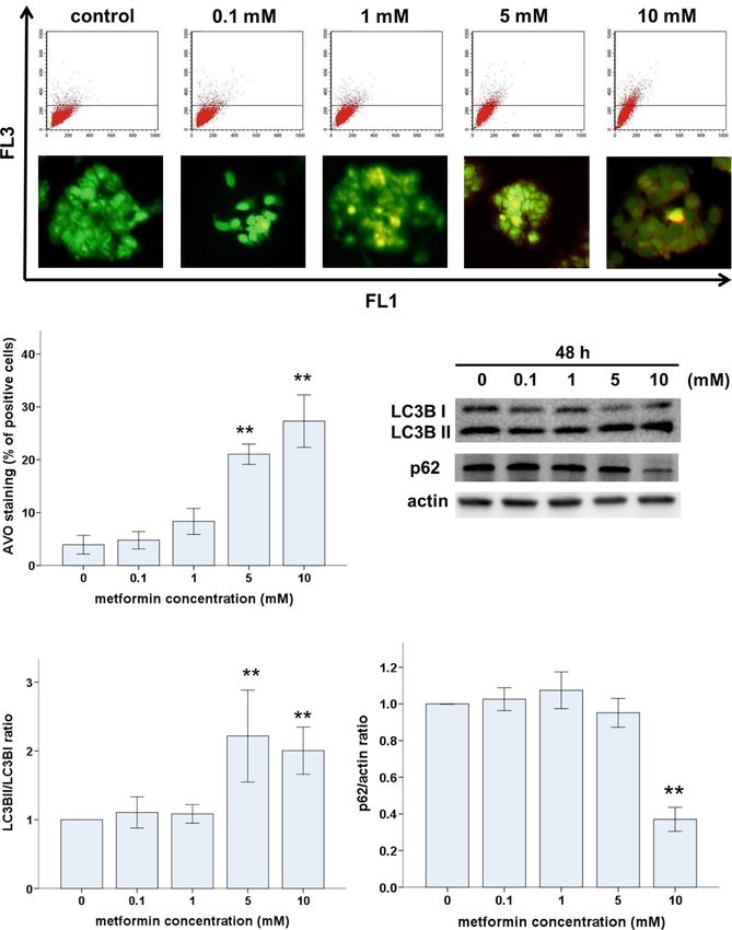
Takahashi et al. Cancer Cell International 2014, 14:53                                                             Page 2 of 12
http://www.cancerci.com/content/14/1/53

Metformin (1,1-dimethylbiguanide hydrochloride) is a well-      BrdU Flow Kit, and BD MitoScreen (JC-1) were pur-
tolerated drug that has numerous cellular effects in mul-       chased from BD Pharmingen (San Diego, CA, USA).
tiple tissues. The main anti-hyperglycemic effect is believed   Acridine orange (AO) was purchased from Molecular
to be due to the suppression of hepatic glucose production      Probes (Eugene, OR, USA). Lipofectamine 2000 was
[11]. In addition, metformin has been reported to inhibit       purchased from Invitrogen (Carlsbad, CA, USA).
the growth of various cancers [12-18], including endomet-
rial cancer [19]. Metformin activates AMPK, a critical cellu-
                                                                Cell culture, cell viability assay, and colony formation assay
lar energy sensor. Activation of AMPK suppresses the
                                                                The Ishikawa human endometrial adenocarcinoma cell
mTOR; this cascade leads to reduced protein synthesis and
                                                                line was purchased from the European Collection of Cell
cell proliferation [20]. In addition, higher doses of metfor-
                                                                Culture (ECACC, Salisbury, UK). Ishikawa cells were cul-
min (2–5 mM) reportedly induce apoptosis in endometrial
                                                                tured in MEM supplemented with l-glutamine (2 mM), 5%
cancer cell lines [20]. Whether metformin induces other
                                                                (v/v) FBS, 1% NEAA, and ABAM at 37°C in a humidified
forms of cell death such as autophagy is unknown.
                                                                atmosphere with 5% CO2.
   Programmed cell death refers to any type of cell death
                                                                   We performed this work by using only cell line, but not
mediated by an intracellular program [21]. Apoptosis is
                                                                clinical samples. Therefore, this work has been granted ex-
type-I programmed cell death, which is morphologically
                                                                emption from the Ethics Committee of Shiga University of
characterized by cell shrinkage, chromatin condensation,
                                                                Medical Science.
nuclear fragmentation, and formation of apoptotic bodies.
                                                                   The WST-8 assay was used to measure cell viability.
Autophagic cell death is type-II programmed cell death,
                                                                Cells were plated on 96-well plates at a density of 1 ×
which is characterized by the accumulation of multi-
                                                                104 cells/well in 100 μL medium. At 24 h after seeding,
lamellar vesicles that engulf the cytoplasm and organelles
                                                                metformin (0, 0.01, 1, 5, 10, or 20 mM) was added to
[22]. Apoptosis has long been known to play an important
                                                                each well and cells were cultured for an additional 48 h.
role in the response to several chemotherapeutic agents;
                                                                CCK-8 solution (10 μL) was then added to each well,
however, the importance of treatment-induced autophagic
                                                                and the plates were incubated at 37°C for 2 h. The ab-
cell death in tumor regression has only recently been rec-
                                                                sorbance of WST-8 formazan was measured at 450 nm
ognized [23,24]. Metformin induces apoptosis in some
                                                                using a microplate reader.
cancers [12,14,25] and autophagy in other, including mel-
                                                                   To measure colony formation, adherent Ishikawa cells
anoma, lymphoma, and colon cancer [12,17,18]. Multiple
                                                                were trypsinized and 1000 viable cells (depending on
functional relationships between apoptosis and autophagy
                                                                the experiment) were subcultured in 60-mm plates; each
in cancer cells have been reported. Thus, a better un-
                                                                treatment was tested in triplicate. After 24 h, the medium
derstanding of the interactions between apoptosis and
                                                                was replaced with fresh culture medium containing met-
autophagy may be a key to continued improvement of
                                                                formin (0, 0.01, 1, 5, 10, or 20 mM) in a 37°C humidified
cancer treatments.
                                                                atmosphere with 95% air and 5% CO2 and grown for
   Here we used an endometrial cancer cell line to inves-
                                                                2 weeks. The culture medium was replaced every 3 days.
tigate the anti-cancer activity of metformin. We focused
                                                                Cell clones were stained for 15 min with a solution con-
on the role of autophagy and its effects on apoptotic cell
                                                                taining 0.5% crystal violet and 25% methanol in water.
death.
                                                                Stained cells were rinsed three times with tap water to
                                                                remove excess dye. Each dish was then washed and
Methods
                                                                dried, and the number of colonies/plate was macroscop-
Reagents and antibodies
                                                                ically counted. Colonies were defined as those contai-
Metformin (1,1-dimethylbiguanide hydrochloride), 3-
                                                                ning >50 cells by microscopic examination.
methyladenine (3MA), chloroquine (CQ), and siRNA were
purchased from Sigma Aldrich (St. Louis, MI, USA). Anti-
actin antibody was purchased from Sigma; all other anti-        Assessment of cell cycle, apoptosis, and mitochondrial
bodies were purchased from Cell Signaling Technology            membrane potential via flow cytometry
(Beverly, MA, USA). Modified Eagle’s medium (MEM),              To assess cell cycle progression, cells were seeded onto
non-essential amino acids (NEAA), and trypsin/EDTA              60-mm plates and incubated for 24 h to allow for expo-
(0.25% trypsin, 1 mM EDTA) were purchased from Wako             nential growth. Ishikawa cells were incubated with or
Pure Chemical Industries (Osaka, Japan). Antibiotics/anti-      without metformin for an additional 48 h. All cells were
mycotics (ABAM) were purchased from Gibco (Carlsbad,            incubated with 10 μM BrdU (BD Pharmingen) for
CA, USA). Cell counting kit-8 (CCK-8) was purchased             30 min; BrdU-labeled cells were then harvested, fixed,
from Dojindo Laboratories (Tokyo, Japan). Caspase-Glo           permeabilized, and stained with FITC-conjugated anti-
assay kits were purchased from Promega (Madison, WI,            BrdU antibody and 7-AAD, according to the manufac-
USA). FITC Annexin V apoptosis detection kit I, FITC            turer’s instructions. A flow cytometer (BD, FACSCalibur,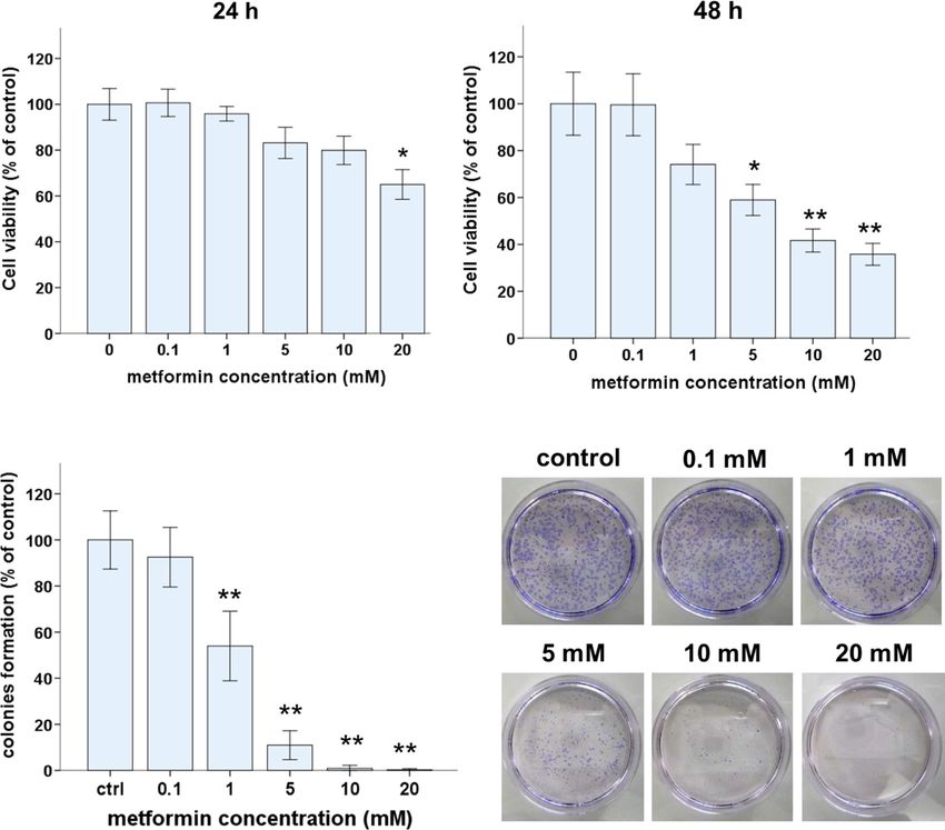
Takahashi et al. Cancer Cell International 2014, 14:53                                                          Page 3 of 12
http://www.cancerci.com/content/14/1/53

San Jose, CA, USA) was used to assess DNA content              siRNA sequence (Sigma-Aldrich; #SIC001) into Ishikawa
and cell cycle phase.                                          cells. Cells were then incubated for 48 h prior to metfor-
  Annexin V-FITC apoptosis detection kits were used            min treatment (0, 5, or 10 mM).
according to the manufacturer’s instructions to measure
apoptosis. Cells were incubated with or without metfor-        Western blot analysis
min for 48 h, collected and washed with PBS, gently re-        Ishikawa cells (2 × 10⁶/dish) were seeded in 100-mm cul-
suspended in annexin V binding buffer, and incubated           ture dishes and cultured for 24 h. After metformin treat-
with annexin V-FITC/7-AAD. Flow cytometry was per-             ment, cells were lysed in RIPA lysis buffer containing a
formed using CellQuest Pro software (BD).                      protease-inhibitor cocktail (“Complete” protease inhibi-
  A mitochondrial membrane potential detection kit was         tor mixture; Roche Applied Science, Indianapolis, IN) on
used according to the manufacturer’s instructions to           ice for 30 min. Suspensions of lysed cells were centrifuged
measure mitochondrial membrane potential (Δψm). In             at 14 000 × g at 4°C for 10 min; supernatants containing
brief, cells were treated with or without metformin, re-       soluble cellular proteins were collected and stored at −80°C
suspended in 0.5 mL of JC-1 solution, and incubated at         until use. BCA protein assay kits were used to measure
37°C for 15 min. Cells were then rinsed before flow cy-        protein concentration. Furthermore, 15 μg of protein was
tometry. A dot plot of red (living cells with intact Δψm)      resuspended in sample buffer and separated on a 4%–20%
versus green fluorescence (cells lacking Δψm) was gener-       tris-glycine gradient gel using the SDS-PAGE system. Re-
ated. Data were expressed as the percentage of cells with      solved proteins were transferred to PVDF membrane,
intact Δψm.                                                    which was blocked with 5% milk in tris-buffered saline/
                                                               0.1% Tween 20. Immunodetection was performed using
Caspase activity                                               each primary antibody. The membranes were incubated
The Caspase-Glo™ 3/7, Caspase-Glo™ 8 or Caspase-Glo™           with donkey anti-rabbit horseradish peroxidase (HRP)-
9 assay kit was used according to the manufacturer’s in-       conjugated secondary antibody (1:5000 dilution). The
structions to measure the activity of caspase 3/7, caspase-    ECL Western Blotting Detection System (GE Health-
8 or caspase-9, respectively. In brief, 50 μL of cell lysate   care, Little Chalfont, UK) was used to detect signals,
(cytosolic extracts, 20 μg) was incubated in 50 μL of          which were visualized using a LAS-4000 mini (GE
Caspase-Glo reagent at room temperature for 1 h.               Healthcare). Actin was used as the loading control.
After incubation, the luminescence of each sample was
measured in a plate-reading luminometer (Tecan, Linz,          Statistical analysis
Austria).                                                      All data points represent the mean of at least three inde-
                                                               pendent measurements and are expressed as the mean ±
Detection and quantification of autophagic cells by            standard deviation. SPSS ver. 20 was used to perform
staining with acridine orange                                  one-way ANOVA and Tukey’s post hoc test or Student’s
To identify autophagic cells, the volume of the cellular       t-test, as appropriate. A significance threshold of p < 0.05
acidic compartment was visualized by AO staining [26].         was used.
Cells were seeded in 60-mm culture dishes and treated
as described above. After 48 h of treatment with or with-      Results
out metformin, cells were incubated with medium con-           Metformin inhibits growth of Ishikawa endometrial
taining 5 μg/mL AO for 15 min. The AO medium was               cancer cells
then removed, cells were washed once with PBS, and             WST-8 and colony formation assays were used to assess
fresh medium was added. Fluorescence micrographs               the effects of metformin on the viability of Ishikawa
were taken using an Olympus inverted fluorescence micro-       endometrial cancer cells. The number of viable cells de-
scope (Olympus Corp., Tokyo, Japan). All images presented      creased with increasing concentrations of metformin for
are at the same magnification. Flow cytometry was used to      24- or 48-h treatments (Figure 1A). After 24 h, 20 mM
determine the number of cells with acidic vesicular or-        of metformin significantly reduced the number of viable
ganelles (AVOs). Cells were trypsinized and harvested;         cells but 0.01–10 mM metformin did not. After 48 h,
BD FACSCalibur and BD CellQuest Pro software was used          metformin at 5 mM or more significantly reduced the
to analyze the cells. A minimum of 10,000 cells within the     number of viable cells. At 48 h, IC50 of metformin was
gated region was analyzed for each treatment.                  6.78 mM.
                                                                 The ability of metformin-treated and control Ishikawa
RNA interference                                               cells to form colonies on 60-mm culture plates within
Lipofectamine 2000 reagent and the Invitrogen protocol         two weeks was examined. Metformin at concentrations
were used to introduce Beclin-1 siRNA (Sigma-Aldrich;          as low as 1 mM, significantly reduced colony formation
seq1 #SASI_Hs02_00336256) or a scramble control                (Figure 1B), and the inhibitory effect of metformin on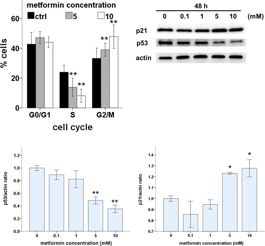
Takahashi et al. Cancer Cell International 2014, 14:53                                                                                       Page 4 of 12
http://www.cancerci.com/content/14/1/53

                     A

                     B

 Figure 1 Metformin inhibits Ishikawa cell growth and induces cell death. (A) WST-8 assays were used to measure the viability of Ishikawa
 cells after treatment with metformin (0, 0.1, 1, 5, 10, or 20 mM) for 24 or 48 h. (B) Colony formation assays were used to measure the colonogenicity
 of Ishikawa cells after metformin treatment. The number of untreated cells was set as 100%. The data are mean values from triplicate experiments.
 *p < 0.05, **p < 0.01, one way ANOVA, post hoc comparisons, Tukey’s test. Columns, mean; error bars, SD.

colony formation was dose dependent. Metformin at                              metformin induced p21 expression, which led to cell cycle
5 mM or more reduced colony formation to 10% of that                           arrest in G1 and G2/M via a p53-independent pathway.
of untreated control cells. Based on these results and those
in several published reports [13,17,27,28], 5 or 10 mM                         Metformin induces apoptosis of Ishikawa endometrial
metformin was used in the following experiments.                               cancer cells via intrinsic and extrinsic pathways
                                                                               To assess whether the induction of apoptosis also contrib-
Metformin induces cell cycle arrest and modulates cell                         uted to metformin-mediated inhibition of Ishikawa cell
cycle proteins in Ishikawa endometrial cancer cells                            growth, the proportion of apoptotic cells was measured.
To investigate the underlying mechanisms of metformin-                         After cells were incubated with or without metformin
induced growth inhibition in Ishikawa cells, we first                          (5 or 10 mM) for 48 h, the proportion of apoptotic cells
evaluated the effect of metformin on cell proliferation                        was measured by flow cytometric of annexin V expression
and cell-cycle progression. Cell-cycle profiles were analyzed                  and JC-1 staining, which indicates the presence of a mito-
after 48 h of metformin treatment. There were significantly                    chondrial membrane potential (Figure 3A and 3B). Our
fewer S-phase cells and significantly more G2/M cells in                       results demonstrate that the proportion of apoptotic cells
metformin-treated cultures compared with those in                              was higher in metformin-treated cultures compared with
control cultures, and these effects were dose dependent                        that in controls.
(Figure 2A). Furthermore, we used western blots to as-                           To understand the mechanism by which metformin
sess the effects of metformin on the expression of two cell                    induced apoptosis in Ishikawa cells, we examined pro-
cycle regulators, p53 and p21 (Figure 2B). Expression of                       apoptotic activity. Apoptosis can be activated through two
p53 decreased in a dose-dependent manner with metformin                        main pathways: the intrinsic mitochondria-dependent
treatment (Figure 2C). The induction of p21, a cell-cycle                      pathway and the extrinsic death-receptor-dependent path-
blocker, increased in a dose-dependent manner with met-                        way. Caspase-8 is predominantly activated by signals from
formin treatment (Figure 2D). These results indicate that                      the extrinsic death-receptor pathway, while caspase-9
Takahashi et al. Cancer Cell International 2014, 14:53 Page 5 of 12
http://www.cancerci.com/content/14/1/53
A B
C D
Figure 2 Metformin induces cell cycle arrest. (A) Ishikawa cells were treated with metformin (0, 5, or 10 mM) for 48 h. After fixation, the cell
cycle was analyzed by flow cytometry. Quantitative analysis of percentage gated cells at the G0/G1, S, and G2/M phases are shown. Data are
shown as mean ± SD of three independent replicate measurements. *p < 0.05 and **p < 0.01 vs. untreated cells. (B) Ishikawa cells were treated
with metformin (0, 0.1, 1, 5, or 10 mM) for 48 h. Cell lysates were separated by SDS-PAGE and analyzed on western blots with the indicated antibodies.
Actin was used as a loading control. One representative experiment of three experiments is shown. (C and D) Densitometric quantitation of p21/actin and
p53/actin protein expression levels is shown as fold changes. One representative experiment of three is shown. *p < 0.05 and **p < 0.01 vs. untreated cells.
activation is dependent primarily on the intrinsic mito- lacked bright red fluorescence. In contrast, metformin-
chondrial pathway. Together, pro-apoptotic Bax and anti- treated cells exhibited AVOs, identified as bright red
apoptotic Bcl-2 play an important role in mitochondrial compartments (Figure 4A). The number of AVOs was
outer membrane permeabilization. Metformin treatment significantly higher in metformin-treated cells compared
induced a marked, dose-dependent increase in the Bax/ with that in untreated controls, and this effect was dose
Bcl-2 ratio (Figure 3D). Furthermore, metformin-mediated dependent (Figure 4A). Levels of LC3B (an autophago-
apoptotic death was accompanied by the activation of cas- some component) and p62 (an autophagosome target)
pase, which is the principal apoptosis-executing enzyme. positively and negatively correlate with autophagy, re-
Fluorescence calorimetric analysis demonstrated that met- spectively. Therefore, we used western blots to assess
formin treatment induced the activation of caspase-3/7, -8, LC3B-I to LC3B-II conversion and p62 protein levels.
and -9 (Figure 3E). Consistent with the induction of apop- As expected, metformin treatment induced significant
tosis, western blots revealed that metformin treatment led LC3 I to II conversion (Figure 4C and D) and a decrease
to cleavage of caspase-3 and PARP in Ishikawa cells in a in p62 levels (Figure 4C and E) in a dose-dependent
dose-dependent manner (Figure 3C). manner. Taken together, these results demonstrate that
metformin induced autophagy in Ishikawa cells.
Metformin triggers autophagy in Ishikawa cells
To determine whether metformin induced autophagy in Inhibition of autophagy reduced metformin-induced
Ishikawa cells, we used AO to stain AVOs, including au- apoptosis in Ishikawa cells
tophagic vacuoles. Untreated Ishikawa cells exhibited To determine the relationship between apoptosis and au-
bright green fluorescence in the cytoplasm and nuclei and tophagy in Ishikawa cells, we inhibited autophagy eitherTakahashi et al. Cancer Cell International 2014, 14:53 Page 6 of 12 http://www.cancerci.com/content/14/1/53 A B C D E Figure 3 (See legend on next page.)
Takahashi et al. Cancer Cell International 2014, 14:53 Page 7 of 12
http://www.cancerci.com/content/14/1/53
(See figure on previous page.)
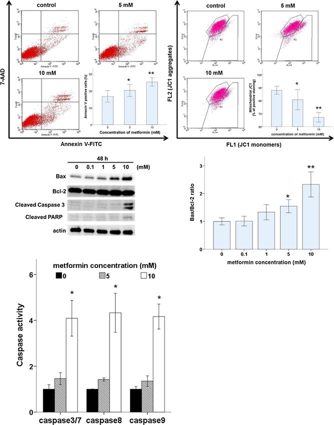
Figure 3 Metformin induces apoptosis in Ishikawa cells. (A) Ishikawa cells were treated with metformin (0, 5, or 10 mM) for 48 h. Cells
were harvested and stained with annexin V-FITC and 7-AAD, and cell apoptosis was analyzed using flow cytometry. *p < 0.05 and **p < 0.01 vs.
untreated cells. (B) Ishikawa cells were treated with metformin (0, 5, or 10 mM) for 48 h, stained with JC-1, incubated, and analyzed using flow
cytometry. *p < 0.05 and **p < 0.01 vs. untreated cells. (C and D) Ishikawa cells were treated with metformin (0, 0.1, 1, 5, or 10 mM) for 48 h. Cell
lysates were separated by SDS-PAGE and analyzed on western blots probed with the indicated antibodies. Actin was used as a loading control.
Densitometric quantitation of Bax/Bcl-2 protein expression ratios are shown as fold changes. One representative experiment of three is shown.
(E) Ishikawa cells were incubated with metformin (0, 5, or 10 mM) for 48 h, followed by assays of caspase-3/7, -8, and -9 activity. *p < 0.05 and
**p < 0.01 vs. untreated cells.
pharmacologically (via 3MA or CQ) or genetically, and metformin induces autophagy and that autophagy and
assessed the effects on metformin-mediated apoptosis. A apoptosis are linked processes.
WST-8 assay showed that 3MA and CQ treatment sig- Several studies have indicated that metformin treatment
nificantly enhanced the viability of metformin-treated decreases cancer cell viability by inducing apoptosis. Can-
(10 mM) cells (Figure 5A). On addition, flow cytometric trell et al. showed that metformin increased activation of
analysis showed that 3MA treatment caused a marked caspase-3 in human endometrial cancer cells in a dose-
decrease in the proportion of metformin-treated (10 mM) dependent manner [19]. Hanna et al. suggested that met-
apoptotic cells (Figure 5B). Moreover, 3MA treatment formin induces apoptosis [20]. Similar to the results of
caused a significant reduction in caspase activity in these studies, we observed that metformin treatment of
metformin-treated (10 mM) cells (Figure 5C). Thus, these Ishikawa endometrial cancer cells induces a significant in-
findings revealed that inhibition of metformin-mediated crease in apoptosis in a dose-dependent manner.
autophagy reduced apoptosis in Ishikawa cells. To elucidate the mechanism of metformin-induced
To confirm these results, we used siRNA to repress ex- apoptosis, we investigated mitochondrial function and
pression of the autophagy regulator Beclin1 in Ishikawa caspase activity in Ishikawa cells. We observed that met-
cells. Beclin1 siRNA knocked down Beclin1 expression by formin treatment altered the expression of Bcl-2 family
approximately 75% (Figure 6A). Upon metformin treat- proteins, PARP cleavage, and the activation of caspase-3/
ment, significantly fewer Annexin-V-positive cells were 7, -8, and -9. Caspase-8 is essential for death-receptor-
observed in Beclin1siRNA cells compared with that in mediated apoptosis, while caspase-9 is essential for
controls (Figure 6B). The inhibition of autophagy by mitochondria-mediated apoptosis. These 2 pathways
Beclin1 siRNA resulted in decreases in caspase-3/7 activ- converge on caspase-3/7 activation, leading to subsequent
ity (Figure 6C), PARP cleavage, and LC3-II and increases activation of other caspases. Our results are similar to
in p62 (Figure 6D), as did pharmacologic inhibition of au- those of previous findings demonstrating that metformin
tophagy by 3MA (Figure 5D). These results demonstrate induces significant increases in apoptosis in pancreatic cell
that the inhibition of autophagy reduced apoptosis associ- lines and that metformin-induced apoptosis is associated
ated with metformin treatment. with PARP cleavage, which is dependent on activation of
caspase-3, -8, and -9 [14]. Thus, metformin may modulate
Discussion apoptotic cell death via extrinsic and intrinsic pathways in
Recent data indicate that metformin may be a useful Ishikawa cells.
anti-proliferation agent for some types of cancer. The In addition, metformin has been shown to induce ar-
potential role of metformin in treating endometrial can- rest of the cell cycle in cancer cell lines [31]. Cantrell
cer has been explored in a number of in vitro studies et al. showed that metformin induces G0/G1 cell cycle
[19,29,30]. However, the anti-tumor effects of metformin arrest in Ishikawa cells [19]. However, we observed that
are not completely understood. Furthermore, the effect metformin blocked cell cycle progression not only in
of metformin on autophagy has not been investigated in G0/G1 but also in the G2/M phase. This apparent dis-
endometrial cancer cells. Here we demonstrate that met- crepancy may result from differences in incubation time,
formin induced caspase-dependent apoptosis and sup- pharmacologic dose or both. G0/G1 cell cycle arrest re-
pressed proliferation by upregulating the cyclin-dependent sulted from a 24-h incubation [19], and G0/G1 and G2/
kinase inhibitor p21 and inducing both G1 and G2/M M phase arrest resulted from a 48-h incubation. These
arrest. In addition, we revealed that metformin pro- findings suggest that metformin may block the cell cycle
moted the formation of AVOs, the conversion of LC3-I at two points. We observed that the cyclin-dependent
to LC3-II, and the degradation of p62. Moreover, both kinase inhibitor p21, which plays an important role in
pharmaco logic and genetic inhibition of autophagy re- cell-cycle arrest, was activated by metformin. Notably,
duced metformin-induced apoptosis. To the best of our p21 is among the genes most consistently induced by
knowledge, this is the first report to demonstrate that metformin [32]. Recent reports indicate that p21 is notTakahashi et al. Cancer Cell International 2014, 14:53 Page 8 of 12 http://www.cancerci.com/content/14/1/53 A B C D E Figure 4 Metformin triggers autophagy in Ishikawa cells. (A) Ishikawa cells were incubated with metformin (0, 0.1, 1, 5, or 10 mM) for 48 h. The presence of acridine orange-stained intracellular vesicles was demonstrated by fluorescent microscopy (lower panel) and flow cytometry, showing an increase in red fluorescence (FL3) intensity (upper panel). (B) Autophagy was quantified by measuring the percentage of cells with bright red fluorescence (FL3-positive cells; data are presented as the mean ± SD of three independent measurements, *p < 0.05). (C) Immunoblot analysis of LC3 conversion and p62 levels in Ishikawa cells treated with metformin (0, 0.1, 1, 5, or 10 mM). (D and E) Densitometric quantitation of LC3B-II/LC3B-I and p62/actin protein expression ratios are shown as fold changes. *p < 0.05 and **p < 0.01 vs. untreated cells.
Takahashi et al. Cancer Cell International 2014, 14:53                                                                                         Page 9 of 12
http://www.cancerci.com/content/14/1/53

only a well-established negative regulator of the G1/S                          independent pathway may have potential as candidate
transition but also an inhibitor of the CDK1/cyclin B                           drugs. Histone deacetylase (HDAC) inhibitors, such as
complex that maintains G2/M arrest [33,34]. These re-                           Psammaplin A, suppress cell proliferation and induce
ports support our supposition that the G2/M phase cell                          apoptosis in Ishikawa cells via p53-independent upregu-
cycle block occurs at 48 h.                                                     lation of p21 expression [36]. Our results indicate that
  Alternatively, it is possible that low doses of metformin                     metformin treatment of Ishikawa cells increased p21 ex-
cause G0/G1 arrest, whereas higher doses cause G2/M ar-                         pression but also decreased mutant p53 expression.
rest. High metformin concentrations induce more p21 ex-                         These findings also indicate that metformin-induced p21
pression; therefore, they may induce apoptosis of cells not                     expression may be regulated through a p53-independent
only in G0/G1 but also in the G2/M cell cycle arrest.                           mechanism. Therefore, we propose that metformin in-
Moreover, p21 expression is induced by both p53-                                duces cell-cycle arrest in Ishikawa endometrial cancer cells
dependent and -independent mechanisms. Mutations in                             both at G0/G1 and G2/M by activating p21 via a p53-
the p53 gene are reportedly evident in >50% of all                              independent pathway.
known cancer types. These mutations are recognized as                             Autophagy is a process where the cytosol and organelles
one of the major events in carcinogenesis, and the Ishi-                        become encased in vacuoles called autophagosomes. Al-
kawa cell line also has a p53 mutation [35]. Therefore,                         though autophagy is primarily a protective process for
agents that induce p21 expression through a p53-                                the cell, it can play a role in cell death [37]. Therefore,

                      A                                                        B

                      C                                                          D

 Figure 5 Pharmacologic inhibition of autophagy reduces metformin-mediated apoptotic cell death. (A) Ishikawa cells were seeded in
 96-well plates and incubated with the indicated dose of metformin (with or without 3MA or CQ) for 48 h; cell viability was determined by WST-8
 assays. Data are presented as the mean ± SD of three independent replicate measurements. *p < 0.05 and **p < 0.01 vs. 3MA or CQ untreated cells.
 (B) Flow cytometry of apoptosis in Ishikawa cells treated with metformin (with or without 3MA) for 48 h. *p < 0.05 and **p < 0.01 vs. 3MA untreated cells.
 (C) Ishikawa cells were incubated with the indicated dose of metformin alone or combined with 3MA for 48 h. Cells were then lysed to measure
 caspase-3/7 activity. *p < 0.05 and **p < 0.01 vs. 3MA untreated cells. (D) Ishikawa cells were treated with the indicated dose of metformin (±3MA) for
 48 h, and cell lysates were subjected to western blot analysis using antibodies against LC3B, p62, and cleaved PARP. Actin was used as a loading control.
Takahashi et al. Cancer Cell International 2014, 14:53 Page 10 of 12
http://www.cancerci.com/content/14/1/53
A B
C D
Figure 6 Genetic inhibition of autophagy reduces metformin-mediated apoptotic cell death. (A) Western blot analysis of Beclin1
expression in siBeclin1 and control Ishikawa cells. (B) Beclin1 expression was blocked in Ishikawa cells by Beclin1 siRNA; cells were incubated with
the indicated concentrations of metformin for 48 h. Cells were then harvested and analyzed by flow cytometry. *p < 0.05 and **p < 0.01 vs.
Control siRNA cells. (C) Beclin1 siRNA Ishikawa cells were incubated with the indicated concentrations of metformin for 48 h. Cells were then
lysed to measure caspase-3/7 activity. *p < 0.05 and **p < 0.01 vs. Control siRNA cells. (D) Beclin1, LC3B, p62, and cleaved PARP expression were
examined in siBeclin1-treated and control cells. Actin expression was used as a loading control.
autophagy is considered to be a double-edged sword. A autophagy and apoptosis are linked, we performed several
recent work highlights the prosurvival role of autophagy experiments following the inhibition or induction of au-
in cancer cells [38]. Alternatively, autophagy may confer tophagy. We observed that both pharmacologic and genetic
a disadvantage on cancer cells [12]. The variability in inhibition of autophagy promoted cancer cell survival and
the effects of autophagy on cancer cells may depend on reduced metformin-induced apoptosis. In addition, our re-
the cell type, cell cycle phase, genetic background, and sults show that inhibition of autophagy decreased the cleav-
microenvironment [39]. When the autophagic capacity age of PARP (Figure 5D and 6D) and the activation of
of cancer cells is reached, apoptosis is promoted [40]. caspase-3/7, -8, and -9 (data not shown). These findings in-
This finding is particularly interesting because metfor- dicate that inhibitors of autophagy enhanced both intrinsic
min can induce autophagy in colon cancer and melan- and extrinsic activation of apoptosis. Taken together, these
oma [12,17], as well as Ishikawa endometrial cancer data suggest that metformin induces autophagic cell death
cells, as demonstrated here. in Ishikawa endometrial cancer cells. To the best of our
Metformin induced apoptosis and autophagy in Ishikawa knowledge, this is the first demonstration that metfor-
endometrial cells. Because autophagy has been implicated min promotes the elimination of endometrial cancer
in the promotion and inhibition of cell survival [21], we cells through concomitant regulation of autophagy and
were interested in the role of autophagy in metformin- apoptosis. These results are based on in vitro studies
mediated apoptosis. To determine whether the processes of only, and further in vivo studies are necessary.Takahashi et al. Cancer Cell International 2014, 14:53 Page 11 of 12
http://www.cancerci.com/content/14/1/53
Conclusions 11. Viollet B, Guigas B, Sanz Garcia N, Leclerc J, Foretz M, Andreelli F: Cellular
We demonstrate that metformin is cytotoxic to Ishikawa and molecular mechanisms of metformin: an overview. Clin Sci (Lond)
2012, 122(6):253–270.
endometrial cancer cells. Several mechanisms underlying 12. Buzzai M, Jones RG, Amaravadi RK, Lum JJ, DeBerardinis RJ, Zhao F, Viollet B,
the anti-tumor effects of metformin in Ishikawa cells are Thompson CB: Systemic treatment with the antidiabetic drug metformin
revealed by the data presented here. Metformin was selectively impairs p53-deficient tumor cell growth. Cancer Res 2007,
67(14):6745–6752.
shown to inhibit Ishikawa endometrial cancer cell prolif- 13. Gotlieb WH, Saumet J, Beauchamp MC, Gu J, Lau S, Pollak MN, Bruchim I: In
eration through the induction of cell cycle arrest and vitro metformin anti-neoplastic activity in epithelial ovarian cancer.
caspase-dependent apoptosis and enhanced autophagic Gynecol Oncol 2008, 110(2):246–250.
14. Wang LW, Li ZS, Zou DW, Jin ZD, Gao J, Xu GM: Metformin induces
flux. In addition, we showed that pharmacological or apoptosis of pancreatic cancer cells. World J Gastroenterol 2008,
genetic inhibition of autophagy decreased metformin- 14(47):7192–7198.
induced apoptotic cell death. These observations indi- 15. Zhuang Y, Miskimins WK: Cell cycle arrest in Metformin treated breast
cancer cells involves activation of AMPK, downregulation of cyclin D1,
cate that metformin may be a promising agent for the and requires p27Kip1 or p21Cip1. J Mol Signal 2008, 3:18.
treatment of early endometrial cancer. In addition, our 16. Alimova IN, Liu B, Fan Z, Edgerton SM, Dillon T, Lind SE, Thor AD:
findings may provide insight into the role of autophagy Metformin inhibits breast cancer cell growth, colony formation and
induces cell cycle arrest in vitro. Cell Cycle 2009, 8(6):909–915.
in anti-cancer therapies. 17. Tomic T, Botton T, Cerezo M, Robert G, Luciano F, Puissant A, Gounon P,
Allegra M, Bertolotto C, Bereder JM, Tartare-Deckert S, Bahadoran P, Auberger
Abbreviations P, Ballotti R, Rocchi S: Metformin inhibits melanoma development through
AMPK: Adenosine-monophosphate-activated protein kinase; autophagy and apoptosis mechanisms. Cell Death Dis 2011, 2:e199.
mTOR: Mammalian target of rapamycin; FITC: Fluorescein isothiocyanate; 18. Shi WY, Xiao D, Wang L, Dong LH, Yan ZX, Shen ZX, Chen SJ, Chen Y, Zhao
WST-8 assay: Modified 3-(4,5-dimethylthiazol-2-yl)-2,5-diphenyltetrazolium WL: Therapeutic metformin/AMPK activation blocked lymphoma cell
assay; 7-AAD: 7-Amino-Actinomycin D; FACS: Fluorescence activated cell growth via inhibition of mTOR pathway and induction of autophagy. Cell
sorting; AO: Acridine orange; AVOs: Acidic vesicular organelles; Death Dis 2012, 3:e275.
RIPA: Radioimmunoprecipitation assay buffer; PARP: poly (ADP-ribose) 19. Cantrell LA, Zhou C, Mendivil A, Malloy KM, Gehrig PA, Bae-Jump VL: Metformin
polymerase; CDK1: Cyclin-dependent kinase 1. is a potent inhibitor of endometrial cancer cell proliferation–implications for
a novel treatment strategy. Gynecol Oncol 2010, 116(1):92–98.
Competing interests 20. Hanna RK, Zhou C, Malloy KM, Sun L, Zhong Y, Gehrig PA, Bae-Jump VL:
The authors declare that they have no conflicts of interest concerning the Metformin potentiates the effects of paclitaxel in endometrial cancer
work presented here. cells through inhibition of cell proliferation and modulation of the mTOR
pathway. Gynecol Oncol 2012, 125(2):458–469.
Authors’ contributions 21. Eisenberg-Lerner A, Bialik S, Simon HU, Kimchi A: Life and death partners:
AT designed and performed research, analyzed data, and wrote manuscript; apoptosis, autophagy and the cross-talk between them. Cell Death Differ
AT and AY assisted with flow cytomety analysis; FK assisted in witing and 2009, 16(7):966–975.
editing paper; NK, KT and TM assisted in research design, oversaw data 22. Tanida I, Minematsu-Ikeguchi N, Ueno T, Kominami E: Lysosomal turnover,
analysis. All authors read and approved the final manuscript. but not a cellular level, of endogenous LC3 is a marker for autophagy.
Autophagy 2005, 1(2):84–91.
Acknowledgments 23. Jing K, Song KS, Shin S, Kim N, Jeong S, Oh HR, Park JH, Seo KS, Heo JY, Han
We thank the Central Research Laboratory of Shiga University of Medical J, Park JI, Han C, Wu T, Kweon GR, Park SK, Yoon WH, Hwang BD, Lim K:
Science for valuable technical assistance. The authors would like to thank Docosahexaenoic acid induces autophagy through p53/AMPK/mTOR
Enago (www.enago.jp) for the English language review. signaling and promotes apoptosis in human cancer cells harboring
wild-type p53. Autophagy 2011, 7(11):1348–1358.
Received: 17 March 2014 Accepted: 11 June 2014 24. Wang N, Pan W, Zhu M, Zhang M, Hao X, Liang G, Feng Y: Fangchinoline
Published: 16 June 2014 induces autophagic cell death via p53/sestrin2/AMPK signalling in human
hepatocellular carcinoma cells. Br J Pharmacol 2011, 164(2b):731–742.
References 25. Liu B, Fan Z, Edgerton SM, Deng XS, Alimova IN, Lind SE, Thor AD:
1. Siegel R, Naishadham D, Jemal A: Cancer statistics, 2012. CA Cancer J Clin Metformin induces unique biological and molecular responses in triple
2012, 62(1):10–29. negative breast cancer cells. Cell Cycle 2009, 8(13):2031–2040.
2. Ushijima K: Current status of gynecologic cancer in Japan. J Gynecol Oncol 26. Paglin S, Hollister T, Delohery T, Hackett N, McMahill M, Sphicas E, Domingo D,
2009, 20(2):67–71. Yahalom J: A novel response of cancer cells to radiation involves autophagy
3. Rose PG: Endometrial carcinoma. N Engl J Med 1996, 335(9):640–649. and formation of acidic vesicles. Cancer Res 2001, 61(2):439–444.
4. Amant F, Moerman P, Neven P, Timmerman D, Van Limbergen E, Vergote I: 27. Luo Q, Hu D, Hu S, Yan M, Sun Z, Chen F: In vitro and in vivo anti-tumor
Endometrial cancer. Lancet 2005, 366(9484):491–505. effect of metformin as a novel therapeutic agent in human oral
5. Crissman JD, Azoury RS, Barnes AE, Schellhas HF: Endometrial carcinoma in squamous cell carcinoma. BMC Cancer 2012, 12:517.
women 40 years of age or younger. Obstet Gynecol 1981, 57(6):699–704. 28. Kato K, Gong J, Iwama H, Kitanaka A, Tani J, Miyoshi H, Nomura K, Mimura S,
6. Gallup DG, Stock RJ: Adenocarcinoma of the endometrium in women Kobayashi M, Aritomo Y, Kobara H, Mori H, Himoto T, Okano K, Suzuki Y, Murao
40 years of age or younger. Obstet Gynecol 1984, 64(3):417–420. K, Masaki T: The antidiabetic drug metformin inhibits gastric cancer cell
7. Duska LR, Garrett A, Rueda BR, Haas J, Chang Y, Fuller AF: Endometrial cancer proliferation in vitro and in vivo. Mol Cancer Ther 2012, 11(3):549–560.
in women 40 years old or younger. Gynecol Oncol 2001, 83(2):388–393. 29. Xie Y, Wang YL, Yu L, Hu Q, Ji L, Zhang Y, Liao QP: Metformin promotes
8. Ota T, Yoshida M, Kimura M, Kinoshita K: Clinicopathologic study of progesterone receptor expression via inhibition of mammalian target of
uterine endometrial carcinoma in young women aged 40 years and rapamycin (mTOR) in endometrial cancer cells. J Steroid Biochem Mol Biol
younger. Int J Gynecol Cancer 2005, 15(4):657–662. 2011, 126(3–5):113–120.
9. Ehrlich CE, Young PC, Stehman FB, Sutton GP, Alford WM: Steroid receptors 30. Zhang Z, Dong L, Sui L, Yang Y, Liu X, Yu Y, Zhu Y, Feng Y: Metformin
and clinical outcome in patients with adenocarcinoma of the reverses progestin resistance in endometrial cancer cells by
endometrium. Am J Obstet Gynecol 1988, 158(4):796–807. downregulating GloI expression. Int J Gynecol Cancer 2011, 21(2):213–221.
10. Soliman PT, Oh JC, Schmeler KM, Sun CC, Slomovitz BM, Gershenson DM, 31. Ben Sahra I, Le Marchand-Brustel Y, Tanti JF, Bost F: Metformin in cancer
Burke TW, Lu KH: Risk factors for young premenopausal women with therapy: a new perspective for an old antidiabetic drug? Mol Cancer Ther
endometrial cancer. Obstet Gynecol 2005, 105(3):575–580. 2010, 9(5):1092–1099.Takahashi et al. Cancer Cell International 2014, 14:53 Page 12 of 12
http://www.cancerci.com/content/14/1/53
32. Rattan R, Giri S, Hartmann LC, Shridhar V: Metformin attenuates ovarian
cancer cell growth in an AMP-kinase dispensable manner. J Cell Mol Med
2011, 15(1):166–178.
33. Vermeulen K, Van Bockstaele DR, Berneman ZN: The cell cycle: a review of
regulation, deregulation and therapeutic targets in cancer. Cell Prolif
2003, 36(3):131–149.
34. Kawabe T: G2 checkpoint abrogators as anticancer drugs. Mol Cancer Ther
2004, 3(4):513–519.
35. Murai Y, Hayashi S, Takahashi H, Tsuneyama K, Takano Y: Correlation
between DNA alterations and p53 and p16 protein expression in cancer
cell lines. Pathol Res Pract 2005, 201(2):109–115.
36. Ahn MY, Jung JH, Na YJ, Kim HS: A natural histone deacetylase inhibitor,
Psammaplin A, induces cell cycle arrest and apoptosis in human
endometrial cancer cells. Gynecol Oncol 2008, 108(1):27–33.
37. Mizushima N, Levine B, Cuervo AM, Klionsky DJ: Autophagy fights disease
through cellular self-digestion. Nature 2008, 451(7182):1069–1075.
38. Amaravadi RK, Yu D, Lum JJ, Bui T, Christophorou MA, Evan GI,
Thomas-Tikhonenko A, Thompson CB: Autophagy inhibition enhances
therapy-induced apoptosis in a Myc-induced model of lymphoma.
J Clin Invest 2007, 117(2):326–336.
39. Műzes G, Sipos F: Anti-tumor immunity, autophagy and chemotherapy.
World J Gastroenterol 2012, 18(45):6537–6540.
40. Maiuri MC, Zalckvar E, Kimchi A, Kroemer G: Self-eating and self-killing:
crosstalk between autophagy and apoptosis. Nat Rev Mol Cell Biol 2007,
8(9):741–752.
doi:10.1186/1475-2867-14-53
Cite this article as: Takahashi et al.: Metformin impairs growth of
endometrial cancer cells via cell cycle arrest and concomitant
autophagy and apoptosis. Cancer Cell International 2014 14:53.
Submit your next manuscript to BioMed Central
and take full advantage of:
• Convenient online submission
• Thorough peer review
• No space constraints or color figure charges
• Immediate publication on acceptance
• Inclusion in PubMed, CAS, Scopus and Google Scholar
• Research which is freely available for redistribution
Submit your manuscript at
www.biomedcentral.com/submitYou can also read