Effects of cholera toxin on innate and adaptive immunity
←
→
Page content transcription
If your browser does not render page correctly, please read the page content below
Effects of cholera toxin on innate and adaptive immunity and
its application as an immunomodulatory agent
Ed C. Lavelle,1 Andrew Jarnicki, Edel McNeela, Michelle E. Armstrong, Sarah C. Higgins,
Olive Leavy, and Kingston H. G. Mills
Immune Regulation Research Group, Department of Biochemistry, Trinity College, Dublin, Ireland
Abstract: Cholera toxin (CT) is a potent vaccine transferase activity that is responsible for toxicity and a pen-
adjuvant when administered via parenteral, muco- tameric B oligomer (CTB) that is necessary for internalization
sal, or transcutaneous routes. It also inhibits innate of the globular A subunit after binding to cell-surface receptors
inflammatory responses induced by pathogen-de- [1– 6]. In the case of mucosal immunization, specific interac-
rived molecules, such as lipopolysaccharide (LPS). tion of the B subunit with its receptor on epithelial cells is
We demonstrated previously that CT promotes the necessary for uptake from the lumen of the gastrointestinal or
induction of regulatory type 1 T cells (Tr1) as well respiratory tract. Although CTB can bind to a number of
as T helper type 2 cells (Th2). T cells from mice galactose-containing molecules, it binds with high affinity to
immunized with antigen in the presence of CT pro- the glycosphingolipid, GM1-ganglioside [Gal(1-3)GalNAc
duced high levels of interleukin (IL)-10 and IL-5 (1-4)(NeuAc(␣2-3))Gal(1-4)Glc(1-1)ceramide; ref. 7]. CTB
and low levels of IL-4 and interferon-␥ (IFN-␥). also binds to GD1b-ganglioside but with a lower affinity [5].
Here, we demonstrate that immunization with an- Recent evidence indicates that the adjuvant activity of CT and
tigen in the presence of CT induced a population of its ability to activate dendritic cells (DC) are dependent on its
antigen-specific CD4ⴙ T cells that produced IL-10 specific interaction with GM1 ganglioside [8].
in the absence of IL-4, in addition to cells that The CT A subunit is composed of a globular A1 domain and
coexpressed IL-4 and IL-10 or produced IL-4 an A2 domain that interacts with the B subunit. The ADP-
only. CT-generated Tr1 cells inhibited antigen-spe- ribosyltransferase activity is facilitated following proteolytic
cific proliferation as well as IFN-␥ production by cleavage of the trypsin-sensitive loop between the two domains
Th1 cells, and this suppression was cell contact- and reduction of the disulphide bond [7]. The A1 fragment
independent. It is interesting that coincubation enters the cytosol, and ADP ribosylates the stimulatory ␣
with Th1 cells significantly enhanced IL-10 pro- subunit of a guanosine 5⬘-triphosphate (GTP)-binding protein
duction by the Tr1 cells. As IL-10 can promote the (Gs) that causes permanent activation of the adenylate cyclase,
differentiation of Tr1 cells, we investigated cyto- resulting in an elevation in intracellular cyclic adenosine
kine production by dendritic cells (DC) following monophosphate (cAMP) concentration [6, 9]. The C terminus of
exposure to CT. Previous data showed that CT can the A2 fragment contains a sequence motif associated with
modulate the expression of costimulatory mole- retrieval of proteins from the trans-Golgi network to the endo-
cules and inhibit the production of chemokines and plasmic reticulum [10]. This may be important in delivering
cytokines, including IL-12 and tumor necrosis fac- the A1 fragment to the correct cellular compartment [4]. The
tor ␣ and enhance IL-10 production. Here, we basal ADP-ribosyltransferase activity of the toxin is enhanced
show that CT synergizes with LPS to induce IL-6 by interaction with GTP-binding proteins, known as ADP-
and IL-1 in addition to IL-10 production by im- ribosylation factors [11]. These factors play a crucial role in
mature DC. Therefore, CT may promote the in- vesicular membrane trafficking and contribute to the mainte-
duction of Th2 and Tr1 cells in part via selective nance of organelle integrity and the assembly of coat proteins.
modulation of DC cytokine production and co-
stimulatory molecule expression. J. Leukoc. Biol.
75: 756 –763; 2004. IMMUNOGENICITY AND ADJUVANTICITY
OF CT
Key Words: regulatory T cell 䡠 dendritic cell 䡠 innate immunity
䡠 vaccination 䡠 tolerance/suppression/anergy 䡠 Th1/Th2 cell
CT is a powerful parenteral and mucosal immunogen; low
doses of the toxin can induce strong antitoxin, secretory, and
INTRODUCTION
1
Correspondence: Immune Regulation Research Group, Department of
Cholera toxin (CT) from Vibrio cholerae is a member of the AB Biochemistry, Trinity College, Dublin 2, Ireland. E-mail: lavellee@tcd.ie
class of bacterial toxins. It is composed of an enzymatically Received November 4, 2003; accepted November 11, 2003; doi: 10.1189/
active A subunit with adenosine-diphosphate (ADP)-ribosyl- jlb.1103534.
756 Journal of Leukocyte Biology Volume 75, May 2004 http://www.jleukbio.org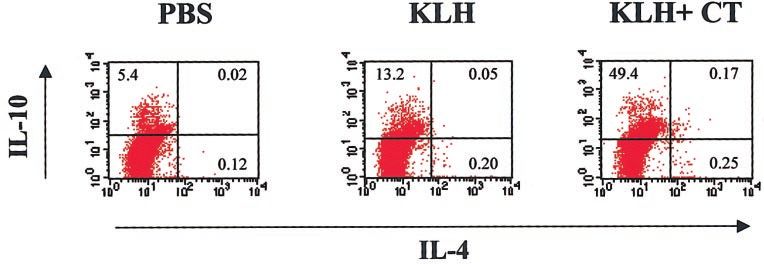
systemic antibody responses [12]. In addition, immunization tion of mice with KLH alone or in the presence of CT resulted
with antigen in the presence of CT via parenteral, mucosal, and in the induction of CD4⫹ T cells producing IL-4 and IL-10.
transcutaneous routes results in substantial enhancement of However, there was a considerable increase in the percentage
mucosal immunoglobulin A (IgA) and serum IgG responses to of IL-10-producing CD4⫹ T cells in the mice immunized with
the coadministered antigen [4]. CT also activates cellular im- KLH in the presence of CT. Although some of these CD4⫹ T
mune responses to coadministered antigens and enhances the cells produced IL-4 and IL-10, there was a large population of
induction of CD4⫹ T helper and class I-restricted cytolytic T CD4⫹ T cells that produced IL-10, independently of IL-4. The
lymphocyte responses [13–15]. Most studies indicate that CT phenotype and ontogeny of these IL-10-producing, antigen-
promotes a strong T helper cell type 2 (Th2)-biased response to specific CD4⫹ T cells are uncertain at present, but these may
itself and to bystander antigens. This conclusion is based on T represent a distinct T cell population. Tr1 cells may arise from
cell production of interleukin (IL)-4, IL-5, and IL-10 with little naı̈ve cells in lymph nodes following presentation by DC, in
interferon-␥ (IFN-␥) [16 –21] and is supported by evidence that which IL-10 production is enhanced, and IL-12 production is
IgE [16, 18, 22] and higher titers of IgG1 than IgG2a [16, 18, inhibited [32, 33]. Alternatively, Tr1 cells may be derived from
21–27] are induced after immunization with antigens in the conventional Th2 cells that have lost their ability to produce
presence of CT. However, other studies have reported mixed IL-4 but retain their ability to secrete IL-10 [35]. In addition to
Th1/Th2 (with the production of IFN-␥ and IL-4) responses our evidence that parenteral immunization with the CT holo-
following oral [28 –30] and intranasal immunization [31] with toxin can generate a population of Tr1 cells, mucosal immu-
antigens in the presence of CT. Our data indicate that although nization with CTB-antigen conjugates induces regulatory T
some IFN-␥ is produced, the response is Th2-biased, but in cells capable of suppressing autoimmune diseases mediated by
addition, CT induces a population of IL-10-producing T cells Th1 cells [36, 37].
that have regulatory activity. Experiments were performed to determine whether the an-
tigen-specific, IL-10-producing Tr1 cells induced in the pres-
ence of CT could exhibit regulatory activity against Th1 cells.
INDUCTION OF ANTIGEN-SPECIFIC A Th1 cell line was generated from spleens of mice immunized
REGULATORY T CELLS BY CT with KLH in the presence of the Toll-like receptor 9 (TLR9)
ligand, CpG oligodeoxynucleotide (CpG). These T cells prolif-
Although CT is able to activate the production of a number of erated strongly and produced high levels of IFN-␥ when re-
cytokines associated with Th2 and to a lesser degree, Th1 cells, stimulated with KLH (Fig. 1). In contrast, the CT-generated
it also induces a population of IL-10-producing T cells with Tr1 cells proliferated very poorly and produced no IFN-␥. This
suppressor activity. T cell lines established from the spleens of concurs with reports that under these conditions, CD4⫹CD25⫹
mice immunized with antigens in the presence of CT secreted Tr cells are difficult to expand in vitro [38]. However, recent
variable concentrations of the cytokines IL-4, IL-5, and IL-10. data suggest that in contrast to reports of anergy in vitro,
Antigen-specific T cell clones established from these lines CD4⫹CD25⫹ Tr cells can proliferate in response to antigen in
included clones that produced IL-10 in the absence of IL-4 vivo [39]. Furthermore, CD4⫹CD25⫹ Tr cells are able to
[32]. This is consistent with a number of studies that have expand on incubation with antigen-loaded, mature DC in vitro
reported that type 1 T regulatory (Tr1) cells secrete high levels [40]. The poor proliferation of the CT-generated Tr1 cells may
of IL-10 but in addition, may also secrete IL-5, with low or also be an in vitro phenomenon related to the type of antigen-
undetectable IL-4 [33, 34]. To demonstrate that distinct pop- presenting cell (APC), the absence of growth factors, or the
ulations of antigen-specific IL-4- and IL-10-secreting T cells presence of suppressive cytokines such as IL-10. Coincubation
were generated in vivo, intracellular staining was performed on of Th1 with Tr1 cells resulted in a significant suppression of
T cells from spleens of mice immunized subcutaneously with Th1 cell proliferation, although this was only achieved at a
antigen in the presence or absence of CT (Fig. 1). Immuniza- high ratio of Tr1-to-Th1 cells (Fig. 2). Furthermore, a Tr1-
Fig. 1. CT induces IL-10-producing
CD4⫹ Tr1 cells with a regulatory pheno-
type. Single spleen-cell suspensions from
mice immunized with phosphate-buffered
saline (PBS), keyhole limpet hemocyanin
(KLH), or KLH ⫹ CT were stimulated
with KLH (10 g/ml) for 3 days. Cells
were then further stimulated for 6 h with
phorbol 12-myristate 13-acetate (10 ng/
ml) and ionomycin (1 g/ml). Brefeldin A
(10 g/ml) was added for the final 4 h,
after which cells were washed and
blocked with 50% fetal calf serum. Sur-
face and intracellular staining was per-
formed using the Caltag Fix and Perm Cell permeabilization kit (GAS003, Caltag Laboratories, South San Francisco, CA). Cells were surface-stained with rat
anti-mouse CD4 and intracellularly, with rat anti-mouse IL-4 and IL-10. All antibodies were purchased from Caltag Laboratories. Staining was assessed by
FACSCalibur (BD Biosciences, San Jose, CA), and data were analyzed using CellQuest software for MacIntosh.
Lavelle et al. Modulation of immune responses by cholera toxin 757of the Tr1 cells may be up-regulated in a controlled manner on
contact with inflammatory Th1 cells.
ROLE OF DC IN CT-INDUCED IMMUNE
RESPONSES
The mechanism used by CT to promote the induction of spe-
cific T cell subtypes in vivo has not been fully elucidated but
is likely to involve direct interactions with APC and lympho-
cytes. DC are pivotal in the initiation of T cell responses and
in the instruction of antigen-specific, naı̈ve T cells [41]. It has
been suggested that plasmacytoid and myeloid DC promote
Th1 and Th2 cell responses, respectively [42], whereas imma-
ture DC have been implicated in the induction of anergic or Tr
cells, partly through the lack of costimulation signals and
consequent downstream effects [41]. Alternatively, the same
subtype of DC may selectively enhance the development of
distinct T cell subtypes, depending on the dose and type of
antigen or immunomodulatory molecules and the environment
pertaining at the time of maturation [32, 33, 43– 45]. Adoptive-
transfer experiments have also shown that modulation of DC
with particular pathogen-derived molecules can induce polar-
ized Th1 or Th2 responses in vivo [32, 46]. We demonstrated
that DC pulsed with antigen in the presence of CT induced a
Tr1/Th2 response in vivo, characterized by high levels of
antigen-specific IL-10 [32]. This indicates that direct effects of
CT on DC can at least partly explain its immunological actions.
CT can promote DC maturation alone or in the presence of
additional stimuli such as lipopolysaccharide (LPS) or a com-
bination of IL-1 and tumor necrosis factor ␣ (TNF-␣) [44, 45].
Fig. 2. Immunization with antigen in the presence of CT induces Tr1 cells We have previously found that exposure of DC to CT enhanced
that suppress proliferation and IFN-␥ production by Th1 cells. A KLH- surface expression of CD80 and to a lesser extent, CD86 and
specific, Tr1-type T cell line (1⫻104–1⫻106/ml) generated from mice immu- OX40 and reduced the surface expression of the chemokine
nized with KLH in the presence of CT and a KLH-specific Th1 cell line
receptor CCR5 as well as CD40 and intercellular adhesion
(1⫻105/ml) generated from mice immunized with KLH in the presence of CpG
were cultured alone or together (separated via a semipermeable membrane) in molecule-1 (ICAM-1; Table 1) [32]. This contrasts with Th1-
the presence of APC (irradiated syngeneic spleen cells; 2⫻106/ml) and KLH promoting molecules such as CpG, polyinosinic-polycytidylic
(50 g/ml). Supernatants were collected after 72 h for analysis of IFN-␥ and acid (poly I:C), and LPS, which enhance expression of each of
IL-10 and replaced with fresh medium, 3H-thymidine was added, and cells these surface-expressed molecules [43, 44]. Costimulatory
were incubated at 37° for 4 h. Cells were harvested, and proliferation was
molecule expression plays an important role in the ability of
determined by measuring thymidine incorporation. Results are means (⫹SD)
for triplicate cultures. **, P ⬍ 0.01; ***, P ⬍ 0.001, Th1 versus Th1 ⫹ Tr1 DC to promote distinct T cell responses. It has been suggested
at the same Th1 cell number. Cytokine concentrations were compared by that expression of major histocompatibility complex (MHC)
one-way ANOVA. Where significant differences were found, the Tukey- class II, CD80, and CD86 can influence the ability of DC to
Kramer multiple comparisons test was used to identify differences between direct naı̈ve T cells into Th1 or Th2 subtypes [47] and that the
individual groups.
primary response was strictly dependent on these interactions.
As CT does not inhibit MHC class II expression and enhances
CD80 and CD86 expression on DC, this may fulfill the primary
induced inhibition of IFN-␥ production by the Th1 cells was requirements for the DC to activate T cells. Additionally,
observed (Fig. 2). The inhibition of Th1 cell proliferation and selective inhibition of the expression of costimulatory mole-
IFN-␥ production did not require cell-to-cell contact, as sup- cules such as CD40 and ICAM-1 may play a role in the
pression occurred when cells were separated with a semiper- polarization of Tr1/Th2 responses by the toxin. Ineffective
meable membrane. The Tr1 cells produced high levels of IL-10 CD40 ligation has been associated with T cell unresponsive-
on stimulation with KLH, and this was significantly enhanced ness and reduced type 1 cytokines but enhanced IL-10 pro-
(at the lower Tr1:Th1 cell ratios) when the Tr1 cells were duction [48, 49]. Thus, the ability of CT to selectively modulate
coincubated with Th1 cells (Fig. 2). This effect was not cell DC costimulatory molecule expression may promote the induc-
contact-dependent and indicates that the suppressive activity tion of Tr1 and Th2 cells and block Th1 differentiation.
758 Journal of Leukocyte Biology Volume 75, May 2004 http://www.jleukbio.orgTABLE 1. Summary of the Effects of CT on DC Cytokine, [59]. The propensity of the lung microenvironment to generate
Chemokine, and Costimulatory Molecule Expression Th2-dominated responses thus appears to be associated with
IL-10 and IL-6 induction. However, these cytokines may also
Induced Enhanced Inhibited induce the differentiation of T cells into Tr cells [33]. There is
Cytokines some evidence that signaling through the IL-1 receptor 1 may
MIP-2 ⫹ also promote Th2 responses [60]. As much of the work on
IL-1 ⫹ cytokine-mediated differentiation of Th cells did not attempt to
IL-6 ⫹ separate Th2 from Tr1 responses, it is presently unclear
IL-10 ⫹
IL-12 p70 ⫹ whether factors that drive Th2 differentiation also play a role in
IL-12 p40 ⫹ promoting Tr1 responses.
TNF-␣ ⫹ In contrast to the up-regulated secretion of the above cyto-
MIP-1␣ ⫹ kines, CT inhibited the production of the proinflammatory
MIP-1 ⫹ cytokines IL-12 and TNF-␣ and inflammatory chemokines
MCP-1 ⫹
Costimulatory/surface-expressed MIP-1␣, MIP-1, and MCP-1, induced in response to the TLR
molecules ligands LPS (Table 1), CpG, and poly I:C [32]. In human DC,
CD80 ⫹ ⫹
CD86 ⫹
OX40 ⫹ ⫹
CD14 ⫹
CD40 ⫹
ICAM-1 ⫹
The columns refer to the direct effects of CT (Induced) or modulation of
LPS-mediated effects (Enhanced/Inhibited) on DC. CT was added to DC at 1
g/ml alone or 1 h prior to addition of Escherichia coli LPS (0.1–10 ng/ml).
Cytokine expression was determined by enzyme-linked immunosorbent assay
(ELISA) and costimulatory molecule expression by flow cytometry. MIP-2,
macrophage-inflammatory protein-2; MCP-1, monocyte chemoattractant pro-
tein-1.
Although the polarization of naı̈ve T cells is multifactorial,
the cytokine environment pertaining at the time of primary
activation appears central [50]. A number of cytokines pro-
duced by cells of the innate-immune system and/or T cells are
involved in the selective activation of Th1 and Th2 cells [51].
Secretion of IL-12 by DC and macrophages enhances Th1
responses [43]. In contrast, IL-4 as well as IL-6 and IL-10 can
promote the differentiation of Th2 cells [52, 53]. Indeed, IL-10
was required for optimal generation of Th2 cells by CD8␣– DC
[54]. IL-10, IL-4, and transforming growth factor- (TGF-)
may be involved in driving the differentiation of Tr cells [35,
55, 56]. We were unable to detect the production of IL-6, IL-4,
or IL-10 by DC exposed to CT alone in vitro. However, in the
presence of low doses of LPS, CT significantly enhanced IL-10
production (Fig. 3) [32]. In addition, CT synergized with LPS
to enhance the production of IL-6 and IL-1 (Fig. 3). The
enhancement of IL-6 was only evident with low concentrations
of LPS, and IL-10 and IL-1 production was enhanced over a
wide range of LPS concentrations (Fig. 3). The enhancement of
LPS-mediated IL-6 production by CT was also detected at the
transcriptional level (data not shown).
Fig. 3. CT synergizes with LPS to induce IL-10, IL-6, and IL-1 production
Enhancement of LPS-induced IL-6 and IL-1 production by by DC. Bone marrow-derived, immature DC were incubated for 12 h with LPS
CT was previously demonstrated in murine macrophages [57]. (0.1–10 ng/ml) in the presence (■) or absence (䊐) of CT (1 g/ml) or with CT
IL-6 can promote Th2 differentiation via the activation of alone. Cytokine concentrations were determined in supernatants by ELISA.
nuclear factor of activated T cells and induction of early IL-4 Results are means (⫹SD) of triplicate assays and are representative of four
production by CD4⫹ T cells [58] and can inhibit IFN-␥ pro- experiments. *, P ⬍ 0.05; **, P ⬍ 0.01; ***, P ⬍ 0.001, CT ⫹ LPS versus
LPS at the same concentration. Cytokine concentrations were compared by
duction and Th1 differentiation. Following intranasal immuni- one-way ANOVA. Where significant differences were found, the Tukey-
zation, antigen-loaded, pulmonary DC produced IL-6 and IL- Kramer multiple comparisons test was used to identify differences between
10, which was proposed to promote Th2 differentiation in situ individual groups.
Lavelle et al. Modulation of immune responses by cholera toxin 759CT inhibited the production of IL-12 and the expression of cells and down-regulation of chemokines in the CNS. However,
IL-12 receptor chains and TNF-␣, leading to a suppression of the inclusion of CT in this system abrogated the protective
Th1 cell differentiation [45, 61]. Forskolin, a pharmacological effects of CTB. Antigen–CTB conjugates have also recently
activator of adenylate cyclase, can also inhibit the production been shown to be protective in a number of other autoimmune
of bioactive IL-12, TNF-␣, and MIP-1␣ and can enhance IL-10 models. Oral delivery of CTB conjugated to a heat-shock
production by DC, implicating cAMP as a determining factor in protein 60 kDa-derived peptide prevented mucosally induced
the inhibitory action of CT on inflammatory cytokines (E. C. uveitis in rats, an effect that was associated with enhanced
Lavelle et al., unpublished data). IL-10 and TGF- and reduced IL-12 and IFN-␥ production
Although our recent studies point to a dominant role for the [36]. Furthermore, oral administration of a CTB-insulin conju-
elevation in intracellular cAMP concentrations in the inhibi- gate prevented diabetes in nonobese diabetic mice, which was
tory effects of CT, other workers have found that CTB can also associated with a reduction in IFN-␥ production and Tr cell
modulate DC activation. Pretreatment of monocytes and mac- migration into pancreatic islets [68]. Oral administration of
rophages with relatively high concentrations of CTB reduced recombinant CTB also prevented IL-12-mediated murine ex-
their subsequent responsiveness to LPS [62]. LPS-induced perimental trinitrobenzene sulfonic acid-induced colitis [69].
production of TNF-␣, IL-6, IL-12 p70, and nitric oxide was Decreased IL-12 and IFN-␥ production was documented, but
inhibited, and IL-10 production was enhanced. This suggests in this case, there was no elevation in IL-10 or TGF-. CTB
that in addition to the cAMP-mediated enhancement of LPS- conjugates were also effective in the induction of tolerance to
driven IL-10 reported by our group and others, the B subunit type II collagen, leading to a suppression of chondritis in a
may also enhance IL-10 production by cells of the innate- model of autoimmune ear disease [70]. Oral administration of
immune system. However, the inhibitory effect of CTB on allogeneic antigen linked to CTB induced, immunological tol-
macrophage IL-6 production is in contrast to the enhanced erance against allograft rejection [66]. It was shown that even
effect of CT on IL-6 production by macrophages [57] and DC. without conjugation, CTB could potentiate oral tolerance in-
Antibodies to IL-10 and TGF- prevented the inhibitory effect duction to insulin [71]. In addition to this work on CTB, there
of CTB on LPS-induced IL-6 and TNF-␣ production [62]. is extensive evidence that the related B subunit of E. coli
However, the effect of anti-IL-10 and TGF- antibodies on heat-labile enterotoxin can potentiate mucosal tolerance and
IL-6 production was greater than on TNF-␣ production, indi- prevent the induction of autoimmune inflammatory conditions
cating a role for additional factors in the inhibition of TNF-␣ including collagen-induced arthritis [4, 72]. Thus, it appears
by CTB. that the A–B holotoxins and their purified B chains have
potential in treatment of inflammatory conditions via their
anti-inflammatory properties and through the induction of Tr
cells.
CT AS A POTENTIAL THERAPEUTIC FOR
AUTOIMMUNE DISEASES
The anti-inflammatory effects of CT and its ability to promote CONCLUSIONS
the induction of antigen-specific Tr cells underline its potential
for the treatment of diseases mediated by Th1 cells. It has been CT is a powerful mucosal adjuvant but also has potential as an
reported that nasal administration of high doses of CT can immunomodulatory and anti-inflammatory agent. We recently
suppress clinical signs of experimental autoimmune encepha- demonstrated that in addition to its well-documented Th2-
lomyelitis (EAE), a murine model of multiple sclerosis [63]. promoting activity, CT enhances the induction of a population
The concentrations of IFN-␥ and IL-12 in the central nervous of IL-10-producing Tr cells. Adoptive transfer of myeloid DC
system (CNS) of CT-treated mice were lower than in controls. pulsed with antigen in the presence of CT can induce antigen-
CT was also shown to potentiate tolerance to bovine peripheral specific T cells with a similar cytokine profile to that observed
nerve myelin in the experimental autoimmune neuritis model following direct immunization [32], indicating that the adju-
of human inflammatory demyelinating neuropathies [64]. There vant and modulatory activities of the toxin in vivo are at least
is also earlier evidence that CT could induce tolerance to partly attributable to its effects on DC. CT can enhance the
allografts in mice [65]. Thus, in a number of T cell-mediated, expression of a number of costimulatory molecules on DC but
autoimmune conditions, mucosal administration of CT has in contrast to Th1-driving molecules such as CpG and poly I:C,
been shown to enhance the induction of tolerance and alleviate inhibited the expression of CD40 and ICAM-1. In addition, in
disease symptoms. vitro studies with macrophages and DC have shown that CT can
Although it is conceptually attractive to attribute the anti- enhance the secretion of IL-10, IL-6, and IL-1 in the pres-
inflammatory effects of CT to the elevated cAMP levels medi- ence of limiting doses of LPS. These selective inhibitory and
ated by the A subunit, a large body of evidence now exists synergistic effects of CT on DC may explain the ability of CT
indicating that mucosal delivery of the CTB subunit can inde- to promote the induction of antigen-specific Tr1 and Th2 cells
pendently exert anti-inflammatory effects. Oral delivery of CTB (Fig. 4). The use of CT and E. coli heat-labile enterotoxin (LT)
conjugated to myelin basic protein protected mice against the as therapeutic agents is hampered by their toxicity, and it is not
development of EAE [66, 67]. It was proposed that the inhib- yet completely clear which of the beneficial adjuvant and
itory effect was a result of the induction of TGF--producing Tr immunomodulatory activities are retained in nontoxic mutants
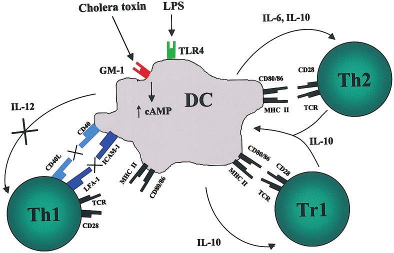
760 Journal of Leukocyte Biology Volume 75, May 2004 http://www.jleukbio.orgFig. 4. Proposed model of the mod-
ulatory effects of CT on innate- and
adaptive-immune responses. CT binds
to GM-1 gangliosides on DC with high
affinity, leading to entry of the toxin
and enhancement of intracellular
cAMP concentrations via the actions
of the A subunit. These toxin-medi-
ated effects selectively inhibit induc-
tion of the costimulatory molecules
CD40 and ICAM-1, which play a role
in Th1 cell differentiation. In contrast,
the toxin enhances expression of other
costimulatory molecules, particularly
CD80 and CD86. In addition, CT in-
hibits the induction of IL-12, which is
important in the differentiation of na-
ı̈ve T cells toward a Th1 phenotype
and in the presence of a second signal
(e.g., LPS), enhances the production of
other cytokines such as IL-10 and
IL-6, which are important for Tr1/Th2
cell differentiation. TCR, T cell recep-
tor; LFA-1, lymphocyte function-asso-
ciated antigen-1.
or subunits. However, the B subunits and site-directed mutants 10. Pelham, H. R. (1992) The Florey Lecture, 1992. The secretion of proteins
by cells. Proc. R. Soc. Lond. B. Biol. Sci. 250, 1–10.
of CT and LT, which are currently in clinical trials, have shown
11. Tsai, S. C., Noda, M., Adamik, R., Chang, P. P., Chen, H. C., Moss, J.,
potential as adjuvants and therapeutics for a number of im- Vaughan, M. (1988) Stimulation of choleragen enzymatic activities by
mune-mediated diseases. GTP and two soluble proteins purified from bovine brain. J. Biol. Chem.
263, 1768 –1772.
12. Lavelle, E. C., Grant, G., Pusztai, A., Pfuller, U., O’Hagan, D. T. (2000)
Mucosal immunogenicity of plant lectins in mice. Immunology 99, 30 –
ACKNOWLEDGMENT 37.
13. Simmons, C. P., Hussell, T., Sparer, T., Walzl, G., Openshaw, P., Dougan,
G. (2001) Mucosal delivery of a respiratory syncytial virus CTL peptide
Science Foundation Ireland (Grant 00/P1.1/B045) supported
with enterotoxin-based adjuvants elicits protective, immunopathogenic,
this work. and immunoregulatory antiviral CD8⫹ T cell responses. J. Immunol. 166,
1106 –1113.
14. Simmons, C. P., Mastroeni, P., Fowler, R., Ghaem-maghami, M., Lycke,
N., Pizza, M., Rappuoli, R., Dougan, G. (1999) MHC class I-restricted
REFERENCES cytotoxic lymphocyte responses induced by enterotoxin-based mucosal
adjuvants. J. Immunol. 163, 6502– 6510.
1. Zhang, R. G., Scott, D. L., Westbrook, M. L., Nance, S., Spangler, B. D., 15. Bowen, J. C., Nair, S. K., Reddy, R., Rouse, B. T. (1994) Cholera toxin
Shipley, G. G., Westbrook, E. M. (1995) The three-dimensional crystal acts as a potent adjuvant for the induction of cytotoxic T-lymphocyte
structure of cholera toxin. J. Mol. Biol. 251, 563–573. responses with non-replicating antigens. Immunology 81, 338 –342.
2. Rappuoli, R., Pizza, M., Douce, G., Dougan, G. (1999) Structure and 16. Simecka, J. W., Jackson, R. J., Kiyono, H., McGhee, J. R. (2000) Muco-
mucosal adjuvanticity of cholera and Escherichia coli heat-labile entero- sally induced immunoglobulin E-associated inflammation in the respira-
toxins. Immunol. Today 20, 493–500. tory tract. Infect. Immun. 68, 672– 679.
3. Pizza, M., Giuliani, M. M., Fontana, M. R., Monaci, E., Douce, G., 17. Yamamoto, M., Rennert, P., McGhee, J. R., Kweon, M. N., Yamamoto, S.,
Dougan, G., Mills, K. H., Rappuoli, R., Del Giudice, G. (2001) Mucosal Dohi, T., Otake, S., Bluethmann, H., Fujihashi, K., Kiyono, H. (2000)
vaccines: nontoxic derivatives of LT and CT as mucosal adjuvants. Vaccine Alternate mucosal immune system: organized Peyer’s patches are not
19, 2534 –2541. required for IgA responses in the gastrointestinal tract. J. Immunol. 164,
4. Williams, N. A., Hirst, T. R., Nashar, T. O. (1999) Immune modulation by 5184 –5191.
the cholera-like enterotoxins: from adjuvant to therapeutic. Immunol. 18. Yamamoto, S., Kiyono, H., Yamamoto, M., Imaoka, K., Fujihashi, K., Van
Today 20, 95–101.
Ginkel, F. W., Noda, M., Takeda, Y., McGhee, J. R. (1997) A nontoxic
5. Holmgren, J., Lonnroth, I., Svennerholm, L. (1973) Tissue receptor for
mutant of cholera toxin elicits Th2-type responses for enhanced mucosal
cholera exotoxin: postulated structure from studies with GM1 ganglioside
immunity. Proc. Natl. Acad. Sci. USA 94, 5267–5272.
and related glycolipids. Infect. Immun. 8, 208 –214.
6. Spangler, B. D. (1992) Structure and function of cholera toxin and the 19. Yamamoto, S., Takeda, Y., Yamamoto, M., Kurazono, H., Imaoka, K.,
related Escherichia coli heat-labile enterotoxin. Microbiol. Rev. 56, 622– Fujihashi, K., Noda, M., Kiyono, H., McGhee, J. R. (1997) Mutants in the
647. ADP-ribosyltransferase cleft of cholera toxin lack diarrheagenicity but
7. Gill, D. M., Rappaport, R. S. (1979) Origin of the enzymatically active A1 retain adjuvanticity. J. Exp. Med. 185, 1203–1210.
fragment of cholera toxin. J. Infect. Dis. 139, 674 – 680. 20. Clarke, C. J., Wilson, A. D., Williams, N. A., Stokes, C. R. (1991) Mucosal
8. Kawamura, Y. I., Kawashima, R., Shirai, Y., Kato, R., Hamabata, T., priming of T-lymphocyte responses to fed protein antigens using cholera
Yamamoto, M., Furukawa, K., Fujihashi, K., McGhee, J. R., Hayashi, H., toxin as an adjuvant. Immunology 72, 323–328.
Dohi, T. (2003) Cholera toxin activates dendritic cells through depen- 21. Xu-Amano, J., Kiyono, H., Jackson, R. J., Staats, H. F., Fujihashi, K.,
dence on GM1-ganglioside which is mediated by NF-B translocation. Burrows, P. D., Elson, C. O., Pillai, S., McGhee, J. R. (1993) Helper T cell
Eur. J. Immunol. 33, 3205–3212. subsets for immunoglobulin A responses: oral immunization with tetanus
9. Field, M., Rao, M. C., Chang, E. B. (1989) Intestinal electrolyte transport toxoid and cholera toxin as adjuvant selectively induces Th2 cells in
and diarrheal disease (2). N. Engl. J. Med. 321, 879 – 883. mucosa associated tissues. J. Exp. Med. 178, 1309 –1320.
Lavelle et al. Modulation of immune responses by cholera toxin 76122. Marinaro, M., Staats, H. F., Hiroi, T., Jackson, R. J., Coste, M., Boyaka, 42. Rissoan, M. C., Soumelis, V., Kadowaki, N., Grouard, G., Briere, F., de
P. N., Okahashi, N., Yamamoto, M., Kiyono, H., Bluethmann, H., et al. Waal Malefyt, R., Liu, Y. J. (1999) Reciprocal control of T helper cell and
(1995) Mucosal adjuvant effect of cholera toxin in mice results from dendritic cell differentiation. Science 283, 1183–1186.
induction of T helper 2 (Th2) cells and IL-4. J. Immunol. 155, 4621– 43. Boonstra, A., Asselin-Paturel, C., Gilliet, M., Crain, C., Trinchieri, G.,
4629. Liu, Y. J., O’Garra, A. (2003) Flexibility of mouse classical and plasma-
23. Douce, G., Fontana, M., Pizza, M., Rappuoli, R., Dougan, G. (1997) cytoid-derived dendritic cells in directing T helper type 1 and 2 cell
Intranasal immunogenicity and adjuvanticity of site-directed mutant de- development: dependency on antigen dose and differential Toll-like re-
rivatives of cholera toxin. Infect. Immun. 65, 2821–2828. ceptor ligation. J. Exp. Med. 197, 101–109.
24. Pierre, P., Denis, O., Bazin, H., Mbongolo Mbella, E., Vaerman, J. P. 44. de Jong, E. C., Vieira, P. L., Kalinski, P., Schuitemaker, J. H., Tanaka, Y.,
(1992) Modulation of oral tolerance to ovalbumin by cholera toxin and its Wierenga, E. A., Yazdanbakhsh, M., Kapsenberg, M. L. (2002) Microbial
B subunit. Eur. J. Immunol. 22, 3179 –3182. compounds selectively induce Th1 cell-promoting or Th2 cell-promoting
25. Glenn, G. M., Scharton-Kersten, T., Vassell, R., Mallett, C. P., Hale, T. L., dendritic cells in vitro with diverse Th cell-polarizing signals. J. Immunol.
Alving, C. R. (1998) Transcutaneous immunization with cholera toxin 168, 1704 –1709.
protects mice against lethal mucosal toxin challenge. J. Immunol. 161, 45. Gagliardi, M. C., Sallusto, F., Marinaro, M., Langenkamp, A., Lanzavec-
3211–3214. chia, A., De Magistris, M. T. (2000) Cholera toxin induces maturation of
26. Ruedl, C., Rieser, C., Kofler, N., Wick, G., Wolf, H. (1996) Humoral and human dendritic cells and licences them for Th2 priming. Eur. J. Immu-
cellular immune responses in the murine respiratory tract following oral nol. 30, 2394 –2403.
immunization with cholera toxin or Escherichia coli heat-labile entero- 46. MacDonald, A. S., Straw, A. D., Dalton, N. M., Pearce, E. J. (2002) Cutting
toxin. Vaccine 14, 792–798. edge: Th2 response induction by dendritic cells: a role for CD40. J. Im-
27. Richards, C. M., Shimeld, C., Williams, N. A., Hill, T. J. (1998) Induction munol. 168, 537–540.
of mucosal immunity against herpes simplex virus type 1 in the mouse 47. Lespagnard, L., Mettens, P., De Smedt, T., Bazin, H., Urbain, J., Leo, O.,
protects against ocular infection and establishment of latency. J. Infect. Moser, M. (1998) The immune response induced in vivo by dendritic cells
Dis. 177, 1451–1457. is dependent on B7–1 or B7–2, but the inhibition of both signals does not
28. Hornquist, E., Lycke, N. (1993) Cholera toxin adjuvant greatly promotes lead to tolerance. Int. Immunol. 10, 295–304.
antigen priming of T cells. Eur. J. Immunol. 23, 2136 –2143. 48. Martin, E., O’Sullivan, B., Low, P., Thomas, R. (2003) Antigen-specific
29. Pacheco, S. E., Gibbs, R. A., Ansari-Lari, A., Rogers, P. (2000) Intranasal suppression of a primed immune response by dendritic cells mediated by
immunization with HIV reverse transcriptase: effect of dose in the induc- regulatory T cells secreting interleukin-10. Immunity 18, 155–167.
tion of helper T cell type 1 and 2 immunity. AIDS Res. Hum. Retroviruses 49. Fontana, S., Moratto, D., Mangal, S., De Francesco, M., Vermi, W.,
16, 2009 –2017. Ferrari, S., Facchetti, F., Kutukculer, N., Fiorini, C., Duse, M., Das, P. K.,
30. Schaffeler, M. P., Brokenshire, J. S., Snider, D. P. (1997) Detection of Notarangelo, L. D., Plebani, A., Badolato, R. (2003) Functional defects of
precursor Th cells in mesenteric lymph nodes after oral immunization with dendritic cells in patients with CD40 deficiency. Blood 102, 4099 – 4106.
protein antigen and cholera toxin. Int. Immunol. 9, 1555–1562. 50. O’Garra, A. (1998) Cytokines induce the development of functionally
31. Yanagita, M., Hiroi, T., Kitagaki, N., Hamada, S., Ito, H. O., Shimauchi, heterogeneous T helper cell subsets. Immunity 8, 275–283.
51. Murphy, K. M., Reiner, S. L. (2002) The lineage decisions of helper T
H., Murakami, S., Okada, H., Kiyono, H. (1999) Nasopharyngeal-associ-
cells. Nat. Rev. Immunol. 2, 933–944.
ated lymphoreticular tissue (NALT) immunity: fimbriae-specific Th1 and
52. Iwasaki, A., Kelsall, B. L. (1999) Freshly isolated Peyer’s patch, but not
Th2 cell-regulated IgA responses for the inhibition of bacterial attachment
spleen, dendritic cells produce interleukin 10 and induce the differenti-
to epithelial cells and subsequent inflammatory cytokine production.
ation of T helper type 2 cells. J. Exp. Med. 190, 229 –239.
J. Immunol. 162, 3559 –3565.
53. Rincon, M., Anguita, J., Nakamura, T., Fikrig, E., Flavell, R. A. (1997)
32. Lavelle, E. C., McNeela, E., Armstrong, M. E., Leavy, O., Higgins, S. C.,
Interleukin (IL)-6 directs the differentiation of IL-4-producing CD4⫹ T
Mills, K. H. (2003) Cholera toxin promotes the induction of regulatory T
cells. J. Exp. Med. 185, 461– 469.
cells specific for bystander antigens by modulating dendritic cell activa-
54. Maldonado-Lopez, R., Maliszewski, C., Urbain, J., Moser, M. (2001)
tion. J. Immunol. 171, 2384 –2392.
Cytokines regulate the capacity of CD8␣⫹ and CD8␣⫹ dendritic cells to
33. McGuirk, P., Mills, K. H. (2002) Pathogen-specific regulatory T cells prime Th1/Th2 cells in vivo. J. Immunol. 167, 4345– 4350.
provoke a shift in the Th1/Th2 paradigm in immunity to infectious 55. Chen, Y., Kuchroo, V. K., Inobe, J., Hafler, D. A., Weiner, H. L. (1994)
diseases. Trends Immunol. 23, 450 – 455. Regulatory T cell clones induced by oral tolerance: suppression of auto-
34. Doetze, A., Satoguina, J., Burchard, G., Rau, T., Loliger, C., Fleischer, B., immune encephalomyelitis. Science 265, 1237–1240.
Hoerauf, A. (2000) Antigen-specific cellular hyporesponsiveness in a 56. Seder, R. A., Marth, T., Sieve, M. C., Strober, W., Letterio, J. J., Roberts,
chronic human helminth infection is mediated by T(h)3/T(r)1-type cyto- A. B., Kelsall, B. (1998) Factors involved in the differentiation of TGF-
kines IL-10 and transforming growth factor- but not by a T(h)1 to T(h)2 -producing cells from naive CD4⫹ T cells: IL-4 and IFN-␥ have opposing
shift. Int. Immunol. 12, 623– 630. effects, while TGF- positively regulates its own production. J. Immunol.
35. Mendel, I., Shevach, E. M. (2002) The IL-10-producing competence of 160, 5719 –5728.
Th2 cells generated in vitro is IL-4 dependent. Eur. J. Immunol. 32, 57. Cong, Y., Oliver, A. O., Elson, C. O. (2001) Effects of cholera toxin on
3216 –3224. macrophage production of co-stimulatory cytokines. Eur. J. Immunol. 31,
36. Phipps, P. A., Stanford, M. R., Sun, J. B., Xiao, B. G., Holmgren, J., 64 –71.
Shinnick, T., Hasan, A., Mizushima, Y., Lehner, T. (2003) Prevention of 58. Diehl, S., Rincon, M. (2002) The two faces of IL-6 on Th1/Th2 differen-
mucosally induced uveitis with a HSP60-derived peptide linked to cholera tiation. Mol. Immunol. 39, 531–536.
toxin B subunit. Eur. J. Immunol. 33, 224 –232. 59. Constant, S. L., Brogdon, J. L., Piggott, D. A., Herrick, C. A., Visintin, I.,
37. Sun, J. B., Xiao, B. G., Lindblad, M., Li, B. L., Link, H., Czerkinsky, C., Ruddle, N. H., Bottomly, K. (2002) Resident lung antigen-presenting cells
Holmgren, J. (2000) Oral administration of cholera toxin B subunit con- have the capacity to promote Th2 T cell differentiation in situ. J. Clin.
jugated to myelin basic protein protects against experimental autoimmune Invest. 110, 1441–1448.
encephalomyelitis by inducing transforming growth factor--secreting 60. Nakae, S., Komiyama, Y., Yokoyama, H., Nambu, A., Umeda, M., Iwase,
cells and suppressing chemokine expression. Int. Immunol. 12, 1449 – M., Homma, I., Sudo, K., Horai, R., Asano, M., Iwakura, Y. (2003) IL-1 is
1457. required for allergen-specific Th2 cell activation and the development of
38. Thornton, A. M., Shevach, E. M. (1998) CD4⫹CD25⫹ immunoregulatory airway hypersensitivity response. Int. Immunol. 15, 483– 490.
T cells suppress polyclonal T cell activation in vitro by inhibiting inter- 61. Braun, M. C., He, J., Wu, C. Y., Kelsall, B. L. (1999) Cholera toxin
leukin 2 production. J. Exp. Med. 188, 287–296. suppresses interleukin (IL)-12 production and IL-12 receptor 1 and 2
39. Walker, L. S., Chodos, A., Eggena, M., Dooms, H., Abbas, A. K. (2003) chain expression. J. Exp. Med. 189, 541–552.
Antigen-dependent proliferation of CD4⫹ CD25⫹ regulatory T cells in 62. Burkart, V., Kim, Y. E., Hartmann, B., Ghiea, I., Syldath, U., Kauer, M.,
vivo. J. Exp. Med. 198, 249 –258. Fingberg, W., Hanifi-Moghaddam, P., Muller, S., Kolb, H. (2002) Cholera
40. Yamazaki, S., Iyoda, T., Tarbell, K., Olson, K., Velinzon, K., Inaba, K., toxin B pretreatment of macrophages and monocytes diminishes their
Steinman, R. M. (2003) Direct expansion of functional CD25⫹ CD4⫹ proinflammatory responsiveness to lipopolysaccharide. J. Immunol. 168,
regulatory T cells by antigen-processing dendritic cells. J. Exp. Med. 1730 –1737.
198, 235–247. 63. Yura, M., Takahashi, I., Terawaki, S., Hiroi, T., Kweon, M. N., Yuki, Y.,
41. Jonuleit, H., Schmitt, E., Steinbrink, K., Enk, A. H. (2001) Dendritic cells Kiyono, H. (2001) Nasal administration of cholera toxin (CT) suppresses
as a tool to induce anergic and regulatory T cells. Trends Immunol. 22, clinical signs of experimental autoimmune encephalomyelitis (EAE). Vac-
394 – 400. cine 20, 134 –139.
762 Journal of Leukocyte Biology Volume 75, May 2004 http://www.jleukbio.org64. Gaupp, S., Hartung, H. P., Toyka, K., Jung, S. (1997) Modulation of 69. Boirivant, M., Fuss, I. J., Ferroni, L., De Pascale, M., Strober, W. (2001)
experimental autoimmune neuritis in Lewis rats by oral application of Oral administration of recombinant cholera toxin subunit B inhibits IL-
myelin antigens. J. Neuroimmunol. 79, 129 –137. 12-mediated murine experimental (trinitrobenzene sulfonic acid) colitis.
65. Tsuru, S., Taniguchi, M., Shinomiya, N., Fujisawa, H., Zinnaka, Y., J. Immunol. 166, 3522–3532.
Nomoto, K. (1987) Cholera toxin-induced tolerance to allografts in mice. 70. Kim, N., Cheng, K. C., Kwon, S. S., Mora, R., Barbieri, M., Yoo, T. J.
Immunology 61, 77– 83. (2001) Oral administration of collagen conjugated with cholera toxin
66. Sun, J. B., Li, B. L., Czerkinsky, C., Holmgren, J. (2000) Enhanced induces tolerance to type II collagen and suppresses chondritis in an
immunological tolerance against allograft rejection by oral administration animal model of autoimmune ear disease. Ann. Otol. Rhinol. Laryngol.
of allogeneic antigen linked to cholera toxin B subunit. Clin. Immunol.
110, 646 – 654.
97, 130 –139.
67. Sun, J. B., Rask, C., Olsson, T., Holmgren, J., Czerkinsky, C. (1996) 71. Bregenholt, S., Wang, M., Wolfe, T., Hughes, A., Baerentzen, L., Dyrberg,
Treatment of experimental autoimmune encephalomyelitis by feeding my- T., von Herrath, M. G., Petersen, J. S. (2003) The cholera toxin B subunit
elin basic protein conjugated to cholera toxin B subunit. Proc. Natl. Acad. is a mucosal adjuvant for oral tolerance induction in type 1 diabetes.
Sci. USA 93, 7196 –7201. Scand. J. Immunol. 57, 432– 438.
68. Aspord, C., Czerkinsky, C., Durand, A., Stefanutti, A., Thivolet, C. (2002) 72. Luross, J. A., Heaton, T., Hirst, T. R., Day, M. J., Williams, N. A. (2002)
␣4 Integrins and L-selectin differently orchestrate T-cell activity during Escherichia coli heat-labile enterotoxin B subunit prevents autoimmune
diabetes prevention following oral administration of CTB-insulin. J. Au- arthritis through induction of regulatory CD4⫹ T cells. Arthritis Rheum.
toimmun. 19, 223–232. 46, 1671–1682.
Lavelle et al. Modulation of immune responses by cholera toxin 763You can also read