Understanding the Differentiation, Expansion, Recruitment and Suppressive Activities of Myeloid-Derived Suppressor Cells in Cancers - Core
←
→
Page content transcription
If your browser does not render page correctly, please read the page content below
International Journal of
            Molecular Sciences

Review
Understanding the Differentiation, Expansion,
Recruitment and Suppressive Activities of
Myeloid-Derived Suppressor Cells in Cancers
Hui Xuan Lim 1 , Tae Sung Kim 2                and Chit Laa Poh 1, *
 1    Centre for Virus and Vaccine Research, School of Science and Technology, Sunway University,
      Bandar Sunway, Kuala Lumpur, Selangor 47500, Malaysia; huixuanl@sunway.edu.my
 2    Division of Life Sciences, College of Life Sciences and Biotechnology, Korea University, Seoul 136-701, Korea;
      tskim@korea.ac.kr
 *    Correspondence: pohcl@sunway.edu.my; Tel.: +603-7491-8622 (ext. 7338)

 Received: 11 February 2020; Accepted: 13 March 2020; Published: 20 May 2020

 Abstract: There has been a great interest in myeloid-derived suppressor cells (MDSCs) due to their
 biological functions in tumor-mediated immune escape by suppressing antitumor immune responses.
 These cells arise from altered myelopoiesis in response to the tumor-derived factors. The most
 recognized function of MDSCs is suppressing anti-tumor immune responses by impairing T cell
 functions, and these cells are the most important players in cancer dissemination and metastasis.
 Therefore, understanding the factors and the mechanism of MDSC differentiation, expansion,
 and recruitment into the tumor microenvironment can lead to its control. However, most of the
 studies only defined MDSCs with no further characterization of granulocytic and monocytic subsets.
 In this review, we discuss the mechanisms by which specific MDSC subsets contribute to cancers.
 A better understanding of MDSC subset development and the specific molecular mechanism is
 needed to identify treatment targets. The understanding of the specific molecular mechanisms
 responsible for MDSC accumulation would enable more precise therapeutic targeting of these cells.

 Keywords: MDSC subsets; G-MDSCs; M-MDSCs; cancers; immunosuppression

1. Developmental Origin of Myeloid-Derived Suppressor Cells
     Myeloid-derived suppressor cells (MDSCs) represent a population of heterogeneous myeloid
lineage cells that have the potent immunosuppressive activity of T cell activation and function.
They comprise macrophages, granulocytes, and dendritic cells in immature stages of development.
Hematopoietic stem cells give rise to myeloid progenitor and precursor cells in the bone marrow.
These immature myeloid cells (IMCs) migrate to peripheral lymphoid organs and differentiate
into mature granulocytes, macrophages, or dendritic cells. MDSCs arise from common myeloid
progenitors and are arrested in an immature phase of differentiation. Various sources of immunological
stress, including cancer, chronic inflammation, trauma, and autoimmune disorder, can inhibit the
differentiation and promote the expansion of IMCs. IMCs can be activated by tumor-derived factors and
host cytokines, which lead to the generation of MDSCs with potent immunosuppressive potential [1].
Healthy people do not have MDSCs, but in pathological conditions, MDSCs can be detected in the
bone marrow, spleen, blood, tumor, and lymph nodes. The frequency of circulating MDSC increases
dramatically in cancer, autoimmunity, infection, always correlates with the disease severity and
worsens the survival rates.

Int. J. Mol. Sci. 2020, 21, 3599; doi:10.3390/ijms21103599                               www.mdpi.com/journal/ijms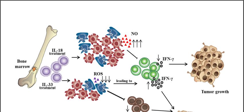
Int. J. Mol. Sci. 2020, 21, 3599                                                                   2 of 13

2. MDSC Surface Markers and Subsets
      MDSCs are characterized by the co-expression of surface markers GR-1 and CD11b in mice.
Normal mouse bone marrow contains 20–30% of cells with this phenotype, but only approximately
2–4% of cells are present in the spleen, and these cells are absent from the lymph nodes. In naive mice,
CD11b+ GR1+ lacks immunosuppressive activity, but these cells have strong immunosuppressive effects
on T cell response in tumor-bearing mice. Since GR-1 antibodies can bind to two separate epitopes,
Ly6G and Ly6C, these epitope-specific antibodies have been used to distinguish two MDSC subsets:
granulocytic MDSCs (G-MDSCs), which have a CD11b+ Ly6G+ Ly6Clow phenotype, whilst monocytic
MDSCs (M-MDSCs) have a CD11b+ Ly6G− Ly6Chigh phenotype [2]. CD49d was suggested by Haile
et al., 2010, to be an alternative marker for Gr-1 to differentiate G-MDSCs (CD11b+ CD49d− ) and
M-MDSCs (CD11b+ CD49d+ ) [3] (Table 1). Evidence indicates that both MDSC subsets expanded
in most of the murine tumor model, but the expansion of G-MDSCs was much greater than the
M-MDSCs and represented more than 80% of all MDSCs [4,5]. The frequency of G-MDSCs was also
greater than M-MDSCs in the peripheral blood and tumor tissue of pancreatic cancer patients [6].
However, studies have shown that M-MDSCs have higher suppressive activity than G-MDSCs on a
single-cell basis [7]. Additionally, M-MDSCs acquired the ability to differentiate into tumor-associated
macrophages (TAMs), which produced immunosuppressive cytokines that protected the tumor from the
immune system and immunotherapy [8]. Another subset of MDSCs resembles eosinophils, Eo-MDSC,
(CD11b+ Syglec-F+ CCR3low IL-5Ralow SSC-Ahigh ) was identified in mice with chronic Staphylococcus
aureus infection [5]. Human MDSC was firstly identified in hepatocellular carcinoma and non-Hodgkin’s
lymphoma patients with phenotypes CD14+ HLA-DRlow/− [9,10]. Other phenotypic markers for human
MDSC subsets in the peripheral blood include CD11b+ CD14– CD15+ or CD11b+ CD14− CD66b+ for
G-MDSC, CD11b+ CD14+ HLA-DR−/low CD15− for M-MDSC, and Lin− HLA-DR− CD33+ for more
immature MDSC progenitors (Table 1) [11]. However, some of the markers mentioned earlier
overlapped with other cell populations. Hence, phenotypic characterization in combination with
immune-suppressive activity is the optimal strategy for identifying MDSCs.

                       Table 1. Phenotype and functional proteins of murine and human MDSCs.

                         MDSC Subsets                       Phenotype                References
                                   MDSC             CD11b+ GR1+
           Murine                  G-MDSC           CD11b+ Ly6G+ Ly6Clow                 [2]
                                   M-MDSC           CD11b+ Ly6Gneg Ly6Chigh
                                   G-MDSC           CD11b+ CD49−
           Murine                                                                        [3]
                                   M-MDSC           CD11b+ CD49+
                                                    CD14+ HLA-DRlow/−
                                   MDSC
                                                    CD14− CD11b+ CD33+ CD15+
           Human                   G-MDSC                                                [10]
                                                    CD11b+
                                   M-MDSC
                                                    HLA-DRlow/− CD14+
                                   G-MDSC           CD11b+ CD14– CD15+
           Human                                    CD11b+ CD14– CD66b+                  [11]
                                   M-MDSC           CD11b+ CD14+ HLA-DR−/low CD15−
                                   MDSC             Lin− HLA-DR− CD11b+ CD33+
           Human                   G-MDSC           HLA-DR− CD11b+ CD14− CD15+ CD33+ [12]
                                   M-MDSC           HLA-DR− CD11b+ CD14+ CD15− CD33+

     G-MDSCs and neutrophils are phenotypically and morphologically similar. The main feature of
G-MDSCs, which differs from neutrophils, is their suppressive activity. Recently, more approaches were
used to distinguish these cells based on genomic, proteomic, and biochemical characteristics. Clinically,
an elevated neutrophil/lymphocyte ratio (NLR) has been reported to relate to poor prognosis in several
cancers including prostate cancer, gastric cancer, lung cancer, and ovarian cancer patients [13–16].
Int. J. Mol. Sci. 2020, 21, 3599                                                                     3 of 13

G-MDSCs could be considered as pathologically activated neutrophils. Chen et al., 2018, reported that
the NLR positively correlated with MDSC levels in the circulation and the prognosis of head and neck
squamous cell carcinoma [17]. Other studies have also reported that the MDSC levels correlated with
NLR in metastatic prostate cancer and urothelial carcinoma patients [12,18]. However, these authors
did not specify which MDSC subset (granulocytic or monocytic myeloid cells) contributed to the
overall NLR.

3. Factors Affecting MDSC Differentiation and Expansion
      MDSCs participate in immunosuppression by inhibiting the effector function of T cells in the
tumor microenvironment, thereby influencing the effectiveness of cancer immunotherapy. The effort to
improve the ability of effector T cells to kill tumors will not be sufficient in the immunosuppressive tumor
microenvironment consisting of MDSCs, tumor-associated macrophages (TAMs), cancer-associated
fibroblasts (CAFs), and T regulatory cells (Tregs). The strategy that alters the differentiation, expansion,
and function of MDSCs can partially restore anti-tumor immunity. The differentiation of MDSCs could
be driven by various mediators including GM-CSF, G-CSF, M-CSF, VEGF, SCF, IL-6, and IL-13 [19,20].
Immunosuppressive cytokines such as soluble tumor necrosis factor (sTNF), IL-1β, transforming growth
factor β (TGF-β), and IL-10 could subvert the immunosurveillance [21,22]. For example, sTNF binding
phosphorylated the signal transducer and activator of transcription 3 (STAT3), inducing the proliferation
and differentiation of myeloid precursors into MDSCs [23]. TGF-β increased the expansion of the
M-MDSC population, the expression of immunosuppressive molecules by MDSCs, and the ability of
MDSCs to suppress CD4+ T cell proliferation [24]. IL-10 produced by myeloid-derived suppressor
cells is critical for the induction of Tregs, which provides a link between different suppressive cells
in the tumor microenvironment [25]. Besides, IL-18 was shown to promote the differentiation of
CD11b− bone marrow progenitor cells into M-MDSCs. IL-18–induced MDSCs showed enhanced
suppression of CD4+ T cell proliferation and IFN-γ secretion along with a significant increase of
M-MDSC suppressive function, including NO production and arginase 1 expression [26]. However,
IL-33 was shown to reduce the differentiation of lineage negative bone marrow precursor cells into
G-MDSCs. IL-33 treatment of hematopoietic CD11b− cells sorted from the bone marrow resulted
in a marginal decrease in the percentage of G-MDSCs. Importantly, IL-33 treatment significantly
impaired the immunosuppressive capacity of MDSCs by reduced inhibition of T cell proliferation and
IFN-γ production and also decreased the capacity to induce the differentiation or expansion of Treg
cells (Figure 1) [27]. Additionally, aminoacyl-tRNA synthetase-interacting multifunctional protein 1
(AIMP1), a novel pleiotropic cytokine, was shown to inhibit the expansion of MDSCs and tumor growth
by reducing the MDSCs in tumor tissues. AIMP1 was suggested to inhibit the immunosuppressive
function of M-MDSCs due to the reduction of NO production and arginase activity [28].
      Other molecules including prostaglandin E2, S100A8/9 proteins, toll-like receptor agonists,
tumor-derived exosome-associated Hsp72, inflammasome component NLRP3, complement
component C5a, and vasoactive intestinal peptide have also been shown to contribute to MDSC
differentiation [1,29–35]. For example, tumor-derived factors promoted MDSC differentiation by
inducing the intracellular production of PGE2 [36,37]. COX-2 induction is associated with an increased
production of PGE2; therefore, COX-2 blockade was shown to suppress tumor by reducing the
MDSC-attracting chemokine CCL2 and the number of G-MDSCs in the tumor microenvironment [38].
Most of these soluble factors have been identified to be secreted by a wide range of cancer cell
lines in vitro and then distributed through the circulation to bone marrow. Hypoxia-inducible factor
(HIF)-1α was found to alter the function of MDSC dramatically in the tumor microenvironment and
redirected their differentiation towards tumor-associated macrophages [39,40]. The transcription
factor NFIA has been shown to diminish the expression of miR-223 with crucial functions in myeloid
lineage development. Conditional deletion of the NFIA gene in the myeloid lineage precludes MDSC
development. NFIA-deficient Gr1+ CD11b+ myeloid cells are not immunosuppressive and differentiate
normally into macrophages and dendritic cells. NFIA could attenuate monocytic and granulocytic
Int. J. Mol. Sci. 2020, 21, 3599 4 of 13
differentiation by downregulating the expression of the M-CSF receptor and the G-CSF receptor on
human hematopoietic progenitors [41].
Int. J. Mol. Sci. 2020, 21, x FOR PEER REVIEW 4 of 13
Figure
Figure 1. roles
1. The The roles of interleukin-18
of interleukin-18 andand interleukin-33
interleukin-33 ononthe
thedifferentiation
differentiationof
ofbone
bone marrow
marrow cells
cells into
into myeloid-derived suppressor cell subsets. ↑: increase level, ↓: decrease level.
myeloid-derived suppressor cell subsets. ↑: increase level, ↓: decrease level.
4. Factors
4. Factors Affecting
Affecting MDSCMDSC Recruitment
Recruitment
The major role of chemokines is to act as a chemoattractant to guide the migration of cells.
The major role of chemokines is to act as a chemoattractant to guide the migration of cells.
MDSCs were demonstrated to be recruited to the tumor site by chemokines CCL2, CXCL5, and
MDSCs were demonstrated to be recruited to the tumor site by chemokines CCL2, CXCL5,
CXCL12 [21]. The importance of CXCL-1, CCL5, and CCL7 in MDSC enrichment was also
and demonstrated
CXCL12 [21].in The importance of CXCL-1, CCL5, and CCL7 in MDSC enrichment was also
murine colon and liver carcinoma models [33]. PGE2 was reported to promote the
demonstrated
accumulationin murine
of human colon andin
MDSCs liver
the carcinoma
ovarian andmodels [33]. PGE2
gastric cancer was reported
microenvironment byto promote the
enhancing
accumulation of human MDSCs in the ovarian and gastric cancer microenvironment
the production of CXCL12 and CXCR4 expression [42]. CCL2-CCR2 signaling has been shown by enhancing
to
the production of CXCL12 and CXCR4 expression [42]. CCL2-CCR2 signaling
recruit MDSCs into the tumor microenvironment to suppress antitumor immune responses [43]. has been shown
It to
was
recruit also reported
MDSCs that CCL5
into the tumor promoted VEGF-dependent
microenvironment tumor angiogenesis
to suppress antitumor in the human
immune responses [43]. It was
osteosarcoma
also reported microenvironment
that CCL5 by activating thetumor
promoted VEGF-dependent hypoxia-inducible
angiogenesisfactor
in the(HIF)-1α signaling
human osteosarcoma
cascades [44]. by activating the hypoxia-inducible factor (HIF)-1α signaling cascades [44].
microenvironment
Studies indicated that the types of chemokines responsible for the recruitment of MDSC into
Studies indicated that the types of chemokines responsible for the recruitment of MDSC into the
the tumor site were dependent on the different MDSC subsets and tumor models. CCL2 signaling
tumor site were dependent on the different MDSC subsets and tumor models. CCL2 signaling has
has been shown to accumulate M-MDSCs in multiple tumor models [45]. M-MDSCs have been
been shown
showntotodepend
accumulate M-MDSCs in multiple tumor models [45]. M-MDSCs have been shown
on CCR2-mediated signals in regulating the entry of CD8+ T cells into the tumor
to depend on CCR2-mediated signals in regulating the et
entry of CD8 + T cells into the tumor site in
site in melanoma patients [46]. Additionally, Schlecker al., 2012, reported that tumor-infiltrating
melanoma
M-MDSCs patients [46]. Additionally,
produced high levels of Schlecker
the CCR5 et al., 2012,
ligands CCL3,reported
CCL4, that
and tumor-infiltrating
CCL5 and recruitedM-MDSCs
high
produced
numbershighoflevels of the
Tregs into theCCR5
tumor ligands CCL3, CCL4,
microenvironment [47]. and
OtherCCL5 and recruited
investigators high numbers
also reported the role of
Tregs of CCL3,
into the CCL5,
tumorand CX3CL1 in the migration
microenvironment of M-MDSC
[47]. Other [48]. also reported the role of CCL3, CCL5,
investigators
G-MDSCs are recruited primarily
and CX3CL1 in the migration of M-MDSC [48]. by CXC chemokines, which include CXCL1, CXCL2, and
G-MDSCs are recruited primarily by CXC chemokines, whichG-MDSC
CXCL5. Their receptor CXCR2 was specifically expressed in purified CXCL2,
include CXCL1, from RET.AAD
and CXCL5.
tumor with spontaneous melanoma. RET.AAD mice are transgenic for the human RET oncogene
Their receptor CXCR2 was specifically expressed in G-MDSC purified from RET.AAD tumor with
and the chimeric mouse/human MHC antigen AAD. Genetic deletion of CXCR2 impaired the
spontaneous melanoma. RET.AAD mice are transgenic for the human RET oncogene and the chimeric
recruitment of G-MDSCs to the primary tumor in vivo [49]. The knockdown of CCL15 in colorectal
mouse/human
cancer cellsMHC antigen
was shown to AAD. Genetic
diminish CCR1deletion of CXCR2
+ accumulation, impaired
and tumor the was
growth recruitment of G-MDSCs
suppressed. Most
to the primary tumor in vivo [49]. The knockdown of CCL15 in colorectal cancer cells was shown to
diminish CCR1+ accumulation, and tumor growth was suppressed. Most of the CCR1+ cells wereInt. J. Mol. Sci. 2020, 21, 3599 5 of 13
G-MDSCs, and the CCL15 levels in the sera of colorectal cancer patients were significantly higher than
those in controls [50].
5. Suppressive Mechanisms of MDSC
MDSCs have potent immunosuppressive activities and can impair both the innate and adaptive
immune responses. They affect the innate immunity by secreting immunosuppressive cytokines such
as IL-10 and TGF-β, driving macrophages to exhibit a suppressive M2 phenotype, and negatively
regulate the maturation of natural killer cells. MDSCs can inhibit DC maturation by reducing antigen
uptake and prevent migration of immature and mature DCs. They can block the ability of DCs to
induce IFN-γ-producing T cells and skewing DC cytokine production towards an anti-inflammatory
phenotype [51]. MDSCs are known to inhibit adaptive immunity by suppressing T cell activation,
proliferation, and function. The depletion of the essential amino acid L-arginine was reported to lead
to the loss of the T cell receptor (TCR)z chain, resulting in T cell anergy [49]. Besides, MDSCs were
shown to promote the formation of Treg cells and the differentiation of fibroblasts to cancer-associated
fibroblasts (CAFs) [52].
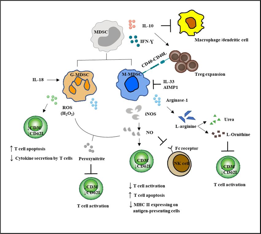
G-MDSCs and M-MDSCs inhibit T cell function via different mechanisms (Figure 2). G-MDSC has
increased NADPH oxidase (Nox) activity, which results in high levels of ROS, but low levels of
nitric oxide (NO) production. M-MDSCs express high levels of NO, but show low ROS production.
Both G-MDSC and M-MDSC subsets express arginase 1. ROS produced by G-MDSCs in high
concentrations not only induced T cell apoptosis, but has been demonstrated to cause T cell anergy by
downregulating the expression of TCR ζ-chain, leading to impaired TCR signaling [5,11,53]. Besides,
ROS form peroxynitrite, which when reacted with NO, nitrosylated the TCR and could result in T cell
anergy. NO production by M-MDSCs could induce T cell anergy and nitrosylate important mediators
of the IL-2 pathway [54]. Recently, NO production was shown to impair Fc receptor-mediated natural
killer cell function, leading to impaired response to monoclonal antibody therapy in cancer [55].
Another important immunosuppressive mediator arginase 1 was able to convert L-arginine into
L-ornithine and urea, leading to the depletion of L-arginine. The lack of L-arginine caused a translational
blockade in infiltrating T cells and led to cell cycle arrest in G0-G1 [56].
Recently, long noncoding RNA Pvt1 (lncRNA Pvt1) was suggested to be a potent antitumor
immunotherapy target. The knockdown of lncRNA Pvt1 significantly reduced the immunosuppressive
activity of G-MDSCs such as decreased levels of Arg1 and ROS in G-MDSCs and delayed tumor
progression in tumor-bearing mice [57]. Veglia et al., 2019, discovered that fatty acid transport protein
2 (FATP2) controlled the suppressive activity of G-MDSCs via increased uptake of arachidonic acid and
the synthesis of PGE2. Overexpression of FATP2 in G-MDSCs was induced by GM-CSF, through the
activation of the STAT5 transcription factor. Inhibition of FATP2 abrogated the activity of G-MDSCs
and substantially delayed tumor progression [58]. The Olfr29-ps1 pseudogene was reported to be
expressed in MDSCs and upregulated by the proinflammatory cytokine IL-6. Olfr29-ps1 promoted the
immunosuppressive function and differentiation of M-MDSCs through downregulation of miR-214-3p,
thereby releasing the expression of its target gene MyD88 in response to inflammatory factors [59].
Additionally, MDSCs were reported to exert their immunosuppressive effects via the upregulation
of programmed death-ligand 1 (PD-L1) [40]. The binding of PD-L1 to the programmed cell death
protein 1 (PD-1) receptor expressed on T cells caused exhaustion of T cells, and they lost their ability
to produce interferon IFN-γ and IL-2 [60]. Recently, Strauss et al., 2020, discovered the role of PD-1
expressed by myeloid cells in dampening antitumor immunity. Deleting PD-1 from myeloid cells when
compared to deleting it from T cells in mice led to a more significant reduction in tumor growth [61].
Moreover, MDSCs were shown to express the death receptor CD95 and induced T cell apoptosis via
CD95 ligands expressed on activated T cells [62].
MDSCs are known to affect both the innate and adaptive immune responses [30]. These cells
induce anergy of NK cells as an immune evasion mechanism [63]. Macrophages are also downregulated
by MDSCs, which results in a decrease in IL-12 production and an increase in IL-10 synthesis [64].Int. J. Mol. Sci. 2020, 21, 3599 6 of 13
Int. J. Mol. Sci. 2020, 21, x FOR PEER REVIEW 6 of 13
M-MDSCs, but not G-MDSCs, could promote the differentiation of Treg from CD4+
T cells. The lack
synthesis [64]. M-MDSCs, but not G-MDSCs, could promote the differentiation of Treg from CD4+ T
of CD40 on MDSCs resulted in decreasing either the expansion or de novo
cells. The lack of CD40 on MDSCs resulted in decreasing either the expansion or de novo
production of Treg,
suggesting
productionthatofthe interaction
Treg, suggestingof co-stimulatory
that molecules
the interaction of CD40molecules
co-stimulatory and CD40LCD40isand
crucial
CD40L foris Treg
development [65]. Additionally, the signal stimulated MDSCs to acquire immunosuppressive
crucial for Treg development [65]. Additionally, the signal stimulated MDSCs to acquire properties
that were mediated through
immunosuppressive STAT1,that
properties STAT3,
wereSTAT6, andthrough
mediated NF-κB transcription
STAT1, STAT3, factors.
STAT6,The
andmechanisms
NF-κB
transcription
regulating factors. of
the activation TheM-MDSCs
mechanisms regulating
depend the activation
on STAT1, STAT6, of M-MDSCs
and dependnot
NF-κB. STAT3 on only
STAT1,plays a
majorSTAT6,
role inand
the NF-κB.
expansionSTAT3of not
bothonly
MDSCplayssubsets,
a major but
role is
inalso
the expansion
involved of
in both MDSC subsets,
the suppressive but of
abilities
is also[29].
G-MDSCs involved in the suppressive abilities of G-MDSCs [29].
Figure 2. MDSC-mediated immunosuppression in innate and adaptive immune responses. MDSCs
Figure 2. MDSC-mediated immunosuppression in innate and adaptive immune responses. MDSCs
suppressed the activation of macrophages and the antigen-presenting ability of the dendritic cells.
suppressed
MDSCs the activation
enhanced of macrophages
Treg expansion and theNK
and suppressed antigen-presenting ability
cell cytotoxicity. Direct of the
actions dendritic
of MDSCs on cells.
MDSCs enhanced
T cells Treg expansion
are by increased NO and and
ROS suppressed
secretion andNK cell cytotoxicity.
decreased Direct actions
L-arginine production. T barofrefers
MDSCsto on
T cells are by increased NO and ROS secretion and decreased L-arginine production. T bar refers
inhibition.
to inhibition.
6. MDSC in Cancer Progression
6. MDSC in Cancer Progression
Several lines of evidence indicated that MDSCs were associated with tumor progression. The
Several
levels oflines of evidence
MDSCs indicated that
were profoundly MDSCs
correlated were
with theassociated
extent of with
tumortumor progression.
burden The levels
and the overall
survival of the tumor-bearing host. Administration of MDSCs in the murine
of MDSCs were profoundly correlated with the extent tumor burden and the overall survival tumor models wasof the
found to significantly promote tumor growth [66–68]. The detrimental effects of MDSCs
tumor-bearing host. Administration of MDSCs in the murine tumor models was found to significantly in tumor
progression
promote have been
tumor growth well The
[66–68]. described as the effects
detrimental depletion of Gr-1+ in
of MDSCs cells
tumorin tumor-bearing micebeen
progression have
strikingly inhibited tumor growth, reduced
+ cancer cell dissemination a d metastasis, and prolonged
well described as the depletion of Gr-1 cells in tumor-bearing mice strikingly inhibited tumor growth,
survival [63,69]. The reduction of murine MDSC numbers was shown to facilitate the rejection of
reduced cancer cell dissemination a d metastasis, and prolonged survival [63,69]. The reduction of
murine MDSC numbers was shown to facilitate the rejection of established metastatic disease after the
removal of primary tumors [70]. A study in renal cell carcinoma showed that surgical resection ofInt. J. Mol. Sci. 2020, 21, 3599 7 of 13
primary cancer lesion contributed to the reduction of MDSC, which indicated the unknown factors
derived from cancer tissue affected MDSC maintenance [71]. Kawano et al., 2015, reported a statistically
significant higher frequency of circulating MDSCs in the blood of advanced and recurrent patients with
cervical cancers compared to healthy patients. Therefore, circulating MDSCs have been validated as a
predictive marker for cancer immunotherapy. Further characterization of MDSC subsets demonstrated
that the frequency of both M-MDSCs and G-MDSCs was significantly elevated in the blood of patients
from advanced glioma and cervical cancer [12,72]. Recently, a study suggested that G-MDSCs could
serve as a potential biomarker for disease progression of cervical cancers. The frequency of circulating
G-MDSCs was found to correlate with disease prognosis, while the percentage of M-MDSCs was only
elevated in patients with advanced cervical cancers. For patients with early and locally advanced
cervical cancers, the frequency of circulating G-MDSCs, but not M-MDSCs correlated with tumor
recurrence. The levels of circulating G-MDSCs also negatively correlated with the densities of CD8+ T
cells and the suppression of T cell proliferation [12]. M-MDSCs were reported as prognosis markers
in the, colorectal, gastric, and pancreatic cancer [73,74]. Increased levels of M-MDSCs in advanced
non-small cell lung cancer (NSCLC) patients were associated with an unfavorable clinical outcome [75].
The lower quantity of M-MDSCs in metastatic melanoma patients following treatment with ipilimumab
were more likely to achieve prolonged survival [76]. Therefore, MDSC could serve as a predictive
marker for immunotherapy.
MDSCs and their immunosuppressive functions might be eliminated via several approaches such
as deactivation of MDSCs, promoting the differentiation of MDSCs into mature cells, blocking the
development of MDSCs, and depletion of MDSCs [77]. MDSCs could be inactivated by blocking
the NO, ROS, and arginase secretion such as by using phosphodiesterase inhibitors, nitroaspirins,
synthetic triterpenoids, COX2 inhibitors, ARG1 inhibitors, anti-glycan antibodies, CSF-1R, IL-17,
and histamine inhibitors. Agents that block the development of MDSCs include N-bisphosphonates,
modulators of tyrosine kinases, and STAT3 inhibitors. MDSCs could also be depleted with gemcitabine,
HSP90 (heat shock protein 90) inhibitors, and paclitaxel (Table 2). Some FDA-approved compounds
such as ATRA (All-trans retinoic acid), PDE5 (phosphodiesterase type 5) inhibitors, COX-2 inhibitors,
or bisphosphonates are already in clinical trials for evaluating their ability to inhibit MDSCs and
enhance anti-tumor immunity in humans (Table 2) [78]. However, major anti-tumor effects may
not be expected by only targeting MDSCs. It has been found that gemcitabine and anti-GR-1 Ab,
when administered together with DNA vaccine, could induce a strong antitumor immune response,
which was accompanied by reduced self-tolerance in a preclinical HER2-expressing mouse tumor
model [79]. ATRA is a promising agent that promoted the differentiation of M-MDSCs into mature
cells, and when used together with a dendritic cell (DC) vaccine against p53, substantial improvement
of the CD8+ T cell responses was observed in late-stage small cell lung cancer patients [80].
The combination of MDSC targeting with immune checkpoint inhibitors has been applied in
preclinical tumor models and cancer patients. The FDA approved immune checkpoint inhibitors
including one CTLA-4 inhibitor (ipilimumab), three PD-1 inhibitors (nivolumab, pembrolizumab,
and cemiplimab), and three PD-L1 inhibitors (atezolizumab, durvalumab, and avelumab).
Serine/threonine protein kinase CK2 inhibition blocked MDSC differentiation and substantially
increased anti-tumor efficacy when combined with anti-CTLA-4 blockade in mice [81]. Treatment of
tumor-bearing mice with Sema4D mAb in combination with either CTLA-4 or PD-1 blockade enhanced
the rejection of tumors or tumor growth delay, resulting in prolonged survival with either treatment [82].
A recent study has shown that the combination of anti-CXCR4, which decreased M-MDSC, and anti-PD-1
therapy improved the overall survival in a mouse glioma model [83]. Furthermore, ATRA decreased
the frequency of circulating MDSCs in melanoma patients treated in combination with ipilimumab,
and this combination is still on-going in a clinical trial to treat Stage IV melanoma patients [84,85].Int. J. Mol. Sci. 2020, 21, 3599 8 of 13
Table 2. Strategies for myeloid-derived suppressor cell (MDSC) targeting.
Strategy Mechanism of Action Examples Clinical Trial
N-Bisphosphonates Zoledronic acid Phase 3-completed
Blocking MDSC Sunitinib Phase 2-completed
Multi-kinase inhibitors
development Sorafenib Phase 3-completed
Cucurbitacin B N/A
JAK2/STAT3 inhibitors
JSI-124 N/A
Blocking antibodies Anti-VEGF antibodies NCT03503604
ATRA NCT024403778
Vitamin A N/A
Vitamins
Differentiation of MDSC Vitamin D3 N/A
into mature cells Vitamin E N/A
Cytokines IL-12 N/A
Others CpG N/A
Sildenafil NCT02544880
PDE5 inhibitors
Tadalafil NCT01697800
NO-aspirins (NCX-4016)
NO inhibitors Phase1-completed
MDSC deactivation L-NAME
Synthetic triterpenoids
ROS inhibitors Phase 2-completed
(omaveloxolone)
COX2 inhibitors N/A
Arginase inhibitors NOHA N/A
L-NAME N/A
Recruitment and migration Anti-glycan antibodies
NCT03557970
inhibitor CSF-1R inhibitors
Histamine inhibitor
Others (ranitidine) NCT03145012
Anti-IL-17 antibodies
Gemcitabine NCT01803152
Cisplatin NCT02432378
Cytotoxic agents
5-Fluorouracil N/A
MDSC depletion
Paclitaxel N/A
HSP90 inhibitors 17-DMAG Phase 1-completed
Peptide-FC fusion proteins N/A N/A
N/A refers to no available information. ATRA: All-trans retinoic acid; PDE5: phosphodiesterase type 5; NCX: Nitric
Oxide-Aspirin; L-NAME: L-NG -Nitroarginine methyl ester; NOHA: N(omega)-hydroxy-l-arginine; HSP90: heat
shock protein 90; 17-DMAG: 17-Dimethylaminoethylamino-17-demethoxygeldanamycin.
7. Conclusions
Successful immunotherapeutic approaches that utilize the host immune system to inhibit tumor
growth could lead to increased patient survival. These approaches are non-toxic and usually involve
either regulation of the secretion of soluble factors such as cytokines, chemokines, and tumor-derived
factors by immune cells or a reduction in the activity of immune regulatory cells such as regulatory
T (Treg) cells and myeloid-derived suppressor cells (MDSCs). The depletion of MDSCs normally
involved cytotoxic agents, but an alternative approach might be better by altering the development,
differentiation, and functions of MDSCs. PD-1 expression from myeloid cells plays a critical role
in preventing the differentiation of effector myeloid cells and promoting the formation of MDSCs;
thus, blocking PD-1 signaling in myeloid cells appears to be a requirement for antitumor immunity.
Additionally, combinatorial approaches to target precisely the suppressive activity of MDSCs or MDSC
subsets using anti-IL-18, inhibition of FATP2, inactivating the long non-coding RNA Pvt1, the NFIA
gene, or the Olfr19-ps1 pseudogene might enhance the existing immunotherapeutic strategies by the
administration of immune checkpoint inhibitors including the CTLA-4, PD-1, and PD-L1 inhibitors.
Funding: This study was funded by the Sunway University Internal Grant 2020 (GRTIN-RSF-SST-CVVR-01-2020)
to the Centre for Virus and Vaccine Research (CVVR).
Conflicts of Interest: The authors declare that there is no conflict of interest. The funders had no role in the
design of the study; in the collection, analyses, or interpretation of data; in the writing of the manuscript, or in the
decision to publish the results.Int. J. Mol. Sci. 2020, 21, 3599 9 of 13
References
1. Ribechini, E.; Greifenberg, V.; Sandwick, S.; Lutz, M. Subsets, expansion and activation of myeloid-derived
suppressor cells. Med. Microbiol. Immunol. 2010, 199, 273–281. [CrossRef] [PubMed]
2. Movahedi, K.; Guilliams, M.; Van den Bossche, J.; Van den Bergh, R.; Gysemans, C.; Beschin, A.;
De Baetselier, P.; Van Ginderachter, J.A. Identification of discrete tumor-induced myeloid-derived suppressor
cell subpopulations with distinct T cell-suppressive activity. Blood 2008, 111, 4233–4244. [CrossRef] [PubMed]
3. Haile, L.A.; Gamrekelashvili, J.; Manns, M.P.; Korangy, F.; Greten, T.F. CD49d Is a New Marker for Distinct
Myeloid-Derived Suppressor Cell Subpopulations in Mice. J. Immunol. 2010, 185, 203–210. [CrossRef]
[PubMed]
4. Youn, J.-I.; Nagaraj, S.; Collazo, M.; Gabrilovich, D.I. Subsets of Myeloid-Derived Suppressor Cells in
Tumor-Bearing Mice. J. Immunol. 2008, 181, 5791–5802. [CrossRef] [PubMed]
5. Gabrilovich, D.I.; Ostrand-Rosenberg, S.; Bronte, V. Coordinated regulation of myeloid cells by tumours.
Nat. Rev. Immunol. 2012, 12, 253–268. [CrossRef]
6. Khaled, Y.S.; Ammori, B.J.; Elkord, E. Increased levels of granulocytic myeloid-derived suppressor cells
in peripheral blood and tumour tissue of pancreatic cancer patients. J. Immunol. Res. 2014, 2014, 879897.
[CrossRef]
7. Kumar, V.; Patel, S.; Tcyganov, E.; Gabrilovich, D.I. The Nature of Myeloid-Derived Suppressor Cells in the
Tumor Microenvironment. Trends Immunol. 2016, 37, 208–220. [CrossRef]
8. Tesi, R.J. MDSC the Most Important Cell You Have Never Heard Of. Trends Pharmacol. Sci. 2019, 40, 4–7.
[CrossRef]
9. Hoechst, B.; Ormandy, L.A.; Ballmaier, M.; Lehner, F.; Krüger, C.; Manns, M.P.; Greten, T.F.; Korangy, F.
A new population of myeloid-derived suppressor cells in hepatocellular carcinoma patients induces
CD4(+)CD25(+)Foxp3(+) T cells. Gastroenterology 2008, 135, 234–243. [CrossRef]
10. Lin, Y.; Gustafson, M.P.; Bulur, P.A.; Gastineau, D.A.; Witzig, T.E.; Dietz, A.B. Immunosuppressive
CD14+HLA-DR(low)/- monocytes in B-cell non-Hodgkin lymphoma. Blood 2011, 117, 872–881. [CrossRef]
11. Bronte, V.; Brandau, S.; Chen, S.H.; Colombo, M.P.; Frey, A.B.; Greten, T.F.; Mandruzzato, S.; Murray, P.J.;
Ochoa, A.; Ostrand-Rosenberg, S.; et al. Recommendations for myeloid-derived suppressor cell nomenclature
and characterization standards. Nat. Commun. 2016, 7, 12150. [CrossRef] [PubMed]
12. Liang, Y.; Lu, B.; Zhao, P.; Lu, W. Increased circulating GrMyeloid-derived suppressor cells correlated
with tumor burden and survival in locally advanced cervical cancer patient. J. Cancer 2019, 10, 1341–1348.
[CrossRef]
13. Piccard, H.; Muschel, R.J.; Opdenakker, G. On the dual roles and polarized phenotypes of neutrophils in
tumor development and progression. Crit. Rev. Oncol. Hematol. 2012, 82, 296–309. [CrossRef] [PubMed]
14. Yu, Y.; Qian, L.; Cui, J. Value of neutrophil-to-lymphocyte ratio for predicting lung cancer prognosis:
A meta-analysis of 7,219 patients. Mol. Clin. Oncol. 2017, 7, 498–506. [CrossRef] [PubMed]
15. Zhang, G.-M.; Zhu, Y.; Ma, X.-C.; Qin, X.-J.; Wan, F.-N.; Dai, B.; Sun, L.-J.; Ye, D.-W.
Pretreatment Neutrophil-to-Lymphocyte Ratio: A Predictor of Advanced Prostate Cancer and Biochemical
Recurrence in Patients Receiving Radical Prostatectomy. Medicine (Baltimore) 2015, 94, e1473. [CrossRef]
[PubMed]
16. Chen, S.; Zhang, L.; Yan, G.; Cheng, S.; Fathy, A.H.; Yan, N.; Zhao, Y. Neutrophil-to-Lymphocyte Ratio Is a
Potential Prognostic Biomarker in Patients with Ovarian Cancer: A Meta-Analysis. Biomed. Res. Int. 2017,
2017, 7943467. [CrossRef]
17. Chen, M.-F.; Tsai, M.-S.; Chen, W.-C.; Chen, P.-T. Predictive Value of the Pretreatment
Neutrophil-to-Lymphocyte Ratio in Head and Neck Squamous Cell Carcinoma. J. Clin. Med. 2018,
7, 294. [CrossRef] [PubMed]
18. Basu, A.; Kollengode, K.A.; Rafatnia, A.; Manoli, H.; Danenberg, G.; Chakravartty, E.; Epstein, A.L.;
Pinski, J.K. Relationship between neutrophil lymphocyte ratio (NLR) and MDSC concentration in localized
and metastatic castration resistant prostate cancer (mCRPC) patients. J. Clin. Oncol. 2018, 36, 338. [CrossRef]
19. Morales, J.; Kmieciak, M.; Knutson, K.; Bear, H.; Manjili, M. GM-CSF is one of the main breast
tumor-derived soluble factors involved in the differentiation of CD11b-Gr1- bone marrow progenitor
cells into myeloid-derived suppressor cells. Breast Cancer Res. Treat. 2010, 123, 39–49. [CrossRef]Int. J. Mol. Sci. 2020, 21, 3599 10 of 13
20. Dolcetti, L.; Peranzoni, E.; Ugel, S.; Marigo, I.; Fernandez Gomez, A.; Mesa, C.; Geilich, M.; Winkels, G.;
Traggiai, E.; Casati, A.; et al. Hierarchy of immunosuppressive strength among myeloid-derived suppressor
cell subsets is determined by GM-CSF. Eur. J. Immunol. 2010, 40, 22–35. [CrossRef]
21. Elkabets, M.; Ribeiro, V.S.G.; Dinarello, C.A.; Ostrand-Rosenberg, S.; Di Santo, J.P.; Apte, R.N.;
Vosshenrich, C.A.J. IL-1β regulates a novel myeloid-derived suppressor cell subset that impairs NK
cell development and function. Eur. J. Immunol. 2010, 40, 3347–3357. [CrossRef] [PubMed]
22. Zhao, X.; Rong, L.; Zhao, X.; Li, X.; Liu, X.; Deng, J.; Wu, H.; Xu, X.; Erben, U.; Wu, P.; et al. TNF signaling
drives myeloid-derived suppressor cell accumulation. J. Clin. Investig. 2012, 122, 4094–4104. [CrossRef]
23. Sobo-Vujanovic, A.; Vujanovic, L.; DeLeo, A.B.; Concha-Benavente, F.; Ferris, R.L.; Lin, Y.; Vujanovic, N.L.
Inhibition of Soluble Tumor Necrosis Factor Prevents Chemically Induced Carcinogenesis in Mice. Cancer
Immunol. Res. 2016, 4, 441–451. [CrossRef] [PubMed]
24. Lee, C.R.; Lee, W.; Cho, S.K.; Park, S.G. Characterization of Multiple Cytokine Combinations and TGF-beta
on Differentiation and Functions of Myeloid-Derived Suppressor Cells. Int. J. Mol. Sci. 2018, 19. [CrossRef]
25. Bah, I.; Kumbhare, A.; Nguyen, L.; McCall, C.E.; El Gazzar, M. IL-10 induces an immune repressor pathway
in sepsis by promoting S100A9 nuclear localization and MDSC development. Cell Immunol. 2018, 332, 32–38.
[CrossRef] [PubMed]
26. Lim, H.X.; Hong, H.J.; Cho, D.; Kim, T.S. IL-18 enhances immunosuppressive responses by promoting
differentiation into monocytic myeloid-derived suppressor cells. J. Immunol. (Baltim. Md. 1950) 2014, 193,
5453–5460. [CrossRef]
27. Lim, H.X.; Choi, S.; Cho, D.; Kim, T.S. IL-33 inhibits the differentiation and immunosuppressive activity of
granulocytic myeloid-derived suppressor cells in tumor-bearing mice. Immunol. Cell Biol. 2017, 95, 99–107.
[CrossRef]
28. Hong, H.J.; Lim, H.X.; Song, J.H.; Lee, A.; Kim, E.; Cho, D.; Cohen, E.P.; Kim, T.S. Aminoacyl-tRNA
synthetase-interacting multifunctional protein 1 suppresses tumor growth in breast cancer-bearing mice by
negatively regulating myeloid-derived suppressor cell functions. Cancer Immunol. Immunother. 2016, 65,
61–72. [CrossRef]
29. Gabrilovich, D.I.; Nagaraj, S. Myeloid-derived suppressor cells as regulators of the immune system.
Nat. Rev. Immunol. 2009, 9, 162–174. [CrossRef]
30. Ostrand-Rosenberg, S.; Sinha, P. Myeloid-derived suppressor cells: Linking inflammation and cancer.
J. Immunol. (Baltim. Md. 1950) 2009, 182, 4499–4506. [CrossRef]
31. Van Deventer, H.W.; Burgents, J.E.; Wu, Q.P.; Woodford, R.M.; Brickey, W.J.; Allen, I.C.; McElvania-Tekippe, E.;
Serody, J.S.; Ting, J.P. The inflammasome component NLRP3 impairs antitumor vaccine by enhancing the
accumulation of tumor-associated myeloid-derived suppressor cells. Cancer Res. 2010, 70, 10161–10169.
[CrossRef] [PubMed]
32. Chalmin, F.; Ladoire, S.; Mignot, G.; Vincent, J.; Bruchard, M.; Remy-Martin, J.P.; Boireau, W.; Rouleau, A.;
Simon, B.; Lanneau, D.; et al. Membrane-associated Hsp72 from tumor-derived exosomes mediates
STAT3-dependent immunosuppressive function of mouse and human myeloid-derived suppressor cells.
J. Clin. Investig. 2010, 120, 457–471. [CrossRef] [PubMed]
33. Valenti, R.; Huber, V.; Filipazzi, P.; Pilla, L.; Sovena, G.; Villa, A.; Corbelli, A.; Fais, S.; Parmiani, G.; Rivoltini, L.
Human tumor-released microvesicles promote the differentiation of myeloid cells with transforming growth
factor-beta-mediated suppressive activity on T lymphocytes. Cancer Res. 2006, 66, 9290–9298. [CrossRef]
[PubMed]
34. Valenti, R.; Huber, V.; Iero, M.; Filipazzi, P.; Parmiani, G.; Rivoltini, L. Tumor-released microvesicles as
vehicles of immunosuppression. Cancer Res. 2007, 67, 2912–2915. [CrossRef] [PubMed]
35. Ichikawa, M.; Williams, R.; Wang, L.; Vogl, T.; Srikrishna, G. S100A8/A9 activate key genes and pathways in
colon tumor progression. Mol. Cancer Res. 2011, 9, 133–148. [CrossRef] [PubMed]
36. Obermajer, N.; Muthuswamy, R.; Lesnock, J.; Edwards, R.P.; Kalinski, P. Positive feedback between PGE2
and COX2 redirects the differentiation of human dendritic cells toward stable myeloid-derived suppressor
cells. Blood 2011, 118, 5498–5505. [CrossRef]
37. Lechner, M.G.; Liebertz, D.J.; Epstein, A.L. Characterization of cytokine-induced myeloid-derived suppressor
cells from normal human peripheral blood mononuclear cells. J. Immunol. (Baltim. Md. 1950) 2010, 185,
2273–2284. [CrossRef]Int. J. Mol. Sci. 2020, 21, 3599 11 of 13
38. Fujita, M.; Kohanbash, G.; Fellows-Mayle, W.; Hamilton, R.L.; Komohara, Y.; Decker, S.A.; Ohlfest, J.R.;
Okada, H. COX-2 blockade suppresses gliomagenesis by inhibiting myeloid-derived suppressor cells.
Cancer Res. 2011, 71, 2664–2674. [CrossRef]
39. Corzo, C.A.; Condamine, T.; Lu, L.; Cotter, M.J.; Youn, J.I.; Cheng, P.; Cho, H.I.; Celis, E.; Quiceno, D.G.;
Padhya, T.; et al. HIF-1alpha regulates function and differentiation of myeloid-derived suppressor cells in
the tumor microenvironment. J. Exp. Med. 2010, 207, 2439–2453. [CrossRef]
40. Noman, M.Z.; Desantis, G.; Janji, B.; Hasmim, M.; Karray, S.; Dessen, P.; Bronte, V.; Chouaib, S. PD-L1
is a novel direct target of HIF-1alpha, and its blockade under hypoxia enhanced MDSC-mediated T cell
activation. J. Exp. Med. 2014, 211, 781–790. [CrossRef]
41. Dai, J.; Kumbhare, A.; Williams, D.A.; Youssef, D.; Yao, Z.Q.; McCall, C.E.; El Gazzar, M. Nfia deletion in
myeloid cells blocks expansion of myeloid-derived suppressor cells during sepsis. Innate Immun. 2018, 24,
54–65. [CrossRef] [PubMed]
42. Obermajer, N.; Muthuswamy, R.; Odunsi, K.; Edwards, R.P.; Kalinski, P. PGE(2)-induced CXCL12 production
and CXCR4 expression controls the accumulation of human MDSCs in ovarian cancer environment.
Cancer Res. 2011, 71, 7463–7470. [CrossRef] [PubMed]
43. Lim, S.Y.; Yuzhalin, A.E.; Gordon-Weeks, A.N.; Muschel, R.J. Targeting the CCL2-CCR2 signaling axis in
cancer metastasis. Oncotarget 2016, 7, 28697–28710. [CrossRef] [PubMed]
44. Wang, S.W.; Liu, S.C.; Sun, H.L.; Huang, T.Y.; Chan, C.H.; Yang, C.Y.; Yeh, H.I.; Huang, Y.L.; Chou, W.Y.;
Lin, Y.M.; et al. CCL5/CCR5 axis induces vascular endothelial growth factor-mediated tumor angiogenesis
in human osteosarcoma microenvironment. Carcinogenesis 2015, 36, 104–114. [CrossRef] [PubMed]
45. Zhang, J.; Patel, L.; Pienta, K.J. CC chemokine ligand 2 (CCL2) promotes prostate cancer tumorigenesis and
metastasis. Cytokine Growth Factor Rev. 2010, 21, 41–48. [CrossRef] [PubMed]
46. Lesokhin, A.M.; Hohl, T.M.; Kitano, S.; Cortez, C.; Hirschhorn-Cymerman, D.; Avogadri, F.; Rizzuto, G.A.;
Lazarus, J.J.; Pamer, E.G.; Houghton, A.N.; et al. Monocytic CCR2(+) myeloid-derived suppressor cells
promote immune escape by limiting activated CD8 T-cell infiltration into the tumor microenvironment.
Cancer Res. 2012, 72, 876–886. [CrossRef]
47. Schlecker, E.; Stojanovic, A.; Eisen, C.; Quack, C.; Falk, C.S.; Umansky, V.; Cerwenka, A. Tumor-Infiltrating
Monocytic Myeloid-Derived Suppressor Cells Mediate CCR5-Dependent Recruitment of Regulatory T Cells
Favoring Tumor Growth. J. Immunol. 2012, 189, 5602. [CrossRef]
48. Gama, L.; Shirk, E.N.; Russell, J.N.; Carvalho, K.I.; Li, M.; Queen, S.E.; Kalil, J.; Zink, M.C.; Clements, J.E.;
Kallas, E.G. Expansion of a subset of CD14highCD16negCCR2low/neg monocytes functionally similar to
myeloid-derived suppressor cells during SIV and HIV infection. J. Leukoc. Biol. 2012, 91, 803–816. [CrossRef]
49. Srivastava, M.K.; Sinha, P.; Clements, V.K.; Rodriguez, P.; Ostrand-Rosenberg, S. Myeloid-derived suppressor
cells inhibit T-cell activation by depleting cystine and cysteine. Cancer Res. 2010, 70, 68–77. [CrossRef]
50. Inamoto, S.; Itatani, Y.; Yamamoto, T.; Minamiguchi, S.; Hirai, H.; Iwamoto, M.; Hasegawa, S.; Taketo, M.M.;
Sakai, Y.; Kawada, K. Loss of SMAD4 Promotes Colorectal Cancer Progression by Accumulation of
Myeloid-Derived Suppressor Cells through the CCL15-CCR1 Chemokine Axis. Clin. Cancer Res. 2016, 22,
492–501. [CrossRef]
51. Poschke, I.; Mao, Y.; Adamson, L.; Salazar-Onfray, F.; Masucci, G.; Kiessling, R. Myeloid-derived suppressor
cells impair the quality of dendritic cell vaccines. Cancer Immunol. Immunother. 2012, 61, 827–838. [CrossRef]
[PubMed]
52. Alkasalias, T.; Moyano-Galceran, L.; Arsenian-Henriksson, M.; Lehti, K. Fibroblasts in the Tumor
Microenvironment: Shield or Spear? Int. J. Mol. Sci. 2018, 19, 1532. [CrossRef] [PubMed]
53. Baniyash, M. TCR ζ-chain downregulation: Curtailing an excessive inflammatory immune response.
Nat. Rev. Immunol. 2004, 4, 675–687. [CrossRef] [PubMed]
54. Mazzoni, A.; Bronte, V.; Visintin, A.; Spitzer, J.H.; Apolloni, E.; Serafini, P.; Zanovello, P.; Segal, D.M.
Myeloid suppressor lines inhibit T cell responses by an NO-dependent mechanism. J. Immunol. (Baltim.
Md. 1950) 2002, 168, 689–695. [CrossRef] [PubMed]
55. Stiff, A.; Trikha, P.; Mundy-Bosse, B.; McMichael, E.; Mace, T.A.; Benner, B.; Kendra, K.; Campbell, A.;
Gautam, S.; Abood, D.; et al. Nitric Oxide Production by Myeloid-Derived Suppressor Cells Plays a Role
in Impairing Fc Receptor-Mediated Natural Killer Cell Function. Clin. Cancer Res. 2018, 24, 1891–1904.
[CrossRef]Int. J. Mol. Sci. 2020, 21, 3599 12 of 13
56. Rodriguez, P.C.; Quiceno, D.G.; Ochoa, A.C. L-arginine availability regulates T-lymphocyte cell-cycle
progression. Blood 2007, 109, 1568–1573. [CrossRef]
57. Zheng, Y.; Tian, X.; Wang, T.; Xia, X.; Cao, F.; Tian, J.; Xu, P.; Ma, J.; Xu, H.; Wang, S. Long noncoding
RNA Pvt1 regulates the immunosuppression activity of granulocytic myeloid-derived suppressor cells in
tumor-bearing mice. Mol. Cancer 2019, 18, 61. [CrossRef]
58. Veglia, F.; Tyurin, V.A.; Blasi, M.; De Leo, A.; Kossenkov, A.V.; Donthireddy, L.; To, T.K.J.; Schug, Z.; Basu, S.;
Wang, F.; et al. Fatty acid transport protein 2 reprograms neutrophils in cancer. Nature 2019, 569, 73–78.
[CrossRef]
59. Shang, W.; Gao, Y.; Tang, Z.; Zhang, Y.; Yang, R. The Pseudogene Olfr29-ps1 Promotes the Suppressive
Function and Differentiation of Monocytic MDSCs. Cancer Immunol. Res. 2019, 7, 813–827. [CrossRef]
60. Berger, K.N.; Pu, J.J. PD-1 pathway and its clinical application: A 20year journey after discovery of the
complete human PD-1 gene. Gene 2018, 638, 20–25. [CrossRef]
61. Strauss, L.; Mahmoud, M.A.A.; Weaver, J.D.; Tijaro-Ovalle, N.M.; Christofides, A.; Wang, Q.; Pal, R.; Yuan, M.;
Asara, J.; Patsoukis, N.; et al. Targeted deletion of PD-1 in myeloid cells induces antitumor immunity. Sci.
Immunol. 2020, 5, eaay1863. [CrossRef] [PubMed]
62. Sinha, P.; Chornoguz, O.; Clements, V.K.; Artemenko, K.A.; Zubarev, R.A.; Ostrand-Rosenberg, S.
Myeloid-derived suppressor cells express the death receptor Fas and apoptose in response to T cell-expressed
FasL. Blood 2011, 117, 5381–5390. [CrossRef] [PubMed]
63. Li, H.; Han, Y.; Guo, Q.; Zhang, M.; Cao, X. Cancer-expanded myeloid-derived suppressor cells induce
anergy of NK cells through membrane-bound TGF-beta 1. J. Immunol. (Baltim. Md. 1950) 2009, 182, 240–249.
[CrossRef] [PubMed]
64. Sinha, P.; Clements, V.K.; Bunt, S.K.; Albelda, S.M.; Ostrand-Rosenberg, S. Cross-talk between myeloid-derived
suppressor cells and macrophages subverts tumor immunity toward a type 2 response. J. Immunol. (Baltim.
Md. 1950) 2007, 179, 977–983. [CrossRef]
65. Pan, P.Y.; Ma, G.; Weber, K.J.; Ozao-Choy, J.; Wang, G.; Yin, B.; Divino, C.M.; Chen, S.H. Immune stimulatory
receptor CD40 is required for T-cell suppression and T regulatory cell activation mediated by myeloid-derived
suppressor cells in cancer. Cancer Res. 2010, 70, 99–108. [CrossRef]
66. Yang, L.; DeBusk, L.M.; Fukuda, K.; Fingleton, B.; Green-Jarvis, B.; Shyr, Y.; Matrisian, L.M.; Carbone, D.P.;
Lin, P.C. Expansion of myeloid immune suppressor Gr+CD11b+ cells in tumor-bearing host directly promotes
tumor angiogenesis. Cancer Cell 2004, 6, 409–421. [CrossRef]
67. Balwit, J.M.; Hwu, P.; Urba, W.J.; Marincola, F.M. The iSBTc/SITC primer on tumor immunology and biological
therapy of cancer: A summary of the 2010 program. J. Transl. Med. 2011, 9, 18. [CrossRef]
68. Vincent, J.; Mignot, G.; Chalmin, F.; Ladoire, S.; Bruchard, M.; Chevriaux, A.; Martin, F.; Apetoh, L.; Rebe, C.;
Ghiringhelli, F. 5-Fluorouracil selectively kills tumor-associated myeloid-derived suppressor cells resulting
in enhanced T cell-dependent antitumor immunity. Cancer Res. 2010, 70, 3052–3061. [CrossRef] [PubMed]
69. Zhang, Y.; Liu, Q.; Zhang, M.; Yu, Y.; Liu, X.; Cao, X. Fas signal promotes lung cancer growth by recruiting
myeloid-derived suppressor cells via cancer cell-derived PGE2. J. Immunol. (Baltim. Md. 1950) 2009, 182,
3801–3808. [CrossRef]
70. Sinha, P.; Clements, V.K.; Ostrand-Rosenberg, S. Reduction of myeloid-derived suppressor cells and induction
of M1 macrophages facilitate the rejection of established metastatic disease. J. Immunol. (Baltim. Md. 1950)
2005, 174, 636–645. [CrossRef]
71. Motoshima, T.; Komohara, Y.; Horlad, H.; Tsukamoto, H.; Fujita, M.; Saito, Y.; Tanoue, K.; Kasejima, Y.;
Sugiyama, Y.; Kawano, Y.; et al. CXCL10 and CCL2 mRNA expression in monocytes is inversely correlated
with the HLA-DR lower fraction of monocytes in patients with renal cell carcinoma. Oncol. Lett. 2016, 11,
1911–1916. [CrossRef] [PubMed]
72. Gielen, P.R.; Schulte, B.M.; Kers-Rebel, E.D.; Verrijp, K.; Bossman, S.A.; Ter Laan, M.; Wesseling, P.; Adema, G.J.
Elevated levels of polymorphonuclear myeloid-derived suppressor cells in patients with glioblastoma highly
express S100A8/9 and arginase and suppress T cell function. Neuro Oncol. 2016, 18, 1253–1264. [CrossRef]
[PubMed]
73. Solito, S.; Falisi, E.; Diaz-Montero, C.M.; Doni, A.; Pinton, L.; Rosato, A.; Francescato, S.; Basso, G.;
Zanovello, P.; Onicescu, G.; et al. A human promyelocytic-like population is responsible for the immune
suppression mediated by myeloid-derived suppressor cells. Blood 2011, 118, 2254–2265. [CrossRef] [PubMed]Int. J. Mol. Sci. 2020, 21, 3599 13 of 13
74. Gabitass, R.F.; Annels, N.E.; Stocken, D.D.; Pandha, H.A.; Middleton, G.W. Elevated myeloid-derived
suppressor cells in pancreatic, esophageal and gastric cancer are an independent prognostic factor and are
associated with significant elevation of the Th2 cytokine interleukin-13. Cancer Immunol. Immunother. CII
2011, 60, 1419–1430. [CrossRef] [PubMed]
75. Vetsika, E.-K.; Koinis, F.; Gioulbasani, M.; Aggouraki, D.; Koutoulaki, A.; Skalidaki, E.; Mavroudis, D.;
Georgoulias, V.; Kotsakis, A. A Circulating Subpopulation of Monocytic Myeloid-Derived Suppressor Cells as
an Independent Prognostic/Predictive Factor in Untreated Non-Small Lung Cancer Patients. J. Immunol. Res.
2014, 2014, 12. [CrossRef]
76. Kitano, S.; Postow, M.A.; Ziegler, C.G.K.; Kuk, D.; Panageas, K.S.; Cortez, C.; Rasalan, T.; Adamow, M.; Yuan, J.;
Wong, P.; et al. Computational algorithm-driven evaluation of monocytic myeloid-derived suppressor cell
frequency for prediction of clinical outcomes. Cancer Immunol. Res. 2014, 2, 812–821. [CrossRef]
77. Wesolowski, R.; Markowitz, J.; Carson, W.E., III. Myeloid derived suppressor cells—A new therapeutic target
in the treatment of cancer. J. Immunother. Cancer 2013, 1, 10. [CrossRef]
78. Fleming, V.; Hu, X.; Weber, R.; Nagibin, V.; Groth, C.; Altevogt, P.; Utikal, J.; Umansky, V.
Targeting Myeloid-Derived Suppressor Cells to Bypass Tumor-Induced Immunosuppression. Front. Immunol.
2018, 9, 398. [CrossRef]
79. Danishmalik, S.N.; Sin, J.I. Therapeutic Tumor Control of HER2 DNA Vaccines Is Achieved by an Alteration of
Tumor Cells and Tumor Microenvironment by Gemcitabine and Anti-Gr-1 Ab Treatment in a HER2-Expressing
Tumor Model. DNA Cell Biol. 2017, 36, 801–811. [CrossRef]
80. Iclozan, C.; Antonia, S.; Chiappori, A.; Chen, D.T.; Gabrilovich, D. Therapeutic regulation of myeloid-derived
suppressor cells and immune response to cancer vaccine in patients with extensive stage small cell lung
cancer. Cancer Immunol. Immunother. 2013, 62, 909–918. [CrossRef]
81. Hashimoto, A.; Gao, C.; Mastio, J.; Kossenkov, A.; Abrams, S.I.; Purandare, A.V.; Desilva, H.; Wee, S.; Hunt, J.;
Jure-Kunkel, M.; et al. Inhibition of Casein Kinase 2 Disrupts Differentiation of Myeloid Cells in Cancer and
Enhances the Efficacy of Immunotherapy in Mice. Cancer Res. 2018, 78, 5644–5655. [CrossRef] [PubMed]
82. Clavijo, P.E.; Friedman, J.; Robbins, Y.; Moore, E.C.; Smith, E.; Zauderer, M.; Evans, E.E.; Allen, C.T.
Semaphorin4D Inhibition Improves Response to Immune-Checkpoint Blockade via Attenuation of MDSC
Recruitment and Function. Cancer Immunol. Res. 2019, 7, 282. [CrossRef] [PubMed]
83. Wu, A.; Maxwell, R.; Xia, Y.; Cardarelli, P.; Oyasu, M.; Belcaid, Z.; Kim, E.; Hung, A.; Luksik, A.S.;
Garzon-Muvdi, T.; et al. Combination anti-CXCR4 and anti-PD-1 immunotherapy provides survival benefit
in glioblastoma through immune cell modulation of tumor microenvironment. J. Neurooncol. 2019, 143,
241–249. [CrossRef] [PubMed]
84. Tobin, R.P.; Davis, D.; Jordan, K.R.; McCarter, M.D. The clinical evidence for targeting human myeloid-derived
suppressor cells in cancer patients. J. Leukoc. Biol. 2017, 102, 381–391. [CrossRef]
85. Tobin, R.P.; Jordan, K.R.; Robinson, W.A.; Davis, D.; Borges, V.F.; Gonzalez, R.; Lewis, K.D.; McCarter, M.D.
Targeting myeloid-derived suppressor cells using all-trans retinoic acid in melanoma patients treated with
Ipilimumab. Int. Immunopharmacol. 2018, 63, 282–291. [CrossRef]
© 2020 by the authors. Licensee MDPI, Basel, Switzerland. This article is an open access
article distributed under the terms and conditions of the Creative Commons Attribution
(CC BY) license (http://creativecommons.org/licenses/by/4.0/).You can also read