Extracellular vesicles from dHL 60 cells as delivery vehicles for diverse therapeutics
←
→
Page content transcription
If your browser does not render page correctly, please read the page content below
www.nature.com/scientificreports
OPEN Extracellular vesicles from dHL‑60
cells as delivery vehicles for diverse
therapeutics
Jun‑Kyu Kim1,6, Young‑Jin Youn1,6, Yu‑Bin Lee1, Sun‑Hwa Kim1, Dong‑Keun Song2,
Minsang Shin3, Hee Kyung Jin4,5, Jae‑sung Bae1,5, Sanjeeb Shrestha1* & Chang‑Won Hong1*
Extracellular vesicles (EVs) are membrane-derived heterogeneous vesicles that mediate intercellular
communications. They have recently been considered as ideal vehicles for drug-delivery systems, and
immune cells are suggested as a potential source for drug-loaded EVs. In this study, we investigated
the possibility of neutrophils as a source for drug-loaded EVs. Neutrophil-like differentiated human
promyelocytic leukemia cells (dHL-60) produced massive amounts of EVs within 1 h. The dHL-60
cells are also easily loaded with various cargoes such as antibiotics (penicillin), anticancer drug
(paclitaxel), chemoattractant (MCP-1), miRNA, and Cas9. The EVs derived from the dHL-60 cells
showed efficient incorporation of these cargoes and significant effector functions, such as bactericidal
activity, monocyte chemotaxis, and macrophage polarization. Our results suggest that neutrophils or
neutrophil-like promyelocytic cells could be an attractive source for drug-delivery EVs.
Abbreviations
EV Extracellular vesicles
NDEV Neutrophil-derived extracellular vesicles
HL-60 Human promyelocytic leukemia cells
dHL-60 Neutrophil-like differentiated human promyelocytic leukemia cells
ATRA All-trans-retinoic acid
fMLP N-formyl-methionyl-leucyl-phenylalanine
MCP-1 Monocyte chemoattractant protein 1
iNOS Inducible nitric oxide synthase
Extracellular vesicles (EVs) are membrane-derived heterogeneous vesicles that are enclosed by lipid bilayers1.
Almost all kinds of living cells generate EVs, ranging in size from nanometers (exosomes) to micrometers
(apoptotic bodies) m eters1,2. The EVs mediate intercellular communications by transferring packaged biological
information, such as proteins, lipids, and nucleic a cids1–6. Recently, EV is considered to be an ideal target for a
drug-delivery vehicle because of its unique characteristics, such as long systemic circulation time, low degra-
dation rate, low systemic toxicity, robust cargo preservation, and easy absorption by target cells7–9. Therefore,
they have been utilized to deliver of therapeutic small-molecule drugs for specific diseases, tissue repair, and
regeneration7–10. However, limitations remain for developing EVs as drug-delivering vehicles. Therefore, many
studies are trying to develop a method to shorten the time required for EV generation, force cells to generate
EVs, and enhance the specificity of EVs against target cells. Recently, studies have focused on developing of
drug-delivery using EVs derived from immune cells to avoid possible harmful effects to h ost11–14.
Neutrophils are highly mobile immune cells recruited to the inflammatory site to fight against invading patho-
gens. They are equipped with various receptors and ligands that enable them to interact with a specific target cell.
Neutrophils also generate EVs during the inflammatory process. Neutrophil-derived EVs (NDEVs) are classified
into three major groups upon the site of generation: exosomes, microvesicles, and t rails15. NDEVs contain vari-
ous molecules, such as RNA, proteins, and lipids. They actively participate in intercellular communications and
1
Department of Physiology, School of Medicine, Kyungpook National University, 680 Gukchaebosang‑ro,
Daegu 41944, Republic of Korea. 2Department of Pharmacology, College of Medicine, Hallym University,
Chuncheon 24252, Republic of Korea. 3Department of Microbiology, School of Medicine, Kyungpook National
University, Daegu 41944, Republic of Korea. 4Department of Laboratory Animal Medicine, College of Veterinary
Medicine, Kyungpook National University, Daegu 41944, Republic of Korea. 5KNU Alzheimer’s Disease Research
Institute, Kyungpook National University, Daegu 41566, Republic of Korea. 6These authors contributed equally:
Jun-Kyu Kim and Young-Jin Youn. *email: sanjeebs2012@gmail.com; cwhong@knu.ac.kr
Scientific Reports | (2021) 11:8289 | https://doi.org/10.1038/s41598-021-87891-8 1
Vol.:(0123456789)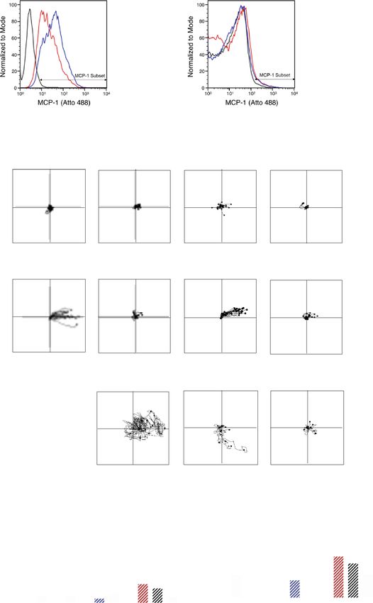
www.nature.com/scientificreports/
modulation of disease c onditions15. Activated neutrophils release tremendous amounts of EVs than cancer cells or
nonimmune cells15, within few minutes after external stimulation. Moreover, NDEVs are equipped with various
ligands and receptors that target pathogens and other immune cells. Therefore, NDEVs could be an attractive
vehicle for developing a drug-delivery system targeting specific pathogens or immune cells. However, the short
life span of neutrophils has limited the development of a drug-delivery system using NDEVs. HL-60 cells, pro-
myelocytic leukemia cells, are widely used as a model system to study neutrophil f unction16,17. Neutrophil-like
cells are easily differentiated from HL-60 cells. These neutrophil-like differentiated HL-60 (dHL-60) cells have
neutrophil characteristics, such as reactive oxygen species (ROS) generation, neutrophil extracellular trap (NET)
formation, chemotaxis, and bactericidal a ctivity17–20. Moreover, they have a short-life span due to spontaneous
apoptosis21 which is an advantage over the EVs generated from general cancer cells. Therefore, recent studies
employed dHL-60 cells to investigate NDEVs22,23 and found the beneficial effects on inflammatory diseases, such
as acute lung injury and s troke22,23.
Here, we investigated the incorporation of various cargoes in dHL-60 cell-derived EVs. The dHL-60 cells
produce large amounts of EVs within a short time and EVs derived from the dHL-60 cells showed similar char-
acteristics to NDEVs. In addition, the dHL-60 cell-derived EVs were successfully loaded with various cargoes,
such as antibiotics, anti-cancer drugs, and Cas-9, and these cargo-loaded EVs showed efficient therapeutic effects
according to the types of cargo.
Materials and methods
Differentiation of HL‑60 cells. Human myeloblastic cells (HL-60 cells) were cultured and maintained in
RPMI 1640 (Gibco) supplemented with 5% heat-inactivated fetal bovine serum (Hyclone) and 1 × antibiotic/
antimycotic (Gibco) at 37 °C and 5% C O2. Human myeloblastic cells (HL-60 cells), Jukart cells, and THP-1
cells were cultured and maintained in RPMI 1640 (Gibco) supplemented with 10% heat-inactivated fetal bovine
serum (Hyclone) and 1 × antibiotic/antimycotic (Gibco) at 37 °C and 5% CO2. The MCF-7 and COLO205 cells
were cultured and maintained in RPMI 1640 (Gibco) supplemented with 10% heat-inactivated fetal bovine
serum (Hyclone) and 1 × antibiotic/antimycotic (Gibco) at 37 °C and 5% CO2. For the differentiation of neutro-
phil-like differentiated HL-60 cells, 1 × 10⁵ cells/ml were treated with 1 μM all-transretionic acid (ATRA, Sigma-
Aldrich). The cultures were washed and reseeded in fresh media followed by ATRA treatment every third day
up to the sixth day. For differentiation of THP-1, 2 × 10⁶ cells were treated with phorbol 12-myristate 13-acetate
(PMA, Sigma-Aldrich, 50 ng) for 48 h.
Live‑imaging of EVs formation. Naïve HL-60 cells or dHL-60 cells (106 cells) were stained with calcein-
AM (2 μg/ml, Merk Millipore) and seeded in a confocal dish (SPL Life Sciences) pre-coated with 0.1% poly-L-
lysine (Sigma-Aldrich). The cells were visualized for EVs formation by fluorescence microscopy (Olympus IX83,
Olympus) in the presence or absence of 1 μM fMLP (Sigma-Aldrich).
Isolation of EVs. EVs were isolated from supernatants collected after the culture of naïve HL-60 or dHL-60
cells in the presence or absence of fMLP. The supernatants were collected, and the EVs were purified by cen-
trifugation at 2500 rpm, followed by filtration through a 1.2 μm filter (Ministart Syringe Filter). The filtered EVs
containing supernatants were ultracentrifuged at 100,000 × g for 1 h at 4 °C. The pelleted EVs were resuspended
into 1 × PBS and stored at − 70 °C.
The quantification of EVs. The EVs were isolated from naïve HL-60, dHL-60, or HEK293 cells prestained
with calcein-AM (5 μg/ml). The fluorescence of calcein was measured using a spectrophotometer (Spectromax
M2/e fluorescence microplate reader, Molecular devices).
Nanoparticle‑tracking analysis (NTA). The isolated EVs were resuspended into 1 × PBS and further
diluted. NTA analysis was performed using a Nanosight LM10 nanoparticle characterization system (Malvern
Instruments). The NTA analytical software version 3 was used for capturing and analyzing data.
Scanning electron microscopic analysis. The EVs were absorbed on the surface of the filter disc, incu-
bated in 3% glutaraldehyde (Sigma-Aldrich), dehydrated using ethanol (Merk Millipore), dried with liquid CO2,
and sputtered with osmium. Samples were visualized using a Hitachi SU8229 scanning electron microscope
(Hitachi).
The separation of NDEVs. Human blood experiment was approved by the Institutional Research Board
of Kyungpook National University and proceeded according to the institutional guidelines. Venous blood was
taken from healthy adult volunteers (age ≥ 18). All participants provided informed consent following the Dec-
laration of Helsinki. Neutrophils were purified using density gradient as described previously24. Isolated neu-
trophils (5 × 10⁶ cells) were stimulated with either fMLP (1 μM) or S. aureus (1 × 10⁶ cells) for 20 min. S. aureus
(109 cells) were opsonized for 30 min with autologous serum (100 μl). The supernatants were collected, and EVs
were isolated as previously d escribed25. In brief, remnant cells and debris were removed and filtered through a
1.2 μm filter (Minisart Syringe Filter, Sartorius) and further concentrated using ultracentrifugation at 100,000 × g
for 60 min. The concentrated EVs were dissolved in 100 μl RPMI. The supernatants were exposed to penicil-
lin–streptomycin (Pen-Strep, Sigma-Aldrich) for 30 min before centrifugation to eliminate remnant bacteria in
NDEVs.
Scientific Reports | (2021) 11:8289 | https://doi.org/10.1038/s41598-021-87891-8 2
Vol:.(1234567890)
www.nature.com/scientificreports/
Cargo loading in EVs. Either naïve HL-60 or dHL-60 cells were transfected with various cargoes (i.e.,
penicillin, Sigma-Aldrich; paclitaxel, Invitrogen; monocyte chemoattractant protein-1, MCP-1, Sinobiologicals;
miR-16 mimic, Abm; miR-16 precursor, Ambion; Cas-9-GFP, provided by Prof. Minsang-Shin) using NEPA21
electroporator (NEPA gene) at 225 V. To validate the transfection efficacy, the cargoes were conjugated with vari-
ous fluorescent: BOCILLIN-FL penicillin, Invitrogen; oregon green conjugated paclitaxel, Flutax-2, Invitrogen;
Atto 488 MCP-1, Atto 488 labeling kit, Sigma-Aldrich; miRNA, labeled using ULYSIS Nucleic acid labeling kit,
Molecular Probes; Cas9-GFP. The transfected cells were analyzed using a flow cytometer to determine the fluo-
rescence levels of each cargo. For comparison, the HEK293 cells were transfected with cargoes using a NEPA21
electroporator and the EVs were isolated.
Bactericidal activity of EVs. S. aureus (ATCC 25,923) was opsonized with human serum for 30 min at
37 °C. Serum was obtained from clotted blood by centrifugation. The opsonized S. aureus (1 × 10⁶ cells/ml) were
exposed to the EVs ( 108 particles) for 30 min at 37 °C. Bacteria were then spread into an LB agar (Difco) and
colony-forming units were counted following incubation for 18 h at 37 °C. Bacteria were exposed to EVs (108
particles) in the presence of annexin A5 (AnxA5) or lysed EVs to inhibit the effector function of penicillin-
loaded EVs. The EVs were lysed with SDS following a previous study26. The EVs were lysed with 0.025% SDS,
vortexed for 1 min, centrifuged at 14,000g for 10 min, and the supernatants were removed. The pellets were
washed twice with ice-chilled 1 × PBS and resuspended.
Chemotaxis of monocytes against EVs. Monocytes were isolated from the peripheral blood mono-
nuclear cells layer obtained after histopaque centrifugation and purified using a percoll solution as described
previously27. The chemotaxis of monocytes against the EVs was determined using a μ-slide chamber (ibidi)
following the manufacturer’s instructions. One side of the chemotaxis chamber was loaded with the EVs, and
monocyte were allowed to migrate. For comparison, one side of the chemotaxis chamber was loaded with MCP-
1, and monocyte chemotaxis was assessed. For the inhibition of monocyte chemotaxis against EVs, monocytes
were pretreated with CCR2 inhibitor (1 μg/ml, Santacruz) for 30 min and allowed to migrate toward EVs in the
presence of the CCR2 inhibitor. The cell movements were visualized and captured by the Nikon Eclipse Ni-U
microscope using × 20 objective. The chemotaxis was then analyzed using ImageJ and chemotaxis/migration
tool (ibidi).
Tumoricidal effects of drug‑loaded EVs. MCF-7 (Korean Cell Line Bank) and COLO205 (Korean Cell
Line Bank) were exposed to either vehicle or paclitaxel-loaded EVs for 24 h. The viability of cancer cells was
assayed using the MTT (3-[4,5-dimethylthiazol-2-yl]-2,5 diphenyltetrazolium bromide) assay kit (Invitrogen)
following the manufacturer’s instructions.
Polarization of differentiated THP‑1. THP-1 cells were purchased from Korean Cell Line Bank.
THP-1 cells were maintained at 5 × 10⁶ cells/ml in RPMI (Gibco) supplemented with 10% heat-inactivated FBS
(HyClone, GE healthcare Life Sciences) with 1% penicillin/streptomycin (Sigma-Aldrich). THP-1 cells were
differentiated into M0 macrophages by treatment with PMA (100 ng/ml) for 48 h. M0-differentiated THP-1
cells (106 cells) to EVs. For comparison, M0-differentiated THP-1 cells were stimulated with either LPS (100 ng/
ml) or IFN-γ (10 ng/ml) for M1 polarization or IL-4 (10 ng/ml) and recombinant human IL-12 (10 ng/ml,
Sinobiologicals) for M2 polarization. RNA was extracted from polarized-THP-1 cells using TRIzol (Ambion),
and cDNA was synthesized using the R T2 First strand kit according to manufacturer’s recommended protocols
(Qiagen). RT-qPCR was performed by using the cDNA (100 ng), primer (1 μl), and R T2 SYBR Green qPCR
2
mastermix (Qiagen) using Qiagen rotor. Predesigned RT qPCR primer assays (Qiagen) were used to determine
the expression level of iNOS (NOS2, PPH00173F) and Arginase-1 (PPH20977A). Gene expression levels were
calculated as log2(2−ΔΔct) using GAPDH (PPH00150F) as a control. For some experiments, M0-differentiated
THP-1 cells were exposed to the EVs in presence of AnxA5 or lysed EVs.
The loading of Cas9 in EVs. MCF7, Jukart (Korean Cell Line Bank), and differentiated THP-1 (THP-1,
Korean Cell Line Bank) were exposed to either vehicle or Cas9-loaded EVs for 2 h at 37 °C. The cells were col-
lected and fixed. A flow cytometer examined the expression of Cas9-GFP in the harvested cells.
Statistical analysis. Data are presented as the mean ± SEM for continuous variables and as the number (%)
for the categorical variables. Data are compared using the Student’s t-test (unpaired, two-sided) and a p value of
less than 0.05 was considered statistically significant. The statistical data were analyzed by Graphpad prism 7.0
(GraphPad Software Inc).
Results
The dHL‑60 release a heterogeneous population of EVs. The HL-60 cells were allowed to differenti-
ate into neutrophil-like dHL-60 cells by treatment with all-trans-retinoic acid (ATRA). The ATRA-treated HL-60
cells showed increased segmentation of nuclear lobes (Supplementary Fig. 1a) without significant changes in
apoptosis (Supplementary Fig. 1b). dHL-60 cells also showed increased expressions of CD11b, CD18, and CD54
with decreased expressions of CD15 and CD33 (Supplementary Fig. 2a). These results are in accordance with
previous studies that reported surface marker expressions of myeloid leukemic cells toward neutrophils28,29. We
further confirmed the effector functions of the dHL-60 cells. The dHL-60 cells successfully generated reactive
Scientific Reports | (2021) 11:8289 | https://doi.org/10.1038/s41598-021-87891-8 3
Vol.:(0123456789)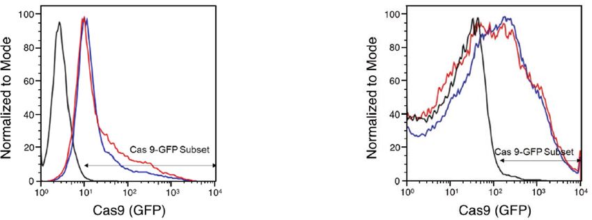
www.nature.com/scientificreports/
oxygen species (ROS) in response to PMA stimulation and showed spontaneous NET formation (Supplemen-
tary Fig. 2b,c) but did not enhance the expression of markers for granules (Supplementary Fig. 2d).
Next, we examined whether dHL-60 cells generate EVs. The spontaneous generation of EVs from cells is
a prolonged process. Hence, various methods are developed to force cells to generate EVs30. We employed
a biological approach using N-formyl-methionyl-leucyl- phenylalanine (fMLP), a bacterial-derived protein,
because neutrophils generate EVs in response to fMLP stimulation31–33. The naïve HL-60 and dHL-60 cells were
loaded with calcein-AM, stimulated with fMLP for 90 min, and live-cell fluorescence images were obtained
using fluorescence microscopy. The dHL-60 cells showed a significantly enhanced production of EVs compared
to those from the naïve HL-60 cells (Fig. 1a, upper panel). They showed numerous protrusions that extended
from membranes (Fig. 1a, arrows), which were finally detached from cell membranes. The EVs derived from the
naïve HL-60 cells and dHL-60 cells were isolated, and fluorescence was measured using a spectrofluorometer.
The dHL-60 cells produced significantly large amounts of EVs in response to fMLP stimulation compared to
those from the naïve HL-60 or HEK293 cells (Fig. 1b). The nanoparticle tracking analysis (NTA) showed that the
EVs generated from the naïve HL-60 and dHL-60 cells are heterogenous in size ranging from 50 to 600 nm in
diameter (mean diameter of 170.5 ± 49.4 nm versus 246.8 ± 19.5 nm) compared to the NDEVs that were isolated
from fMLP-stimulated neutrophils (Fig. 1c). We further examined the morphology of the EVs derived from the
naïve HL-60 and dHL-60 cells using electron microscopy. Scanning electron microscopic (SEM) analysis identi-
fied that both EVs have an oval-shaped morphology with heterogeneous diameters (Fig. 1d). Next, we evaluated
the functions of EVs isolated from the naïve HL-60 cells and dHL-60 cells. Both EVs did not show bactericidal
activity (Supplementary Fig. 3a) and did not induce chemotaxis of monocytes (Supplementary Fig. 3b).
These results suggest that dHL-60 cells generate massive amounts of EVs that are phenotypically similar to
NDEVs with negligible effector functions. Therefore, we thought they could be an attractive vehicle for drug
delivery and investigated their possible therapeutic use on drug-delivery systems based on the dHL-60-derived
EVs.
The delivery of anti‑bacterial and anti‑cancer drugs using the dHL‑60 derived EVs. Recent stud-
ies showed that antibiotics and anticancer drugs are efficiently derived through EVs13,34. Therefore, we evaluated
the delivery of these drugs through the dHL-60 cell-derived EVs. The naïve HL-60 and dHL-60 cells were loaded
with fluorescence-tagged penicillin and paclitaxel using an electroporator. Flowcytometric analysis showed that
both drugs were successfully incorporated into parent cells (Left panels in Fig. 2a,b). The EVs derived from
both cells showed significant amounts of fluorescence from drugs derived from the parent cells (Right panels
in Fig. 2a,b). The dHL-60 cells showed higher efficacy of incorporating both drugs, and only paclitaxel showed
significantly higher incorporation in the dHL-60 cell-derived EVs.
We further confirmed the effectiveness of these drug-loaded EVs. The bactericidal activity of the drug-
loaded EVs was examined. The NDEVs isolated from neutrophils stimulated with S. aureus showed significant
bactericidal activity as previously reported25. Interestingly, the penicillin-loaded EVs derived from the dHL-60
cells showed significant bactericidal activity against S. aureus, whereas the penicillin-loaded EVs derived from
the HL-60 cells did not (Fig. 2c). The EVs derived from HEK293 cells did not show bactericidal activity against
S. aureus (Fig. 2c). We further confirmed the effects of lysed EVs on the bactericidal activity of the penicillin-
loaded EVs. The lysed EVs did not show bactericidal activity against S. aureus (Fig. 2c). We further inhibited the
bactericidal activity of the EVs derived from the penicillin-loaded dHL-60 cells using annexin A5 (AnxA5), an
inhibitor for phosphatidylserine expressed on the EV membrane. The AnxA5 completely inhibited the bacteri-
cidal activity of the EVs derived from the penicillin-loaded dHL-60 cells (Fig. 2c).
We further examined the anticancer effects of the drug-loaded EVs. The anti-cancer effect of the drug-loaded
EVs was compared to the EVs isolated from the paclitaxel-transfected HEK293 cells. Both paclitaxel-loaded EVs
showed significant anticancer effects on COLO205 and MCF-7 (Fig. 2d). However, the paclitaxel-loaded EVs
derived from the dHL-60 cells showed higher cytotoxic effects against both cancer cells compared to those from
HL-60 cells and paclitaxel-loaded HEK293 cells (Fig. 2d). Collectively, these results suggest that the EVs derived
from the dHL-60 cells could be an effective vehicle for antibiotics and anti-cancer drugs.
Delivery of MCP‑1 using the dHL‑60 derived microvesicles. Next, we evaluated whether diffus-
ible molecules such as chemotactic proteins can be delivered through EVs. We transfected fluorescence-tagged
monocyte chemoattractant protein-1 (MCP-1, 1 mg/ml) in the naïve HL-60 or dHL-60 cells using an electropo-
rator. Flowcytometric analysis showed that MCP-1 was effectively incorporated in both cells (Fig. 3a, left panel)
and EVs were derived from both cells (Fig. 3a, right panel). Although transfection efficacy of the HL-60 cells
with MCP-1 was significantly higher than that of the dHL60 cells, dHL-60 cell-derived EVs showed higher rates
of MCP-1 incorporation.
To confirm the successful incorporation of MCP-1 in EVs, we examined the effects of MCP-1 incorporated
EVs on monocyte chemotaxis. Both MCP-1 loaded EVs effectively induced chemotaxis of monocytes with
increased directionality and distance coverage (Fig. 3b). Moreover, monocyte showed a slightly higher rate of
chemotaxis against EVs isolated from the dHL-60 cells loaded with MCP-1 than against either an equal concen-
tration of MCP-1 or NDEVs (Fig. 3b). EVs isolated from HEK293 cells loaded MCP-1 did not induce chemotaxis
of monocytes (Fig. 3b).
We further examined the effect of the pharmacological inhibition of C–C chemokine receptor 2 (CCR2),
a receptor for MCP-1, on monocyte chemotaxis against MCP-1-loaded EVs. The CCR2 antagonist (CCR2i)
completely inhibited monocyte chemotaxis against MCP-1-loaded EVs (Fig. 3b). These results suggest that a
diffusible chemoattractant can be successfully incorporated into dHL-60 cell-derived EVs.
Scientific Reports | (2021) 11:8289 | https://doi.org/10.1038/s41598-021-87891-8 4
Vol:.(1234567890)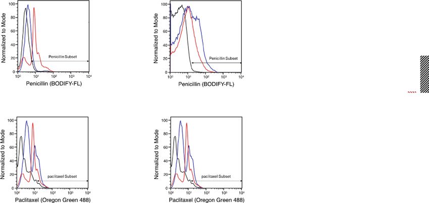
www.nature.com/scientificreports/

                                  Figure 1.  The naïve HL-60 cells and dHL-60 cells release a heterogeneous population of EVs. (a) Representative images
                                  showing EVs released from HL-60 cells and dHL-60 cells stimulated with fMLP. The arrow indicates membrane protrusions
                                  during EVs release. (b) Quantification of relative amounts of EVs derived from the naïve HL-60 and dHL-60 cells pre-stained
                                  with calcein-AM in the presence or absence of fMLP stimulation. (c) A representative plot showing the size and concentration
                                  of EVs derived from the HL-60 cells, dHL-60 cells, and neutrophils stimulated with fMLP. (d) Scanning electron microscopic
                                  analysis of EVs generated from HL-60 and dHL-60 cells. The data shown are the mean ± SEM. *P < 0.05; **P < 0.01.

Scientific Reports |   (2021) 11:8289 |                 https://doi.org/10.1038/s41598-021-87891-8                                                           5

                                                                                                                                                       Vol.:(0123456789)
www.nature.com/scientificreports/
Figure 2. The delivery of penicillin and paclitaxel using dHL-60 cell-derived EVs. (a) Left panels, fluorescence
levels of penicillin in the HL-60 and dHL-60 cells. Right panels, fluorescence levels of penicillin in the EVs
derived from the HL-60 and dHL-60 cells. (b) Left panels, fluorescence levels of paclitaxel in the HL-60 and
dHL-60 cells. Right panels, fluorescence levels of paclitaxel in EVs derived from the HL-60 and dHL-60 cells. (c)
Bactericidal activity of penicillin-loaded EVs against S. aureus. (d) Cytotoxicity of paclitaxel-loaded EVs against
COLO205 and MCF-7. All data represent three independent experiments; mean ± SEM. *p < 0.05; **P < 0.01;
***P < 0.001.
The delivery of micro‑RNA using the dHL‑60 derived microvesicles. MicroRNAs (miRNAs) are
small non-coding RNA molecules that mediate the post-translational regulation of gene expressions35,36. To
examine the possibility of EVs as a miRNA-delivery vehicle, we examined the incorporation of miRNAs in the
dHL-60 cell-derived EVs. The naïve HL-60 and dHL-60 cells were transfected with fluorescence-labeled synthetic
miR-16 (5 nM). The miR-16 is known to induce apoptosis of tumor cells, induce polarization of macrophages
into the M1 phenotype, and activate CD4+ T cells by downregulation of PD-L137,38. The miR-16 (precursor and
mimic) were successfully incorporated into both cells and loaded in both EVs (Fig. 4a,b). The miR-16 loaded
EVs significantly inhibited the proliferation of MCF-7 and Jurkat cells but not the THP-1-derived macrophages
(dTHP1) (Fig. 4c). However, miR-16 loaded EVs significantly enhanced the expression of iNOS and attenuated
the expression of arginase (Fig. 4d). We further confirmed the uptake of the dHL-60 derived EVs by the target
cells. EVs were isolated from the dHL-60 cells transfected with miR-16 and M0-differentiated THP-1 cells were
exposed to EVs loaded with miR-16. Then, RNA was extracted from the THP-1 cells, cDNA was synthesized,
and subsequently used as a template for qPCR using the miR-16 primer. The THP-1 cells exposed to EVs derived
from the dHL-60 cells loaded with miR-16 showed significantly high levels of miR-16 (Supplementary Fig. 4d).
We finally confirmed the requirement of interaction between dHL-60 derived EVs and target cells. The expres-
sion of iNOS in EV-exposed THP-1 cells was completely inhibited by the EV lysis, anti-AnxA1 antibody, and
AnxA5 (Fig. 4e). These results suggest that EVs derived from HL-60 cells and dHL-60 cells could be utilized to
deliver miRNAs with functional modalities.
The delivery of Cas9‑RNP using the dHL‑60 derived microvesicles. Although clustered regularly
interspaced short palindromic repeats (CRISPR)/CRISPR-associated protein-9 nuclease (Cas9) provides robust
genome editing, efficient methods for delivering them into target cells are still required. To date, electropora-
tion is the primary choice for delivering CRISPR/Cas9 to target cells39,40. We examined whether EVs derived
from the HL-60 cells could be efficient vehicles for delivering Cas9. The naïve HL-60 and the dHL-60 cells were
transfected with GFP-tagged Cas9. The Cas9-GFP were successfully incorporated in both cells, and EVs were
derived from both cells (Fig. 5a). Next, we examined the delivery of Cas9-GFP-loaded EVs against target cells.
The dTHP-1 cells, MCF-7 cells, and Jurkat cells were exposed to the Cas9-GFP-loaded EVs, and the GFP fluores-
cence levels were measured. The MCF-7 cells showed the highest GFP fluorescence, and the Jurkat cells showed
negligible GFP fluorescence levels (Fig. 5b). These results suggest that the dHL-60 cell-derived EVs could be
candidate vehicles for CRISPR/Cas9.
Discussion
In this study, we found that the HL-60 and dHL-60 cells generated EVs in response to external stimulation. These
EVs were successfully loaded with various cargoes and showed functional modalities. However, the dHL-60 cells
produced significantly larger amounts of EVs than HL-60 cells, and most cargoes were more efficiently loaded
in EVs derived from the dHL-60 cells. Moreover, the cargoes-loaded EVs derived from the dHL-60 cells showed
higher effector functions than those from the HL-60 cells. These results suggest that the EVs derived from the
dHL-60 cells could be attractive vehicles for drug delivery.
In recent years, substantial effort has been undertaken to develop a drug-delivery system using cell-derived
EVs. Although extensive studies, including clinical trials, are ongoing to develop drug-delivering EVs, several
limitations still remain. Therefore, recent studies have searched for an alternative source of drug-delivering
EVs and immune cells are strong c andidates11,12. Immune cells actively release EVs upon external stimulation,
Scientific Reports | (2021) 11:8289 | https://doi.org/10.1038/s41598-021-87891-8 6
Vol:.(1234567890)www.nature.com/scientificreports/
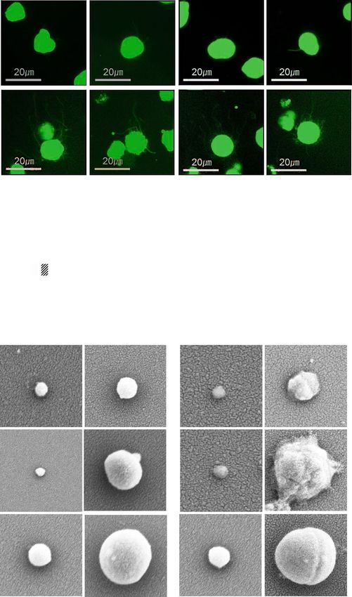
Figure 3. The delivery of MCP-1 using dHL-60 cell-derived EVs. (a) Left panels, fluorescence levels of MCP-1
in the HL-60 and dHL-60 cells. Right panels, fluorescence levels of MCP-1 in EVs derived from the HL-60 and
dHL-60 cells. (b) Monocyte migration tracking analysis against EVs. Bar graphs denote Euclidean distance and
relative directionality. All data represent three independent experiments; mean ± SEM. *P < 0.05; **P < 0.01;
***P < 0.001.
Scientific Reports | (2021) 11:8289 | https://doi.org/10.1038/s41598-021-87891-8 7
Vol.:(0123456789)www.nature.com/scientificreports/
Figure 4. The delivery of miRNA-16 using dHL-60 cell-derived EVs. (a) Left panels, fluorescence levels of
the miRNA-16 precursor in the HL-60 and dHL-60 cells. Right panels, fluorescence levels of the miRNA-16
precursor in EVs derived from the HL-60 and dHL-60 cells. (b) Left panels, fluorescence levels of the miRNA-
16 mimic in the HL-60 and dHL-60 cells. Right panels, fluorescence levels of the miRNA-16 mimic in EVs
derived from the HL-60 and dHL-60 cells. (c) The effects of EVs on the proliferation of the MCF-7, Jurkat, and
dTHP-1 cells. (d) The effects of EVs on expressions of iNOS and arginase in MCF-7, Jurkat, and dTHP-1 cells.
(e) The expression of iNOS in the dTHP-1 cells exposed to the HL-60 and dHL-60 cells. All data represent three
independent experiments; mean ± SEM. *P < 0.05; **P < 0.01; ***P < 0.001.
Figure 5. The delivery of Cas 9-GFP using dHL-60 cell-derived EVs. (a) Left panels, fluorescence levels of Cas9
in the HL-60 and dHL-60 cells. Right panels, fluorescence levels of Cas9 in EVs derived from the HL-60 and
dHL-60 cells. (b) Fluorescence levels of Cas9 in the MCF-7, Jurkat, and dTHP-1 cells exposed to EVs. All data
represent three independent experiments; mean ± SEM. *P < 0.05; **P < 0.01.
especially at inflammatory sites. Immune cell-derived EVs are equipped with multiple receptors and ligands
derived from their parent cells and contain multiple effector molecules such as miRNAs. Therefore, they are con-
sidered to be an appropriate drug-delivering EVs without possible toxic r eactions11,15. In the current manuscript,
we suggest that the neutrophil-like dHL-60 cells could be another candidate for the source of drug-delivering
EVs. The dHL-60 cells generate massive amounts of EVs spontaneously (Fig. 1b), and the sizes of EVs are large
enough to load various cargoes (Fig. 1c). Although the phenotype of the dHL-60-derived EVs is similar to
NDEVs, they lack the effector function of NDEVs, such as bactericidal activity (Supplementary Fig. 3a) and
chemoattractive effects on monocytes (Supplementary Fig. 3b).
Another issue to be solved in developing a drug-delivery system using EVs is cargo-loading techniques. The
most common method is passive cargo loading by incubating small cargoes with isolated EVs. However, this
Scientific Reports | (2021) 11:8289 | https://doi.org/10.1038/s41598-021-87891-8 8
Vol:.(1234567890)www.nature.com/scientificreports/
method is time-consuming and has a low loading efficiency. Moreover, some cargoes adhere to surfaces of the
EVs rather than being adequately incorporated. Therefore, active loading techniques are employed to increase
the efficiency of cargo loading. The cargoes were loaded using electroporation or membrane disruption (e.g.,
sonication, osmotic shock, freeze–thaw, and membrane permeabilization) and these techniques guaranteed
high drug-loading efficiency. However, these techniques lead to disturbances in the membrane integrity of EVs
and extensive molecule a ggregates1,8, leading to decreased loading efficiency13,41–44. Therefore, the endogenous
loading method—isolation of the cargo-loaded EVs from the parent cells passively incubated with cargoes—was
developed, and this technique limits these drawbacks without compromised membrane integrity. However, low
cargo-loading efficiency in this method should be s olved42,44.
We used well-established endogenous cargo loading into HL-60 cells using transfection rather than merely
incubating cells with the desired cargo44. The HL-60 cells are efficiently transfected cells with known transfection
techniques. Therefore, they are used to evaluate general transfection efficiency for transfection materials. We
employed mild biphasic shocks in the HL-60 cells, which greatly enhanced the overall cargo-loading efficiency of
small molecules (Figs. 2 and 3) and even large proteins (Fig. 4) to reduce possible membrane damage by transfec-
tion. The aggregation and disruption of EVs were significantly reduced because possible harmful steps, such as
freeze/thaw and sonication, were missing in our method. In addition, we found that the dHL-60 cells generated
higher amounts of EVs spontaneously within a given period. The dHL-60 cells showed higher incorporation of
desired cargoes, and the dHL-60-derived EVs showed higher incorporation with higher cargo efficiency (Figs. 2,
3, 4). These results suggest the advantages of EVs derived from the HL-60 and dHL-60 cells as drug-delivery
vehicles. Moreover, the dHL-60 cells generate tremendous amounts of EVs within minutes, and it is an essential
advantage for developing drug-delivery vehicles.
In conclusion, we provided evidence that neutrophil-like dHL-60 cells quickly produce a tremendous amount
of EVs. We also demonstrated that endogenous loading of the dHL-60 cells by electroporation could attribute
desired functionality into isolated EVs. Therefore, our study highlights the increased productivity and efficient
loading of therapeutics into dHL-60 cell-derived EVs. This finding opens the possibility for generating a drug-
delivery system using immune cells, especially neutrophils.
Received: 17 March 2020; Accepted: 22 March 2021
References
1. Mentkowski, K. I., Snitzer, J. D., Rusnak, S. & Lang, J. K. Therapeutic potential of engineered extracellular vesicles. AAPS J. 20, 50.
https://doi.org/10.1208/s12248-018-0211-z (2018).
2. Clemmens, H. & Lambert, D. W. Extracellular vesicles: Translational challenges and opportunities. Biochem. Soc. Trans. 46,
1073–1082. https://doi.org/10.1042/BST20180112 (2018).
3. Buzas, E. I., Toth, E. A., Sodar, B. W. & Szabo-Taylor, K. E. Molecular interactions at the surface of extracellular vesicles. Semin.
Immunopathol. 40, 453–464. https://doi.org/10.1007/s00281-018-0682-0 (2018).
4. Crewe, C. et al. An endothelial-to-adipocyte extracellular vesicle axis governed by metabolic state. Cell 175, 695–708. https://doi.
org/10.1016/j.cell.2018.09.005 (2018).
5. Meldolesi, J. Exosomes and ectosomes in intercellular communication. Curr. Biol. 28, R435–R444. https://doi.org/10.1016/j.cub.
2018.01.059 (2018).
6. Ying, W. et al. Adipose tissue macrophage-derived exosomal mirnas can modulate in vivo and in vitro insulin sensitivity. Cell 171,
372–384. https://doi.org/10.1016/j.cell.2017.08.035 (2017).
7. Piffoux, M. et al. Extracellular vesicles for personalized medicine: The input of physically triggered production, loading and
theranostic properties. Adv. Drug Deliv. Rev. 138, 247–258. https://doi.org/10.1016/j.addr.2018.12.009 (2019).
8. Piffoux, M., Silva, A. K. A., Wilhelm, C., Gazeau, F. & Tareste, D. Modification of extracellular vesicles by fusion with liposomes
for the design of personalized biogenic drug delivery systems. ACS Nano 12, 6830–6842. https://doi.org/10.1021/acsnano.8b020
53 (2018).
9. Pinheiro, A. et al. Extracellular vesicles: Intelligent delivery strategies for therapeutic applications. J. Control Release 289, 56–69.
https://doi.org/10.1016/j.jconrel.2018.09.019 (2018).
10. Kamerkar, S. et al. Exosomes facilitate therapeutic targeting of oncogenic KRAS in pancreatic cancer. Nature 546, 498–503. https://
doi.org/10.1038/nature22341 (2017).
11. Akao, Y. et al. Microvesicle-mediated RNA molecule delivery system using monocytes/macrophages. Mol. Ther. 19, 395–399.
https://doi.org/10.1038/mt.2010.254 (2011).
12. Alvarez-Erviti, L. et al. Delivery of siRNA to the mouse brain by systemic injection of targeted exosomes. Nat. Biotechnol. 29,
341–345. https://doi.org/10.1038/nbt.1807 (2011).
13. Vader, P., Mol, E. A., Pasterkamp, G. & Schiffelers, R. M. Extracellular vesicles for drug delivery. Adv. Drug Deliv. Rev. 106, 148–156.
https://doi.org/10.1016/j.addr.2016.02.006 (2016).
14. Wiklander, O. P. et al. Extracellular vesicle in vivo biodistribution is determined by cell source, route of administration and target-
ing. J. Extracell. Vesicles 4, 26316. https://doi.org/10.3402/jev.v4.26316 (2015).
15. Hong, C. W. Extracellular vesicles of neutrophils. Immune Netw. 18, e43. https://doi.org/10.4110/in.2018.18.e43 (2018).
16. Hauert, A. B., Martinelli, S., Marone, C. & Niggli, V. Differentiated HL-60 cells are a valid model system for the analysis of human
neutrophil migration and chemotaxis. Int. J. Biochem. Cell Biol. 34, 838–854. https://doi.org/10.1016/s1357-2725(02)00010-9
(2002).
17. Yaseen, R. et al. Antimicrobial activity of HL-60 cells compared to primary blood-derived neutrophils against Staphylococcus
aureus. J. Negat. Results Biomed. 16, 2. https://doi.org/10.1186/s12952-017-0067-2 (2017).
18. Vong, L., Lorentz, R. J., Assa, A., Glogauer, M. & Sherman, P. M. Probiotic Lactobacillus rhamnosus inhibits the formation of
neutrophil extracellular traps. J. Immunol. 192, 1870–1877. https://doi.org/10.4049/jimmunol.1302286 (2014).
19. Millius, A. & Weiner, O. D. Chemotaxis in neutrophil-like HL-60 cells. Methods Mol. Biol. 571, 167–177. https://doi.org/10.1007/
978-1-60761-198-1_11 (2009).
20. Brechard, S., Bueb, J. L. & Tschirhart, E. J. Interleukin-8 primes oxidative burst in neutrophil-like HL-60 through changes in
cytosolic calcium. Cell Calcium 37, 531–540. https://doi.org/10.1016/j.ceca.2005.01.019 (2005).
21. Watson, R. W. et al. Granulocytic differentiation of HL-60 cells results in spontaneous apoptosis mediated by increased caspase
expression. FEBS Lett. 412, 603–609. https://doi.org/10.1016/s0014-5793(97)00779-5 (1997).
Scientific Reports | (2021) 11:8289 | https://doi.org/10.1038/s41598-021-87891-8 9
Vol.:(0123456789)www.nature.com/scientificreports/
22. Gao, J., Wang, S. & Wang, Z. High yield, scalable and remotely drug-loaded neutrophil-derived extracellular vesicles (EVs) for
anti-inflammation therapy. Biomaterials 135, 62–73. https://doi.org/10.1016/j.biomaterials.2017.05.003 (2017).
23. Wang, S., Dong, X., Gao, J. & Wang, Z. Targeting inflammatory vasculature by extracellular vesicles. AAPS J. 20, 37. https://doi.
org/10.1208/s12248-018-0200-2 (2018).
24. Hong, C. W. et al. Lysophosphatidylcholine increases neutrophil bactericidal activity by enhancement of azurophil granule-
phagosome fusion via glycine.GlyR alpha 2/TRPM2/p38 MAPK signaling. J. Immunol. 184, 4401–4413. https://doi.org/10.4049/
jimmunol.0902814 (2010).
25. Youn, Y. J. et al. Neutrophil-derived trail is a proinflammatory subtype of neutrophil-derived extracellular vesicles. Theranostics
11, 2770–2787. https://doi.org/10.7150/thno.51756 (2021).
26. Osteikoetxea, X. et al. Differential detergent sensitivity of extracellular vesicle subpopulations. Org. Biomol. Chem. 13, 9775–9782.
https://doi.org/10.1039/c5ob01451d (2015).
27. Repnik, U., Knezevic, M. & Jeras, M. Simple and cost-effective isolation of monocytes from buffy coats. J. Immunol. Methods 278,
283–292. https://doi.org/10.1016/s0022-1759(03)00231-x (2003).
28. Barber, N., Belov, L. & Christopherson, R. I. All-trans retinoic acid induces different immunophenotypic changes on human HL60
and NB4 myeloid leukaemias. Leuk Res. 32, 315–322. https://doi.org/10.1016/j.leukres.2007.04.013 (2008).
29. Trayner, I. D. et al. Changes in antigen expression on differentiating HL60 cells treated with dimethylsulphoxide, all-trans retinoic
acid, alpha1,25-dihydroxyvitamin D3 or 12-O-tetradecanoyl phorbol-13-acetate. Leuk Res. 22, 537–547. https://doi.org/10.1016/
s0145-2126(98)00041-1 (1998).
30. Gao, J., Dong, X. & Wang, Z. Generation, purification and engineering of extracellular vesicles and their biomedical applications.
Methods 177, 114–125. https://doi.org/10.1016/j.ymeth.2019.11.012 (2020).
31. Dalli, J. et al. Heterogeneity in neutrophil microparticles reveals distinct proteome and functional properties. Mol. Cell Proteom.
12, 2205–2219. https://doi.org/10.1074/mcp.M113.028589 (2013).
32. Hess, C., Sadallah, S., Hefti, A., Landmann, R. & Schifferli, J. A. Ectosomes released by human neutrophils are specialized functional
units. J. Immunol. 163, 4564–4573 (1999).
33. Pliyev, B. K., Kalintseva, M. V., Abdulaeva, S. V., Yarygin, K. N. & Savchenko, V. G. Neutrophil microparticles modulate cytokine
production by natural killer cells. Cytokine 65, 126–129. https://doi.org/10.1016/j.cyto.2013.11.010 (2014).
34. Lamichhane, T. N. & Jay, S. M. Production of extracellular vesicles loaded with therapeutic cargo. Methods Mol. Biol. 37–47, 2018.
https://doi.org/10.1007/978-1-4939-8661-3_4 (1831).
35. Forero, A., So, L. & Savan, R. Re-evaluating strategies to define the immunoregulatory roles of miRNAs. Trends Immunol. 38,
558–566. https://doi.org/10.1016/j.it.2017.06.001 (2017).
36. Oliveto, S., Mancino, M., Manfrini, N. & Biffo, S. Role of microRNAs in translation regulation and cancer. World J. Biol. Chem. 8,
45–56. https://doi.org/10.4331/wjbc.v8.i1.45 (2017).
37. Cimmino, A. et al. miR-15 and miR-16 induce apoptosis by targeting BCL2. Proc. Natl. Acad. Sci. U.S.A. 102, 13944–13949. https://
doi.org/10.1073/pnas.0506654102 (2005).
38. Kitadate, A. et al. MicroRNA-16 mediates the regulation of a senescence-apoptosis switch in cutaneous T-cell and other non-
Hodgkin lymphomas. Oncogene 35, 3692–3704. https://doi.org/10.1038/onc.2015.435 (2016).
39. Wang, P. Two distinct approaches for CRISPR-Cas9-mediated gene editing in Cryptococcus neoformans and related species. mSphere
https://doi.org/10.1128/mSphereDirect.00208-18 (2018).
40. Xu, X. et al. Efficient homology-directed gene editing by CRISPR/Cas9 in human stem and primary cells using tube electropora-
tion. Sci. Rep. 8, 11649. https://doi.org/10.1038/s41598-018-30227-w (2018).
41. Kooijmans, S. A. A. et al. Electroporation-induced siRNA precipitation obscures the efficiency of siRNA loading into extracellular
vesicles. J. Control Release 172, 229–238. https://doi.org/10.1016/j.jconrel.2013.08.014 (2013).
42. Lamparski, H. G. et al. Production and characterization of clinical grade exosomes derived from dendritic cells. J. Immunol. Methods
270, 211–226. https://doi.org/10.1016/s0022-1759(02)00330-7 (2002).
43. Stoicheva, N. G. & Hui, S. W. Electrofusion of cell-size liposomes. Biochim. Biophys. Acta 1195, 31–38. https://doi.org/10.1016/
0005-2736(94)90005-1 (1994).
44. Sutaria, D. S., Badawi, M., Phelps, M. A. & Schmittgen, T. D. Achieving the promise of therapeutic extracellular vesicles: The devil
is in details of therapeutic loading. Pharm. Res. 34, 1053–1066. https://doi.org/10.1007/s11095-017-2123-5 (2017).
Acknowledgements
This study was supported by grants from the National Research Foundation of Korea (NRF) (Grant nos.
2017R1C1B2009015, 2017R1A4A1015652, 2020R1A4A2002691, C.W. Hong; 2018R1D1A1B07050421, S.
Shrestha).
Author contributions
J.K.K., Y.J.Y., Y.B.L., S.H.K., and S.S. conducted experiments. J.K.K., Y.J.Y., and S.S. designed experiments, ana-
lyzed results, and wrote the paper. D.K.S., M.S., H.K.J., and J.S.B. assisted in experimental design, contributed
reagents, and provided key advice. S.S. and C.W.H. conceived and designed the research, analyzed data, wrote
the manuscript, provided financial support, and approved the final paper.
Competing interests
The authors declare no competing interests.
Additional information
Supplementary Information The online version contains supplementary material available at https://doi.org/
10.1038/s41598-021-87891-8.
Correspondence and requests for materials should be addressed to S.S. or C.-W.H.
Reprints and permissions information is available at www.nature.com/reprints.
Publisher’s note Springer Nature remains neutral with regard to jurisdictional claims in published maps and
institutional affiliations.
Scientific Reports | (2021) 11:8289 | https://doi.org/10.1038/s41598-021-87891-8 10
Vol:.(1234567890)www.nature.com/scientificreports/
Open Access This article is licensed under a Creative Commons Attribution 4.0 International
License, which permits use, sharing, adaptation, distribution and reproduction in any medium or
format, as long as you give appropriate credit to the original author(s) and the source, provide a link to the
Creative Commons licence, and indicate if changes were made. The images or other third party material in this
article are included in the article’s Creative Commons licence, unless indicated otherwise in a credit line to the
material. If material is not included in the article’s Creative Commons licence and your intended use is not
permitted by statutory regulation or exceeds the permitted use, you will need to obtain permission directly from
the copyright holder. To view a copy of this licence, visit http://creativecommons.org/licenses/by/4.0/.
© The Author(s) 2021
Scientific Reports | (2021) 11:8289 | https://doi.org/10.1038/s41598-021-87891-8 11
Vol.:(0123456789)You can also read