Combined cell and nanoparticle models for TOPAS to study radiation dose enhancement in cell organelles - Nature
←
→
Page content transcription
If your browser does not render page correctly, please read the page content below
www.nature.com/scientificreports
OPEN Combined cell and nanoparticle
models for TOPAS to study
radiation dose enhancement in cell
organelles
Marc Benjamin Hahn1,2* & Julián Mateo Zutta Villate3,4
Dose enhancement by gold nanoparticles (AuNP) increases the biological effectiveness of radiation
damage in biomolecules and tissue. To apply them effectively during cancer therapy their influence
on the locally delivered dose has to be determined. Hereby, the AuNP locations strongly influence
the energy deposit in the nucleus, mitochondria, membrane and the cytosol of the targeted cells. To
estimate these effects, particle scattering simulations are applied. In general, different approaches
for modeling the AuNP and their distribution within the cell are possible. In this work, two newly
developed continuous and discrete-geometric models for simulations of AuNP in cells are presented.
These models are applicable to simulations of internal emitters and external radiation sources. Most
of the current studies on AuNP focus on external beam therapy. In contrast, we apply the presented
models in Monte-Carlo particle scattering simulations to characterize the energy deposit in cell
organelles by radioactive 198AuNP. They emit beta and gamma rays and are therefore considered for
applications with solid tumors. Differences in local dose enhancement between randomly distributed
and nucleus targeted nanoparticles are compared. Hereby nucleus targeted nanoparticels showed
a strong local dose enhancement in the radio sensitive nucleus. These results are the foundation for
future experimental work which aims to obtain a mechanistic understanding of cell death induced by
radioactive 198Au.
The application of metallic nanoparticles (NP) in cancer treatment is considered for different forms of radiation
therapy1,2. Most prominently, they are evaluated for external beam therapy with ionizing r adiation3–5. Addi-
tional therapeutic approaches include brachytherapy in combination with internally placed radioactive e mitters6,
photothermal therapy7–10, and their usage in the form of radioactive gold nanoparticles (AuNP) them self11–16.
All these different forms of application have in common that they benefit from an increase of the local energy
deposit in the surrounding of the NP upon interaction with ionizing radiation. This increase in energy deposit
is caused by the higher scattering cross section of the metallic NP when compared to water or organic matter.
The resulting increase of damaged biomolecules (e.g. DNA in the nucleus or mitochondria, proteins) is based
on a locally increased production of reactive secondary damaging species such as Auger electrons, low energy
electrons (LEE) and reactive oxygen species (ROS)17–22. To assess the effectiveness of AuNP in radiation therapy,
their lethality towards different cell lines have to be evaluated. Especially their energy deposit characteristics in
cell organelles, such as mitochondria, cell membrane and the nucleus, have to be predicted for the respective cell
lines, to obtain a mechanistic understanding of experimentally obtained exposure-survival curves.
For effective therapies, it is of importance to increase the dose delivered to the tumor while sparing healthy
tissue23. The AuNP discussed here, are considered for applications with solid tumors. The optimal delivery strat-
egy depends on the tumor type which has to be t reated2. Active targeting is a promising approach to specifically
targets cancer cells through direct interaction between ligands and receptors of the cells. To distinguish them
from healthy cells, ligands on the surface of NPs can be selected to specifically target overexpressed molecules
which are only located on the surface of cancer c ells24,25. The interaction between ligands and receptors can
induce endocytosis, which allows internalization of AuNPs. Afterwards the AuNP can release their energy into
tumor cells effectively, which leads to an enhancement of the locally absorbed d ose15. The local dose enhancement
1
Bundesanstalt für Materialforschung und -prüfung, 12205 Berlin, Germany. 2Institut für Experimentalphysik,
Freie Universität Berlin, 14195 Berlin, Germany. 3Universidad Nacional de Colombia, Medellín, Colombia.
4
Departamento de Física, Facultad de Ciencias, Pontificia Universidad Javeriana, Bogotá, Colombia. *email:
hahn@physik.fu-berlin.de
Scientific Reports | (2021) 11:6721 | https://doi.org/10.1038/s41598-021-85964-2 1
Vol.:(0123456789)www.nature.com/scientificreports/
effects of AuNP takes place until most of the secondary particles, originating from the radiation-NP interaction,
thermalize3,15. Therefore, it is beneficial to not only introduce the NP into the cancerous tissue, but also to have
them accumulate in the vicinity of the most radiation sensitive parts of the cell, e.g. nucleus or m itochondria1,4.
However, plain AuNP tend to accumulate in the cytoplasm, while the preferential target for radiation is the
nuclear DNA4,26. Thus, a promising way to enhance the effectiveness of radiation induced cell killing is to specifi-
cally target tumor cells and their nucleus13,27. This can be achieved by tailor made AuNP coatings from peptides,
as recently demonstrated by Özçelik and P ratx27. They observed a 3.8-fold reduced cell proliferation for nucleus
targeted AuNP during in vitro irradiations.
To study these effects, Monte-Carlo based particle-scattering simulations are an effective tool to predict energy
deposit in irradiated cells. However, the presence of AuNP in irradiated cells can be modeled in different ways.
For example, a continuous model assumes a homogeneous distribution of gold within the cytosol while neglecting
the nanoscopic structure of the AuNP (e.g. geometry, radius). A discrete-geometric model where individual AuNP
are placed within the cell, provides a more detailed picture together with information on small scale effects by
variation of AuNP location, radii etc.15. Especially nanoparticles targeting the nucleus can be simulated properly
only by a discrete-geometric model1,27. However, such a detailed description increases the computational cost
due to the creation of more complex geometries, higher amount of simulated boundaries and less homogene-
ity. Here, we evaluate the (dis-)advantages of these different approaches to simulate the effects of AuNP on the
dose distribution within a model system, and to provide guidance for future combinations of simulational and
experimental work. Therefore, we extend the recently developed cell models within Topas-nBio28 by adding the
possibility to explicitly include nanoparticles in the cell during Monte-Carlo based particle scattering simulations
with Geant429–31 and Topas32. Hereby, the nanoparticles can be randomly distributed in the cytosol or attached
to the nucleus or mitochondria, emulating different treatment conditions. The newly presented extension will
be applied to a model cell line, Chinese hamster ovary cells (CHO), and radioactive 198AuNP. The inclusion of
198
AuNP in a generalized cell model was chosen since it provides, a computational demanding application for
the the presented cell-nanoparticle extension, as well as a useful guidance for future experimental application of
198
AuNP in different cell lines. The energy deposit characteristics of isolated radioactive 198AuNP in dependence
on their intrinsic and extrinsic properties e.g. AuNP diameter, AuNP density, relative gold mass percentage and
their clustering behavior were investigated in a previous s tudy15. The results showed an increase of the energy
deposit within 150 nm around the AuNP in dependence of the AuNP radius (2.5–20 nm). For the investigated
AuNP clusters, the enhancement of the energy deposit increases locally with the relative gold mass percent-
age. However, to obtain information on the effects on a cell, their locations have to be considered. Therefore,
we extend this previous work by a flexible cellular model to study the effects of randomly distributed as well
as nucleus targeted AuNP. Hereby, we focus on the energy deposit to different cell organelles in dependence of
AuNP location and the computational speed, which is of importance for the simulation of complex geometries
and the possibility to evaluate a huge parameter space.
Results
The cell models. Combined cell and nanoparticle models were developed and are presented in this work.
The cell models were implemented for Topas32 and Topas-nBio which provide an easy to use interface to the
Monte-Carlo particle-scattering framework Geant429–31,33. They share structures, ideas and naming conventions
with the cell classes provided by Topas-nBio28. Both cell models can be obtained from our w ebpage34 and the
continuously updated version from our github account . The presented models have the capability to simulate
35
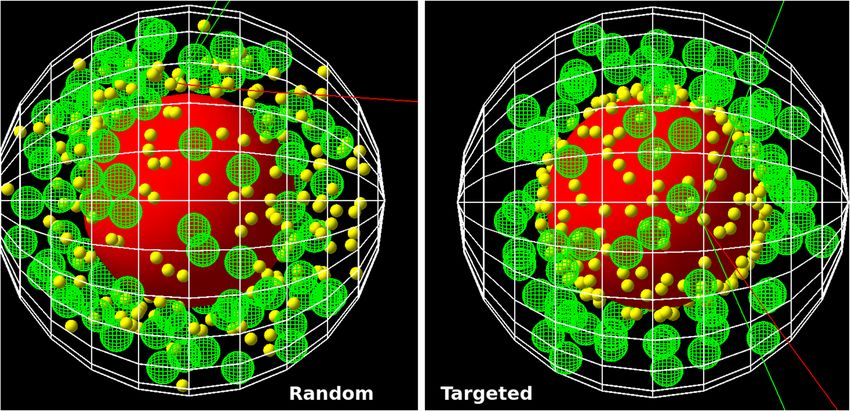
radiation interaction with cells, including the nucleus, mitochondria, the outer cell membrane, as well as ran-
domly placed nanoparticles within the cytosol (Fig. 1 left) and nanoparticles located at the surface of the nucleus
(right) or mitochondria.
s : Ge / C e l l / Type =" T s S p h e r i c a l C e l l S p h e r i c a l N P "
s : Ge / C e l l / M a t e r i a l =" C e l l M a t e r i a l "
s : Ge / C e l l / P a r e n t =" World "
d : Ge / C e l l / C e l l R a d i u s = 6 . 0 um
d : Ge / C e l l / N u c l e u s / N u c l e u s R a d i u s = 3 . 3 um
s : Ge / C e l l / N u c l e u s / M a t e r i a l =" N u c l e u s M a t e r i a l "
i : Ge / C e l l / M i t o c h o n d r i a / N u m b e r O f M i t o c h o n d r i a =90
d : Ge / C e l l / M i t o c h o n d r i a / r = 0 . 4 6 3 um
s : Ge / C e l l / M i t o c h o n d r i a / M a t e r i a l =" M i t o M a t e r i a l "
i : Ge / C e l l / N a n o p a r t i c l e / N u m b e r O f N a n o p a r t i c l e s =1000
i : Ge / C e l l / N a n o p a r t i c l e / N u m b e r O f N a n o p a r t i c l e s A t N u c l e u s =0
i : Ge / C e l l / N a n o p a r t i c l e / N u m b e r O f N a n o p a r t i c l e s A t M i t o c h o n d r i a =0
d : Ge / C e l l / Membrane / T h i c k n e s s = 1 0 . 0 nm
s : Ge / C e l l / Membrane / M a t e r i a l =" M e m b r a n e M a t e r i a l "
d : Ge / C e l l / N a n o p a r t i c l e / r = 2 nm
s : Ge / C e l l / N a n o p a r t i c l e / M a t e r i a l = " Gold "
Listing 1. Example paramter file for the usage of the TsSphericalCellSphericalNP class to include nano particles in the
cytosol during simulations with Topas.
Scientific Reports | (2021) 11:6721 | https://doi.org/10.1038/s41598-021-85964-2 2
Vol:.(1234567890)www.nature.com/scientificreports/
Figure 1. Visualization of the spherical cell model with the nucleus (red) and 90 mitochondria (green)
generated by Topas (3.5)32 and the TsSphericalCellSphericalNP model with the dimensions as given in Table 2.
For illustrative purposes two hundred AuNP (yellow) were generated randomly in the cytosol (left) or targeted
to the nucleus surface (right) and their radius was set to 200 nm. Electron trajectories are shown in red, photons
in green.
The determination of the dose and energy deposit in all these subvolume can be obtained from the simulations
by the scorers provided. The models are easily applied by defining them as standard object within a TOPAS input
parameter file. An example, showing the usage of this class is given in listing 1. A visualization of the TsSpheri-
calCellSphericalNP model is shown in Fig. 1. From a computational point of view, two different realizations of
the cell models were implemented. The first model enables the simulation of cell organelles in form of ellipsoids
(TsSphericalCellNP). Here, the drawback is the need for computational expensive overlap checking. To enable
faster simulations, a second version restricted to spherical NP and organelles was developed. The second version
(TsSphericalCellSphericalNP) provides much faster overlap checking for high number of NP and organelles by
analytical methods.
The TsSphericalCellSphericalNP model. The first model was optimized for the fast generation of spherical cell
geometries (TsSphericalCellSphericalNP) including nanoparticles. It shares some structures, ideas and naming
conventions with the cell classes provided by Topas-nBio28. Additionally, nanoparticles and an outer cell mem-
brane can be simulated explicitly. Here, a new method for the placement and overlap checking of cell organelles
and nanoparticles was implemented by the authors. It improves simulation speed and offers the inclusion of
nanoparticles which reside at the surface of the cell nucleus or mitochondria. In this model only spherical nano-
particles and spherical cell organelles can be included. The restriction to spherical geometries enables a much
quicker (analytical) check for overlaps of the randomly placed objects within the cell compared to the general
approach valid for arbitrary geometries as implemented within Topas itself. The drawback is indeed the loss of
the ability to use nanoparticles or organelles with other than spherical shapes.
The TsSphericalCellNP model. The second implementation provides a more general model which enables inclu-
sion of ellipsoids within different cell geometries. This model (TsSphericalCellNP) was extended by the authors
and is based on the TsSphericalCell class as provided by Topas-nBio28. Since the overlap checking is much faster
with the TsSphericalCellSphericalNP class, when many objects are included in the cell, all simulations presented
in the following were performed with this class.
Comparision of the continuous and discrete‑geometric AuNP models. The energy deposit in
dependence of the Au mass percentage in the cell for simulations of the continuous model and randomly distrib-
uted discrete-geometric AuNP are summarized in Table 1. The absorbed dose after 105 decays of a cell is about
32 Gy-35 Gy, which leads to an expected survival rate of less than 1%36.
For the continuous model the results show an increase of energy with increasing Au mass percentage and
density in the cytosol from 0.001 to 1% of less then 2% deposit within the whole cell and it its parts. The uncer-
tainties given are calculated from the standard deviation for n = 10 simulation runs, each with a different random
number generator seed. In the discrete-geometric model variation of the results is increased, therefore no signifi-
cant differences between the AuNP radii of 3 nm, 4 nm and 5 nm result were observed within the simulational
Scientific Reports | (2021) 11:6721 | https://doi.org/10.1038/s41598-021-85964-2 3
Vol.:(0123456789)www.nature.com/scientificreports/
Cytosol (eV/ Nucleus (eV/ Mito. (eV/ Membrane AuNP (eV/
Model (unit) rAuNP (nm) mAuNP (%) Cell (eV/dec.) dec.) dec.) dec.) (eV/dec.) dec.)
Cont. – 0.001 1810±2 1485±1 232±1 87.3±0.2 5.44±0.02 –
Cont. – 0.01 1812±2 1488±1 232±1 87.1±0.2 5.42±0.02 –
Cont. – 0.1 1814±2 1489±1 233±1 87.3±0.2 5.40±0.02 –
Cont. – 1 1822±2 1497±2 232±1 87.5±0.2 5.44±0.01 –
Disc. 3 0.0024 1906± 8 1537±8 272±4 91±2 4.85±0.05 57±1
Disc. 4 0.0057 1913±15 1544±11 273±6 92±2 4.84±0.01 66±2
Disc. 5 0.011 1911±7 1543±6 273±6 92±2 4.81±0.05 74±1
Table 1. Energy deposit per decay (dec.) in the cell organelles for continuous (Cont.) and discrete-geometric
(Disc.) models. Energy deposit in the cell and cytosol include the energy deposit in the AuNP. Energy deposit
values for mitochondria (Mito.) and AuNP are given as sum over all objects.
Figure 2. Left: Energy deposit per decay in the respective region of the cell for the continuous model with m Au
= 0.1% and the discrete-geometric model for rAuNP = 5 nm. Energy deposit in the cell and cytosol include the
energy deposit in the AuNP. The errorbars represent the standard deviation for n = 10 simulations each. They
are in the size of the line thickness and therefore not visible. Right: Relative change of the energy deposit in the
continuous model with respect to the discrete-geometric model.
uncertainties. In general, the total energy deposit in all parts, except of the membrane, is 4–15% lower when
simulations are performed for the continuous compared to the discrete-geometric model (Fig. 2 right).
Nucleus targeted nanoparticles. The relative accumulation of AuNP at the nucleus was varied between 0
and 100%. The energy deposit in the whole cell as well as in the nucleus increased linearly (both R2 > 0.99) with
AuNP accumulation at the nucleus, while the deposit in the outer cell membrane decreased somewhat (Figs. 3, 4
left). The relative energy deposit ( Erel (x)) is the energy deposit caused by x% targeted AuNP with respect to
Energy deposit without nucleus targeting AuNP (x = 0%). It is calculated by Erel (x) = 100 % · E(x)/E(0 %).
Hereby E(x) is the energy deposit in the respective volumes of organelles caused by x% of the AuNP being
present at the nucleus. E(0 %) represents the case, where 100% of the AuNP were distributed randomly within
the cytosol and 0% at the nucleus. When AuNP are exclusively located on the surface of the nucleus, the energy
deposit within the nucleus increases to 260%. For this case the overall energy deposit in the cell increases about
20%. Effects on the mitochondria are negligible (Fig. 4). These location dependent dose enhancements are inde-
pendent of the relative mass percentage of the AuNP as can be seen in Fig. 4 right. There, AuNP mass percent-
ages between 0.0024% (104 AuNP with r = 3 nm—first datapoint) up to 0.044% (4×10⁴ AuNP with r = 5 nm—last
datapoint) were simulated. Based on the results of our previous s tudy15, the resulting enhancement efficiency,
as shown in Fig. 4, can be expected to be similar for varying AuNP diameter when the same mass percentage is
considered.
Computational cost of the different modeling approaches. In general, the continuous model is
much faster (4 threads, t≈1 hour per run) than the discrete-geometric models (16 threads, t≈1-5 days per
run). Exact values depend on the interplay of many parameters such as the amount and size of NPs, number of
organelles, and the total cellular volume. These differences in simulation speed are mostly due to the reduced
Scientific Reports | (2021) 11:6721 | https://doi.org/10.1038/s41598-021-85964-2 4
Vol:.(1234567890)www.nature.com/scientificreports/
Figure 3. Energy deposit per decay in dependence of the AuNP location. For the whole CHO cell (red) and the
cytosol (light blue), here excluding the AuNP (yellow), the nucleus (dark blue) and the 90 mitochondria (green)
and the cell membrane (black). The errorbars are in the size of the symbols and therefore not visible. For details
see the text.
Figure 4. Left: Relative energy deposit (Erel (x) = 100 % · E(x)/E(0 %)) in dependence of AuNP (r = 5 nm)
located at the nucleus with respect to the case of all AuNP distributed randomly in the cytosol. The total amount
of AuNP in the cell is constant (n = 10,000). The curves represent linear fits. Right: Relative energy deposit in
the whole cell, cytosol and nucleus when all AuNP are located at the nucleus compared to randomly distributed
AuNP for different mass percentages and radii. For details see the text.
complexity in the continuous model, in terms of geometries, boundaries between different materials and time
needed to find initial decay positions. The generation of the geometries and the check for overlaps of subvolumes
is much faster when performed analytically as implemented in the TsSphericalCellSphericalNP class compared to
the TsSphericalCellNP. In both discrete-geometric models the time for the initial generation of the geometry, and
especially the random placement of the NP in the cell, increases exponentially with the number of subvolumes
included. Since the process of overlap checking runs on a single thread only it can take up a significant amount
of the whole simulation time for high numbers of NP (> 100,000). Furthermore, the implementation of volumet-
ric radioactive sources in Topas searches for random points within the the active volume (here, the cell) which
contain the active material (the AuNP). Since the total volume of the cell is much bigger than the total volume
of all AuNP, the search for decay position can take a significant amount of time when there are low numbers
of particles in the cell. However, this applies only for radioactive sources and does not concern applications of
the model to external beam therapy. We note here, that detailed statements about exact simulation speeds and
their comparison depend on a multitude of factors. Thus, all these values should be interpreted as been rough
approximations.
Scientific Reports | (2021) 11:6721 | https://doi.org/10.1038/s41598-021-85964-2 5
Vol.:(0123456789)www.nature.com/scientificreports/
Discussion
Metallic NP are introduced into cancerous tissue during radiation therapy to enhance the local dose. Hereby, it
is beneficial when the NP accumulate in the vicinity of the radiation sensitive parts of the cell, e.g. the nucleus
or mitochondria1,4. To study accumulation effects and analyze related experiments, in silico studies can pro-
vide detailed information about NP localization effects, which are otherwise inaccessible by experiments alone.
Therefore, a detailed model of the sub-cellular geometries and NP is necessary, which was introduced above.
The newly presented combined cell-nanoparticle model was implemented as a TOPAS-extension, which enables
the study of radiation-matter interaction covering all types of radiation and energies applied in cancer t herapy32.
TOPAS was chosen since it is freely available upon request, and provides an easy to use interface to the complex
Geant4-framework29,33. The extension was used in an exemplary application to the computationally demanding
task simulating radioactive 198AuNP. We note that the application of the extension to external beam-therapy is
straight forward and accessible. Here, simulation times are in the range from hours to days for a single run with
105 external photons.
Different simulations to characterize the energy deposit in cell organelles by radioactive 198AuNP were per-
formed. Firstly, discrete-geometric AuNP were randomly placed in the cytosol of CHO cells (Fig. 1 left), rep-
resenting the case where the AuNP do not target the nucleus. The comparison of these results with a simplified
continuous model revealed deviations of up to 15% in the energy deposit values in the radiation sensitive nucleus
(Fig. 2). This outcome can be rationalized when considering the local dose enhancement, which is only present
in the discrete-geometric model. It is strongest within a range up to some ten nanometers around the AuNP.
For example, within the first 25 nm around the source, the energy deposit density per decay is approximately
twice as high for an AuNP with r = 5 nm, when compared to a point source. This enhancement of the local dose
decreases quickly and vanishes at about 175 nm, as calculated in a previous s tudy15. Thus, the AuNP have to
reside as close as possible (preferentially nearer than approximately 100 nm), to the biologically relevant target
(e.g. the cell nucleus or mitochondria)37. Further away, at distances of about 1 µm the energy deposit is only
slightly (< 2%) increased by their presence. Furthermore, the average energy deposit in the cell increased linear
with AuNP presence at the surface of the nucleus (Fig. 3). This can be understood by the longer electron and
photon tracks throughout the cell. The energy deposit in the outer cell membrane decreases due to their on
average higher distance to the nanoparticles and decay positions. When AuNP are exclusively located on the
surface of the nucleus the energy deposit can reach up to 260% compared to the case of randomly distributed
AuNP. Additionally, the overall dose in the cell increases about 20% (Fig. 4). When only 20% of the AuNP are
located at the nucleus an energy deposit enhancement can still be observed. This is owed to the strong energy
deposit enhancement around the AuNP, where the decay takes place. For example, from 50 nm to 150 nm
distance to the AuNP surface, the energy deposit density decreases over t enfold15. Thus, the most important
parameters for increasing the local energy deposit in the nucleus is the location of the AuNP within the cell. In
case of radioactive AuNP, their specific activity, which is determined by the activation protocol used, has to be
taken into account as well. It has to be noted, that the location dependent enhancement of the energy deposit
per decay, does not depend on the Au mass percentage, as it was simulated for AuNP mass percentages up to
0.044% (Fig. 4 right). This shows, that at these AuNP concentrations most of the inelastic scattering events in Au
happen within the AuNP where the initial decay event took place. The combination of these parameters offers
the possibility to adjust and optimize the AuNP behavior for cellular uptake or metabolic b ehavior15,38. Especially
selective permeability into the cell depending on AuNP size, their structure and functionalisation are of interest
to specifically target t umors39. Furthermore, to predicts cell death or mutation, the rate of DNA double-strand
breaks (DSB) has to be estimated in dependence of the local energy deposit in the nucleus. Recent theoretical
studies estimated a threshold value for DSB induction as EtDSB = (76 − 103.54) eV 40. A combined experimen-
tal and simulational study showed that an energy deposit of about E1/2 DSB = 72 eV in the target volume of the
respective sugar-phosphate backbone of DNA leads with a 50% probability to a DSB41. This work accounted for
direct radiation effects by ionization of DNA and the indirect effects mediated by ROS produced from water
radiolysis. Based on this data a microscopic target-model can be implemented to estimate DSB induction, and
related biological response of the cell afterwards. The flexibility of the cell model presented here, allows for an easy
extension in future work, and provides a powerful tool to benchmark theoretical models against experimental
datasets from different cell lines. We note here, that the “biological response” of the cells, in terms of radiation
sensitivity and repair efficiency depends on a multitude of factors, which have to be considered for each cell line
separately. Another important point is the possible toxicity of AuNP. Gold nanoparticles have been widely used
in current medical and biological research, much experimental work has been done which confirms the non-
toxicity of AuNPs, and are considered comparatively safe42. Especially when compared to other treatment agents,
as used in chemotherapy. On the other hand, they are not completely bio-inert and bio-compatible; since toxicity
is directly related to various factors such as size, shape, and surface chemistry42. Toxicity has been observed at
high concentrations greater than 10 µg/mL42. However, concerning AuNP with diameters comparable to the
present study, Alkilany et al.43 demonstrated that for d = 1.4 nm (up to 0.4 µM, 72h) to 18 nm (0.001-0.25 µM,
72h) spherical gold nanoparticles were non-toxic from in-vitro study. Besides, spherical gold nanoparticles with
1.9 nm diameter were found to be non toxic, when administered to mice for a tumor t herapy23. Furthermore,
radioactive gold colloids have already been effectively used in local radioisotope cancer therapy in h umans44. A
detailed discussion of all these factors is beyond the focus of this study.
In future studies, where the energy deposit with nanometer accuracy is of interest, special care has to be taken
to choose appropriate scattering models45. When track-structure code, where every scattering event is explicitly
simulated, is available for the materials under investigation, its usage is recommended. As an alternative, when
only condensed history codes are available, it has to be taken care that simulation parameters, such as production
and range cuts, are sufficiently low to minimize the associated u ncertainties46. For example, the path length of
Scientific Reports | (2021) 11:6721 | https://doi.org/10.1038/s41598-021-85964-2 6
Vol:.(1234567890)www.nature.com/scientificreports/
Type r H C N O S P Au
Unit nm % % % % % % %
Cytosol 6000 10.25 12.25 4.25 73.25-72.25 0.00 0.00 0-1
Nucleus 3300 10.60 9.00 3.20 74.20 0.40 2.60 0
Mitochondria 463 10.60 9.00 3.20 74.20 0.40 2.60 0
Membrane 10 10.25 12.25 4.25 73.25 0.00 0.00 0
Table 2. Geometrical parameters of CHO cells. The dimensions of cytosol, nucleus and mitochondria are
given as radius. The membrane thickness is listed as diameter59. The chemical composition of the different
parts of the cell is given in mass percent and density of 1.0 g/cm360 Ninety mitochondria were simulated57.
secondary electrons with energies below 100 eV can vary up to 10 nm in dependence of the medium, scattering
cross sections and scattering models a pplied47. This has to be considered when more detailed statements about
energy deposit distribution within chromosomes or even smaller structures are m ade46. However, in this study, we
focus on the nucleus which has a diameter of above 6 µm (Table 2), thus, the resulting differences are negligible
for the analysis preformed here. For the AuNP, which are indeed much smaller, simulations should preferentially
be performed with track-structure code. Therefore the simulations of AuNP will benefit greatly from the new
scattering models for gold implemented by Sakata et al. when they become available in Geant4 and T opas48–50.
Summary and outlook
In this work we have presented combine cell-nanoparticle models for Topas/Geant4 to simulate the local dose
enhancement effects caused by the presence of nanoparticles in the cytoplasm, as well as on the nucleus surface
of cells. The models were applied to determine the energy deposit caused by the presence of radioactive AuNP
nanoparticles in CHO cells. Simulations were performed for continuous and a discrete-geometric AuNP models.
These discrete-geometric nanoparticle models enable the simulation of non-homogeneous distribution of AuNP
within the cell. The energy deposit in the cytosol, mitochondria and nucleus were determined in dependence
on AuNP locations. Future work will extend the presented cell models to predict DNA strand-break induction
within the cell. This extension will be based on a recently developed DNA damage mode, which accounts for
direct radiation effects by ionization of DNA and the indirect effects mediated by reactive-oxygen species pro-
duced from water radiolysis. Another possible extension towards more realistic cellular models, is the inclusion
of clustering behavior of mitochondria within the cytosol. In conclusion, it was shown that the type of simula-
tion model and AuNP location within the the cell strongly influences the energy deposit in different organelles.
Thus, AuNP which target the nucleus or mitochondria of cancerous tissue have the potential to greatly enhance
damage in tumours while decreasing side effects on healthy tissue.
Methods
Particle scattering simulations. To obtain the energy deposit in different cell organelles Monte Carlo
simulations (MCS) based on the Geant4 MCS framework (10.06)29–31 in combination with the Topas (3.5)32
interface and the Topas-nBio28 extensions were performed. The radioactive decay of 198Au, the production of sec-
ondary particles, and their interaction with the surrounding matter was simulated. In each simulation 105 decays
of the AuNP (19.32 g/cm3) were simulated by the g4radioactivedecay and g4decay modules. All other scattering
processes were simulated with the processes provide by the g4emstandard_opt4 physics lists and a range cut for
all particles of 2 nm was applied. This physics list provides the most accurate standard and low-energy models
for electron scattering, with a decreased range factor (0.08) to improve the accuracy and the atomic de-excitation
modules were set to ignore the c uts51. The standard models for atomic de-excitation, Auger electron emission,
Auger cascade and fluorescence were enabled. During the simulations the position of the radioactive decay was
chosen randomly within the active material. It has to be taken into account, that the scattering-models applied
in the AuNP region have a recommended low energy limit of 100 eV. This corresponds to a range of secondary
electrons of about 100 nm52,53, which puts some constrains on the accuracy of simulations of nanometer sized
structures. When possible, simulations should preferentially be performed with track-structure code which ena-
bles step-by-step simulations of the scattering interactions, and results in a higher accuracy compared to con-
densed-history code50,54. Thus, this situation will improve in the future when the newly implemented scattering
models for gold as implemented by Sakata et al. become available in future releases of Geant4 and T
opas48–50. As a
model system for testing, CHO cells were selected, since they are readily available and well established organism
used in pharmaceutical production and radiation research36,55,56. Here we note that the amount of mitochondria
within the CHO cells reported varies between approximately 60–200, in dependence on the counting methods
applied57,58. Their geometrical parameters and chemical composition used throughout the simulations are sum-
marized in Table 2.
Scientific Reports | (2021) 11:6721 | https://doi.org/10.1038/s41598-021-85964-2 7
Vol.:(0123456789)www.nature.com/scientificreports/
The discrete‑geometric AuNP models. For the discrete-geometric models (We note here, that the term
discrete refers to the explicit simulation of the AuNP within the medium, and is not to be confused with “discrete
transport” in particle-scattering Monte-Carlo simulations) the simulations were performed with 104 NP per cell
with radii of 3 nm, 4 nm and 5 nm. These values correspond to gold mass percentages in the cell of 2.4 · 10−3 %,
5.7 · 10−3 % and 1.1 · 10−2 %, respectively. The AuNP located in the cytosol are randomly distributed. The AuNP
attached to the nucleus or the mitochondria are placed in direct contact with the respective surface, whereby the
angles describing the location on the surface are chosen randomly. When simulations of mitochondria targeted
NP are performed, which is not the case in the examples presented above, the respective mitochondria are cho-
sen for each AuNP independently and randomly with equal probability. To assess the effect of nucleus targeted
AuNP, we simulated a constant amount AuNP of whom a certain amount was distributed randomly in the
cytosol or at the surface of the nucleus. Hereby the amount at both locations were varied between 0 and 100%.
The continuous Au model. For comparison, the simulations for continuous distribution of Au within the
cytosol were performed with the TsSphericalCellSphericalNP class, which was used without inclusion of discrete-
geometric AuNP. Thereby the chemical compositions tabulated in Table 2 were used for the different parts of
the cell. The gold was added to the cytosol. Gold mass percentages of 0.001%, 0.01%, 0.1%, 1.0% were simulated.
Each simulation was repeated ten times with different seeds for the random number generator to allow for the
calculation of the sample standard deviation, as reported in the following. Here, the position of each radioactive
decay was chosen randomly within the cytosol.
Properties of radioactive gold nanoparticles. The properties of radioactive AuNP, especially the influ-
ence of diameter and clustering behavior on the energy deposits characteristics were described in detail in our
previous work15. Briefly, 198Au has an isotope mass of 198 u and a half life of 2.7 days. With a specific activity of
9.03×10¹⁵ Bq/g, it performs a β − decay with particle energies of 961 keV (99%) and 285 keV(1%) respectively,
as well as γ emissions with energies of 412 keV (96%), 676 keV (www.nature.com/scientificreports/
17. McMahon, S. J. et al. Biological consequences of nanoscale energy deposition near irradiated heavy atom nanoparticles. Sci. Rep.
1, 1–10. https://doi.org/10.1038/srep00018 (2011).
18. Schürmann, R., Vogel, S., Ebel, K. & Bald, I. The physico-chemical basis of DNA radiosensitization: implications for cancer radia-
tion therapy. Chem. A Eur. J. 24, 10271–10279. https://doi.org/10.1002/chem.201800804 (2018).
19. Hahn, M. B. et al. DNA protection by ectoine from ionizing radiation: molecular mechanisms. Phys. Chem. Chem. Phys. 19,
25717–25722. https://doi.org/10.1039/C7CP02860A (2017).
20. Hahn, M. B. et al. Direct electron irradiation of DNA in a fully aqueous environment. Damage determination in combination with
Monte Carlo simulations. Phys. Chem. Chem. Phys. 19, 1798–1805. https://doi.org/10.1039/C6CP07707B (2017).
21. Tran, H. N. et al. Geant4 Monte Carlo simulation of absorbed dose and radiolysis yields enhancement from a gold nanoparticle
under MeV proton irradiation. Nucl. Instrum. Methods Phys. Res. Sect. B Beam Interact. Mater. Atoms 373, 126–139. https://doi.
org/10.1016/j.nimb.2016.01.017 (2016).
22. Hahn, M. B., Smales, G. J., Seitz, H., Solomun, T. & Sturm, H. Ectoine interaction with DNA: influence on ultraviolet radiation
damage. Phys. Chem. Chem. Phys. 22, 6984–6992. https://doi.org/10.1039/D0CP00092B (2020).
23. Hainfeld, J. F., Slatkin, D. N. & Smilowitz, H. M. The use of gold nanoparticles to enhance radiotherapy in mice. Phys. Med. Biol.
49, N309. https://doi.org/10.1088/0031-9155/49/18/N03 (2004).
24. Shi, J., Xiao, Z., Kamaly, N. & Farokhzad, O. C. Self-assembled targeted nanoparticles: evolution of technologies and bench to
bedside translation. Acc. Chem. Res. 44, 1123–1134. https://doi.org/10.1021/ar200054n (2011).
25. Sykes, E. A., Chen, J., Zheng, G. & Chan, W. C. Investigating the impact of nanoparticle size on active and passive tumor targeting
efficiency. ACS Nano 8, 5696–5706 (2014).
26. Chithrani, B. D., Ghazani, A. A. & Chan, W. C. W. Determining the size and shape dependence of gold nanoparticle uptake into
mammalian cells. Nano Lett. 6, 662–668. https://doi.org/10.1021/nl052396o (2006).
27. Özçelik, S. & Pratx, G. Nuclear-targeted gold nanoparticles enhance cancer cell radiosensitization. Nanotechnology 31, 415102.
https://doi.org/10.1088/1361-6528/aba02b (2020).
28. Schuemann, J. et al. TOPAS-nBio: an extension to the TOPAS simulation toolkit for cellular and sub-cellular radiobiology. Radiat.
Res. 191, 125–138. https://doi.org/10.1667/RR15226.1 (2019).
29. Agostinelli, S. et al. Geant4: a simulation toolkit. Nucl. Instrum. Methods Phys. Res. Sect. A Accel. Spectrom. Detect. Assoc. Equip.
506, 250–303. https://doi.org/10.1016/S0168-9002(03)01368-8 (2003).
30. Bernal, M. A. et al. Track structure modeling in liquid water: a review of the Geant4-DNA very low energy extension of the Geant4
Monte Carlo simulation toolkit. Phys. Med. 31, 861–874. https://doi.org/10.1016/j.ejmp.2015.10.087 (2015).
31. Incerti, S. et al. Comparison of GEANT4 very low energy cross section models with experimental data in water. Med. Phys. 37,
4692–4708. https://doi.org/10.1118/1.3476457 (2010).
32. Perl, J., Shin, J., Schumann, J., Faddegon, B. & Paganetti, H. TOPAS: an innovative proton Monte Carlo platform for research and
clinical applications. Med. Phys. 39, 6818–6837. https://doi.org/10.1118/1.4758060 (2012).
33. Allison, J. et al. Recent developments in Geant4. Nucl. Instrum. Methods Phys. Res. Sect. A Accel. Spectrom. Detect. Assoc. Equip.
835, 186–225. https://doi.org/10.1016/j.nima.2016.06.125 (2016).
34. Hahn, M. B. TOPAS Cell Model with Nanoparticles (BAM Eigenverlag, Berlin, 2020). https://doi.org/10.26272/opus4-51150 .
35. Hahn, M. B. https://github.com/BAMresearch/TOPAS-CellModels. Bundesanstalt für Materialforschung und -prüfung (2020).
36. Matsuya, Y. et al. Investigation of dose-rate effects and cell-cycle distribution under protracted exposure to ionizing radiation for
various dose-rates. Sci. Rep.https://doi.org/10.1038/s41598-018-26556-5 (2018).
37. Kirkby, C. & Ghasroddashti, E. Targeting mitochondria in cancer cells using gold nanoparticle-enhanced radiotherapy: a Monte
Carlo study. Med. Phys. 42, 1119–1128. https://doi.org/10.1118/1.4906192 (2015).
38. Chithrani, D. B. et al. Gold nanoparticles as radiation sensitizers in cancer therapy. Radiat. Res. 173, 719–728. https://doi.org/10.
1667/RR1984.1 (2010).
39. Mi, Y., Shao, Z., Vang, J., Kaidar-Person, O. & Wang, A. Z. Application of nanotechnology to cancer radiotherapy. Cancer Nano-
technol. 7, 11. https://doi.org/10.1186/s12645-016-0024-7 (2016).
40. Margis, S. et al. Microdosimetric calculations of the direct DNA damage induced by low energy electrons using the Geant4-DNA
Monte Carlo code. Phys. Med. Biol. 65, 045007. https://doi.org/10.1088/1361-6560/ab6b47 (2020).
41. Hahn, M. B., Meyer, S., Kunte, H.-J., Solomun, T. & Sturm, H. Measurements and simulations of microscopic damage to DNA in
water by 30 keV electrons: a general approach applicable to other radiation sources and biological targets. Phys. Rev. E 95, 052419.
https://doi.org/10.1103/PhysRevE.95.052419 (2017).
42. Lewinski, N., Colvin, V. & Drezek, R. Cytotoxicity of nanoparticles. Small 4, 26–49. https://d oi.o
rg/1 0.1 002/s mll.2 00700 595 (2008).
43. Alkilany, A. M. & Murphy, C. J. Toxicity and cellular uptake of gold nanoparticles: what we have learned so far?. J. Nanopart. Res.
12, 2313–2333. https://doi.org/10.1007/s11051-010-9911-8 (2010).
44. Metz, O., Stoll, W. & Plenert, W. Meningosis prophylaxis with intrathecal 198Au-colloid and methotrexate in childhood acute
lymphocytic leukemia. Cancer 49, 224–228. https://doi.org/10.1002/1097-0142(19820115)49:23.
0.CO;2-O (1982).
45. Nikjoo, D. H., Emfietzoglou, D. & Charlton, D. E. The Auger effect in physical and biological research. Int. J. Radiat. Biol. 84,
1011–1026. https://doi.org/10.1080/09553000802460172 (2008).
46. Lazarakis, P. et al. Investigation of track structure and condensed history physics models for applications in radiation dosimetry
on a micro and nano scale in Geant4. Biomed. Phys. Eng. Express 4, 024001. https://doi.org/10.1088/2057-1976/aaa6aa (2018).
47. Emfietzoglou, D., Papamichael, G. & Nikjoo, H. Monte Carlo electron track structure calculations in liquid water using a new
model dielectric response function. Radiat. Res. 188, 355–368. https://doi.org/10.1667/RR14705.1 (2017).
48. Sakata, D. et al. An implementation of discrete electron transport models for gold in the Geant4 simulation toolkit. J. Appl. Phys.
120, 244901. https://doi.org/10.1063/1.4972191 (2016).
49. Sakata, D. et al. Geant4-DNA track-structure simulations for gold nanoparticles: the importance of electron discrete models in
nanometer volumes. Med. Phys. 45, 2230–2242. https://doi.org/10.1002/mp.12827 (2018).
50. Sakata, D. et al. Electron track structure simulations in a gold nanoparticle using Geant4-DNA. Phys. Med. 63, 98–104. https://
doi.org/10.1016/j.ejmp.2019.05.023 (2019).
51. Basaglia, T. et al. Investigation of Geant4 simulation of electron backscattering. IEEE Trans. Nucl. Sci. 62, 1805–1812. https://doi.
org/10.1109/TNS.2015.2442292 (2015).
52. Byrne, H., McNamara, A. & Kuncic, Z. Impact of nanoparticle clustering on dose radio-enhancement. Radiat. Prot. Dosim.https://
doi.org/10.1093/rpd/ncy218 (2018).
53. Francis, Z., Incerti, S., Karamitros, M., Tran, H. N. & Villagrasa, C. Stopping power and ranges of electrons, protons and alpha
particles in liquid water using the Geant4-DNA package. Nucl. Instrum. Methods Phys. Res. Sect. B Beam Interact. Mater. Atoms
269, 2307–2311. https://doi.org/10.1016/j.nimb.2011.02.031 (2011).
54. Kyriakou, I. et al. Influence of track structure and condensed history physics models of Geant4 to nanoscale electron transport in
liquid water. Phys. Med. 58, 149–154. https://doi.org/10.1016/j.ejmp.2019.01.001 (2019).
55. Dahm-Daphi, C., Sass, W. & Alberti, J. Comparison of biological effects of DNA damage induced by ionizing radiation and hydro-
gen peroxide in CHO cells. Int. J. Radiat. Biol. 76, 67–75. https://doi.org/10.1080/095530000139023 (2000).
56. Xu, X. et al. The genomic sequence of the Chinese hamster ovary (CHO)-K1 cell line. Nat. Biotechnol. 29, 735–741. https://doi.
org/10.1038/nbt.1932 (2011).
Scientific Reports | (2021) 11:6721 | https://doi.org/10.1038/s41598-021-85964-2 9
Vol.:(0123456789)www.nature.com/scientificreports/
57. Ross, D. & Mel, H. Growth dynamics of mitochondria in synchronized Chinese hamster cells: ScienceDirect. Biophys. J. 12,
1562–1572 (1972).
58. Peng, J.-Y. et al. Automatic morphological subtyping reveals new roles of caspases in mitochondrial dynamics. PLOS Comput. Biol.
7, e1002212. https://doi.org/10.1371/journal.pcbi.1002212 (2011).
59. Salimi, E., Braasch, K., Butler, M., Thomson, D. J. & Bridges, G. E. Dielectric model for Chinese hamster ovary cells obtained by
dielectrophoresis cytometry. Biomicrofluidics 10, 014111. https://doi.org/10.1063/1.4940432 (2016).
60. White, D. R., Booz, J., Griffith, R. V., Spokas, J. J. & Wilson, I. J. Report 44. J. Int. Comm. Radiat. Units Meas. os23, NP–NP. https://
doi.org/10.1093/jicru/os23.1.Report44 (1989).
61. Delacroix, D., Guerre, P. J., Leblanc, P. & Hickman, C. Radionuclide and radiation protection data handbook 2002. Radiat. Protect.
Dosim. 98, 1–168. https://doi.org/10.1093/oxfordjournals.rpd.a006705 (2002).
62. Plante, I. & Cucinotta, F. A. Cross sections for the interactions of 1 eV–100 MeV electrons in liquid water and application to Monte-
Carlo simulation of HZE radiation tracks. New J. Phys. 11, 063047. https://doi.org/10.1088/1367-2630/11/6/063047 (2009).
63. Kimling, J. et al. Turkevich method for gold nanoparticle synthesis revisited. J. Phys. Chem. B 110, 15700–15707. https://doi.org/
10.1021/jp061667w (2006).
64. Zutta Villate, J. M., Rojas, J. V., Hahn, M. B. & Puerta, J. A. Synthesis and optimization of radioactive gold nanoparticles for cancer
therapy. J. Radioanal. Nucl. Chem. Manuscript in preparation (2021).
Acknowledgements
Discussions with J.A. Puerta are gratefully acknowledged. MBH acknowledges funding by the Deutsche
Forschungsgemeinschaft (DFG, German Research Foundation)—Project Number 442240902/HA 8528/2-1.
Author contributions
J.M.Z.V. provided the fundamental idea to study radioactive AuNP in cells. M.B.H. implemented the algorithms
and extended cell model in C++ and performed the simulations. Data evaluation and writing of the manuscript
was done by both authors.
Funding
Open Access funding enabled and organized by Projekt DEAL.
Competing interests
The authors declare no competing interests.
Additional information
Correspondence and requests for materials should be addressed to M.B.H.
Reprints and permissions information is available at www.nature.com/reprints.
Publisher’s note Springer Nature remains neutral with regard to jurisdictional claims in published maps and
institutional affiliations.
Open Access This article is licensed under a Creative Commons Attribution 4.0 International
License, which permits use, sharing, adaptation, distribution and reproduction in any medium or
format, as long as you give appropriate credit to the original author(s) and the source, provide a link to the
Creative Commons licence, and indicate if changes were made. The images or other third party material in this
article are included in the article’s Creative Commons licence, unless indicated otherwise in a credit line to the
material. If material is not included in the article’s Creative Commons licence and your intended use is not
permitted by statutory regulation or exceeds the permitted use, you will need to obtain permission directly from
the copyright holder. To view a copy of this licence, visit http://creativecommons.org/licenses/by/4.0/.
© The Author(s) 2021
Scientific Reports | (2021) 11:6721 | https://doi.org/10.1038/s41598-021-85964-2 10
Vol:.(1234567890)You can also read