Amentoflavone triggers cell cycle G2/M arrest by interfering with microtubule dynamics and inducing DNA damage in SKOV3 cells
←
→
Page content transcription
If your browser does not render page correctly, please read the page content below
ONCOLOGY LETTERS 20: 168, 2020

Amentoflavone triggers cell cycle G2/M arrest by interfering with
microtubule dynamics and inducing DNA damage in SKOV3 cells
                  JINLI ZHANG1, AIGUO LI1, HANJING SUN2, XIFENG XIONG1, SHENGNAN QIN1,
                  PENGZHEN WANG1, LIBING DAI1, ZHI ZHANG3, XIAOJIAN LI3 and ZHIHE LIU1

  1
   Guangzhou Institute of Traumatic Surgery; Departments of 2Traditional Chinese Medicine and 3Burn and Plastic Surgery,
              Guangzhou Red Cross Hospital, Jinan University, Guangzhou, Guangdong 510220, P.R. China

                                        Received August 12, 2019; Accepted July 14, 2020

                                                    DOI: 10.3892/ol.2020.12031

Abstract. Ovarian cancer is the seventh most common cancer           the present study suggest that AMF is a potential therapeutic
and the second most common cause of cancer‑associated                agent for the treatment of ovarian cancer. In addition, the
mortality among gynecological malignancies worldwide.                effects of AMF on cell cycle arrest and DNA damage induc‑
The combination of antimitotic agents, such as taxanes, and          tion may be the molecular mechanisms by which AMF might
the DNA‑damaging agents, such as platinum compounds, is              exert its potential therapeutic benefits in ovarian cancer.
the standard treatment for ovarian cancer. However, due to
chemoresistance, development of novel therapeutic strate‑            Introduction
gies for the treatment of ovarian cancer remains critical.
Amentoflavone (AMF) is a biflavonoid derived from the                According to statistics, the incidence rate of ovarian cancer
extracts of Selaginella tamariscina, which has been used             in 2018 was 3.4%, worldwide (1). Ovarian cancer is the eighth
as a Chinese herb for thousands of years. A previous study           most common cancer in female and the second most common
demonstrated that AMF inhibits angiogenesis of endothelial           cause of cancer‑associated mortality among gynecological
cells and induces apoptosis in hypertrophic scar fibroblasts. In     malignancies worldwide (1). A combination of antimitotic
order to check the influence of AMF on cell proliferation, the       agents, such as taxanes, and DNA‑damaging agents, such
effects of AMF on cell cycle and DNA damage were measured            as platinum compounds remains the principle treatment
by cell viability, flow cytometry, immunofluorescence and            for ovarian cancer (2), whereby 60‑85% of patients with
western blotting assays in SKOV3 cells, an ovarian cell line. In     high‑grade ovarian cancer initially respond to this regimen;
the present study, treatment with AMF inhibited ovarian cell         however, the majority of these patients eventually relapse
proliferation, increased P21 expression, decreased CDK1/2            due to chemoresistance (3,4). Furthermore, most patients
expression, interrupted the balance of microtubule dynamics          with high‑grade ovarian cancer are resistant to paclitaxel and
and arrested cells at the G2 phase. Furthermore, treatment           associated microtubule inhibitors (3,4). Thus, development of
with AMF increased the expression levels of phospho‑Histone          novel therapeutic strategies for the treatment of ovarian cancer
H2AX (γ‑H2AX; a variant of histone 2A, that belongs to the           remains critical.
histone 2A family member X) and the DNA repair protein                   Several anticancer drugs exert their effects through the
RAD51 homolog 1 (Rad51), indicating the occurrence of DNA            cell cycle. For example, methotrexate, vinca alkaloids and
damage since γ‑H2AX and Rad51 are both key markers of                bleomycin play function by arresting cells in S phase or G2/M
DNA damage. Consistent with previous findings, the results of        phase. The cell cycle is a complex multi‑step process that is
                                                                     regulated by different mechanisms, including cyclin‑depen‑
                                                                     dent kinase (CDK) pathways, metabolic adaptations and
                                                                     redox‑dependent signaling. CDK complexes play key regula‑
Correspondence to: Dr Zhihe Liu, Guangzhou Institute of              tory roles in cell cycle progression (5). In CDK‑dependent
Traumatic Surgery, Guangzhou Red Cross Hospital, Jinan University,   pathways, the catalytic activities of CDKs are modulated by the
396 Tongfu Zhong, Guangzhou, Guangdong 510220, P.R. China            interactions between cyclins and CDK inhibitors (CKIs) (6).
E‑mail: zliu0731@163.com                                             In this progression, cyclins and CKIs serve as brakes to halt
Dr Xiaojian Li, Department of Burn and Plastic Surgery, Guangzhou    cell cycle progression under unfavorable conditions, such
Red Cross Hospital, Jinan University, 396 Tongfu Zhong, Guangzhou,   as when DNA damage is present (7). P21, a member of the
Guangdong 510220, P.R. China                                         cyclin‑dependent kinase inhibition protein/kinase inhibition
E‑mail: lixj64@163.com                                               protein family of CKIs, is activated following DNA damage
                                                                     and metabolic stress, which arrests cell cycle progression in
Key words: amentoflavone, cell cycle, DNA damage, microtubule        the G1/S and G2/M phases by inhibiting Cyclin D/CDK4 and
dynamics                                                             CDK6, and Cyclin E/CDK2 activities, respectively (8).
                                                                         In addition to cyclin‑CDK complexes, several other cell
                                                                     cycle‑associated targets exist for antitumor therapies. For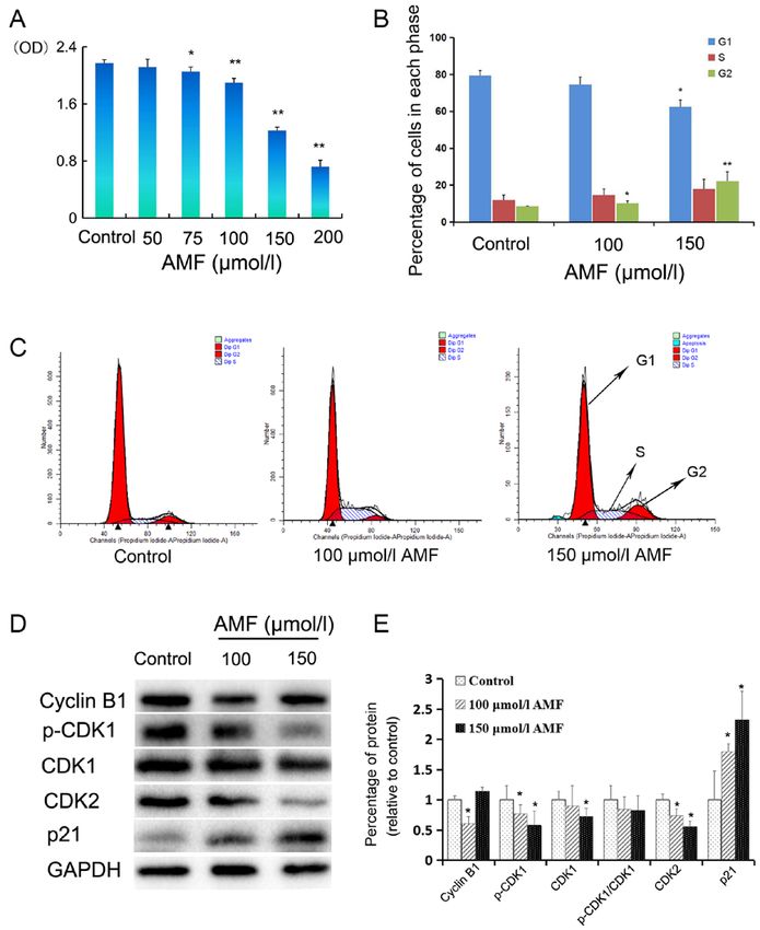
2                        ZHANG et al: AMF INTERRUPTS MICROTUBULE DYNAMICS AND DNA STABILITY

example, taxanes and colchicine can also induce cell cycle         Cell viability assay. SKOV3 cells were seeded in 96‑well
arrest by influencing microtubule (MT) stability (9,10). MTs       plates at a density of 5,000 cells/well (100 µl). After 24 h,
are hollow cylindrical tubes consisting of 13 aligned proto‑       cells were treated with different concentrations of AMF
filaments, formed from repeating α‑tubulin and β ‑tubulin          (0, 50, 75, 100, 150 and 200 µmol/l) for 48 h at 37˚C. Cell
heterodimers (11). MTs undergo polymerization and                  viability was determined via the CellTiter 96 Aqueous One
de‑polymerization, while the dynamic balance between them          Solution Proliferation assay (Promega Corporation) at a
plays a central role in cell meiosis. Disruption of this balance   wavelength of 490 nm, using a multi‑well spectrophotometer
caused by factors, such as low temperature and drugs halts         (Agilent Technologies, Inc.). All experiments were performed
meiosis. Taxanes are MT regulators that block cell meiosis in      in triplicate.
G2/M by binding to tubulin, thus promoting MT polymeriza‑
tion and eventually inducing apoptosis (12). In addition to        Flow cytometric analysis. A total of 1x10⁵ SKOV3 cells/well
directly affecting tubulin, MT regulators can also influence the   were seeded in 6‑well overnight and treated with different
expression of MT‑associated proteins. For example, stathmin        concentrations of AMF (0, 100 and 150 µmol/l) for 48 h at 37˚C.
is a MT de‑polymerizing protein that regulates MT dynamics         Cells were fixed in 70% ethanol overnight at 4˚C, permeabi‑
and spindle assembly through binding to α/β‑tubulin heterodi‑      lized with 0.1% Triton X‑100 (Sigma‑Aldrich; Merck KGaA),
mers (13). The high expression level of stathmin decreased         digested with RNaseA (Thermo Fisher Scientific, Inc.) and
the sensitivity of ovarian cancer to paclitaxel (14). However,     subsequently stained with propidium iodide (BD Biosciences)
taxanes and anti‑stathmin therapy produced a synergistic anti‑     in the dark for 30 min at 37˚C, prior to cell cycle analysis using
cancer effect, and stathmin knockdown, by transfecting the         a FACS Calibur flow cytometer (BD Biosciences) and analyzed
expression construct containing full‑length stathmin cDNA in       using ModFit LT Windows 3.2 (Verity Software House, Inc.).
the antisense orientation, increased taxanes sensitivity (15). A
previous study has demonstrated that p53 induces cell arrest       Immunofluorescence. A total of 1x10⁴ SKOV3 cells/well were
at the G2/M checkpoint by downregulating stathmin, while its       seeded onto coverslips in a six‑well plate. Following incubation
expression is activated following DNA damage (16).                 for 24 h at 37˚C, cells were treated with different concentra‑
    Plant‑derived flavones, such as morelloflavone and             tions of AMF (0, 100 and 150 µmol/l) for 48 h at 37˚C.
ginkgo, have also been reported to play an important role in       Subsequently, cells were fixed with 4% paraformaldehyde for
preventing cancer progression including prostate and lung          15 min at room temperature, permeabilized with 0.25% Triton
cancer cells (17,18). Amentoflavone (AMF) is a biflavonoid         X‑100 for 10 min and blocked with 1% BSA for 30 min at
extracted from the Chinese herb Selaginella tamariscina,           room temperature. Cells were incubated with primary anti‑
which displays several pharmacological properties, including       bodies against phospho‑Histone H2AX (γ‑H2AX; 1:200 v/v;
antitumor, anti‑inflammatory and antiviral effects (19‑22). A      cat. no. 9718), α‑tubulin (1:200 v/v; cat. no. 2144) or β‑tubulin
previous study demonstrated that AMF inhibits angiogenesis         (1:200 v/v; cat. no. 2146), all from Cell Signaling Technology
of endothelial cells and induces apoptosis in hypertrophic         Inc., overnight at 4˚C. Subsequently, cells were incubated
scar fibroblasts (23). Although it has been demonstrated that      with Alexa Fluor 488‑labeled goat anti‑rabbit secondary
AMF inhibits the development of different types of cancer, its     antibody (1:500 v/v; cat. no. 4416; Cell Signaling Technology,
underlying molecular mechanisms in ovarian cancer remain           Inc.) for 1 h at room temperature. Nuclei were stained with
unclear.                                                           0.1 µg/ml DAPI (Santa Cruz Biotechnology Inc.) for 5 min at
    Thus, the present study aimed to investigate the effect        room temperature. Cell images were observed under a Nikon
of AMF on ovarian cancer progression and the underlying            Eclipse E600 fluorescence microscope (magnification, x400;
mechanisms involved in the observed effects. The results           Nikon Corporation) and analyzed using NIS‑Elements D 4.50
demonstrated that AMF decreased ovarian cancer cell viability      software (Nikon Corporation).
and induced cell cycle arrest, by disrupting the balance of MT
dynamics and increasing the levels of DNA damage. Taken            Western blotting. A total of 1x10⁵ SKOV3 cells/well were
together, the results of the present study suggest that AMF may    seeded into 100‑mm cell culture dishes and treated with
act as a therapeutic agent in the treatment of ovarian cancer.     different concentrations of AMF (0, 100 and 150 µmol/l) for
                                                                   48 h at 37˚C. Total protein was extracted using RIPA lysis
Materials and methods                                              buffer (150 mM NaCl, 50 mM Tris with pH 7.4, 1% NP40,
                                                                   0.1% SDS and 0.5% sodium deoxycholate; Beyotime Institute
Cell culture, cell line and reagents. The SKOV3 human              of Biotechnology) supplemented with 10 mM phenylmeth‑
ovarian cancer cell line was purchased from the Cell Bank of       anesulphonyl fluoride (Amresco, Inc.) and 10X phosphatase
Type Culture Collection of the Chinese Academy of Sciences.        inhibitor (Roche Applied Science). Total protein concentration
Cells were maintained in DMEM supplemented with 10%                was determined using the bicinchoninic acid protein assay kit
FBS, 2 mM glutamine (all from Thermo Fisher Scientific,            (Thermo Fisher Scientific, Inc.), 20 µg protein samples per lane
Inc.), 100 units of penicillin/ml and 100 µg of streptomycin/ml    were loaded and separated via 10% SDS‑PAGE and electrob‑
(both from Corning Life Sciences) at 37˚C in a humidified          lotted. The separated proteins were subsequently transferred
atmosphere of 5% CO2 and subcultured every 2‑3 days. AMF           onto polyvinylidene difluoride membranes (Merck KGaA)
was purchased from Shanghai Winherb Medical Science Co.            and blocked with 5% (w/v) non‑fat milk powder in TBST
Ltd., with a purity of 99%. A total of 100 mmol/l stock solution   [10 mM Tris, pH 7.5, 150 mM NaCl and 0.1% (v/v) Tween 20]
of AMF was prepared in dimethyl sulfoxide (Sigma‑Aldrich;          for 2 h at room temperature. Membranes were incubated with
Merck KGaA) and stored at ‑20˚C until further experimentation.     primary antibodies against GAPDH (1:1,000 v/v; cat. no. 2118;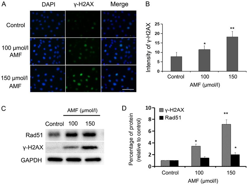
ONCOLOGY LETTERS 20: 168, 2020                                                   3

Cell Signaling Technology Inc.), β‑tubulin (1:1,000 v/v; cat.        mechanisms by which AMF arrests the cell cycle, the effect
no. 2146; Cell Signaling Technology Inc.), Cyclin‑B1 (1:1,000        of AMF on the expression levels of proteins associated with
v/v; cat. no. 12231; Cell Signaling Technology Inc.), CDK2           cell cycle progression was assessed via western blot analysis.
(1:1,000 v/v; cat. no. ab32147; Abcam), p‑CDK1 (1:1,000 v/v;         The expression levels of p‑CDK1 and CDK2 decreased in
cat. no. 4539; Cell Signaling Technology Inc.), P21 (1:1,000         SKOV3 cells treated with 100 or 150 µmol/l AMF for 48 h
v/v; cat. no. 2947; Cell Signaling Technology Inc.), γ‑H2AX          (Fig. 1D and E). Furthermore, CDK1 expression decreased in
(1:1,000 v/v; cat. no. 9718; Cell Signaling Technology Inc.),        SKOV3 cells treated with 100 and 150 µmol/l AMF; however, a
stathmin (1:1,000 v/v; cat. no. ab52630; Abcam), Rad51               significant decrease was only observed in the cells treated with
(1:1,000 v/v; cat. no. ab133534; Abcam) or CDK1 (1:1,000 v/v;        150 µmol/l of AMF (Fig. 1D and E). The p‑CDK1/CDK1 ratio
cat. no. ab18; Abcam) overnight at 4˚C. The membranes were           decreased in cells treated with AMF; however, no significant
washed three times with TBST and subsequently incubated              differences were observed compared with the control cells
with horse radish peroxidase‑conjugated secondary antibodies         (Fig. 1D and E). Notably, cyclin B1 expression was significantly
(1:2,000 v/v; cat. no. 7074 or 7076; Cell Signaling Technology       downregulated in SKOV3 cells treated with 100 µmol/l AMF
Inc.) diluted in TBST for 1 h at room temperature. Membranes         and upregulated in cells treated with 150 µmol/l AMF. P21, a
were re‑washed three times with TBST, and protein bands              well‑known inhibitor of the cell cycle, significantly increased
were visualized using the electrochemiluminescence detection         in AMF‑treated cells compared with AMF‑untreated ovarian
kit (Pierce; Thermo Fisher Scientific, Inc.) and imaged using        cells (Fig. 1E).
the ChemiDoc XRS+ Imaging System (Bio‑Rad Laboratories,
Inc.). Image Lab 3.0 software (Bio‑Rad Laboratories, Inc.) was       AMF interferes with tubulin expression and spindle assembly.
used for semi‑quantitative analysis of band signals.                 MTs are made from tubulin heterodimers and are vital for
                                                                     several cellular processes, such as spindle assembly for cell
Statistical analysis. Statistical analysis was performed using       meiosis (24). MTs have complex polymerization character‑
SPSS software v17.0 (IBM Corp.) and data are presented as            istics and are stable and long lasting during interphase (25).
the mean + standard deviation (SD) of at least three inde‑           Conversely, MTs become short and dynamic during mitosis.
pendent experiments. One‑way analysis of variance and                Stathmin regulates cell cycle progression by influencing the
Student‑Newman‑Keuls post‑hoc test were used to compare              dynamics of MTs (13).
difference between multiple groups. Unpaired Student's t‑test was        The present study assessed the influence of AMF on MT
used to test statistical significance between two groups. P
4 ZHANG et al: AMF INTERRUPTS MICROTUBULE DYNAMICS AND DNA STABILITY Figure 1. AMF decreases SKOV3 cell viability and induces cell cycle arrest. (A) SKOV3 cells were treated with different concentrations (0, 50 75, 100, 150 and 200 µmol/l) of AMF for 48 h and cell viability was assessed via the CellTiter 96 Aqueous One Solution Proliferation assay. The results demonstrated that AMF decreased SKOV3 cell viability in a dose‑dependent manner. (B) Histograms showed the cell cycle distribution at G1, S and G2 phase. Data are presented as the mean ± SD (n=3). SKOV3 cells were treated with different concentrations of AMF (0, 100 and 150 µmol/l) for 48 h and cell cycle distribution was assessed via flow cytometric analysis. (C) Cell cycle analysis by flow cytometry. SKOV3 cells were treated with different concentrations of AMF (0, 100 and 150 µmol/l) for 48 h. (D) The expression levels of cyclin B, p‑CDK1, CDK1, CDK2 and p21 were determined in SKOV3 cells treated with different concentra‑ tions of AMF (0, 100 and 150 µmol/l) for 48 h by western blot. GAPDH was used as the loading control. (E) Protein expression levels from the western blot in (D) relative to the GAPDH control. Data are presented as the mean ± SD (n=3). *P
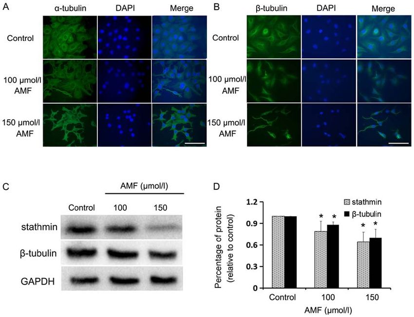
ONCOLOGY LETTERS 20: 168, 2020                                                                     5

Figure 2. AMF interferes with tubulin expression and spindle assembly. (A) Immunofluorescence staining of α‑tubulin (green) in SKOV3 cells treated with
different concentrations of AMF for 48 h. Nuclei (blue) were stained with DAPI (magnification, x400; scale bar, 100 µm). (B) Immunofluorescence staining
of β ‑tubulin (green) in SKOV3 cells treated with different concentrations of AMF for 48 h. Nuclei (blue) were stained with DAPI (magnification, x400;
scale bar, 100 µm). (C) The expression levels of stathmin and β‑tubulin in SKOV3 cells treated with different concentrations of AMF, via western blot analysis.
GAPDH was used as the loading control. (D) Protein expression levels from the western blot in (C) relative to the GAPDH control. Data are presented as the
mean ± standard deviation (n=3). *P
6                              ZHANG et al: AMF INTERRUPTS MICROTUBULE DYNAMICS AND DNA STABILITY

Figure 3. AMF induces DNA damage in SKOV3 cells. (A) Immunofluorescence staining of nuclei (blue) and γ‑H2AX (green) in SKOV3 cells treated with
different concentrations of AMF for 48 h (magnification, x400; scale bar, 100 µm). (B) Fluorescence intensities of γ‑H2AX in SKOV3 cells treated with different
concentrations of AMF for 48 h. Data are presented as the mean ± SD (n=3). (C) Expression levels of γ‑H2AX and Rad51 in SKOV3 cells treated with different
concentrations of AMF, via western blot analysis. GAPDH was used as the loading control. (D) Protein expression levels from the western blot in (C) relative
to the GAPDH control. Data are presented as the mean ± SD (n=3). *P
ONCOLOGY LETTERS 20: 168, 2020                                                         7

repair system in AMF‑treated SKOV3 cells. The present study        References
demonstrated that AMF triggered cell cycle G2/M arrest and
induced DNA damage in ovarian cancer cells, even though it          1. Bray F, Ferlay J, Soerjomataram I, Siegel RL, Torre LA and
                                                                       Jemal A: Global cancer statistics 2018: Globocan estimates
may be better to use several ovarian cancer cell lines than just       of incidence and mortality worldwide for 36 cancers in
to use SKOV3 cell line. We will be aimed at confirming these           185 countries. CA Cancer J Clin 68: 394‑424, 2018.
findings and prove the antitumor effect of AMF in ovarian           2. Bookman MA: Optimal primary therapy of ovarian cancer.
                                                                       Ann Oncol 27 (Suppl 1): i58‑i62, 2016.
cancer cell line derived xenograft mouse models.                    3. Zhang X, Xu B, Sun CY, Wang LM and Miao X: Knockdown of
    In conclusion, the results of the present study demonstrated       CIP2A sensitizes ovarian cancer cells to cisplatin: An in vitro
that AMF inhibited human ovarian cancer cell proliferation             study. Int J Clin Exp Med 8: 16941‑16947, 2015.
                                                                    4. Damia G and Broggini M: Platinum resistance in ovarian cancer:
by triggering cell cycle arrest at the G2/M phase. Furthermore,        Role of DNA repair. Cancers (Basel) 11: 119, 2019.
AMF was demonstrated to interfere with MT dynamics and              5. Dia‑Moralli S, Tarrado‑Castellarnau M, Miranda A and
induce DNA damage. Thus, AMF may act as an antitumor                   Cascante M: Targeting cell cycle regulation in cancer therapy.
                                                                       Pharmacol Ther 138: 255‑271, 2013.
drug by exerting its effects on MT dynamics and inducing            6. Lim S and Kaldis P: Cdks, cyclins and CKIs: Roles beyond cell
DNA damage.                                                            cycle regulation. Development 140: 3079‑3093, 2013.
                                                                    7. Morgan DO: The cell cycle: Principles of control. Primers in
                                                                       biology, New Science, 2007.
Acknowledgements                                                    8. Karimian A, Ahmadi Y and Yousefi B: Multiple functions of p21
                                                                       in cell cycle, apoptosis and transcriptional regulation after DNA
Flow cytometry work was supported by Ms. Fang Su and                   damage. DNA Repair (Amst) 42: 63‑71, 2016.
                                                                    9. Liebmann J, Cook JA, Lipschultz C, Teague D, Fisher J and
Ms. Jing Wei (Guangdong Provincial Key Laboratory of                   Mitchell JB: The influence of Cremophor EL on the cell
Malignant Tumor Epigenetics and Gene Regulation, Sun                   cycle effects of paclitaxcl (Taxol) in human tumor cell lines.
Yat‑Sen Memorial Hospital, Sun Yat‑Sen University, China).             Cancer Chemother Pharmacol 33: 331‑339, 1994.
                                                                   10. Jordan MA, Toso RJ, Thrower D and Wilson L: Mechanism of
                                                                       milotic block and inhibition of cell proliferation by taxol at low
Funding                                                                concentrations. Proc Nati Acad Sci USA 90: 9552‑9556, 1993.
                                                                   11. Desai A and Mitchison TJ: Microtubule polymerization
                                                                       dynamics. Annu Rev Cell Dev Biol 13: 83‑117, 1997.
The present study was funded by Guangdong Bureau of                12. Schiff PB, Fant J and Horwitz SB: Promotion of microtubule
Traditional Chinese Medicine (grant nos. 20181206 and                  assembly in vitro by taxol. Nature 277: 665‑667, 1979.
20191260), the National Natural Science Foundation of China        13. Charbaut E, Curmi PA, Ozon S, Lachkar S, Redeker V and
                                                                       Sobel A: Stathmin family proteins display specific molecular
(grant no. 81272222 and 81902802), the General Science                 and tubulin binding properties. J Biol Chem 276: 16146‑16154,
and Technology Project of Guangzhou Municipal Health                   2001.
Commission (grant no. 20181A010017 and 20191A011019),              14. Su D, Smith SM, Preti M, Schwartz P, Rutherford TJ, Menato G,
                                                                       Danese S, Ma S, Yu H and Katsaros D: Stathmin and tubulin
the Medical Science and Technology Research Foundation                 expression and survival of ovarian cancer patients receiving
of Guangdong (grant nos. B2016018 and A2018063) and the                platinum treatment with and without paclitaxel. Cancer 115:
Guangzhou Science and Technology Program Key Project                   2453‑2463, 2009.
                                                                   15. Watanabe A, Suzuki H, Yokobori T, Tsukagoshi M, Altan B,
(grant no. 201704020145).                                              Kubo N, Suzuki S, Araki K, Wada S, Kashiwabara K, et al:
                                                                       Stathmin1 regulates p27 expression, proliferation and drug resis‑
Availability of data and materials                                     tance, resulting in poor clinical prognosis in cholangiocarcinoma.
                                                                       Cancer Sci 105: 690‑696, 2014.
                                                                   16. Johnsen JI, Aurelio ON, Kwaja Z, Jögensen GE, Pellegata NS,
All data generated or analyzed during this study are included          Plattner R, Stanbridge EJ and Cajot JF: p53‑mediated nega‑
in this published article.                                             tive regulation of stathmin/Op18 expression is associated with
                                                                       G2/M cell‑cycle arrest. Int J Cancer 88: 685‑691, 2000.
                                                                   17. Pang X, Yi T, Yi Z, Cho SG, Qu W, Pinkaew D, Fujise K and
Authors' contributions                                                 Liu M: Morelloflavone, a biflavonoid, inhibits tumor angiogen‑
                                                                       esis by targeting rho GTPases and extracellular signal‑regulated
                                                                       kinase signaling pathways. Cancer Res 69: 518‑525, 2009.
ZL, JZ, AL and XL contributed to the initial conception and        18. Li M, Li B, Xia ZM, Tian Y, Zhang D, Rui WJ, Dong JX
design of the experiments. HS, XX, SQ, PW and LD performed             and Xiao FJ: Anticancer Effects of Five Biflavonoids from
the experiments. ZL and JZ wrote the paper. ZZ revised the             Ginkgo Biloba L. Male Flowers In Vitro. Molecules 24: 1496,
experimental design and analyzed the data. All authors read            2019.
                                                                   19. Banerjee T, Valacchi G, Ziboh VA and van der Vliet A: Inhibition
and approved the final manuscript.                                     of TNFalpha‑induced cyclooxygenase‑2 expression by amento‑
                                                                       flavone through suppression of NF‑kappaB activation in A549
Ethics approval and consent to participate                             cells. Mol Cell Biochem 238: 105‑110, 2002.
                                                                   20. Zhang Z, Sun T, Niu JG, He ZQ, Liu Y and Wang F: Amentoflavone
                                                                       protects hippocampal neurons: Anti‑inflammatory, antioxidative,
Not applicable.                                                        and antiapoptotic effects. Neural Regen Res 10: 1125‑1133, 2015.
                                                                   21. An J, Li Z, Dong Y, Ren J and Huo J: Amentoflavone protects
                                                                       against psoriasis‑like skin lesion through suppression of
Patients consent for publication                                       NF‑κ B‑mediated inflammation and keratinocyte proliferation.
                                                                       Mol Cell Biochem 413: 87‑95, 2016.
Not applicable.                                                    22. Li F, Song XW, Su GF, Wang YL, Wang ZY, Jia XY, Qin SR,
                                                                       Huang L, Wang Y, Zheng K and Wang Y: Amentoflavone inhibits
                                                                       HSV‑1 and ACV‑resistant strain infection by suppressing viral
Competing interests                                                    early infection. Viruses 11: 466, 2019.
                                                                   23. Zhang J, Liu Z, Cao W, Chen L, Xiong X, Qin S, Zhang Z, Li X
                                                                       and Hu CA: Amentoflavone inhibits angiogenesis of endothelial
The authors declare that they have no competing interests.             cells and stimulates apoptosis in hypertrophic scar fifibroblasts.
                                                                       Burns 40: 922‑929, 2014.
8 ZHANG et al: AMF INTERRUPTS MICROTUBULE DYNAMICS AND DNA STABILITY
24. Eshun‑Wilson L, Zhang R, Portran D, Nachury MV, Toso DB, 43. Burbank KS and Mitchison TJ: Microtubule dynamic instability.
Löhr T, Vendruscolo M, Bonomi M, Fraser JS and Nogales E: Curr Biol 16: R516‑R517, 2006.
Effects of α‑tubulin acetylation on microtubule structure and 44. Benbow SJ, Wozniak KM, Kulesh B, Savage A, Slusher BS,
stability. Proc Natl Acad Sci USA 116: 10366‑10371, 2019. Littlefield BA, Jordan MA, Wilson L and Feinstein SC:
25. Ohkawa N, Fujitani K, Tokunaga E, Furuya S and Inokuchi K: The Microtubule‑targeting agents eribulin and paclitaxel differ‑
microtubule destabilizer stathmin mediates the development of entially affect neuronal cell bodies in chemotherapy induced
dendritic arbors in neuronal cells. J Cell Sc 120: 1447‑1456, 2007. peripheral neuropathy. Neurotox Res 32: 151‑162, 2017.
26. Firsanov DV, Solovjeva LV and Svetlova MP: H2AX phosphory‑ 45. Field JJ, Díaz JF and Miller JH: The binding sites of
lation at the sites of DNA double‑strand breaks in cultivated microtubule‑stabilizing agents. Chem Biol 20: 301‑315, 2013.
mammalian cells and tissues. Clin Epigenetics 2: 283‑297, 2011. 46. Cao YN, Zheng LL, Wang D, Liang XX, Gao F and Zhou XL:
27. Chen JJ, Silver DP, Cantor SB, Cantor S, Livingston DM and Recent advances in microtubule‑stabilizing agents. Eur J Med
Scully R: BRCA1, BRCA2, and Rad51 operate in a common Chem 143: 806‑828, 2018.
DNA damage response pathway. Cancer Res 59 (7 Suppl): 47. Prota AE, Bargsten K, Zurwerra D, Field JJ, Diaz JF, Altmann KH
1752S‑1756S, 1999. and Steinmetz MO: Molecular mechanism of action of microtu‑
28. Liu B and Yu S: Amentoflavone suppresses hepatocellular carci‑ bule‑stabilizing anticancer agents. Science 339: 587‑590, 2013.
noma by repressing hexokinase 2 expression through inhibiting 48. Cassimeris L: The oncoprotein 18/stathmin family of microtubule
JAK2/STAT3 signaling. Biomed Pharmacother 107: 243‑253, 2018. destabilizers. Curr Opin Cell Biol 14: 18‑24, 2002.
29. Lee KC, Tsai JJ, Tseng CW, Kuo YC, Chuang YC, Lin SS and 49. Obayashi S, Horiguchi J, Higuchi T, Katayama A, Handa T,
Hsu FT: Amentoflavone inhibits ERK‑modulated tumor progres‑ Altan B, Bai T, Bao P, Bao H, Yokobori T, et al: Stathmin1 expres‑
sion in hepatocellular carcinoma in vitro. In Vivo 32: 549‑554, 2018. sion is associated with aggressive phenotypes and cancer stem
30. Hsu FT, Chiang IT, Kuo YC, Hsia TC, Lin CC, Liu YC and cell marker expression in breast cancer patients. Int J Oncol 51:
Chung JG: Amentoflavone effectively blocked the tumor progres‑ 781‑790, 2017.
sion of glioblastoma via suppression of ERK/NF‑κ B signaling 50. Carney BK and Cassimeris L: Stathmin/oncoprotein 18, a micro‑
pathway. Am J Chin Med 47: 913‑931, 2019. tubule regulatory protein, is required for survival of both normal
31. Guruvayoorappan C and Kuttan G: Effect of amentoflavone on the and cancer cell lines lacking the tumor suppressor p53. Cancer
inhibition of pulmonary metastasis induced by B16F‑10 melanoma Biol Ther 9: 699‑709, 2010.
cells in C57BL/6 mice. Integr Cancer Ther 6: 185‑197, 2007. 51. Alli E, Yang JM and Hait WN: Silencing of stathmin induces
32. Lee JS, Lee MS, Oh WK and Sul JY: Fatty acid synthase inhibi‑ tumor‑suppressor function in breast cancer cell lines harboring
tionby amentoflflavone induces apoptosis and antiproliferation in mutant p53. Oncogene 26: 1003‑1012, 2007.
human breast cancer cells. Biol Pharm Bull 32: 1427‑1432, 2009. 52. Wang R, Dong K, Lin F, Wang X, Gao P, Wei SH, Cheng SY
33. Lee SJ, Kim HJ, Kang JW, Kim JH, Lee DH, Kim MS, Yang Y, and Zhang HZ: Inhibiting proliferation and enhancing
Woo ER, Kim YM, Hong J and Yoon DY: The biflavonoid chemosensitivity to taxanes in osteosarcoma cells by RNA
amentoflavone induces apoptosis via suppressing E7 expression, interference‑mediated downregulation of stathmin expression.
cell cycle arrest at sub‑G1 phase, and mitochondria‑emanated Mol Med 13: 567‑575, 2007.
intrinsic pathways in Human Cervical Cancer Cells. J Med 53. Dumontet C and Jordan MA: Microtubule‑binding agents: A
Food 14: 808‑816, 2011. dynamic field of cancer therapeutics. Nat Rev Drug Discov 9:
34. Pei JS, Liu CC, Hsu YN, Lin LL, Wang SC, Chung JG, Bau DT and 790‑803, 2010.
Lin SS: Amentoflavone induces cell‑cycle arrest and apoptosis in 54. Velic D, Couturier AM, Ferreira MT, Rodrigue A, Poirier GG,
MCF‑7 human breast cancer cells via mitochondria‑dependent Fleury G and Masson GY: DNA damage signalling and repair
pathway. In Vivo 26: 963‑970, 2012. inhibitors: The long‑sought‑after Achilles' heel of cancer.
35. Jung HJ, Park K, Lee IS, Kim HS, Yeo SH, Woo ER and Lee DG: Biomolecules 5: 3204‑3259, 2015.
S‑phase accumulation of Candida albicans by anticandidal effect 55. Lobrich M, Shibata A, Beucher A, Fisher A, Ensminger M,
of amentoflavone isolated from Selaginella tamariscina. Biol Goodarzi AA, Barton O and Jeggo PA: GammaH2AX foci
Pharm Bull 30: 1969‑1971, 2007. analysis for monitoring DNA double‑strand break repair:
36. Liu H, Yue Q and He S: Amentoflavone suppresses tumor growth Strengths, limitations and optimization. Cell Cycle 9: 662‑669,
in ovarian cancer by modulating Skp2. Life Sci 189: 96‑105, 2017. 2010.
37. Dehay C and Kennedy H: Cell‑cycle control and cortical 56. Daley JM, Kwon Y, Niu H and Sung P: Investigations of homolo‑
development. Nat Rev Neurosci 8: 438‑450, 2007. gous recombination pathways and their regulation. Yale J Biol
38. Malumbres M and Barbacid M: Cell cycle, CDKs and cancer: A Med 86: 453‑461, 2013.
changing paradigm. Nat Rev Cancer 9: 153‑166, 2009. 57. Graeser M, McCarthy A, Lord CJ, Savage K, Hills M, Salter J,
39. Marais A, Ji Z, Child ES, Krause E, Mann DJ and Sharrocks AD: Orr N, Parton M, Smith IE, Reis‑Filho JS, et al: A marker of
Cell cycle‑dependent regulation of the forkhead transcription homologous recombination predicts pathologic complete
factor FOXK2 by CDK‑cyclin complexes. J Biol Chem 285: response to neoadjuvant chemotherapy in primary breast cancer.
35728‑35739, 2010. Clin Cancer Res 16: 6159‑6168, 2010.
40. Bertoli C, Skotheim JM and de Bruin RA: Control of cell cycle 58. Baumann P, Benson FE and West SC: Human Rad51 protein
transcription during G1 and S phases. Nat Rev Mol Cell Biol 14: promotes ATP‑dependent homologous pairing and strand
518‑528, 2013. transfer reactions in vitro. Cell 87: 757‑766, 1996.
41. Choi WI, Kim MY, Jeon BN, Koh DI, Yun CO, Li Y, Lee CE,
Oh J, Kim K and Hur MW: Role of promyelocytic leukemia This work is licensed under a Creative Commons
zinc finger (PLZF) in cell proliferation and cyclin‑dependent Attribution-NonCommercial-NoDerivatives 4.0
kinase inhibitor 1A (p21WAF/CDKN1A) gene repression. J Biol International (CC BY-NC-ND 4.0) License.
Chem 289: 18625‑18640, 2014.
42. Tsuda Y, Iimori M, Nakashima Y, Nakanishi R, Ando K,
Ohgaki K, Kitao H, Saeki H and Maehara Y: Mitotic slippage
and the subsequent cell fate after inhibition of Aurora B during
tubulin‑binding agent‑induced mitotic arrest. Sci Rep 17: 16762,
2017.You can also read


