A phase diagram for bacterial swarming - ARTICLE - AVRAHAM BE'ER LAB
←
→
Page content transcription
If your browser does not render page correctly, please read the page content below
ARTICLE
                  https://doi.org/10.1038/s42005-020-0327-1                 OPEN

                  A phase diagram for bacterial swarming
                  Avraham Be’er1,2 ✉, Bella Ilkanaiv1, Renan Gross3, Daniel B. Kearns4, Sebastian Heidenreich5, Markus Bär5 &
                  Gil Ariel 6 ✉
1234567890():,;

                  Bacterial swarming is a rapid mass-migration, in which thousands of cells spread collectively
                  to colonize surfaces. Physically, swarming is a natural example for active particles that use
                  energy to generate motion. Accordingly, understanding the constraints physics imposes on
                  these dynamics is essential for understanding the mechanisms underlying swarming. We
                  present new experiments of swarming Bacillus subtilis mutants with different aspect ratios and
                  at different densities; two physical quantities known to be associated with collective behavior.
                  Analyzing the dynamics reveals a rich phase diagram of qualitatively distinct swarming
                  regimes, describing how cell shape and population density govern the dynamical char-
                  acteristics of the swarm. In particular, we show that under standard conditions, bacteria
                  inhabit a region of phase space that is associated with rapid mixing and robust dynamics, with
                  homogeneous density and no preferred direction of motion. The results suggest that bacteria
                  have adapted their physical properties to optimize the principle functions assumed for
                  swarming.

                  1 ZuckerbergInstitute for Water Research, The Jacob Blaustein Institutes for Desert Research, Ben-Gurion University of the Negev, Sede Boqer Campus,
                  84990 Midreshet Ben-Gurion, Israel. 2 Department of Physics, Ben-Gurion University of the Negev, 84105 Beer-Sheva, Israel. 3 Department of Mathematics,
                  Weizmann Institute of Science, 76100 Rehovot, Israel. 4 Department of Biology, Indiana University, Bloomington, IN 47405, USA. 5 Department of
                  Mathematical Modelling and Data Analysis, Physikalisch-Technische Bundesanstalt Braunschweig und Berlin, Abbestrasse 2-12, 10587 Berlin, Germany.
                  6 Department of Mathematics, Bar-Ilan University, 52000 Ramat-Gan, Israel. ✉email: beera@bgu.ac.il; arielg@math.biu.ac.il

                  COMMUNICATIONS PHYSICS | (2020)3:66 | https://doi.org/10.1038/s42005-020-0327-1 | www.nature.com/commsphys                                           1
ARTICLE COMMUNICATIONS PHYSICS | https://doi.org/10.1038/s42005-020-0327-1
M
icroorganisms such as bacteria, sperm, epithelial, and pieces does not yet fit together into a comprehensive picture. In
cancer cells, as well as immune T cells and even inan- particular, it is not clear how the properties of cells determine the
imate active particles, generate collective flows and dynamical state of a swarm.
demonstrate a wealth of newly discovered emergent dynamical In this paper, monolayer swarms of four strains of B. subtilis
patterns1–8. This report addresses the dynamics of swarming with different aspect ratios—ranging from 5.5 to 19, were ana-
bacteria—a biological state to which some bacterial species lyzed as a function of surface density. A custom algorithm
transition, in which rod-shaped cells, powered by flagellar rota- enabled tracking of individual cell trajectories, which in turn
tion, migrate rapidly on surfaces en masse9–12. Swarming allows allowed a comprehensive analysis of both the individual and
efficient expansion and colonization of new territories, even collective dynamics of bacteria in a swarm. The main results are
under harsh and adverse conditions such as starvation or anti- then expressed in a phase diagram of bacterial swarming. The
biotic stress13,14. Revealing the biological and physical mechan- novel two-dimensional set-up of a thin, single layer of cells, brings
isms underlying bacterial swarming is therefore a key to our out a complex experimentally based phase diagram with various
understanding of how bacteria spread and invade new niches. features that could not be obtained with earlier multilayer
The transition to swarming involves several critical intracel- studies19,20,22,25 or naturally expanding colonies24. Our analysis
lular processes such as an increase in flagellar number and identified five dynamical states for bacterial swarms. Each state
changes in cell aspect ratio, suggesting that these changes pro- showed distinct dynamical features, as detailed below, which are
mote favorable swarming conditions8,9,11,12,15. Quantifying the expressed both at the individual and collective levels.
“quality” of swarming can be done using the tools of statistical
physics by analyzing the dynamical properties of large, out-of-
equilibrium, self-propelled collectives3,16,17. Accordingly, one of Results
the primary goals of such quantification is to obtain a phase Experimental set-up. Small drops of overnight bacterial cultures
diagram that would describe the possible dynamical states of were inoculated at the center of agar plates; each colony grew from
swarms as a function of independent parameters such as density a single strain, with no mixing between strains (see “Methods” for
and cell aspect ratio; density because individual sparsely dis- details). Within a few hours, cells collectively migrated outwards
tributed cells do not swarm, and aspect ratio because this of the initial inoculum, forming a swarm, with a boundary that
often changes in the cell prior to swarming. Thus a phase diagram expanded toward the plate perimeter. When the colonies reached
provides a map that explains how the microscopic mechanical a diameter of 5 cm (approximately 4 h after inoculation), a wide
properties of cells, which are regulated by complex bio-chemical (>2 cm in thickness) and highly active ring-shaped band was
cellular processes, may govern the global dynamical character- formed. High-resolution microscopy was used to observe the
istics of the entire swarm. dynamics within a smaller ring, approximately 300 μm in width,
The surface density, ρ (the fraction of surface covered by close to the colony’s edge; see Fig. 1a and Supplementary Fig. 1.
bacteria), within bacterial cultures in general, and in the swarm in Different regions of this this band showed a range of cell densities.
particular, is typically extremely high (up to 0.8), resulting in a Note that this is different from Jeckel et al.24, who were interested
combination of short-range steric repulsion and long-range in scanning and characterizing different regions and time periods
hydrodynamic interactions. Both forces strongly depend on cell within the expanding colony. Here we concentrate on the most
aspect ratio and particle density, see e.g., refs. 2,8,10,18–24. active part of the swarm for different strains that differ in their
Recently, Jeckel et al.24 studied expanding colonies of swarming aspect ratios. In particular, we study “unnaturally” short and long
Bacillus subtilis and discovered regions in the colony corre- cells, with aspect ratios that are not manifested in freely growing
sponding to single cells, rafts, biofilm, and mixed states. Despite colonies. This point of view, which is complementary to ref. 24,
the recent progress in understanding the physics of active matter allows us to probe the swarming regimes as a function of the two
in general and swarming bacteria in particular, the different independent parameters.
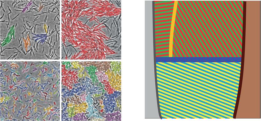
Fig. 1 A phase diagram for bacterial swarming. a Snapshots of monolayer bacterial swarms with different aspect ratios and densities, representative of the
motile phases. Colors represent moving clusters (gray or non-labeled cells are not moving). The scale bar corresponds to 50 μm. For short cells (S phase),
almost all cells are moving coherently. For long cells, spatial ordering into local (SC) or global (LC) clusters is apparent. b The inferred phase diagram as a
function of aspect ratio (α) and surface density (ρ), showing five distinct phases: immotile (IM), swarming (S), small clusters (SCs), large clusters (LCs),
and jammed (J).
2 COMMUNICATIONS PHYSICS | (2020)3:66 | https://doi.org/10.1038/s42005-020-0327-1 | www.nature.com/commsphysCOMMUNICATIONS PHYSICS | https://doi.org/10.1038/s42005-020-0327-1 ARTICLE
Deriving a phase diagram requires measuring a large number (indicating a heavy-tailed distribution) at the large aspect ratios
of collective and individual dynamical statistics for swarms with (Supplementary Fig. 5). Another key statistic, which has been
particular parameter values. As described above, we concentrate theoretically shown to describe different regimes of collective
on cell aspect ratio (α) and surface density (ρ) as the fundamental dynamics, is the distribution of cluster sizes (DCS)18: In the S
mechanical parameters (Fig. 1b). Since the typical expansion rate phase (Fig. 3a, b), the DCS is close to a power law with an
of the colony is about 10 times slower than the typical exponential cut-off. In contrast, in the SC/LC phases, the DCS at
microscopic swarming speed, different regions of the actively low densities (a power law with a cut-off) is different than at
migrating colony can be considered to be in a quasi-steady state. higher ones, where LCs emerge whose sizes are comparable
Therefore, the effect of density can be evaluated by sampling with the observed system size (Fig. 3c, d). Lastly, it has been
regions that occupy a different number of bacteria per unit area, shown that WT swarming bacteria are super-diffusive, with
e.g., refs. 20,24. Cell shape was manipulated genetically by trajectories that are consistent with Lévy walks31,32. Figure 2b
mutating a few of the robust mechanisms that maintain aspect shows that trajectories are super-diffusive at all aspect ratios and
ratio during growth. Artificially long cells were generated using surface densities. However, the associated characteristic exponent
cells mutated for either MinD or MinJ that control proper medial is varying. While the exponent in the S phase is approximately
division in B. subtilis. Artificially short cells were generated by constant (1.6–1.7), it is clearly decreasing in the SC/LC phases.
overexpression of SwrA, the master activator of flagellar Therefore, the super-diffusive property of long cells degrades at
biosynthesis15,26. Overall, four different strains of the same high surface densities, indicating slower mixing and spreading.
species with various average aspect ratios of 5.5, 7 (the wild type At large aspect ratios, the phase diagram is divided into two
(WT)), 13, and 19 (Supplementary Table 1) were compared. distinct regions (Fig. 1b). The transition (as a function of density)
Changes in aspect ratio of a specific strain stemming from is pronounced in the spatial correlation functions (Fig. 4a–d),
different regions in a colony are negligible. A wide range of both in cell directional alignment λ|| and in the velocity (direction
benchmark tests verified that all other motility and expansion- of motion) λν. At low surface densities, the correlation lengths
related parameters were the same (including swimming speeds grow sharply with density, exhibiting small fluctuations (0.3, the correlation lengths are practically
surfactant production). See “Methods,” Supplementary Note 1, constant, with large fluctuations between samples (up to 55%).
and Supplementary Figs. 2 and 3. The transition region is narrow, suggesting a critical phenom-
enon. Further examination reveals that the jump in the standard
deviation of measurements is mostly due to density fluctuations
Quantifying swarming regimes. Figure 2a depicts the mean cell in time (Fig. 4e). A time series analysis reveals a sharp increase in
speed, which is monotonically increasing with density for each the Hurst exponent, which quantifies the roughness of temporal
aspect ratio, a hallmark of collective motion—showing that many fluctuations, indicating that the density varies sharply in time
cells cooperate to produce faster motion, e.g., refs. 2,27,28. The (Fig. 4f, Supplementary Fig. 6). For small aspect ratios, the Hurst
mean speed is not monotonic in the aspect ratio where WT cells exponent is around 0.5 at all densities, as expected for finitely
seem to be optimal in this regard (see also ref. 22). However, correlated series. See Supplementary Note 2 and Supplementary
speeds depend smoothly on cell density, showing no sharp Fig. 6 for details of the effect of the field of view size.
transitions. In addition, Fig. 2a identifies lower and upper density
thresholds, beyond which swarming cannot occur. At very low
densities, cells are practically immotile (IM), suggesting a mini- Interpretation as a self-propelled particle system. From an
mal surface or number density below which cells cannot move29. active matter perspective, our results reveal some successful the-
The minimal speed at which swarming was observed marks the oretical predictions upon comparison to earlier work on
edge of the IM phase depicted in Fig. 1b. At very high densities, simulations17,18,21,23,24,33,34 and experiments with artificial
cells cannot move efficiently due to confinement21,30, suggesting inanimate systems35,36. However, the swarming phase diagram
an additional jammed phase J. This phase is not observed in our also has several unique characteristics that are not observed in
experiments, as we concentrate on the active regions. other active systems. For example, several phases that have been
At intermediate surface densities, our analysis below revealed observed in simulations, such as the bio-nematic and laning
three phases of motile bacteria with distinct dynamical char- phases21,34, were not realized. Other predictions, such as the
acteristics: (i) A swarm phase S at small aspect ratios, in which emergence of bimodal cluster-size distributions (Fig. 3)18,
cells move and flow efficiently, (ii) a low-density phase of long motility-induced spatial segregation into low- and high-density
cells, which consists of small moving clusters (SCs), and (iii) a regions37, large number fluctuations (Supplementary Fig. 7)3,
high-density phase of long cells, in which large moving clusters long-tailed auto-correlations functions (Supplementary Figs. 8
(LCs) of the size of viewing area cause large (but finite) and 9)3, and meso-scale turbulence21, have been inferred from
fluctuations in time. While we find trajectories are always agent-based and continuous models and are found, to some
super-diffusive (Fig. 2b), we first focus on the differences between extent, in the present experiments. See Supplementary Note 3 for
short and long cells. Note that, given our relatively small number further discussions.
of possible aspect ratios, we cannot establish a sharp phase Collective motion of bacteria changes, quite dramatically, if the
transition or its properties (first or second, critical exponents, aspect ratio is increased above a threshold value (around 10 for B.
etc.). Nonetheless, several qualitative differences between short subtilis in our study here). Strikingly, above densities of around
and long cells are apparent: at small aspect ratios (S phase), the 0.25, cells transition into another phase and form LCs that can
swarm is characterized by a unimodal spatial distribution of become of the order of the observation window employed here
surface densities (Fig. 2c, d, Supplementary Fig. 4) and the (see Supplementary Fig. 6c for a variety of window sizes). This
velocities exhibit a Gaussian distribution (kurtosis close to 3, transition is reminiscent of behavior of self-propelled rods with
Supplementary Fig. 5). In contrast, at large aspect ratios (SC and short-range alignment interactions (typically due to volume
LC phases), the swarm is segregated into two populations, exclusion) in simulations and experiments; see ref. 17 and
corresponding to low- and high-density regions (Fig. 2e, f). The references therein. The observed cluster-size distributions are in
proportion of each population changes with the mean surface line with a kinetic theory describing occurrence of LCs as a
density. The distribution of velocities exhibits very large kurtosis specific type of microphase separation characteristic for rod-
COMMUNICATIONS PHYSICS | (2020)3:66 | https://doi.org/10.1038/s42005-020-0327-1 | www.nature.com/commsphys 3ARTICLE COMMUNICATIONS PHYSICS | https://doi.org/10.1038/s42005-020-0327-1 Fig. 2 Comparison between short and long cells. a The denser the cells, the faster they move. The average speed increases with density but is non- monotonic in aspect ratio. Wild-type cells are the fastest. Surface density ρ corresponds to the fraction of the viewing area that is covered by cells. Error bars are the standard deviation and are of the same size as the markers. b The mean squared displacement exponent shows that cell trajectories are super- diffusive. However, the exponent of long cells (small cluster (SC) and large cluster (LC) phases) decreases rapidly with density, showing that their super- diffusive property degrades at high densities. Inset shows the log–log plot of the mean square displacement (MSD) versus time interval from which we have fitted each exponent value. This specific MSD corresponds to the hollow red square. Full lines are the best linear fit, providing the slope value reported in the legend. c–f The distribution of densities among a 10 × 10 partition of the viewing area (15 × 15 μm bins) for different (average) surface densities. c, d At small aspect ratios (S phase), the swarm is characterized by a unimodal spatial distribution, with the mode increasing with mean surface density. e, f At large aspect ratios (SC and LC phases), the swarm is segregated into two populations. The proportion of each population changes with the mean surface density. shaped moving particles like bacteria18,38. DCS is also related to dominated by short-range alignment interaction39. At small the increase in temporal fluctuations. We hypothesize that large aspect ratios, the collective behavior deviates from the self- temporal fluctuations correlate with the occurrence of giant propelled rod paradigm. We rationalize that this is due to long- number fluctuations, which indicate the occurrence of LCs, range hydrodynamic interactions, which suppress the clustering similar to previous findings in bacterial systems19,20 and in and density inhomogeneities. This is in accordance with recent contrast to experimental reports and theoretical predictions, e.g., simulation findings, e.g., with circular microswimmers40 showing for active nematic phases that are assumed to be homogeneous on that hydrodynamic interactions indicate a complex dependency the large scale3,35. of clustering behavior on the shape and the swimming We conclude that, in the SC and LC phases, collective behavior mechanism of the microswimmers41. Thus our observation is dominated by short-range alignment or excluded volume should stimulate more detailed model studies. interactions. This is in line with findings for filamentous, very Independently of the aspect ratio, at low cell densities (
COMMUNICATIONS PHYSICS | https://doi.org/10.1038/s42005-020-0327-1                                                                               ARTICLE

Fig. 3 Distribution of cluster sizes. a, b For short cells (S phase), the distribution of cluster sizes is approximately a power law with an exponential cutoff. c,
d For long cells (small cluster (SC) and large cluster (LC) phases), the distribution is more complex: at low densities, it is a power law. However, at higher
densities, e.g., ρ = 0.6, large clusters emerge whose sizes are comparable with the system size. Error bars are the standard deviation and are of the same
size as the markers.

densities (>0.7) a jamming phase (J) is observed, where cells stop                 scale; individual cells that are placed on the agar with the system
moving, possibly due to lack of space. While jamming of high-                      already above the threshold for surfactant secretion are still
density active systems has been predicted theoretically21,30,34, the               immobile. Indeed, the increase in the average speed as a function
IM phase does not occur in typical active matter systems. This                     of cell density can be explained in terms of collective-motion
includes swimming (not swarming) bacteria in bulk or thin                          models that do not take quorum sensing into account30,47.
films2,42, in driven inanimate particles35,36, or most models of                       Overall, many of the prominent features of the swarm
self-propelled particles, either discrete or continuous, e.g.,                     dynamics, including the non-trivial Hurst exponent (marking
refs. 21,30,34, in which isolated particles typically move. During                 the SC–LC transition) and the lack of phase changes at small
swarming, on the other hand, the reason for the absence of                         aspect ratios cannot be explained by current theories. Therefore,
motion of isolated individuals (or cells at very low densities) is                 the phase diagram, Fig. 1b, and the subsequent detailed statistical
unclear. It has been suggested that the cells are trapped in areas                 analysis of key dynamic quantities provide a rich data set against
that are temporarily too dry8. In such regions, the surface may                    which future models for swarming and swimming bacteria with
exert a large drag force that the thrust of the flagella cannot                     competing alignment and hydrodynamic interactions can be
overcome.                                                                          calibrated or tested.
   Another possibility is that quorum sensing (or some sort of
quorum signaling) may play a role in the onset of swarming.
Indeed, it is known that the quorum sensing in B. subtilis controls                Discussion
surfactin production, which is required for swarming (quorum                       From a biological perspective, the physical properties described
sensing controls many other things including genetic competence                    above may alter, constrain, or even control cells’ ability to move
but these do not appear to be essential for swarming)43–45.                        collectively in an efficient manner, mix within the colony, and
Therefore, introducing quorum signaling to current bacterial                       spread. As a result, it has direct biological consequences in terms
swarming models may be crucial for successfully modeling the                       of the ability of bacteria to swarm efficiently. With a typical aspect
transition between the IM and the motile phases. However, in the                   ratio of 7 and a wide range of densities (0.2–0.7), WT bacteria
swarming regimes that are considered in this work, correspond-                     show rapid movement and highly efficient spreading. Within the
ing to extremely high bacterial densities in the interior of the                   S region, the swarming statistics were not sensitive to the density
colony, surfactin at the colony edge is abundant46, and we do not                  as well as to small changes in the aspect ratio, suggesting that the
expect that quorum sensing plays a major role in the dynamics of                   collective behavior of WT swarming cells is particularly robust to
the cells in the outer band. In particular, the absence of motion of               fluctuations in density and cell shape. Thus the physical robust-
sparsely distributed cells does not seem to be the result of quorum                ness of the swarming phase (S) may be advantageous for main-
signaling effects, which does not fluctuate on the micrometer                       taining efficient swarming, particularly under stress.

COMMUNICATIONS PHYSICS | (2020)3:66 | https://doi.org/10.1038/s42005-020-0327-1 | www.nature.com/commsphys                                                       5
ARTICLE                                                                   COMMUNICATIONS PHYSICS | https://doi.org/10.1038/s42005-020-0327-1

Fig. 4 Characterizing the small cluster (SC) to large cluster (LC) transition. a Average correlation length of cells′ directions and b cells′ velocities.
Standard deviations of the correlation length for c directions and d velocities show a sharp increase within a narrow region of surface densities
corresponding with the SC/LC transition. e The standard deviation of temporal fluctuations in surface density suggests large number of fluctuations in time.
f The Hurst exponent, which indicates the roughness of a time series, shows a sharp jump in the SC/LC transition region. The gray rectangle represents the
transition region. Error bars are the standard deviation and are of the same size as the markers.

   B. subtilis is a representative of a group of bacteria called                 Bacterial swarming is a natural state, i.e., cells appear to enter a
“temperate swarmers” such as Serratia marcescens, Pseudomonas,                swarming state when introduced to a surface. This suggests that
Salmonella, and E. coli that swarm at similar conditions48. Thus              the changes in cells prior to the onset of swarming may be
we expect their phase diagram to be qualitatively similar. Other              advantageous to the colony’s survival. The phase diagram dis-
bacteria, termed “robust swarmers,” such as Proteus mirabilis and             cussed above describes the range of possible dynamical regimes
Vibrio parahaemolyticus, are capable of migrating atop harder                 for the swarm, highlighting the subtle interplay between the
surfaces12. These cells are typically longer (~20 µm) and may                 physical and biological characteristics of the swarm. We find that
show a truncated diagram eliminating the S phase49. In addition,              under standard conditions bacteria inhabit a region of phase
such cells have a “life cycle” of repeated elongation, migration,             space in which the swarm dynamics is highly robust and insen-
division, and sessility, which implies different biological func-             sitive to fluctuations. In this regime, bacteria do not cluster and
tions. Moreover, the difference between phase states may be a                 do not form an orientational order that would bias the bacterial
critical determinant that differentiates temperate from robust                flow toward a particular direction. Global alignment would
swarmers and thus the kinds of surface hardness a bacterium can               reduce the assumed biological function for swarming, which is
traverse.                                                                     rapid isotropic expansion (given no external directional cues). In

6                           COMMUNICATIONS PHYSICS | (2020)3:66 | https://doi.org/10.1038/s42005-020-0327-1 | www.nature.com/commsphys
COMMUNICATIONS PHYSICS | https://doi.org/10.1038/s42005-020-0327-1                                                                                                       ARTICLE

addition, the super-diffusive property of trajectories does not                             correlation is then given by
deteriorate at high surface densities. These conditions are pivotal                                                                 *                            +
                                                                                                                                           1 X f
                                                                                                                    Cv ðrÞ ¼ Z 1
                                                                                                                                                             f
for rapid spreading and mixing of bacteria within the swarm,                                                                                          v  vj
                                                                                                                                        jAf ðrÞj i;j2A i
which may be crucial for efficient growth and colony expansion.                                                                                      r            f
                                                                                                     f
                                                                                            where vi is the velocity of the ith cell in frame f, jAf ðrÞj is the number of elements
Methods                                                                                     in Af ðrÞ, hif denotes averaging over all frames f frames with a given aspect ratio
Growth protocol and observation. B. subtilis is a Gram-positive, rod-shaped,                and with overall density in the range ½ρ  0:01; ρ þ 0:01, and Z is a normalization
flagellated bacterial species used as a model system in many quantitative swarming           constant such that Cv(0) = 1. Similarly, the angle correlation (cell orientation) is
experiments29. Four different variants of B. subtilis 3610 were tested, all with the        given by
same width (~0.8 µm) but varying lengths. The cells were grown on agar plates;                                            *                                      +
monolayer swarming colonies were obtained by growing the colonies on 25 g/l                                                    1         X
                                                                                                                Cθ ¼ Z 1                        cosð2ðθi  θj ÞÞ
Luria Bertani (LB) and 0.5% agar (Difco). Plates were filled with 20 ml of molten                                            jAf ðrÞj ði;jÞ2A ðrÞ
                                                                                                                                              f
agar and aged for 24 h in the laboratory (20 °C and 45% relative humidity) prior to                                                                                  f
inoculation. The cells were incubated at 30 °C and 95% relative humidity for about                   f
                                                                                            where θi is the orientation of the ith cell in frame f. Figure 4 shows both the
5 h. B. subtilis is normally kept at −80 °C in 50% glycerol stocks and grown
                                                                                            correlation lengths obtained by averaging over all experiments in the density range
overnight in LB broth at 30 °C and shaking (200 RPM) prior to plate inoculation
                                                                                            (Fig. 4a, b) or the standard deviation, normalized by the average (Fig. 4c, d).
(5 µl at the center of each plate; OD650 = 1, corresponding to approximately 107
                                                                                                Temporal auto-correlation functions: Auto-correlation functions are defined
cells/ml).
    All mutants were obtained from the same laboratory (Daniel B. Kearns,                   as CðtÞ ¼ Z 1 hrðs þ tÞ  rðsÞis;f , where Z is a normalization constant such that
Indiana)22. Supplementary Table 1 lists the strain name and the mean aspect ratio           C(0) = 1 and his;f denotes averaging with respect to all trajectories in frames at a
with the standard deviation (the table includes strain DS860 too, with which we             given density range and times s along a trajectory. We study three auto-correlation
performed few control tests only). The mean was obtained from 500 randomly                  functions in different vector fields r(t): the instantaneous velocity, normalized
chosen cells in the active part of swarm, close to its edge. In most cases, the large       instantaneous velocity (direction of movement), and cell orientation.
variety of cell lengths in a specific sample is due to proliferation and cell division,          Hurst exponent H: Given a time series, the Hurst exponent quantifies the
thus the mean cell length does not have a Gaussian distribution (the size is                roughness of fluctuations in the series. For a time series X1 ; X2 ; ¼ , we define
bounded in the range 1–2 times the length of a single cell).                                hðnÞ ¼ E½RðnÞ=SðnÞ, where RðnÞ ¼ maxfZ1 ; ¼ ; Zn g  minfZP        1 ; ¼ ; Zn g is the
    An optical microscope (Zeiss Axio Imager Z2; ×5, ×10, ×20, ×40, and ×63 LD-             range of the cumulative (centered) first n observations, ZðnÞ ¼ ni¼1 ðXi  E½Xi Þ,
Phase contrast lenses) equipped with a camera (GX 1050, Allied Vision                       and S(n) is the standard deviation obtained in the first n observations. Assuming
Technologies) was used to capture the microscopic motion at 100 frames/s and                that asymptotically, for large n, hðnÞ  BnH defines the Hurst exponent H. When
1024 × 1024 pixels. For each aspect ratio, at least 30 independent plates were              data are sparse, the Hurst exponent can be     approximated
                                                                                                                                                          using
                                                                                                                                                               the following
created.                                                                                    method, which was used here: Let wðnÞ ¼ std Xk ; ¼ ; Xkþn1 k , i.e., the average
                                                                                            standard deviation of n sequential observations. Then, for large n, wðnÞ  BnH .
Data analysis. In each experimental plate, individual cells were identified using a          Figure 4f was generated using the density obtained in ×40 magnification; see
custom tracking software implemented in Matlab. The number density at each                  additional magnifications for this data in Supplementary Fig. 6c.
frame was estimated by counting the number of cells. Surface density was estimated              Large number fluctuations: Cells with particular aspect ratio and density were
by measuring the area of a threshold filter applied to either the pixel intensity or the     partitioned into 1–30 bins in each dimension. The variance among all bins was
local entropy of the image.                                                                 calculated.
    Snapshots were binned according to densities with a width of 0.02 surface
density. In some of the figures, results with very sparse or very dense bins (e.g., 0.10     Reporting summary. Further information on research design is available in
or 0.80) are not shown due to insufficient data.                                             the Nature Research Reporting Summary linked to this article.
    The tracking algorithm receives as input a video of bacteria given as a sequence
of frame images. It outputs the trajectories of all cells in the video. The algorithm is
separated into two conceptual parts: image analysis and motion reconstruction. In           Data availability
the image analysis step, following standard filtering and sharpening preprocessing,          All relevant data are available from the authors.
the location and orientation of every bacterium in each frame is obtained by
thresholding an intensity histogram and segmenting cells compared to the
background. Cells that overlap or are too close to be distinguished are separated           Code availability
using two custom algorithms. The first estimates the orientation of a bacteria using         All relevant codes are available from the authors.
axis matching. The second applies a skeleton cutting algorithm as suggested in
ref. 50. Finally, the motion reconstruction algorithm matches cells in consecutive          Received: 23 October 2019; Accepted: 3 March 2020;
frames using a nearest-neighbor greedy algorithm to approximate continuous
trajectories.

Measured statistics. The dynamical properties of single cells and the swarm were
quantified using several measurements and observables, which were calculated
from the binned trajectory data.                                                            References
    Speed: Following standard smoothing using Matlab’s malowess command,                    1.  Szabó, B. et al. Phase transition in the collective migration of tissue cells:
speed was obtained by calculating the displacement between adjacent frames.                     Experiment and model. Phys. Rev. E 74, 061908 (2006).
    Mean squared displacement (MSD) exponent of trajectories. Denoting the                  2. Sokolov, A., Aranson, I. S., Kessler, J. O. & Goldstein, R. E. Concentration
trajectory of cell i by ri ðtÞ 2 R2 , the MSD is defined as                                      dependence of the collective dynamics of swimming bacteria. Phys. Rev. Lett.
                                                                                              98, 158102 (2007).
                           MSDðtÞ ¼ kri ðs þ tÞ  ri ðsÞk2 s;i
                                                                                            3. Ramaswamy, S. The mechanics and statistics of active matter. Annu. Rev.
where kk is the Euclidean norm and his;i denotes averaging over all times S and               Condens. Matter Phys. 1, 323 (2010).
                                                                                            4. Marchetti, M. C. et al. Hydrodynamics of soft active matter. Rev. Mod. Phys.
particles i. Assuming that for sufficiently large t, MSDðtÞ  At γ , the exponent γ is
                                                                                                85, 1143 (2013).
obtained using linear regression on a log–log scale.
    The spatial distribution of densities at a given average density ρ was computed as      5. Zhou, S., Sokolov, A., Lavrentovich, O. D. & Aranson, I. S. Living liquid
follows. We collect all frames with a given aspect ratio and with overall density in            crystals. Proc. Natl Acad. Sci. USA 111, 1265 (2014).
the range ½ρ  0:01; ρ þ 0:01. Each frame is divided into 10 × 10 subsections. We          6. Elgeti, J., Winkler, R. G. & Gompper, G. Physics of microswimmers - single
compute the standard deviation, the kurtosis (centered, scaled fourth moment),                  particle motion and collective behavior: a review. Rep. Prog. Phys. 78, 056601
and the histogram of densities within all subsections.                                          (2015).
    Clustering: Two bacteria appearing in the same frame are considered neighbors           7. Blanch-Mercader, C. et al. Turbulent dynamics of epithelial cell cultures. Phys.
if their distance between their centers is less than the mean bacterium length                  Rev. Lett. 120, 208101 (2018).
(which depends on the aspect ratio) and their relative speed is
ARTICLE COMMUNICATIONS PHYSICS | https://doi.org/10.1038/s42005-020-0327-1
11. Kearns, D. B. A field guide to bacterial swarming motility. Nat. Rev. Microbiol. 42. Sokolov, A. & Aranson, I. S. Physical properties of collective motion in
8, 634 (2010). suspensions of bacteria. Phys. Rev. Lett. 109, 248109 (2012).
12. Tuson, H. H., Copeland, M. F., Carey, S., Sacotte, R. & Weibel, D. B. Flagellum 43. Turgay, K., Hahn, J., Burghoorn, J. & Dubnau, D. Competence in Bacillus
density regulates proteus mirabilis swarmer cell motility in viscous subtilis is controlled by regulated proteolysis of a transcription factor. EMBO J.
environments. J. Bacteriol. 195, 368 (2013). 17, 6730 (1998).
13. Lai, S., Tremblay, J. & Deziel, E. Swarming motility: a multicellular behaviour 44. Nakano, M. M. & Zuber, P. The primary role of comA in establishment of the
conferring antimicrobial resistance. Environ. Microbiol 11, 126 (2009). competent state in Bacillus subtilis is to activate expression of srfA. J. Bacteriol.
14. Benisty, S., Ben-Jacb, E., Ariel, G. & Be’er, A. Antibiotic-induced anomalous 173, 7269 (1991).
statistics of collective bacterial swarming. Phys. Rev. Lett. 114, 018105 (2015). 45. D’Souza, C., Nakano, M. M. & Zuber, P. Identification of comS, a gene of the
15. Mukherjee, S. et al. Adaptor-mediated Lon proteolysis restricts Bacillus subtilis srfA operon that regulates the establishment of genetic competence in Bacillus
hyperflagellation. Proc. Natl Acad. Sci. USA 112, 250 (2015). subtilis. Proc. Natl Acad. Sci. USA 91, 9397 (1994). erratum Proc. Natl Acad.
16. Vicsek, T. & Zafeiris, A. Collective motion. Phys. Rep. 517, 71 (2012). Sci. USA 17, 646 (1995).
17. Bär, M., Großmann, R., Heidenreich, S. & Peruani, F. Self-propelled rods: 46. Be’er, A. & Harshey, R. M. Collective motion of surfactant-producing bacteria
insights and perspectives for active matter. Annu. Rev. Condens. Matter Phys. imparts superdiffusivity to their upper surface. Biophys. J. 101, 1017 (2011).
11, 441–466 (2020). 47. Ryan, S. D., Sokolov, A., Berlyand, L. & Aranson, I. S. Correlation properties
18. Peruani, F., Deutsch, A. & Bär, M. Nonequilibrium clustering of self-propelled of collective motion in bacterial suspensions. N. J. Phys. 15, 105021 (2013).
rods. Phys. Rev. E 74, 030904 (2006). 48. Partridge, J. & Harshey, R. M. Swarming: flexible roaming plans. J. Bacteriol.
19. Zhang, H. P., Be’er, A., Florin, E.-L. & Swinney, H. L. Collective motion and 195, 909 (2013).
density fluctuations in bacterial colonies. Proc. Natl Acad. Sci. USA 107, 13626 49. Little, K., Austerman, J., Zheng, J. & Gibbs, K. A. Cell shape and population
(2010). migration are distinct steps of Proteus mirabilis swarming that are decoupled
20. Peruani, F. et al. Collective motion and nonequilibrium cluster formation in on high-percentage agar. J. Bacteriol. 201, e00726-18 (2019).
colonies of gliding bacteria. Phys. Rev. Lett. 108, 098102 (2012). 50. Liu, X., Harvey, C. W., Wang, H., Alber, M. S. & Chen, D. Z. Detecting and
21. Wensink, H. H. et al. Meso-scale turbulence in living fluids. Proc. Natl Acad. tracking motion of myxococcus xanthus bacteria in swarms. In International
Sci. USA 109, 14308 (2012). Conference on Medical Image Computing and Computer-Assisted Intervention
22. Ilkanaiv, B., Kearns, D. B., Ariel, G. & Be’er, A. Effect of cell aspect ratio on (eds Ayache, N., Delingette, H., Golland, P. & Mori, K.) 373–380 (Springer,
swarming bacteria. Phys. Rev. Lett. 118, 158002 (2017). Berlin, Heidelberg, 2012).
23. Shi, X.-q. & Chaté, H. Self-propelled rods: linking alignment-dominated and
repulsion-dominated active matter. Preprint at https://arxiv.org/abs/
1807.00294 (2018). Acknowledgements
24. Jeckel, H. et al. Learning the space-time phase diagram of bacterial swarm Partial support from The Israel Science Foundation’s Grant 373/16 and the Deutsche
expansion. Proc. Natl Acad. Sci. USA 116, 1489 (2019). Forschungsgemeinschaft (The German Research Foundation DFG) Grant No. HE5995/
25. Li, H. et al. Data-driven quantitative modeling of bacterial active nematics. 3–1 and Grant No. BA1222/7–1 are thankfully acknowledged.
Proc. Natl Acad. Sci. USA 116, 777 (2019).
26. Guttenplan, S. B., Shaw, S. & Kearns, D. B. The cell biology of peritrichous Author contributions
flagella in Bacillus subtilis. Mol. Microbiol. 87, 211 (2013). A.B. and G.A. designed the experiments; A.B. and B.I. performed the experiments; D.B.K.
27. Aranson, I. S., Sokolov, A., Kessler, J. O. & Goldstein, R. E. Model for produced the bacterial strains; R.G. wrote the code; A.B., D.B.K., G.A., M.B., S.H., and
dynamical coherence in thin films of self-propelled microorganisms. Phys. R.G. analyzed the data; A.B., D.B.K., G.A., M.B., and S.H. wrote the paper.
Rev. E 75, 040901R (2007).
28. Rabani, A., Ariel, G. & Be’er, A. Collective motion of spherical bacteria. PLoS
ONE 8, e83760 (2013). Competing interests
29. Kearns, D. B. & Losick, R. Swarming motility in undomesticated Bacillus The authors declare no competing interests.
subtilis. Mol. Microbiol. 49, 581 (2003).
30. Ariel, G. et al. Collective dynamics of two-dimensional swimming bacteria:
experiments and models. Phys. Rev. E 98, 032415 (2018).
Additional information
Supplementary information is available for this paper at https://doi.org/10.1038/s42005-
31. Ariel, G. et al. Swarming bacteria migrate by Lévy walk. Nat. Commun. 6, 8396
020-0327-1.
(2015).
32. Ariel, G., Be’er, A. & Reynolds, A. A chaotic model for Lévy walks in
Correspondence and requests for materials should be addressed to A.B’e. or G.A.
swarming bacteria. Phys. Rev. Lett. 118, 228102 (2017).
33. Ginelli, F., Peruani, F., Bär, M. & Chaté, H. Large-scale collective properties of
Reprints and permission information is available at http://www.nature.com/reprints
self-propelled rods. Phys. Rev. Lett. 104, 184502 (2010).
34. Abkenar, M., Marx, K., Auth, T. & Gompper, G. Collective behavior of
Publisher’s note Springer Nature remains neutral with regard to jurisdictional claims in
penetrable self-propelled rods in two dimensions. Phys. Rev. E. 88, 062314
published maps and institutional affiliations.
(2013).
35. Narayan, V., Ramaswamy, S. & Menon, N. Long-lived giant number
fluctuations in a swarming granular nematic. Science 317, 105 (2007).
36. Bricard, A., Caussin, J. B., Desreumaux, N., Dauchot, O. & Bartolo, D.
Emergence of macroscopic directed motion in populations of motile colloids.
Nature 503, 95 (2013). Open Access This article is licensed under a Creative Commons
37. Mishra, S. & Ramaswamy, S. Active nematics are intrinsically phase separated. Attribution 4.0 International License, which permits use, sharing,
Phys. Rev. Lett. 97, 090602 (2006). adaptation, distribution and reproduction in any medium or format, as long as you give
38. Peruani, F. & Bär, M. A kinetic model and scaling properties of non- appropriate credit to the original author(s) and the source, provide a link to the Creative
equilibrium clustering of self-propelled particles. N. J. Phys. 15, 065009 (2013). Commons license, and indicate if changes were made. The images or other third party
39. Nishiguchi, D., Nagai, K. H., Chaté, H. & Sano, M. Long-range nematic order material in this article are included in the article’s Creative Commons license, unless
and anomalous fluctuations in suspensions of swimming filamentous bacteria. indicated otherwise in a credit line to the material. If material is not included in the
Phys. Rev. E 95, 020601(R) (2017). article’s Creative Commons license and your intended use is not permitted by statutory
40. Zöttl, A. & Stark, H. Hydrodynamics determines collective motion and phase regulation or exceeds the permitted use, you will need to obtain permission directly from
behavior of active colloids in quasi-two-dimensional confinement. Phys. Rev. the copyright holder. To view a copy of this license, visit http://creativecommons.org/
Lett. 112, 118101 (2014). licenses/by/4.0/.
41. Theers, M., Westphal, E., Qi, K., Winkler, R. G. & Gompper, G. Clustering of
microswimmers: interplay of shape and hydrodynamics. Soft Matter 14, 8590
(2018). © The Author(s) 2020
8 COMMUNICATIONS PHYSICS | (2020)3:66 | https://doi.org/10.1038/s42005-020-0327-1 | www.nature.com/commsphysYou can also read