Antibacterial Activity and Mechanism of Action of the Silver Ion in
←
→
Page content transcription
If your browser does not render page correctly, please read the page content below
APPLIED AND ENVIRONMENTAL MICROBIOLOGY, Apr. 2008, p. 2171–2178                                                                    Vol. 74, No. 7
0099-2240/08/$08.00⫹0 doi:10.1128/AEM.02001-07
Copyright © 2008, American Society for Microbiology. All Rights Reserved.

   Antibacterial Activity and Mechanism of Action of the Silver Ion in
               Staphylococcus aureus and Escherichia coli䌤
                       Woo Kyung Jung,1† Hye Cheong Koo,1,2* Ki Woo Kim,3 Sook Shin,1
                                    So Hyun Kim,1 and Yong Ho Park1*
      Department of Microbiology1 and KRF Zoonotic Disease Priority Research Institute, College of Veterinary Medicine and
         BK21 Program for Veterinary Science,2 and National Instrumentation Center for Environmental Management,3
                                          Seoul National University, Seoul, Korea
                                            Received 31 August 2007/Accepted 21 January 2008

                                                                                                                                                    Downloaded from http://aem.asm.org/ on March 13, 2020 by guest
             The antibacterial effect and mechanism of action of a silver ion solution that was electrically generated were
          investigated for Staphylococcus aureus and Escherichia coli by analyzing the growth, morphology, and ultra-
          structure of the bacterial cells following treatment with the silver ion solution. Bacteria were exposed to the
          silver ion solution for various lengths of time, and the antibacterial effect of the solution was tested using the
          conventional plate count method and flow cytometric (FC) analysis. Reductions of more than 5 log10 CFU/ml
          of both S. aureus and E. coli bacteria were confirmed after 90 min of treatment with the silver ion solution.
          Significant reduction of S. aureus and E. coli cells was also observed by FC analysis; however, the reduction rate
          determined by FC analysis was less than that determined by the conventional plate count method. These
          differences may be attributed to the presence of bacteria in an active but nonculturable (ABNC) state after
          treatment with the silver ion solution. Transmission electron microscopy showed considerable changes in the
          bacterial cell membranes upon silver ion treatment, which might be the cause or consequence of cell death. In
          conclusion, the results of the present study suggest that silver ions may cause S. aureus and E. coli bacteria to
          reach an ABNC state and eventually die.

   Since ancient times, the silver ion has been known to be                   interaction of silver ions with thiol groups in enzymes and
effective against a broad range of microorganisms. Today, sil-                proteins plays an essential role in its antimicrobial action, al-
ver ions are used to control bacterial growth in a variety of                 though other cellular components, like hydrogen bonding, may
medical applications, including dental work, catheters, and the               also be involved (10). Silver was also proposed to act by bind-
healing of burn wounds (17, 30, 31). Silver ions are also used                ing to key functional groups of enzymes. Silver ions cause the
for a number of nonmedical purposes, such as in electrical                    release of K⫹ ions from bacteria; thus, the bacterial plasma or
appliances (14, 36). The slow-release “nanosilver” linings of                 cytoplasmic membrane, which is associated with many impor-
laundry machines, dishwashers, refrigerators, and toilet seats                tant enzymes, is an important target site for silver ions (9, 22,
are also marketed and advertised. It is clear that we are ex-                 25, 29).
posed to a wide range of mostly unfamiliar uses of silver-                       In addition to their effects on bacterial enzymes, silver ions
containing products intended to function as antimicrobial bio-                caused marked inhibition of bacterial growth and were depos-
cides. Therefore, it is necessary to elucidate the antimicrobial              ited in the vacuole and cell wall as granules (6). They inhibited
activity of the silver ion, which is widely used in these products.           cell division and damaged the cell envelope and contents of
   The mechanism of the antimicrobial action of silver ions is                bacteria (27). Bacterial cells increased in size, and the cyto-
closely related to their interaction with thiol (sulfhydryl)                  plasmic membrane, cytoplasmic contents, and outer cell layers
groups (1, 5, 9, 10), although other target sites remain a pos-               all exhibited structural abnormalities. Finally, silver ions inter-
sibility (27, 34). Amino acids, such as cysteine, and other com-              act with nucleic acids (35); they interact preferentially with the
pounds containing thiol groups, such as sodium thioglycolate,                 bases in DNA rather than with the phosphate groups, although
neutralized the activity of silver against bacteria (18). By con-
                                                                              the significance of this in terms of their lethal action is unclear
trast, disulfide bond-containing amino acids, non-sulfur-con-
                                                                              (12, 24, 34, 37).
taining amino acids, and sulfur-containing compounds, such as
                                                                                 The following silver compounds and silver are listed in Mar-
cystathione, cysteic acid, L-methionine, taurine, sodium bisul-
                                                                              tindale: the Extra Pharmacopoeia: silver metal, silver acetate,
fate, and sodium thiosulfate, were all unable to neutralize the
                                                                              silver nitrate, silver protein, and silver sulfadiazine (26a). The
activity of silver ions. These and other findings imply that the
                                                                              silver ion can be generated by electrolyzing the silver metal or
                                                                              dissolving the silver compounds. It is known that the electri-
  * Corresponding author. Mailing address: Department of Microbi-
                                                                              cally generated silver ion appeared to be superior to the silver
ology, KRF Zoonotic Disease Priority Research Institute, College of           compounds in antimicrobial activity (3, 4). However, most of
Veterinary Medicine, Seoul National University, Sillim-dong, Gwanak-          the aforementioned studies which determined a mechanism of
gu, Seoul 151-742, Korea. Phone: 82-2-880-1257. Fax: 82-2-871-7524.           action of silver used silver ions produced from silver com-
E-mail for Hye Cheong Koo: koohj@snu.ac.kr. E-mail for Yong Ho
Park: yhp@snu.ac.kr.
                                                                              pounds like silver nitrate or silver sulfadiazine, and thus there
  † Present address: Samsung Electronics Co. Ltd., Suwon, Korea.              has been limited research on the electrically generated silver
  䌤
    Published ahead of print on 1 February 2008.                              ion. Recently, a laundry machine that emits electrically gener-

                                                                       2171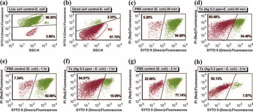
2172       JUNG ET AL.                                                                                                              APPL. ENVIRON. MICROBIOL.

                                                                                                                                                                          Downloaded from http://aem.asm.org/ on March 13, 2020 by guest
  FIG. 1. Viable counts (mean ⫾ standard error) of Staphylococcus
aureus (a) and Escherichia coli (b) bacteria after washing bacteria-
contaminated textile pieces using silver and conventional laundry ma-                FIG. 2. The effect of the silver ion solution on Staphylococcus au-
chines with (W/) or without (W/O) detergent. Each group contained                  reus (a) and Escherichia coli (b) was investigated by conventional plate
three pieces of test textiles. Inoculum, preinoculation bacterial count;           counting. The tested silver ion concentrations were 0.2 ppm, 0.1 ppm,
Ag, result for silver laundry machine (Samsung); Conventional, result              and 0.05 ppm, and PBS was used as a control.
for conventional laundry machine (Samsung). Significant differences
(P ⬍ 0.05) in viable counts of each bacteria between the silver and
conventional laundry machines are denoted with asterisks.
                                                                                   machines was performed as previously described (17a) with minor revision. The
                                                                                   bacteria were enumerated by the conventional plate count method. The test
ated silver ions was developed for hygiene, namely, in order to                    textile (100% cotton) was 5 cm ⫻ 5 cm. Three pieces of test textile were attached
                                                                                   to the edge of a 1-m ⫻ 1-m laundry textile (100% cotton). Each test and laundry
prevent easily transmissible bacterial and fungal skin infections                  textile was autoclaved and dried, and then the test textiles were inoculated with
from being transmitted by contaminated laundry. In particular,                     S. aureus or E. coli. The bacteria were diluted to 109 to 1010 CFU/ml using 0.85%
it can be beneficial to hospitals and homes in which immuno-                       sterile saline. One milliliter of each adjusted bacterial culture was inoculated to
compromised people (the elderly, children, and medical pa-                         the test textiles, and then textiles were washed in each laundry machine.
                                                                                      Two pieces of laundry textile with three pieces of test textile and 28 pieces of
tients) or pets may dwell. Our previous study demonstrated the
                                                                                   laundry textile without test textile, which were used to adjust the weight of the
antifungal activity of a laundry machine that electrically gen-                    laundry to be 3 kg, were processed at the same time with or without detergent
erates silver ions (14). In the present study, we used conven-                     using the silver and the conventional laundry machine. The laundry textile with
tional plate counting, flow cytometry (FC), and transmission                       the test textile attached to it was taken out at the end of the laundry process. The
electron microscopy (TEM) to investigate the antibacterial                         test textile was then removed from the laundry textile and pummeled with 10 ml
                                                                                   of sterile buffered peptone water (Becton Dickinson, Sparks, MD). The buffered
activity and mechanism of action against Staphylococcus aureus                     peptone water rinse solution was then serially diluted with saline, and bacteria
and Escherichia coli bacteria of a silver ion solution generated                   were counted using the conventional plate count method.
from the laundry machine.                                                             Silver ion preparation. A silver ion solution in phosphate-buffered saline
                                                                                   (PBS; pH 7.4) was prepared from the silver laundry machine (Samsung), and this
                                                                                   solution was used in all subsequent experiments (conventional plate counting,
                       MATERIALS AND METHODS
                                                                                   FC analysis, and TEM). The silver ions were produced from two silver plates
   Bacterial strains. S. aureus ATCC 25923 and E. coli ATCC 25922 were used        while PBS was passed through the silver kit, which was made with polypropylene
in this study. The strains were grown in 5% sheep blood agar (Promed, Gyeonggi,    housing. The water from the tap passed through the silver kit housing and went
Korea).                                                                            down to the drum. Both the anode and cathode were 99.9% silver metal plates
   Antibacterial efficacy test of household laundry machines. A silver laundry     with surface areas of 12.5 cm2, and two electrodes were installed parallel to each
machine (Samsung, Gyeonggi, Korea) and a conventional laundry machine              other with 5 mm of distance between them. The volume of the silver kit housing
(Samsung) which was the same as the silver laundry machine except for the fact     was 30 ml. The flow rate through the silver kit housing was regulated to be 10
that it did not emit silver ions were used as the experimental and control         liter/min, and the electric current was controlled at 80 mA by changing the input
machines, respectively. The silver laundry machine is designed to release silver   voltages from 2 to 24 V. The electric current was applied only during water
ions twice during the laundry process: once during the main washing step (for 30   supply. The concentration of the silver was determined by inductively coupled
min) and once during the final rinsing step (for 20 min). Powerclean Max (Oxy,     plasma mass spectrometry (ELAN 6100; Perkin-Elmer SCIEX, Norwalk, CT) at
Seoul, Korea) was used as the detergent.                                           the National Center for Inter-University Research Facilities, Seoul National
   The method for testing the antibacterial properties of household laundry        University, and it was approximately 0.2 ppm.
VOL. 74, 2008 ANTIBACTERIAL ACTIVITY OF ELECTRICALLY GENERATED Ag IONS 2173
Downloaded from http://aem.asm.org/ on March 13, 2020 by guest
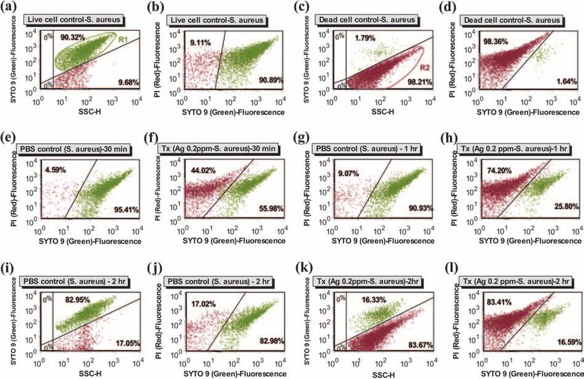
FIG. 3. Representative dot plot profiles of Staphylococcus aureus cells treated with PBS (e, g, i, and j) or silver ion solution (0.2 ppm) (Tx; f,
h, k, and l) for 30 min, 1 h, and 2 h analyzed by FC after staining with SYTO 9 and PI. For controls, suspensions of fresh live (untreated) (a and
b) and dead (70% isopropyl alcohol treated) (c and d) cells were also analyzed. The quadrants show the division between live cells in gate 1 (a;
R1-green) and damaged or dead cells in gate 2 (c; R2-red) with the relative frequencies of cells in each gate before treatment with PBS or silver
ion solution. All of the profiles were analyzed with gates placed on 1 and 2. SSC-H, side-scatter height.
Determination of antibacterial effect of silver ions by conventional plate supplemented with 90 l of sterile 1.0 M phosphate buffer (pH 8.0) and 10 l of
counting. The silver ion solution made with PBS was autoclaved at 121°C for 15 50 mM EDTA. Then, carboxyfluorescein diacetate (CFDA; Molecular Probes,
min and tested for its antibacterial efficacy. The concentrations of silver ions Inc.) stock solution in dimethyl sulfoxide was added to the sample at a final
tested were 0.2, 0.1, and 0.05 ppm. Ninety-nine milliliters of the test solution and concentration of 10 M, and the sample incubated at 35°C in the dark for 10 min
1 ml of the bacterial suspension in PBS were mixed to a final bacterial concen- (11). Following incubation, the cells were washed and resuspended in sterile 1.0
tration of 105 to 106 CFU/ml. The mixture of solution and bacteria was incubated M phosphate buffer (pH 8.0), and esterase-active bacteria were enumerated by
at 37°C with shaking and counted at 30-min intervals from 30 to 180 min and then the enhanced-green-fluorescence intensity as determined by FC analysis. Posi-
again at 24 h using the conventional plate count method, with serial 10-fold tive-control live cells and negative-control dead cells were prepared and stained
dilutions with saline plated on plate count agar (Becton Dickinson). as described above.
FC analysis of antibacterial effect of silver ions. After the bacterial suspen- TEM. Unstained cells of S. aureus and E. coli were observed for the presence
sions (105 to 106 CFU/ml) were treated with silver ion solution (0.2 ppm) or PBS of electron-dense precipitates by TEM. The two bacterial strains were diluted to
for 30 min, 1 h, 1.5 h, 2 h, and 3 h, the bacterial cells (S. aureus or E. coli) were a final concentration of 105 to 106 CFU/ml with silver ion solution (0.2 ppm) or
washed two times with PBS and resuspended in SYTO 9 and propidium iodide PBS. The mixture of solution and bacteria was incubated at 37°C for 2 h with
(PI) from a Live/Dead BacLight bacterial viability kit (Molecular Probes, Inc., shaking, centrifuged at 1,320 ⫻ g for 30 min to obtain cell pellets, and then
Eugene, OR) (2, 28). The suspension was incubated for 15 min in the dark at diluted with 1 ml of PBS. A drop of the mixture was placed on a glow-discharged
room temperature. In the control group, suspensions of fresh live (untreated) Formvar-coated copper grid for 1 min. The excess liquid was drained off with a
and dead (70% isopropyl alcohol treated) cells were stained as described above, filter paper, and the preparation was air dried for 5 min. The specimens were
and the green and red fluorescence generated by SYTO 9 and PI staining, examined with an energy-filtering TEM (LIBRA 120; Carl Zeiss, Oberkochen,
respectively, as well as the size (side scatter height) were also read by FC analysis. Germany) operated at an accelerating voltage of 120 kV. Zero-loss energy-
After reading the parameters of the live and dead cell controls, with the resulting filtered images were recorded with a 4 K slow-scan charge-coupled-device cam-
live cells in gate 1 (R1-green) and damaged or dead cells in gate 2 (R2-red) as era (4000 SP; Gatan, Pleasanton, CA).
discriminated by FC analysis, the relative frequencies of cells in each gate before In addition, the detailed ultrastructural changes induced by the silver ion
treatment with silver ion solution or PBS were determined, with all of the treatment in embedded bacterial cells were examined. The cell pellets of the two
experimental profiles being analyzed with gates 1 and 2 by FC analysis. The green bacterial strains were fixed with modified Karnovsky’s fixative consisting of 2%
fluorescence of the SYTO 9 dyes (FL1) was collected using a 530-nm ⫾ 30-nm (vol/vol) glutaraldehyde and 2% (vol/vol) paraformaldehyde in 0.05 M sodium
band-pass filter. The red fluorescence emitted from PI (FL3) was collected using cacodylate buffer (pH 7.2) at 4°C for 2 h (15). They were then washed three times
a 650-nm ⫾ 13-nm band-pass filter. The proportions of live and dead cells were with the same buffer for a period of 10 min. The specimens were postfixed with
determined and analyzed by using a FACSCalibur with the CellQuest program 1% (wt/vol) osmium tetroxide in the same buffer at 4°C for 2 h and washed briefly
(Becton Dickinson Immunocytometry Systems, San Jose, CA) and FCS Express with distilled water twice. The postfixed specimens were dehydrated in a graded
software (De Novo Software, Ontario, CA), respectively. ethanol series (once in 30, 50, 70, 80, and 95% and three times in 100% for 10
For the enumeration of esterase-active bacteria, 900 l of bacterial cells, which min each). The specimens were further treated with propylene oxide twice each
were treated with silver ion solution or PBS and washed as described above, were for 10 min as a transitional fluid and then embedded in Spurr’s resin (33).2174 JUNG ET AL. APPL. ENVIRON. MICROBIOL.
Downloaded from http://aem.asm.org/ on March 13, 2020 by guest
FIG. 4. Representative dot plot profiles of Escherichia coli cells treated with PBS (c, e, and g) or silver ion solution (0.2 ppm) (Tx; d, f, and h)
for 30 min, 1 h, and 2 h analyzed by FC after staining with SYTO 9 and PI. For controls, suspensions of fresh live (untreated) (a) and dead (70%
isopropyl alcohol-treated) (b) cells were analyzed. The quadrants show the division between live cells in gate 1 (a; R1-green) and damaged or dead
cells in gate 2 (b; R2-red) with the relative frequencies of cells in each gate before treatment with PBS or silver ion solution. All the profiles were
analyzed with gates placed on 1 and 2. SSC-H, side-scatter height.
Ultrathin sections (approximately 60-nm thickness) were cut with a diamond shown in Fig. 2. The total number of S. aureus bacteria was
knife using an ultramicrotome (MT-X; RMC Inc., Tucson, AZ) and then reduced by over 5 log10 CFU/ml after treatment with the orig-
mounted on bare copper grids. They were stained with 2% uranyl acetate and
Reynolds’ lead citrate (26) for 7 min each, followed by examination with the
inal silver ion solution (0.2 ppm) for 90 min, demonstrating
electron microscope. that the antibacterial activity of the silver ion solution was
Statistical analysis. The data from triplicate experiments are presented as the significantly greater than that of PBS treatment (P ⬍ 0.05).
mean ⫾ standard error of the mean. An unpaired t test analysis was performed The E. coli bacterial count was reduced from the inoculum size
using Origin 6.1 (OriginLab, Northampton, MA) to compare the viable bacterial
counts within different samples that underwent different washing treatments
(105 CFU/ml) to the limit of detection (⬍20 CFU/ml) within
(detergent and laundry machines) and to compare the viable bacterial counts 30 min at a silver ion concentration of 0.2 ppm. All of the
between the silver ion treatment and nontreatment groups. The proportions of tested silver ion solutions (0.2, 0.1, and 0.05 ppm) significantly
live or dead E. coli or S. aureus determined by FC analysis in the silver ion eliminated E. coli cells in comparison to PBS treatment (P ⬍
treatment groups treated for different periods of time (30 min, 1 h, 1.5 h, 2 h, and
3 h) were compared with those in the control (PBS) group using the Kruskal-
0.05).
Wallis one-way analysis of variance by rank. Significant differences in the data FC analysis in conjunction with a BacLight kit was also
that originated from the same group but were determined at different times were performed to examine the antibacterial effect of the original
analyzed by the Wilcoxon signed-rank test using Analyze-it software (Analyze-it silver ion solution (0.2 ppm) against S. aureus and E. coli
Software Ltd., Leeds, United Kingdom). The level of significance was set at a P
value of ⬍0.05.
bacteria in terms of damage to the cell membrane, shown in
different colors (green in live cells and red in damaged or dead
cells). In addition, CFDA staining was used for the enumera-
RESULTS tion of esterase-active bacteria because CFDA is cell permeant
Antibacterial efficacy of household laundry machines. The and undergoes hydrolysis of the diacetate groups into fluores-
efficacy test results of the two laundry machines against S. cent carboxyfluorescein by intracellular nonspecific esterases.
aureus and E. coli conducted with or without detergent are Based on the side light scatter and green (FL1) fluorescence,
shown in Fig. 1. The S. aureus bacterial count was significantly the R1 and R2 gates were used to identify live and damaged or
reduced by the silver laundry machines with detergent in com- dead cells, respectively. The proportions of damaged or dead
parison to the results with the conventional laundry machine cells (both S. aureus and E. coli) in the silver ion solution-
(P ⬍ 0.05). All of the inoculated E. coli bacteria were elimi- treated groups were significantly greater (P ⬍ 0.05) at 30 min,
nated when detergent was used in both the silver and conven- 1 h, 1.5 h, and 2 h of treatment than with the control (PBS)
tional laundry machines. In the absence of detergent, E. coli groups (Fig. 3 and 4). Longer treatment times (from 30 min to
was significantly reduced by the silver laundry machine in com- 2 h) had a positive effect on the antibacterial effect of the silver
parison to the results with the conventional laundry machine ion solution (P ⬍ 0.05); however, there were no significant
(P ⬍ 0.05). differences in the proportions of live or dead cells when both S.
Effect of the silver ions on the bacterial reduction rate. The aureus and E. coli cells were treated with the silver ion solution
antibacterial effects of the silver ion solution at different con- for 2 or 3 h (P ⬎ 0.05).
centrations of silver ions against S. aureus and E. coli bacteria The antibacterial-efficacy results determined by conven-
as determined by the conventional plate count technique are tional plate count and FC analyses are compared in Fig. 5. ForVOL. 74, 2008 ANTIBACTERIAL ACTIVITY OF ELECTRICALLY GENERATED Ag IONS 2175
Downloaded from http://aem.asm.org/ on March 13, 2020 by guest
FIG. 5. Comparative analysis of the antibacterial efficacy of the silver ion solution (0.2 ppm) against Staphylococcus aureus (a) and Escherichia
coli (b) bacteria as determined using the conventional plate count method and FC analysis. PBS was used as a control. Antibacterial efficacy was
calculated using the following formula: antibacterial efficacy ⫽ [(A ⫺ B)/A] ⫻ 100, where A is the preinoculation bacterial count (CFU/ml) and
B is the bacterial count after treatment with silver ion solution or PBS (CFU/ml).
the PBS-treated control group and silver ion-treated experi- release of their cellular contents into the surrounding environ-
mental groups tested, both the BacLight kit and the CFDA ment, and finally became disrupted (Fig. 6b to d). It was com-
assay gave similar antibacterial efficacies (P ⬎ 0.05). The num- mon to find electron-dense particles or precipitates around
ber of physiologically active bacteria enumerated by FC anal- damaged bacterial cells that were electron translucent in com-
ysis in conjunction with the BacLight kit or the CFDA assay parison to undamaged cells. In cross section, the untreated
was relatively higher than the bacterial count determined by cells of S. aureus showed normal cell characteristics and ho-
conventional plate counting (P ⬍ 0.05), except for E. coli mogeneous electron density in the cytoplasm. Their cell walls
bacteria after 2 and 3 h of treatment. This difference appeared and membranes were intact, showing a well-preserved pepti-
to be nonlinear across different treatment times, suggesting doglycan layer and cytoplasmic membrane (Fig. 7a and b).
that the difference in antibacterial efficacy determined by the However, significant morphological changes were observed in
two analyses decreased as the silver ion treatment times ap- S. aureus cells treated with the silver ion solution. They showed
proached 2 and 3 h. lysed cells with broken walls and membranes and decreases
Morphological changes in S. aureus and E. coli cells after and heterogeneity in electron density in the cytoplasm (Fig. 7c
silver ion treatment. TEM analysis of unstained bacteria and d). The localized separation of the cell membrane from the
showed the external morphological features of the two bacte- cell wall could be discerned.
rial strains. The untreated S. aureus cells retained their coccal E. coli cells diluted in PBS showed normal morphology hav-
morphology (ca. 600 nm in diameter) and seemed to be normal ing many filaments, such as flagella and fimbriae (Fig. 8a). The
(Fig. 6a). In contrast, S. aureus cells treated with the silver ion fimbriae were peritrichous, approximately 7 nm wide, and up
solution for 2 h appeared to undergo lysis, resulting in the to 900 nm long. Meanwhile, the bacterial cells after silver ion2176 JUNG ET AL. APPL. ENVIRON. MICROBIOL.
Downloaded from http://aem.asm.org/ on March 13, 2020 by guest
FIG. 6. External morphology of unstained Staphylococcus aureus cells
observed by TEM. (a) Untreated bacteria. (b, c, and d) Bacteria treated with FIG. 8. External morphology of unstained Escherichia coli ob-
silver ion solution (0.2 ppm). Electron-dense particles were found around served by TEM. (a) Untreated bacteria. An arrow and an arrowhead
damaged cells (arrows). Note the release of cellular contents (arrowheads). indicate fimbriae and a flagellum, respectively. (b, c, and d) Bacteria
treated with silver ion solution (0.2 ppm).
treatment for 2 h appeared to be seriously damaged (Fig. 8b to
d). The cells showed aberrant morphology; they were cracked
and ruptured. Electron-dense particles or precipitates were structure of the untreated E. coli cells appeared to be normal,
also observed around damaged bacterial cells. The internal showing a multilayered cell surface consisting of an outer
membrane, a peptidoglycan layer in the periplasmic space, and
a cytoplasmic membrane (Fig. 9a and b). Damaged cells
showed either localized or complete separation of the cell
membrane from the cell wall (Fig. 9c). The cellular degrada-
tion was also accompanied by electron-translucent cytoplasm
and cellular disruption in the damaged cells (Fig. 9d).
DISCUSSION
The electrically generated silver ion solution exhibited good
bactericidal efficacy against S. aureus and E. coli both in ex-
periments using the silver laundry machine with contaminated
fabric and in those using the silver ion suspension generated
from the silver laundry machine. The efficacy of the silver ion
solution showed better activity against the gram-negative E.
coli than against the gram-positive S. aureus. This was pos-
sibly due to the thickness of the peptidoglycan layer, which
may prevent the action of the silver ions through the bacterial
cell wall, and this result was consonant with the results of other
studies (8, 23). Although the S. aureus and E. coli bacteria were
effectively eliminated from the contaminated fabric by the sil-
ver washing course, it was not confirmed that the silver ions
killed the bacteria. It is possible that the bacteria were re-
moved from the fabric by the washing course. Therefore, the
antibacterial effect of the silver ions was confirmed by the
FIG. 7. Internal structure of Staphylococcus aureus observed by conventional plate count, FC, and TEM analyses in this study.
TEM. (a and b) Untreated bacteria. (c and d) Bacteria treated with
silver ion solution (0.2 ppm). Black and white arrows indicate pepti-
The number of bacteria determined by conventional plate
doglycan layer and cytoplasmic membrane, respectively. Note the sep- counting, which counts only culturable colonies in media, was
aration of cell membrane from the cell wall (arrowheads). significantly lower than the number determined by FC analysis,VOL. 74, 2008 ANTIBACTERIAL ACTIVITY OF ELECTRICALLY GENERATED Ag IONS 2177
tents were then released from the cell wall, and the cell wall
was degraded. These phenomena suggest possible antibacterial
mechanisms by which silver ions inhibit bacterial growth, as
well as cellular responses of both the gram-positive and gram-
negative bacteria to the silver ion treatment. Although the
mechanisms underlying the antibacterial actions of silver are
still not fully understood, several previous reports (20, 23, 32)
showed that the interaction between silver and the constituents
of the bacterial membrane caused structural changes and dam-
age to the membranes and intracellular metabolic activity
which might be the cause or consequence of cell death, as
demonstrated in this study. Analytical electron microscopy re-
mains to be done to identify the elemental composition of the
electron-dense particles or precipitates around damaged bac-
Downloaded from http://aem.asm.org/ on March 13, 2020 by guest
terial cells. In conclusion, the results of the present study
clearly show that the electrically generated silver ion solution
exerts its antibacterial effect by inducing bacteria into a state of
ABNC, in which the mechanisms required for the uptake and
utilization of substrates leading to cell division were disrupted
at the initial stage and caused the cells to undergo morpho-
logical changes and die at the later stage. These findings sug-
gest that the use of the silver ion solution may have valuable
applications in various fields, such as the manufacture of
FIG. 9. Internal structure of Escherichia coli observed by TEM. (a household appliances and medical devices.
and b) Untreated bacteria. (c and d) Bacteria treated with silver ion
solution (0.2 ppm). Arrows indicate outer membrane, peptidoglycan ACKNOWLEDGMENTS
layer, and cytoplasmic membrane from the outside of the cell. Arrow-
heads indicate separation of the cell membrane from the cell wall. We thank Young Hwan Paik for his technical assistance.
This study was supported by Korea Research Foundation grants
(KRF-2006-005-J02903 and KRF-2007-331-E00254), a grant from the
Technology Development Program for Agriculture and Forestry pro-
suggesting that the cell membrane and intracellular esterase vided by the Ministry of Agriculture and Forestry (grant no. 305003–3),
and the Korea Bio-Hub Program of the Korea Ministry of Commerce,
activity of the bacteria treated with the silver ion solution might Industry Energy (2005-B0000002). Additional support was provided by
be damaged. Bacteria in the environment are exposed to var- the Research Institute of Veterinary Science, Department of Veteri-
ious conditions that lead to survival stress. To counter this nary Microbiology, College of Veterinary Medicine, and the BK21
condition, some bacteria are capable of maintaining metabolic Program for Veterinary Science, Seoul National University.
activity while developing recalcitrance to culture. Such a state REFERENCES
in bacteria is often defined as an “active but nonculturable
1. Belly, R. T., and G. C. Kydd. 1982. Silver resistance in microorganisms. Dev.
(ABNC)” state, a state in which the bacteria exhibit measur- Ind. Microbiol. 23:567–577.
able traits of physiological activity but fail to grow to a detect- 2. Ben-Amor, K., H. Heilig, H. Smidt, E. E. Vaughan, T. Abee, and W. M. de
Vos. 2005. Genetic diversity of viable, injured, and dead fecal bacteria as-
able level (16). A state of ABNC or sublethal injury of bacteria sessed by fluorescence-activated cell sorting and 16S rRNA gene analysis.
seems to be induced by exposure to silver ions, thus rendering Appl. Environ. Microbiol. 71:4679–4689.
bacteria nonculturable in media (7, 21). This may serve as a 3. Berger, T. J., J. A. Spadaro, R. Bierman, S. E. Chapin, and R. O. Becker.
1976. Antifungal properties of electrically generated metallic ions. Antimi-
possible explanation for the discrepancy in the results deter- crob. Agents Chemother. 10:856–860.
mined by the two methods used in this study, and this obser- 4. Berger, T. J., J. A. Spadaro, S. E. Chapin, and R. O. Becker. 1976. Electri-
vation is consistent with the findings of other studies (11, 13). cally generated silver ions: quantitative effects on bacterial and mammalian
cells. Antimicrob. Agents Chemother. 9:357–358.
This finding may be expected because bacteria previously ex- 5. Bragg, P. D., and D. J. Rainnie. 1974. The effect of silver ions on the
posed to environmental stresses may only be able to divide a respiratory chain of Escherichia coli. Can. J. Microbiol. 20:883–889.
6. Brown, T., and D. Smith. 1976. The effects of silver nitrate on the growth and
limited number of times, which would give a positive result in ultrastructure of the yeast Cryptococcus albidus. Microbios Lett. 3:155–162.
the FC analysis, but they would be unable to produce visible 7. du Preez, M., R. Kfir, and P. Coubrough. 1995. Investigation of injury of
colonies on solid media. coliforms after chlorination. Water Sci. Technol. 31:115–118.
8. Feng, Q. L., J. Wu, G. Q. Chen, F. Z. Cui, T. N. Kim, and J. O. Kim. 2000.
The differences between the results of the conventional plate A mechanistic study of the antibacterial effect of silver ions on Escherichia
count and FC analyses were nonlinear, and the difference rate coli and Staphylococcus aureus. J. Biomed. Mater. Res. 52:662–668.
between the results of the two methods was reduced as time 9. Fuhrmann, G. F., and A. Rothstein. 1968. The mechanism of the partial
inhibition of fermentation in yeast by nickel ions. Biochim. Biophys. Acta
progressed. The reason for this aspect might be that the bac- 163:331–338.
teria in the ABNC state started to die after 2 h of treatment 10. Furr, J. R., A. D. Russell, T. D. Turner, and A. Andrews. 1994. Antibacterial
activity of Actisorb Plus, Actisorb and silver nitrate. J. Hosp. Infect. 27:201–
with the silver ions. 208.
Similar phenomena were also observed in the silver ion- 11. Hoefel, D., W. L. Grooby, P. T. Monis, S. Andrews, and C. P. Saint. 2003.
treated cells of S. aureus and E. coli by the TEM studies. Enumeration of water-borne bacteria using viability assays and flow cytom-
etry: a comparison to culture-based techniques. J. Microbiol. Methods 55:
Following the silver ion treatment, the cytoplasm membrane 585–597.
shrank and became separated from the cell wall. Cellular con- 12. Izatt, R. M., J. J. Christensen, and J. H. Rytting. 1971. Sites and thermo-2178 JUNG ET AL. APPL. ENVIRON. MICROBIOL.
dynamic quantities associated with proton and metal ion interaction with Escherichia coli. II. Characteristics of uptake and energy coupling with
ribonucleic acid, deoxyribonucleic acid, and their constituent bases, nucleo- transport in membrane preparations. J. Biol. Chem. 247:6332–6339.
sides, and nucleotides. Chem. Rev. 71:439–481. 26. Reynolds, E. S. 1963. The use of lead citrate at high pH as an electron-
13. Joux, F., and P. Lebaron. 1997. Ecological implications of an improved direct opaque stain in electron microscopy. J. Cell Biol. 17:208–212.
viable count method for aquatic bacteria. Appl. Environ. Microbiol. 63: 26a.Reynolds, J. E. F. 1993. Martindale extra pharmacopoeia, p. 201, 1412.
3643–3647. Pharmaceutical Press, London, England.
14. Jung, W. K., S. H. Kim, H. C. Koo, S. Shin, J. M. Kim, Y. K. Park, S. Y. 27. Richards, R. M. E., H. A. Odelola, and B. Anderson. 1984. Effect of silver on
Hwang, H. Yang, and Y. H. Park. 2007. Antifungal activity of the silver ion whole cells and spheroplasts of a silver resistant Pseudomonas aeruginosa.
against contaminated fabric. Mycoses 50:265–269. Microbios 39:151–158.
15. Karnovsky, M. J. 1965. A formaldehyde-glutaraldehyde fixative of high os- 28. Sachidanandham, R., K. Y. Gin, and C. L. Poh. 2005. Monitoring of active
molality for use in electron microscopy. J. Cell Biol. 27:137–138. but non-culturable bacterial cells by flow cytometry. Biotechnol. Bioeng.
16. Kell, D. B., A. S. Kaprelyants, D. H. Weichart, C. R. Harwood, and M. R. 89:24–31.
Barer. 1998. Viability and activity in readily culturable bacteria: a review and 29. Schreurs, W. J., and H. Rosenberg. 1982. Effect of silver ions on transport
discussion of the practical issues. Antonie van Leeuwenhoek 73:169–187. and retention of phosphate by Escherichia coli. J. Bacteriol. 152:7–13.
17. Klasen, H. J. 2000. Historical review of the use of silver in the treatment of 30. Silver, S., and L. T. Phung. 1996. Bacterial heavy metal resistance: new
burns. I. Early uses. Burns 26:117–130. surprises. Annu. Rev. Microbiol. 50:753–789.
17a.Korea Testing and Research Institute. 2001. SC9608-01. The method for 31. Slawson, R. M., M. I. Van Dyke, H. Lee, and J. T. Trevors. 1992. Germanium
testing the antibacterial properties of a household laundry machine. Korea, and silver resistance, accumulation, and toxicity in microorganisms. Plasmid
Testing and Research Institute, Seoul. 27:72–79.
Downloaded from http://aem.asm.org/ on March 13, 2020 by guest
18. Liau, S. Y., D. C. Read, W. J. Pugh, J. R. Furr, and A. D. Russell. 1997. 32. Sondi, I., and B. Salopek-Sondi. 2004. Silver nanoparticles as antimicrobial
Interaction of silver nitrate with readily identifiable groups: relationship to agent: a case study on E. coli as a model for Gram-negative bacteria. J.
the antibacterial action of silver ions. Lett. Appl. Microbiol. 25:279–283. Colloid Interface Sci. 275:177–182.
19. Reference deleted. 33. Spurr, A. R. 1969. A low-viscosity epoxy resin embedding medium for elec-
20. McDonnell, G., and A. D. Russell. 1999. Antiseptics and disinfectants: ac- tron microscopy. J. Ultrastruct. Res. 26:31–43.
tivity, action, and resistance. Clin. Microbiol. Rev. 12:147–179. 34. Thurman, R. B., and C. P. Gerba. 1989. The molecular mechanisms of
21. McFeters, G. A., M. W. LeChevallier, A. Singh, and J. S. Kippin. 1986. copper and silver ion disinfection of bacteria and viruses. CRC Crit. Rev.
Health significance and occurrence of injured bacteria in drinking water. Environ. Control 18:295–315.
Water Sci. Technol. 18:227–231. 35. Yakabe, Y., T. Sano, H. Ushio, and T. Yasunaga. 1980. Kinetic studies of the
22. Miller, L. P., and S. E. A. McCallan. 1957. Toxic action of metal ions to interaction between silver ion and deoxyribonucleic acid. Chem. Lett. 4:373–
fungus spores. Agric. Food Chem. 5:116–122. 376.
23. Pal, S., Y. K. Tak, and J. M. Song. 2007. Does the antibacterial activity of 36. Yamanaka, M., K. Hara, and J. Kudo. 2005. Bactericidal actions of a silver
silver nanoparticles depend on the shape of the nanoparticle? A study of the ion solution on Escherichia coli, studied by energy-filtering transmission
gram-negative bacterium Escherichia coli. Appl. Environ. Microbiol. 73: electron microscopy and proteomic analysis. Appl. Environ. Microbiol. 71:
1712–1720. 7589–7593.
24. Rahn, R. O., and L. C. Landry. 1973. Ultraviolet irradiation of nucleic acids 37. Zavriev, S. K., L. E. Minchenkova, M. Vorlickova, A. M. Kolchinsky, M. V.
complexed with heavy atoms. II. Phosphorescence and photodimerization of Volkenstein, and V. I. Ivanov. 1979. Circular dichroism anisotrophy of DNA
DNA complexed with Ag. Photochem. Photobiol. 18:29–38. with different modifications at N7 of guanine. Biochim. Biophys. Acta 564:
25. Rayman, M. K., T. C. Lo, and B. D. Sanwal. 1972. Transport of succinate in 212–224.You can also read