NORAD orchestrates endometrial cancer progression by sequestering FUBP1 nuclear localization to promote cell apoptosis - Nature
←
→
Page content transcription
If your browser does not render page correctly, please read the page content below
Han et al. Cell Death and Disease (2020)11:473
                                    https://doi.org/10.1038/s41419-020-2674-y                                                                                                              Cell Death & Disease

                                     ARTICLE                                                                                                                                                    Open Access

                                    NORAD orchestrates endometrial cancer
                                    progression by sequestering FUBP1 nuclear
                                    localization to promote cell apoptosis
                                    Tong Han1, Yukang Wu2, Xiang Hu1, Yaqi Chen2, Wenwen Jia2, Qizhi He3, Yiding Bian1, Mengfei Wang1, Xudong Guo2,
                                    Jiuhong Kang2 and Xiaoping Wan1

                                      Abstract
                                      Long noncoding RNAs (lncRNAs) are emerging as critical regulators in tumor initiation and progression. However, the
                                      biological mechanisms and potential clinical application of lncRNA NORAD in endometrial cancer (EC) remain
                                      unknown. Herein, we identified NORAD underwent promoter hypermethylation-associated downregulation in EC.
                                      Epigenetic inactivation of NORAD was correlated with EC progression (FIGO stage) and poor outcome. Overexpression
                                      of NORAD significantly inhibited cell growth and promoted apoptosis in EC cells. Mechanistic studies revealed that
                                      multiple regions of NORAD served as a platform for binding with the central domain of anti-apoptotic factor FUBP1.
                                      Our findings further indicated that the NORAD/FUBP1 interaction attenuated FUBP1 nuclear localization and thus
                                      impaired the occupancies of FUBP1 on its target pro-apoptotic gene promoters, resulting in apoptosis induction in EC.
                                      Moreover, knockdown of NORAD promoted tumor growth in the xenograft mice model. While, introduction of
                                      NORAD-4 fragment, which bound with FUBP1, successfully reversed tumor growth and apoptosis inhibition mediated
                  1234567890():,;
1234567890():,;
                  1234567890():,;
1234567890():,;

                                      by NORAD knockdown in vivo. Our findings provide mechanistic insight into the critical roles of NORAD as a tumor
                                      suppressor in EC progression. NORAD could possibly serve as a novel prognostic biomarker and provide the rationale
                                      for EC therapy.

                                    Introduction                                                                                        enough for diagnosis and prognosis prediction in EC,
                                      Endometrial cancer (EC), originating from the endo-                                               resulting in the dilemma of risk stratification and further
                                    metrium, is the most common malignant gynecological                                                 application of adjuvant individualized therapies at early
                                    cancer in women, and its incidence is steadily increasing                                           EC stage2,3. Therefore, it is of great importance to explore
                                    around the world without improved 5-year survival1,2. No                                            the underlying mechanisms in EC progression.
                                    acknowledged biomarkers are sensitive and specific                                                     Emerging evidences support the notion that long non-
                                                                                                                                        coding RNAs (lncRNAs), a minimum length of 200
                                                                                                                                        nucleotides, are considered as drivers of multiple cancer
                                    Correspondence: Xudong Guo (gxd.108@163.com) or                                                     phenotypes, including tumor cells sustaining prolifera-
                                    Jiuhong Kang (jhkang@tongji.edu.cn) or Xiaoping Wan (wanxiaoping@tongji.
                                                                                                                                        tion, viability, motility, and angiogenesis4–6. In view of the
                                    edu.cn)
                                    1
                                     Department of Gynecology, Shanghai First Maternity and Infant Hospital,                            biological function and specific expression in tumor tis-
                                    Tongji University School of Medicine, Shanghai 200040, China
                                    2
                                                                                                                                        sues, lncRNAs are served as biomarkers for tumor diag-
                                     Clinical and Translational Research Center of Shanghai First Maternity and
                                                                                                                                        nosis and therapeutic targets7,8. Recent studies found that
                                    Infant Hospital, Shanghai Key Laboratory of Signaling and Disease Research,
                                    Collaborative Innovation Center for Brain Science, School of Life Sciences and                      a highly conserved and abundantly expressed lncRNA,
                                    Technology, Institute for Advanced Study, Tongji University, Shanghai 200092,                       NORAD, could maintain genomic stability by decoying
                                    China
                                                                                                                                        PUMILIO1/2 or binding with RBMX to regulate DNA
                                    Full list of author information is available at the end of the article
                                    These authors contributed equally: Tong Han, Yukang Wu                                              replication and repair9–11. Genome instability was
                                    Edited by G. Blandino

                                    © The Author(s) 2020
                                                        Open Access This article is licensed under a Creative Commons Attribution 4.0 International License, which permits use, sharing, adaptation, distribution and reproduction
                                                        in any medium or format, as long as you give appropriate credit to the original author(s) and the source, provide a link to the Creative Commons license, and indicate if
                                    changes were made. The images or other third party material in this article are included in the article’s Creative Commons license, unless indicated otherwise in a credit line to the material. If
                                    material is not included in the article’s Creative Commons license and your intended use is not permitted by statutory regulation or exceeds the permitted use, you will need to obtain
                                    permission directly from the copyright holder. To view a copy of this license, visit http://creativecommons.org/licenses/by/4.0/.

                                    Official journal of the Cell Death Differentiation Association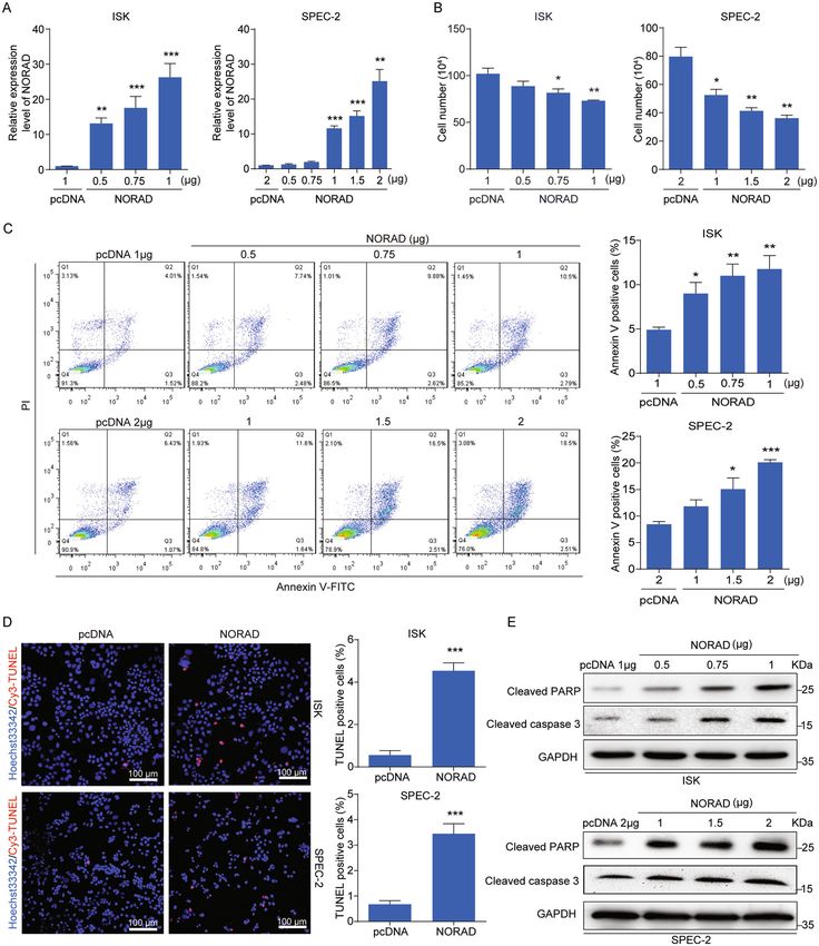
Han et al. Cell Death and Disease (2020)11:473                                                                 Page 2 of 14

recognized as one of the cancer hallmarks and involved in       such as estrogen receptor (ER) expression, etc. NORAD
tumor initiation and progression12. Several studies have        expression was decreased after 17β-estrogen treatment in
revealed that NORAD had effects on tumor cell pro-              ISK (ER-positive) and SPEC-2 (ER-negative) cells in a
liferation, apoptosis, and migration via binding with           dose- and time-dependent manner (Supplementary Fig.
miRNAs or proteins13. NORAD was identified as an                 S1a, b), consistent with no correlation of NORAD and ER
oncogene in pancreatic and ovarian cancer14,15, while its       expression. To determine the predictive value of NORAD
roles in lung and breast cancers have been controversial,       in clinical outcomes, we also evaluated the correlation
indicating a context-dependent role in cancer progres-          between NORAD expression and the 5-year overall sur-
sion16. The function and mechanism of NORAD involved            vival of EC patients, except the patients undergoing hor-
in regulating EC formation and progression remain               mone therapy or radiation prior surgery. The results
unexplored.                                                     illustrated that low NORAD expression predicted a poor
   Far upstream element-binding protein 1 (FUBP1) par-          prognosis in endometrioid endometrial adenocarcinoma
ticipated in diverse biological cellular processes as a DNA-    (the major subtype of EC) (Fig. 1c).
and RNA-binding protein17,18. Mounting evidences sug-              To investigate the mechanism of NORAD down-
gested that FUBP1 was upregulated and served as a proto-        regulation in EC, bioinformatic analysis of the NORAD
oncogene in solid cancers19,20. FUBP1 repressed p21             promoter showed that there was a CpG island (-1300 to
mRNA stabilization and regulated pro-apoptotic genes            -1475) located upstream of the transcription start site
transcription, served as an anti-apoptotic factor in hepa-      (TSS) of NORAD (Fig. 1d). The CpG island hyper-
tocellular carcinoma21. Among gyneocological cancers,           methylation phenotype (CIMP) has been established as
FUBP1 was associated with progression-free survival in          one of the hallmarks in many cancers23,24. Bisulfite
ovarian cancer22. However, there is still an important gap      sequencing PCR (BSP) was performed to investigate the
in the understanding of the role of FUBP1 in EC.                CpG methylation status of normal, peri-tumor, and
   Our study demonstrated that NORAD was gradually              tumor tissues, including early-stage and advanced-stage
decreased with the progression of EC due to promoter            tissues. Our results found that the methylation levels at
hypermethylation, and associated with clinical outcome.         the NORAD promoter were enhanced in tumor tissues
NORAD could promote EC cell apoptosis in vitro and              compared with those in normal tissues and gradually
knockdown of NORAD resulted in tumor malignant                  increased with the progression of EC (Fig. 1d). To
growth in vivo. Mechanistically, NORAD bound with               confirm these findings, we further treated ISK and
FUBP1 and impaired its nuclear localization. Conse-             SPEC-2 cells with the methyltransferase inhibitor Aza-
quently, the NORAD/FUBP1 interaction impeded FUBP1              citidine (Aza). We found that Aza treatment with
enrichment on its target gene promoters, resulting in           increasing concentration and time significantly inhibited
apoptosis induction.                                            the methylation of NORAD promoter (Fig. 1e), resulting
                                                                in rescued NORAD expression in these two cell types
Results                                                         (Fig. 1f). These results verified that the promoter
NORAD is downregulated in EC due to promoter                    hypermethylation-associated suppression of NORAD
hypermethylation and correlated with progression and            occurred in EC.
survival of EC patients                                            Overall, our study demonstrated that NORAD was
   We first analyzed the RNA-seq data of 544 EC tissues          downregulated due to its promoter hypermethylation in
and 23 normal endometrial tissues in The Cancer Gen-            EC and potentially served as a biomarker for EC pro-
ome Atlas (TCGA) and found that the expression level of         gression and prognosis.
NORAD was lower in tumor tissues than that in normal
tissues (Fig. 1a). We further collected 20 normal endo-         Overexpression of NORAD inhibits cell growth and
metrial tissues, 54 peri-tumor tissues, and 56 tumor tis-       promotes apoptosis in EC cells
sues of EC patients and classified into early stage (stage I,      To explore the exact function of NORAD in EC, we
II) and advanced stage (stage III, IV) according to Fed-        transfected NORAD into ISK and SPEC-2 cells (Fig. 2a).
eration of Gynecology and Obstetrics (FIGO) stage. We           Notably, we observed that overexpression of NORAD
found that NORAD expression was gradually decreased             significantly inhibited the cell population (Fig. 2b). The
with the progression of EC compared with that in normal         flow-cytometry analysis (FACS) revealed that NORAD
endometrial tissues (Fig. 1b).                                  triggered EC cells apoptosis (Fig. 2c), but had no sig-
   In addition, we correlated the NORAD expression level        nificant effect on cell-cycle progression (Supplementary
with the clinicopathological characteristics of EC patients     Fig. S2a, b), indicating that the impairment of cell growth
via a chi-square test (Table 1). Our results showed that        might primarily resulted from the induction of apoptosis
the expression level of NORAD was correlated with FIGO          by NORAD. In line with this observation, the induced
stages and patient age, rather than other clinical factors,     apoptosis by NORAD overexpression was also judged by

Official journal of the Cell Death Differentiation Association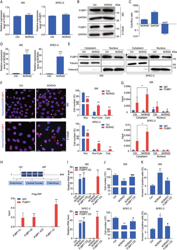
Han et al. Cell Death and Disease (2020)11:473                  Page 3 of 14

  Fig. 1 (See legend on next page.)

Official journal of the Cell Death Differentiation Association
Han et al. Cell Death and Disease (2020)11:473 Page 4 of 14
(see figure on previous page)
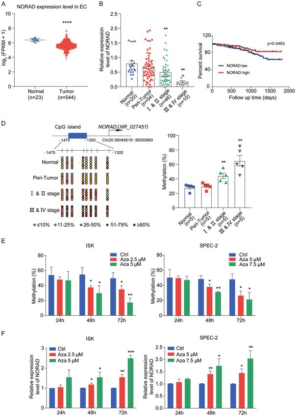
Fig. 1 Downregulation of NORAD due to promoter hypermethylation is correlated with progression and prognosis of EC. a Relative NORAD
expression in the EC patient cohort compared with that in normal endometrial tissues according to TCGA dataset. b The NORAD expression level in
20 normal endometrial tissues, 56 EC (including 46 I & II stage and 10 III & IV stage patients), and 54 peri-tumor tissues was detected by qRT-PCR. c
The survival data from TCGA EC patient cohort containing 308 endometrioid endometrial adenocarcinomas (the major subtype of EC) were analyzed
by Kaplan–Meier analysis. d Methylation status of the CpG sites at the promoter of NORAD in normal (n = 5), peri-tumor (n = 5), I & II stage (n = 5),
and III & IV stage EC patients (n = 5) was investigated by bisulfite sequencing. The average percentages of unmethylated and methylated CpGs of 10
clones from each patient were presented by different colors according to the methylated degree. e The methylation analysis of NORAD promoter in
ISK and SPEC-2 cells with the distinct doses and times of Azacitidine (Aza) treatment, performed by bisulfite sequencing. f Restored expression of
NORAD after treatment with Aza in EC cells at different doses and times. The results were determined from triplicates, and the error bars represented
as the mean ± SEM in patients’ samples, and the mean ± SD in EC cell lines, *P < 0.05, **P < 0.01, ***P < 0.001, ****P < 0.0001. NORAD noncoding RNA
activated by DNA damage, EC endometrial cancer, Aza azacitidine.
Table 1 The relation between the expression level of NORAD and clinicopathologic characteristics.
Clinicopathological data No. of patients NORAD/GAPDH expression χ2 P-value
n Low High
Total 56
FIGO stage
I & II stage 46 20 (43.5%) 26 (56.5%) 4.38 0.05
Grade II 6 5 (83.3%) 1 (16.7%)
Grade III 5 3 (60%) 2 (40%)
ER
Positive 51 24 (47.1%) 27 (52.9%) 1.41 >0.05
Negative 3 3 (100%) 0 (0%)
Histological type
Endometrioid 48 22 (45.8%) 26 (54.2%) 1.31 >0.05
Nonendometrioid 8 6 (75%) 2 (25%)
Age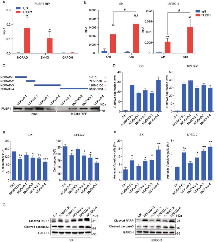
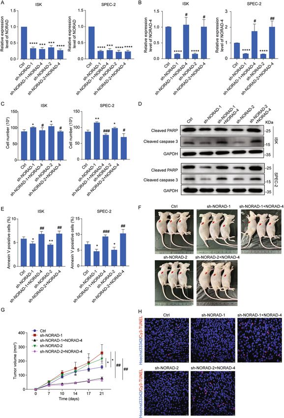
Han et al. Cell Death and Disease (2020)11:473                                                                                              Page 5 of 14

  Fig. 2 NORAD promotes apoptosis in EC cells. a qRT-PCR analysis for the expression level of NORAD in ISK and SPEC-2 EC cell lines with different
  doses transfection of NORAD, in comparison with the empty vectors. b Cell-counting assays for the control and ectopic NORAD-expressing ISK and
  SPEC-2 cells after 48 h transfection. c Increased percentage of apoptosis in the ectopic NORAD-expressing EC cells after 48 h transfection via FACS
  analysis. d TUNEL assays for apoptotic cells in the control and NORAD-expressing EC groups (left). Statistics of the TUNEL-Cy3 positive cells are shown
  (right). Scale bar, 100 μm. e Activated expression of cleaved PARP and cleaved caspase-3 was visualized by western blot. The results were determined
  from triplicates, and the error bars represented as the mean ± SD, *P < 0.05, **P < 0.01, ***P < 0.001. TUNEL TdT-mediated dUTP Nick-end labeling,
  GAPDH glyceraldehyde-3-phosphate dehydrogenase.

Official journal of the Cell Death Differentiation Association
Han et al. Cell Death and Disease (2020)11:473                                                                 Page 6 of 14

(Supplementary Fig. S3b). We found that Aza treatment           were multiple FUBP1-binding sites distributed on
did not affect the health of mice by weight supervision         NORAD. To elicit the significance of the NORAD/FUBP1
(Supplementary Fig. S3c), but substantially impaired            interaction, we transfected the full-length and four frag-
tumor growth after 17 days, and eventually achieved             ments of NORAD into EC cells (Fig. 3d) and found that
55.7% tumor inhibition at day 27 (Supplementary Fig.            the overexpression of NORAD full-length, NORAD-2,
S3d). Subsequently, TUNEL and immunofluorescence                 NORAD-3, and NORAD-4, which bound to FUBP1, sig-
staining also demonstrated that Aza treatment apparently        nificantly inhibited the number of EC cells (Fig. 3e) and
induced cell apoptosis in EC-derived tumors (Supple-            enhanced cell apoptosis (Fig. 3f, g). However, the
mentary Fig. S3e, f). Furthermore, we detected the              NORAD-1 fragment with no interaction of FUBP1 had no
decreased methylation level at the NORAD promoter               impact on the cell growth and apoptosis of EC cells (Fig.
after Aza treatment, while the NORAD expression was             3e–g). These results implied that the interaction with
consequently increased treated by Aza in the PDX model          FUBP1 was responsible for NORAD to promote cell
(Supplementary Fig. S3g, h).                                    apoptosis in EC.
  Taken together, our results demonstrated that both
endogenous and exogenous NORAD expression pro-                  NORAD-4 rescues apoptosis inhibition and tumor growth
moted EC cell apoptosis as a tumor suppressor.                  mediated by knockdown of NORAD in vitro and in vivo
                                                                  To further understand the key role of NORAD in EC
The interaction of NORAD and FUBP1 enhances cell                progression, we first constructed the NORAD knockdown
apoptosis in EC                                                 cell lines rescued by NORAD-4 fragment (bound with
  To elucidate the mechanism of NORAD in apoptosis              FUBP1) (Fig. 4a, b). Knockdown of NORAD promoted
induction, we attempted to identify its cooperative pro-        cell growth (Fig. 4c) and inhibited cell apoptosis per-
teins by exploring the mass spectrometry (MS) data10,11.        formed by FACS and detection of apoptotic proteins in
Among various partners of NORAD, we focused on the              EC cells (Fig. 4d, e). However, introduction of NORAD-4
far upstream element-binding protein 1 (FUBP1), pos-            fragment successfully reversed cell growth and apoptosis
sessing multiple binding regions on NORAD10,11, which is        inhibition in NORAD knockdown cells (Fig. 4c–e). In
critical for antagonizing apoptosis and promoting cell          addition, we introduced the NORAD-4 rescued cell lines
survival in hepatocellular and colorectal carcinoma20,21.       into the xenograft mice model. Knockdown of NORAD
FUBP1 expression was upregulated in the EC tissues              resulted in excessive tumor growth and reduced apoptosis
compared with normal tissues according to TCGA data             (Fig. 4f–h). While introduction of NORAD-4 fragment
(Supplementary Fig. S4a), which was consistently con-           significantly impaired the tumor growth and cell apop-
firmed in the tissue sections of EC patients by immuno-          tosis inhibition mediated by knockdown of NORAD
histochemistry (Supplementary Fig. S4b). In addition,           in vivo (Fig. 4f–h). In conclusion, NORAD played a key
knockdown of FUBP1 resulted in the inhibition of cell           role in EC progression via interacting with FUBP1.
growth and induction of apoptosis (Supplementary Fig.
S4c–g), indicating that FUBP1 played an important role in       NORAD affects the cytosol–nuclear trafficking of FUBP1
resistance to apoptosis in EC as well. To validate the          through its central domain
interaction of NORAD and FUBP1, we overexpressed                  We next focused on investigating how the NORAD/
FUBP1 in 293FT cells and performed RNA immunopre-               FUBP1 interaction induced apoptosis. We first noticed
cipitation (RIP) to detect FUBP1 binding with NORAD             that overexpression of NORAD was unable to modulate
and SNHG1 (FUBP1-binding RNA)25 (Fig. 3a). Moreover,            the mRNA or protein level of FUBP1 (Fig. 5a, b). FUBP1 is
we performed RIP assays in EC cells under the condition         often recognized as a nuclear protein based on its
of endogenous NORAD expression rescued by Aza, which            recognition ability of FUSE element17, while increasing
showed the increased binding of NORAD on FUBP1 (Fig.            evidences indicate that FUBP1 can interact with cyto-
3b). To further clarify the specific regions of NORAD            plasmic RNAs26,27. Cell fractionation followed by RT-
responsible for FUBP1 binding, we divided NORAD into            qPCR revealed that NORAD was predominately located
four fragments (NORAD-1, 2, 3, and 4) according to the          in the cytoplasm (Fig. 5c), suggesting that the interaction
predicted peaks of the FUBP1-binding regions in the MS          of NORAD and FUBP1 might occur in the cytoplasm.
data10. These four fragments of NORAD were tagged with          Notably, we identified that overexpression of NORAD
an MS2 sequence and co-transfected with FUBP1 and               attenuated FUBP1 nuclear accumulation by subcellular
MS2bp-YFP (fused by MS2-binding protein and yellow              fractionation followed by western blot (Fig. 5d, e). We
fluorescent protein) into 293FT cells to perform RNA             subsequently performed immunofluorescence staining to
pull-down assays (Fig. 3c). Our results showed that three       analyze the ratio of FUBP1 distribution only in the cyto-
fragments of NORAD (NORAD-2, 3, and 4), rather than             plasm, nucleus, or both (Fig. 5f). The statistics indicated
NORAD-1, could bind to FUBP1, suggesting that there             that overexpression of NORAD apparently impaired the

Official journal of the Cell Death Differentiation Association
Han et al. Cell Death and Disease (2020)11:473 Page 7 of 14 Fig. 3 The binding of NORAD and FUBP1 is essential for NORAD to induce apoptosis. a qRT-PCR analysis for the binding of NORAD, SNHG1, and GAPDH expression by an anti-FUBP1 antibody compared with that by an IgG control antibody in 293FT cells. SNHG1 and GAPDH served as the positive and negative controls, respectively. b qRT-PCR analysis for the binding of NORAD on FUBP1 in EC cells with Aza treatment by RIP assays. c Western blot for the FUBP1 protein (lower panel) pulled down by truncated NORAD (NORAD-1, -2, -3, and -4; upper panel). d The expression levels of full-length NORAD and fragments transfected in ISK and SPEC-2 cells were detected by qRT-PCR. e Cell-counting assays for ISK and SPEC-2 cells transfected with full-length NORAD and fragments, respectively. f The percentage of apoptotic cells transfected with full-length NORAD and fragments, evaluated by FACS analysis. g Western blot for the expression of cleaved PARP and cleaved caspase-3 after ectopic expression of full- length NORAD and fragments. The results were determined from triplicates, and the error bars represented as the mean ± SD, */# P < 0.05, **P < 0.01, ***P < 0.001. FUBP1 far upstream element-binding protein 1, SNHG1 small nucleolar RNA host gene 1, RIP RNA immunoprecipitation, YFP yellow fluorescent protein. Official journal of the Cell Death Differentiation Association
Han et al. Cell Death and Disease (2020)11:473                  Page 8 of 14

  Fig. 4 (See legend on next page.)

Official journal of the Cell Death Differentiation Association
Han et al. Cell Death and Disease (2020)11:473                                                                                          Page 9 of 14

  (see figure on previous page)
  Fig. 4 NORAD-4 rescues the apoptosis inhibition and tumor growth mediated by knockdown of NORAD in vitro and in vivo. a, b The
  expression level of NORAD (a) and NORAD-4 (b) was detected by qRT-PCR in NORAD knockdown cell lines and in NORAD knockdown rescued by
  NORAD-4 fragment cell lines. c Cell-counting assays for the NORAD knockdown and rescued by NORAD-4 cell lines. d The expression levels of
  apoptotic associated markers were detected by western blot in the NORAD knockdown and rescued by NORAD-4 cell lines. e The percentage of
  apoptotic cells in the NORAD knockdown and rescued by NORAD-4 cell lines was analyzed by FACS. f SPEC-2-derived cell lines (sh-NORAD-1/2, sh-
  NORAD-1/2 + NORAD-4) were subcutaneously injected into the hind flanks of nude mice. g Tumor volume was monitored from day 0 to day 21 post
  injection. h Apoptosis in tumor tissues was presented by TUNEL assay. Scale bar, 10 μm. The results were determined from triplicates, and the error
  bars represented as the mean ± SD, */#P < 0.05, */##P < 0.01, ***/###P < 0.001, ****P < 0.0001.

nuclear localization of FUBP1, suggesting that the con-                      that these pro-apoptotic genes were co-regulated by
vergence     of      NORAD      and    FUBP1       altered                   NORAD and FUBP1. Chromatin Immunoprecipitation
FUBP1 subcellular localization. Consequently, we per-                        (ChIP) assay identified that overexpression of NORAD
formed the subcellular fractionation followed by RIP                         significantly reduced the FUBP1 occupancies on these
assays. Our results identified that overexpression of                         four gene promoters (Fig. 6e). Consistently, the enrich-
NORAD significantly enhanced the interaction with                             ment of RNA polymerase II on these gene promoters was
FUBP1 in the cytoplasm, resulting in the impairment of                       enhanced after NORAD overexpression (Fig. 6f). More-
FUBP1 localized in nucleus (Fig. 5g). Next, to elucidate                     over, we transfected NORAD-1 (without interaction with
the specific region of FUBP1 binding with NORAD, we                           FUBP1), which had no impact on the expression, FUBP1
constructed three depletion mutants of FUBP1                                 occupancy, or transcription activity of these downstream
(FUBP1 ΔN, ΔCD, and ΔC) according to its functional                          target genes (Fig. 6d–f). Taken together, we demonstrated
domains (including the N-terminal inhibitory domain,                         that the interaction of NORAD and FUBP1 affected the
central domain, and C-terminal transactivation                               nuclear distribution of FUBP1 and facilitated its down-
domain)17,18. Our results showed that only the deletion of                   stream pro-apoptotic gene transcription, eventually
FUBP1 central domain abolished its interaction with                          resulting in apoptosis induction in EC cells.
NORAD (Fig. 5h). The central domain of FUBP1 pos-
sesses a dual role in DNA and RNA binding17,18.                              Discussion
Accordingly, we ascertained that NORAD prevented the                           LncRNAs have been recently emerged as important
nuclear translocation of FUBP1 by binding with its central                   players in modulating tumor initiation, progression, and
domain as a “decoy”. Then, we co-transfected NORAD                           the assessment of prognosis4–6. NORAD was recently
and the central domain of FUBP1 fragment (FUBP1 CD)                          discovered to promote cancer cells proliferation, invasion,
into ISK and SPEC-2 cells (Fig. 5i). We found that over-                     and metastasis in various cancers (such as bladder, pan-
expression of the FUBP1 CD fragment significantly                             creatic, cervical cancer, etc.)13,14,28,29. While, NORAD
reversed the cell growth inhibition and apoptosis induc-                     retained controversial roles in liver, lung, and breast
tion mediated by NORAD (Fig. 5j, k), indicating that the                     cancer16,30, implying that NORAD acted on tumorigenesis
dominant-negative fragment of FUBP1 CD competitively                         and progression in a context-dependent manner. In our
bound with NORAD in the cytosol to facilitate endo-                          study, we analyzed the public data in TCGA and collected
genous FUBP1 translocation into the nucleus, where it                        the tumor tissues of EC patients, which illustrated that
rescued cell viability.                                                      NORAD was downregulated due to promoter hyper-
                                                                             methylation in EC patients compared with normal tissues.
NORAD/FUBP1 interaction regulates the downstream pro-                        Moreover, epigenetic inactivation of NORAD was rele-
apoptotic genes                                                              vant to adverse progression and poor prognosis. There-
  We further explored the downstream targets of FUBP1                        fore, for the first time, we propose NORAD as a potential
to execute its anti-apoptotic effect. Gene set enrichment                    molecular marker for the clinical assessment of EC pro-
analysis (GSEA) showed that upon the high and low                            gression and prognosis.
expression of FUBP1 in liver cancer, downstream genes                          Normal cells suffering from external or internal stress
were evidently relevant to the apoptosis pathway (Fig. 6a,                   will trigger DNA damage and repair31. Once defective
b). Previous studies have reported that FUBP1 suppressed                     cells failed to repair effectively, cell death pathways
the transcription of the pro-apoptotic genes (such as BIK,                   (apoptosis, necrosis, or autophagy) would be activated to
NOXA, TRAIL, and TNFA)21. Our findings showed that                            eliminate the negative effects of genomic toxicity on
knockdown of FUBP1 indeed upregulated the expression                         cells32,33. However, during the process of tumor initiation,
level of these four genes (Fig. 6c), which were also pro-                    these defective cells could bypass the cell death pathway
moted by NORAD overexpression (Fig. 6d), indicating                          to complete the malignant transition, which might be the

Official journal of the Cell Death Differentiation Association
Han et al. Cell Death and Disease (2020)11:473 Page 10 of 14 Fig. 5 (See legend on next page.) Official journal of the Cell Death Differentiation Association
Han et al. Cell Death and Disease (2020)11:473                                                                                           Page 11 of 14

  (see figure on previous page)
  Fig. 5 NORAD impairs the nuclear localization of FUBP1 through its central domain. a, b The FUBP1 expression level after the introduction of
  NORAD in ISK and SPEC-2 cells via qRT-PCR (a) and western blot (b). c The subcellular distribution of NORAD was analyzed by qRT-PCR. GAPDH and
  XIST genes were used as controls for the cytoplasmic and nuclear fractions, respectively. d The expression level of NORAD was detected by qRT-PCR.
  e The fractionation of FUBP1 was visualized by western blot after ectopic expression of NORAD in ISK and SPEC-2 cells. GAPDH and Histone 3
  indicated the cytoplasmic and nuclear fractions, respectively. f Immunofluorescence assays indicated the altered localization of FUBP1 (red) after
  introduction of NORAD in ISK and SPEC-2 cells (left). Quantifications of the percentages of FUBP1 presented only in the nucleus, in the cytoplasm, and
  both in the nucleus and cytoplasm are shown (right). White arrows represented the cells which FUBP1 was distributed both in the cytoplasm and
  nucleus. Yellow arrows represented the cells in which FUBP1 was distributed only in the cytoplasm. Scale bar, 25 μm. g The subcellular fractionation
  followed by RIP assays was performed to analyze the interaction of NORAD and FUBP1 in the cytoplasmic and nuclear lysates of NORAD
  overexpressing cells. h qRT-PCR analysis of NORAD immunoprecipitated by Flag-tagged full-length and three deleted mutations of FUBP1 in
  293FT cells compared with the IgG control. i The expression level of NORAD and FUBP1 CD truncation was detected by qRT-PCR. j Cell-counting
  assays of the rescued cell growth by FUBP1 CD in the NORAD-expressing ISK and SPEC-2 cells. k FACS analysis of the rescued percentage of apoptotic
  cells by FUBP1 CD in the NORAD-expressing ISK and SPEC-2 cells. The results were determined from triplicates, and the error bars represented as the
  mean ± SD, */#P < 0.05, **P < 0.01, ***/###P < 0.001. XIST X inactivation-specific transcript.

important cause of tumor formation31. Previous studies                        was reported to bind to the FUBP1 promoter and activate
revealed that NORAD bound with PUMILIO1/2 or                                  its transcription in erythroid differentiation36. FUBP1 was
RBMX to maintain genomic stability9–11. However, inac-                        also regulated by the PI3K/AKT/mTOR pathway and cas-
tivation of NORAD downregulated the expression of                             pase protein in liver cancer37 or targeted by miR-16 in
genes associated with DNA damage and repair, such as                          breast and gastric cancer38. In addition, FUBP1 was found
RBMX and PARP1, resulting in genomic instability10. Our                       to be ubiquitinated by p38 in lung cell differentiation or
study indicated that the expression of NORAD was                              identified as a substrate of parkin in Parkinson’s disease39,40.
decreased in the transition from normal endometrial tis-                      Intriguingly, our results showed that NORAD had no
sue to EC tissue. Knockdown of NORAD could promote                            impact on the transcription or stability of FUBP1 but
tumor growth and prevent cell apoptosis in vitro and                          attenuated FUBP1 nuclear enrichment, which impaired its
in vivo. While, both exogenous NORAD overexpression                           occupancies on the promoters of downstream pro-
and rescued endogenous NORAD expression by Aza                                apoptotic genes. Our results further found that FUBP1
could inhibit cell growth and promote cell apoptosis, as a                    interacted with NORAD through its central domain
tumor suppressor. Thus, these results indicated that                          (DNA-/RNA-binding region). Overexpression of the
NORAD was critically involved in the balance between                          FUBP1 central domain could competitively bind with
cell proliferation and apoptosis evasion in EC progression.                   NORAD and facilitate endogenous FUBP1 trafficking into
   Cytoplasmic distributed lncRNAs can execute their reg-                     the nucleus to reverse the cell apoptosis induction mediated
ulatory roles through binding with proteins to affect their                   by NORAD. FUBP1, primarily located in the nucleus, was
function, subcellular localization, or protein–protein inter-                 prevented to be imported into the nucleus due to caspase-3/
action4,34,35. NORAD contains multiple repetitive motifs                      7 cleavage during the breast and cervical cancer cells
and serves as a molecular decoy for the PUMILIO protein,                      apoptosis41. Herein, we provided an optional mechanism
indicating that NORAD might function as a platform for                        that NORAD decoyed FUBP1 in the cytosol and impaired
assembling proteins10. In view of FUBP1 function in EC and                    its translocation to the nucleus, which was responsible for
its multiple binding regions on NORAD in the MS data10,11,                    apoptosis induction in EC.
we confirmed the interaction of NORAD and FUBP1 under                             In conclusion, we elucidate that epigenetic inactivation of
exogenous and endogenous conditions in EC, and identified                      NORAD affects the cytosol–nucleus trafficking of the anti-
FUBP1 binding with at least three regions of NORAD. We                        apoptotic factor FUBP1 and the expression of its target pro-
also revealed that binding with FUBP1 was essential for                       apoptotic genes, resulting in EC cells evasion from apop-
NORAD to induce apoptosis in EC cells. Furthermore,                           tosis. In this regard, investigating NORAD crosstalk will
introduction of NORAD-4 fragment (bound with FUBP1)                           lead us to significant insights into the mechanism of EC
could reverse cell growth and apoptosis inhibition mediated                   progression. Moreover, we are the first to highlight the
by knockdown of NORAD in vitro and in vivo. These                             predictive clinical value of NORAD as an EC diagnostic and
findings suggested that NORAD had the capacity to interact                     prognostic biomarker and the possibility of developing
with multiple FUBP1 proteins as a decoy to regulate cell                      NORAD-targeted therapy.
apoptosis.
   To date, FUBP1 was ascertained to be upregulated in                        Materials and methods
colorectal and hepatocellular cancer20,21. Our study also                     Sample collections from patients
revealed that FUBP1 was upregulated in EC, and knock-                           EC (n = 56) tissues were collected from patients who
down of FUBP1 remarkably enhanced cell apoptosis. TAL1                        underwent hysterectomy at the Tongji University Affiliated

Official journal of the Cell Death Differentiation Association
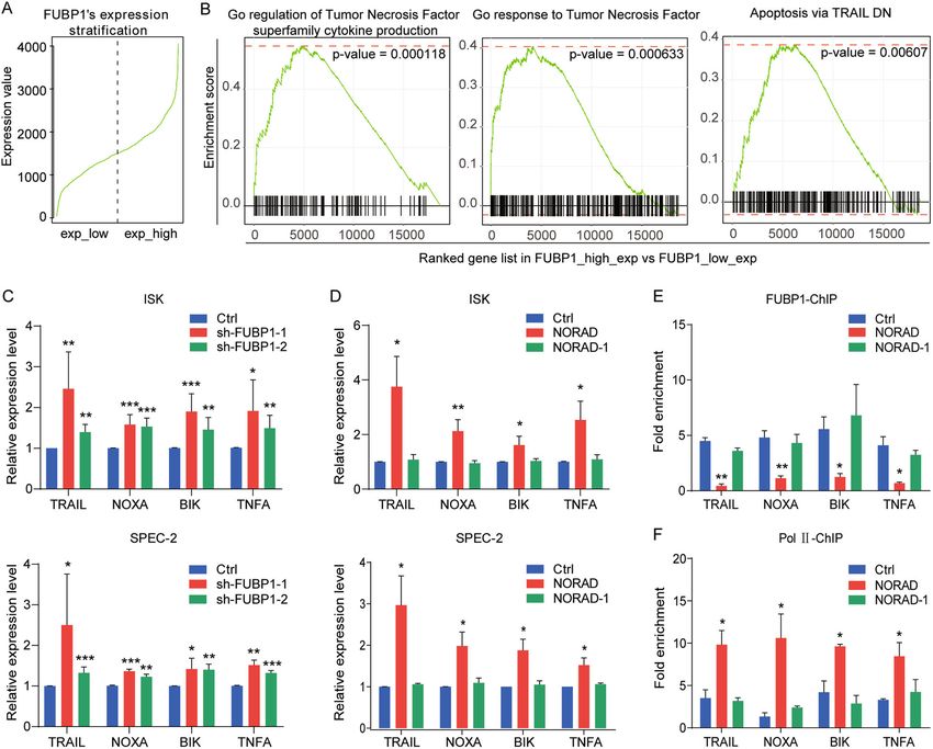
Han et al. Cell Death and Disease (2020)11:473                                                                                            Page 12 of 14

  Fig. 6 The NORAD/FUBP1 interaction results in the upregulation of downstream pro-apoptotic genes. a RNA-seq data of liver cancer from
  TCGA to classify FUBP1 high- and low-expressed groups. b GSEA for the FUBP1-related pathways in liver cancer. c qRT-PCR analysis for the expression
  of four FUBP1 downstream targets (TRAIL, NOXA, BIK, and TNFA) by knockdown of FUBP1. d qRT-PCR analysis for the expression of four FUBP1
  downstream target genes by the introduction of full-length NORAD or the NORAD-1 fragment, which was not bound to FUBP1. e, f ChIP-qPCR
  analysis for the FUBP1 (e) and RNA polymerase II (f) occupancies at the promoters of four target genes (TRAIL, NOXA, BIK, and TNFA) after transfecting
  full-length NORAD or NORAD-1 fragment. The fold enrichment was relative to the input DNA. The results were determined from triplicates, and the
  error bars represented as the mean ± SD, *P < 0.05, **P < 0.01, ***P < 0.001. TNF tumor necrosis factor, TRAIL TNF-related apoptosis-inducing ligand,
  NOXA PMAIP1, phorbol-12-myristate-13-acetate-induced protein 1, BIK BCL2 interacting killer, ChIP chromatin immunoprecipitation, Pol II RNA
  polymerase II.

Shanghai First Maternity and Infant Hospital (Shanghai,                        patients was provided in Supplementary Table S1. The
China) from 2015 to 2019. Peri-tumor endometrial tissues                       research project was approved by the Human Investigation
(n = 54) were sampled 1–2 cm away from tumors in the                           Ethical Committee of Tongji University Affiliated Shanghai
surgeries42. Normal endometrial specimen (n = 20) were                         First Maternity and Infant Hospital.
collected from women undergoing non-maligmant diseases
(uterine leiomyoma) with no underlying endometrial                             RNA immunoprecipitation (RIP)
pathology43. The histology of all tissues was verified by two                     RIP was performed as previously described44. A total of 5 ×
independent pathologists. No patients had undergone                            106 cells were lysed with lysate buffer. Protein A Magnetic
endocrine therapy, radiotherapy, or chemotherapy before                        Beads (161-4013, Bio-Rad) and Protein G Magnetic Beads
surgery. All patients have signed the informed consent form                    (161-4023, Bio-Rad) were incubated with 3 μg of antibodies,
before collection. The detailed clinical information of these                  rotating for at least 6 h. Lysates were added to the prepared

Official journal of the Cell Death Differentiation Association
Han et al. Cell Death and Disease (2020)11:473                                                                                                           Page 13 of 14

beads in RIP buffer, rotating overnight for immunoprecipi-                       Publisher’s note
tation. Finally, RNA was extracted with RNAiso Plus                              Springer Nature remains neutral with regard to jurisdictional claims in
                                                                                 published maps and institutional affiliations.
Reagent. The antibodies in RIP assay were followed as: anti-
FUBP1 (ab192867, Abcam), anti-IgG-Rb (#2729, Cell Sig-                           Supplementary Information accompanies this paper at (https://doi.org/
naling Technology), and anti-flag-Rb (14793s, Cell Signaling                      10.1038/s41419-020-2674-y).
Technology).
                                                                                 Received: 17 December 2019 Revised: 3 June 2020 Accepted: 8 June 2020

Xenograft mice model
   Five-week-old female BALB/c nude mice were purchased
from the National Resource Center for Rodent Laboratory                          References
Animals of China. The mice used in animal studies were                            1. Siegel, R. L., Miller, K. D. & Jemal, A. Cancer statistics, 2020. CA Cancer J. Clin. 70,
randomly and blindly allocated into experimental and                                  7–30 (2020).
                                                                                  2. Brooks, R. A. et al. Current recommendations and recent progress in endo-
control group. Five SPEC-2-derived cell lines (Ctrl, sh-                              metrial cancer. CA Cancer J. Clin. 69, 258–279 (2019).
NORAD-1, sh-NORAD-2, sh-NORAD-1 + NORAD-4, sh-                                    3. Morice, P., Leary, A., Creutzberg, C., Abu-Rustum, N. & Darai, E. Endometrial
NORAD-2 + NORAD-4), suspended at the concentration                                    cancer. Lancet 387, 1094–1108 (2016).
                                                                                  4. Schmitt, A. M. & Chang, H. Y. Long noncoding RNAs in cancer pathways.
of 1 × 10⁷ cells in 100 μL of PBS, were subcutaneously                                Cancer Cell 29, 452–463 (2016).
injected into the hind flanks of nude mice (n = 3, each                            5. Gutschner, T. & Diederichs, S. The hallmarks of cancer: a long non-coding RNA
group). On the 7th day after injection, mice were monitored                           point of view. RNA Biol. 9, 703–719 (2012).
                                                                                  6. Prensner, J. R. & Chinnaiyan, A. M. The emergence of lncRNAs in cancer
and the tumor volume was calculated using the formula 1/2                             biology. Cancer Discov. 1, 391–3407 (2011).
(length × width2) twice a week. The mice were sacrificed at                        7. Bussemakers, M. J. et al. DD3: a new prostate-specific gene, highly over-
day 21 post injection. These studies were approved by the                             expressed in prostate cancer. Cancer Res. 59, 5975–5979 (1999).
                                                                                  8. Wei, J. T. et al. Can urinary PCA3 supplement PSA in the early detection of
Institutional Animal Care and Use Committee of Tongji                                 prostate cancer? J. Clin. Oncol. 32, 4066–4072 (2014).
University (no. TJLAC-019-103).                                                   9. Tichon, A. et al. A conserved abundant cytoplasmic long noncoding RNA
                                                                                      modulates repression by Pumilio proteins in human cells. Nat. Commun. 7,
                                                                                      12209 (2016).
Statistical analysis                                                             10. Lee, S. et al. Noncoding RNA NORAD regulates genomic stability by
  The statistical analyses were performed with GraphPad                              sequestering PUMILIO proteins. Cell 164, 69–80 (2016).
Prism 7 software. The results were from triplicate experi-                       11. Munschauer, M. et al. The NORAD lncRNA assembles a topoisomerase com-
                                                                                     plex critical for genome stability. Nature 561, 132–136 (2018).
ments, and the data was presented as the mean ± SEM or                           12. Hanahan, D. & Weinberg, R. A. Hallmarks of cancer: the next generation. Cell
mean ± SD. The significance of mean values was deter-                                 144, 646–674 (2011).
mined by unpaired two-tailed Student’s t test. Pearson’s chi-                    13. Yang, Z. et al. Noncoding RNA activated by DNA damage (NORAD):
                                                                                     biologic function and mechanisms in human cancers. Clini. Chim. Acta
square test and nonparametric test were used to analyze the                          489, 5–9 (2019).
clinical variables. The survival times of different groups of                    14. Li, H. et al. Long noncoding RNA NORAD, a novel competing endo-
patients were analyzed using the Kaplan-method with the                              genous RNA, enhances the hypoxia-induced epithelial-mesenchymal
                                                                                     transition to promote metastasis in pancreatic cancer. Mol. Cancer 16,
log-rank test. */#, **/##, ***/###, and **** represent P < 0.05, P                   169 (2017).
< 0.01, P < 0.001, and P < 0.0001, respectively.                                 15. Yang, X., Yan, Y., Chen, Y., Li, J. & Yang, J. Involvement of NORAD/miR-608/
                                                                                     STAT3 axis in carcinostasis effects of physcion 8-O-beta-glucopyranoside on
                                                                                     ovarian cancer cells. Artif. Cell Nanomed. B 47, 2855–2865 (2019).
Acknowledgements                                                                 16. Tan, B. S. et al. LncRNA NORAD is repressed by the YAP pathway and sup-
We are grateful for Dr. Joshua T. Mendell’s laboratory (University of Texas          presses lung and breast cancer metastasis by sequestering S100P. Oncogene
Southwestern Medical Center) for providing the plasmid of pcDNA3.1-NORAD.            38, 5612–5626 (2019).
This project was supported by grants from Shanghai Municipal Medical and         17. Zhang, J. & Chen, Q. M. Far upstream element binding protein 1: a com-
Health Discipline Construction Projects (No.2017ZZ02015 to X.W.), Ministry of        mander of transcription, translation and beyond. Oncogene 32, 2907–2916
Science and Technology of China [2016YFA0101300 and 2018YFA0800100],                 (2013).
Chinese National Natural Science Foundation [81672574, 81972438, 81530042,       18. Quinn, L. M. FUBP/KH domain proteins in transcription: Back to the future.
31830059, 31970599, and 31701110], and Fundamental Research Funds for the            Transcription 8, 185–192 (2017).
Central Universities [22120200105].                                              19. Singer, S. et al. Coordinated expression of stathmin family members by far
                                                                                     upstream sequence element-binding protein-1 increases motility in non-small
                                                                                     cell lung cancer. Cancer Res. 69, 2234–2243 (2009).
Author details
1                                                                                20. Jia, M. Y. & Wang, Y. J. Far upstream element-binding protein 1 (FUBP1)
 Department of Gynecology, Shanghai First Maternity and Infant Hospital,
                                                                                     expression differs between human colorectal cancer and non-cancerous tis-
Tongji University School of Medicine, Shanghai 200040, China. 2Clinical and
                                                                                     sue. Neoplasma 61, 533–540 (2014).
Translational Research Center of Shanghai First Maternity and Infant Hospital,
                                                                                 21. Rabenhorst, U. et al. Overexpression of the far upstream element binding
Shanghai Key Laboratory of Signaling and Disease Research, Collaborative
                                                                                     protein 1 in hepatocellular carcinoma is required for tumor growth. Hepa-
Innovation Center for Brain Science, School of Life Sciences and Technology,
                                                                                     tology 50, 1121–1129 (2009).
Institute for Advanced Study, Tongji University, Shanghai 200092, China.
3                                                                                22. Pénzváltó, Z. et al. MEK1 is associated with carboplatin resistanceand is a
 Department of Pathology, Shanghai First Maternity and Infant Hospital, Tongji
                                                                                     prognostic biomarker in epithelial ovarian cancer. BMC Cancer 14, 837 (2014).
University School of Medicine, Shanghai 200040, China
                                                                                 23. Shen, H. & Laird, P. W. Interplay between the cancer genome and epigenome.
                                                                                     Cell 153, 38–55 (2013).
Conflict of interest                                                              24. Jones, P. A. & Baylin, S. B. The fundamental role of epigenetic events in cancer.
The authors declare that they have no conflict of interest.                           Nat. Rev. Genet. 3, 415–428 (2002).

Official journal of the Cell Death Differentiation Association
Han et al. Cell Death and Disease (2020)11:473 Page 14 of 14
25. Sun, Y. et al. The long noncoding RNA SNHG1 promotes tumor growth 35. Willingham, A. T. et al. A strategy for probing the function of noncoding RNAs
through regulating transcription of both local and distal genes. Oncogene 36, finds a repressor of NFAT. Science 309, 1570–1573 (2005).
6774–6783 (2017). 36. Steiner, M. et al. FUSE binding protein 1 (FUBP1) expression is upre-
26. Sully, G. et al. Structural and functional dissection of a conserved destabilizing gulated by T-cell acute lymphocytic leukemia protein 1 (TAL1) and
element of cyclo-oxygenase-2 mRNA: evidence against the involvement of required for efficient erythroid differentiation. PLoS ONE 14, e0210515
AUF-1 [AU-rich element/poly(U)-binding/degradation factor-1], AUF-2, triste- (2019).
traprolin, HuR (Hu antigen R) or FBP1 (far-upstream-sequence-element-bind- 37. Samarin, J. et al. PI3K/AKT/mTOR-dependent stabilization of oncogenic far-
ing protein 1). Biochem. J 377, 629–639 (2004).. upstream element binding proteins in hepatocellular carcinoma cells. Hepa-
27. Zhang, Z., Harris, D. & Pandey, V. N. The FUSE binding protein is a cellular tology 63, 813–826 (2016).
factor required for efficient replication of hepatitis C virus. J. Virol. 82, 38. Venturutti, L. et al. MiR-16 mediates trastuzumab and lapatinib
5761–5773 (2008). response in ErbB-2-positive breast and gastric cancer via its novel
28. Huo, H. et al. Long non-coding RNA NORAD upregulate SIP1 expression to targets CCNJ and FUBP1. Oncogene 35, 6189–6202 (2016).
promote cell proliferation and invasion in cervical cancer. Biomed. Pharmac- 39. Ko, H. S., Kim, S. W., Sriram, S. R., Dawson, V. L. & Dawson, T. M. Identification of
other. 106, 1454–1460 (2018). far upstream element-binding protein-1 as an authentic Parkin substrate. J.
29. Li, Q. et al. High expression of long noncoding RNA NORAD indicates a poor Biol. Chem. 281, 16193–16196 (2006).
prognosis and promotes clinical progression and metastasis in bladder cancer. 40. Kim, M. J. et al. Downregulation of FUSE-binding protein and c-myc by tRNA
Urol. Oncol. 36, e15–10 e22 (2018). synthetase cofactor p38 is required for lung cell differentiation. Nat. Genet. 34,
30. Hu, B. et al. Long non-coding RNA 657 suppresses hepatocellular carcinoma 30–36 (2003).
cell growth by acting as a molecular sponge of miR-106a-5p to regulate PTEN 41. Jang, M. et al. Far upstream element-binding protein-1, a novel caspase
expression. Int. J. Biochem. Cell B 92, 34–42 (2017). substrate, acts as a cross-talker between apoptosis and the c-myc oncogene.
31. Roos, W. P., Thomas, A. D. & Kaina, B. DNA damage and the balance between Oncogene 28, 1529–1536 (2009).
survival and death in cancer biology. Nat. Rev. Cancer 16, 20–33 (2016). 42. Chen, J. et al. Snail recruits Ring1B to mediate transcriptional
32. Tubbs, A. & Nussenzweig, A. Endogenous DNA damage as a source of repression and cell migration in pancreatic cancer cells. Cancer Res.
genomic instability in cancer. Cell 168, 644–656 (2017). 74, 4353–4363 (2014).
33. Zhivotovsky, B. & Kroemer, G. Apoptosis and genomic instability. Nat. Rev. Mol. 43. Xu, Q. et al. MELK promotes endometrial carcinoma progression via activating
Cell Biol. 5, 752–762 (2004). mTOR signaling pathway. eBioMedicine 51, 102609 (2020).
34. Sharma, S. et al. Dephosphorylation of the nuclear factor of activated T cells 44. Guo, X. et al. A Linc1405/Eomes complex promotes cardiac
(NFAT) transcription factor is regulated by an RNA-protein scaffold complex. mesoderm specification and cardiogenesis. Cell Stem Cell 22, 93–908
Proc. Natl Acad. Sci. USA 108, 11381–11386 (2011). e6 (2018).
Official journal of the Cell Death Differentiation AssociationYou can also read