BIOACTIVE PEPTIDE ISOLATED FROM SESAME SEEDS INHIBITS CELL PROLIFERATION AND INDUCES APOPTOSIS AND AUTOPHAGY IN LEUKEMIC CELLS
←
→
Page content transcription
If your browser does not render page correctly, please read the page content below
EXCLI Journal 2021;20:709-721 – ISSN 1611-2156
                                     Received: January 18, 2021, accepted: March 17, 2021, published: March 23, 2021

                                                 Original article:

    BIOACTIVE PEPTIDE ISOLATED FROM SESAME SEEDS INHIBITS
        CELL PROLIFERATION AND INDUCES APOPTOSIS AND
                AUTOPHAGY IN LEUKEMIC CELLS

Kamolchanok Deesrisak1 , Yodying Yingchutrakul2 , Sucheewin Krobthong2,
Sittiruk Roytrakul3 , Chawalit Chatupheeraphat1, Paweena Subkorn1,
Usanarat Anurathapan4 , Dalina Tanyong1*
1
     Department of Clinical Microscopy, Faculty of Medical Technology, Mahidol University,
     Nakhon Pathom 73170, Thailand
2
     Proteomics Research Team, National Omics Center, National Science and Technology
     Development Agency, Pathum Thani 12120, Thailand
3
     Functional Proteomics Technology Laboratory, Functional Ingredients and Food
     Innovation Research Group, National Center for Genetic Engineering and Biotechnology,
     National Science and Technology for Development Agency, Pathum Thani 12120,
     Thailand
4
     Department of Pediatrics, Faculty of Medicine Ramathibodi Hospital, Mahidol University,
     Bangkok 10400, Thailand

* Corresponding author: Dalina Tanyong, Department of Clinical Microscopy, Faculty
  of Medical Technology, Mahidol University, 999 Phuttamonthon sai 4 Road, Salaya,
  Phuttamonthon, Nakhon Pathom 73170, Thailand; Tel: +662-441-4371-5 Ext. 2836;
  Fax: +662-441-4380; E-mail: dalina.itc@mahidol.ac.th

http://dx.doi.org/10.17179/excli2021-3406

This is an Open Access article distributed under the terms of the Creative Commons Attribution License
(http://creativecommons.org/licenses/by/4.0/).

                                                      ABSTRACT
Leukemia is the most common type of hematological malignancies. Several natural products including bioactive
peptides have been explored and studied for their anti-leukemic activities. In the present study, anti-leukemic
peptide, IGTLILM (IM-7), was isolated and identified from the protein hydrolysate of sesame seeds by reverse
phase-solid phase extraction, off-gel fractionation and nano LC-MS/MS. The cytotoxic effects of IM-7 were stud-
ied in MOLT-4 and NB4 acute leukemic cell lines using an MTT assay. The induction of apoptosis and autophagy
was investigated by flow cytometry using Annexin V-FITC/PI staining and anti-LC3/FITC antibodies, respec-
tively. The mRNA alterations of apoptotic and autophagic-related genes were determined by reverse transcription-
quantitative PCR. The present study found that IM-7 inhibited the proliferation of MOLT-4 and NB4 cells in dose-
dependent manner, but it showed a minimal effect on healthy mononuclear cells. IM-7 activated apoptosis and
autophagy through the upregulation of CASP3, ULK1 and BECN1 and the downregulation of BCL2. In addition,
IM-7 enhanced the cytotoxic effect of the anti-leukemic drug, daunorubicin. The findings suggested that IM-7 was
potent to suppress the proliferation of MOLT-4 and NB4 leukemic cells and induce apoptosis and autophagy
through the regulation of caspase 3-Bcl-2 and ULK1-Beclin1, respectively.

Keywords: Apoptosis, autophagy, bioactive peptide, leukemia, sesame

                                                            709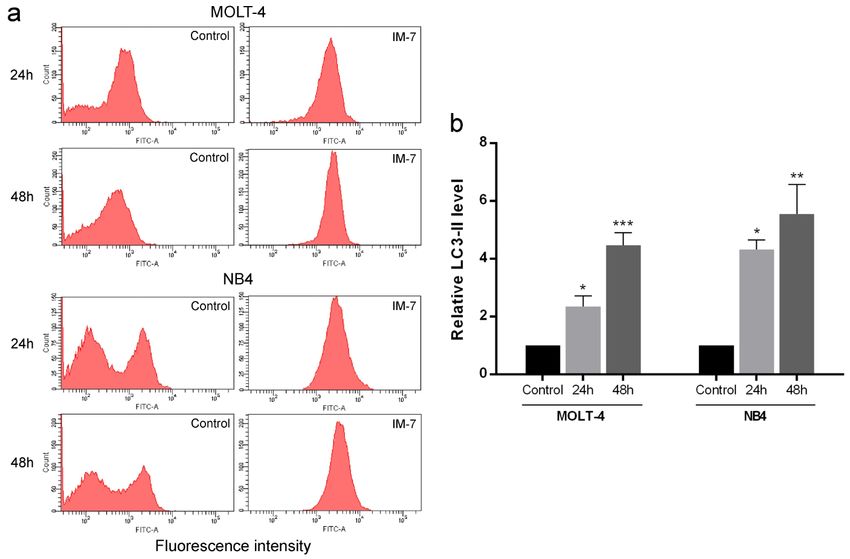
EXCLI Journal 2021;20:709-721 – ISSN 1611-2156
                             Received: January 18, 2021, accepted: March 17, 2021, published: March 23, 2021

             INTRODUCTION                             death has been used for eliminating the num-
                                                      ber of leukemic cells (Cassier et al., 2017).
     Leukemia is one of the hematological ma-
                                                      Likewise, autophagy as known as type II pro-
lignancies characterized by uncontrolled pro-
                                                      grammed cell death is an alternative approach
duction of abnormal leukocytes. Comparing
                                                      for leukemia treatment (Zhang et al., 2013).
to all sites of cancer, 2.4 % cases and 3.2 %
                                                      GX15-070 (obatoclax) has been reported to
deaths of leukemia were reported (Bray et al.,
                                                      inhibit the proliferation of acute lymphocytic
2018). Chemotherapy is the main treatment
                                                      leukemia via the induction of apoptosis and
for most types of leukemia. However, the
                                                      autophagy (Heidari et al., 2010). Same as
complications from intensive chemotherapy
                                                      resveratrol, the natural polyphenol found in
are the limitation and may lead to relapse in
                                                      several plants, it has been shown to induce the
some patients (Jarfelt et al., 2016; Yilmaz et
                                                      apoptotic and autophagic death in leukemic
al., 2019). Over the years, therapeutic pep-
                                                      cells (Fan et al., 2018; Zhang et al., 2018).
tides have been studied and developed as
                                                          Thus, the present study purposed to iso-
drugs due to their advantages, including small
                                                      late the anti-leukemic peptides from sesame
size, ease of synthesis and modification, tu-
                                                      seeds and examine the underlying mecha-
mor-penetrating ability and low toxicity
                                                      nisms related to apoptosis and autophagy in
(Thundimadathil, 2012). The anti-leukemic
                                                      MOLT-4 and NB4 leukemic cell lines.
peptides extracted from natural sources, in-
cluding plants and animals, have been re-
ported. For example, cyclotides extracted                   MATERIALS AND METHODS
from Violaceae families and Psychotria lep-
                                                      Preparation of sesame protein hydrolysate
tothyrsa have been shown to possess dose-de-               Black sesame seeds were purchased from
pendent cytotoxicity in U-937 cell line               local supermarket (Nakhon Pathom, Thai-
(Gerlach et al., 2010; Herrmann et al., 2008;         land). To obtain protein hydrolysate, sesame
Svangård et al., 2007; Yeshak et al., 2011).          seeds were ground and dissolved in 10 mM
Moreover, cationic peptide-CM4 from hemo-             sodium acetate buffer pH 4.0 with pepsin hy-
lymph of the silkworm Bombyx mori has been            drolysis at 37 °C for 18 h. Following the in-
reported to inhibit the proliferation of leuke-       cubation period, the enzyme activity was
mic cells (Chen et al., 2010).                        stopped by boiling for 10 min. The protein hy-
     Sesame (Sesamum indicum L.) is one of            drolysate in supernatant was collected by cen-
the oilseed crops commonly used in various            trifugation at 5,000 rpm for 20 min, and stored
types of food and pharmaceutical products.            at 4 °C for further purification.
Sesame seeds contain the valuable nutrients
and bioactive chemical agents such as sesa-
                                                      Determination of protein concentration by
min, sesamol and sesaminol that have exhib-
                                                      the Bradford assay
ited the therapeutic activities including anti-           The protein concentration was determined
oxidant, anti-inflammatory and anticancer ac-         using the Bradford assay (Bradford, 1976).
tivities (Pathak et al., 2017). Besides, the pro-     The bovine serum albumin was used for set-
teins and peptides derived from sesame seeds          ting a standard curve.
have been reported for the medicinal proper-
ties, including antioxidant (Liu and Chiang,
                                                      Purification of sesame peptides by reverse
2008; Lu et al., 2019), anti-bacterial (Das et
                                                      phase-solid phase extraction
al., 2012) and anti-hypertensive (Nakano et               The protein hydrolysate from sesame
al., 2006) activities. However, there is no re-       seeds was filtered through a 3 kDa cutoff
port about the effect of sesame peptides on           membrane and fractionated by reverse phase-
leukemia.                                             solid phase extraction (RP-SPE) using Sep-
     Since leukemia is characterized by the ex-       Pak C18 cartridge (Waters, Milford, MA,
cessive production of blood cells, the induc-         USA). The cartridge was pre-conditioned by
tion of apoptosis or type I programmed cell

                                                 710
EXCLI Journal 2021;20:709-721 – ISSN 1611-2156
Received: January 18, 2021, accepted: March 17, 2021, published: March 23, 2021
acetonitrile and equilibrated by water. Then, acetonitrile in 0.1 % formic acid within 20
the cartridge was loaded by sesame peptides min. The raw data were analyzed by DeCyder
and washed with water. The elution was done MS 2.0 software (GE Healthcare, Piscataway,
with 10 % stepwise concentration of acetoni- NJ, USA) using PepDetect module for pep-
trile at 1 ml/min of flow rate. Each 5 ml of tide detection and quantitation. The analyzed
eluates was pooled, removed acetonitrile us- data were searched against the NCBI protein
ing vacuum concentrator and determined the database of Sesamum indicum L. (35,825 pro-
cytotoxicity in MOLT-4 and NB4 leukemic teins) for peptide matching by Mascot soft-
cell lines. The effective samples were selected ware (Matrix Science, London, UK) with in-
for subsequent purification. terrogation of taxonomy (Sesamum indicum),
variation modification (oxidation) and pep-
Purification of sesame peptides by off-gel tide charge (1+, 2+ and 3+). The peptides with
fractionation high ID score which indicates the similarity
The samples from RP-SPE that showed between analyzed peptides and databases
the cytotoxic effects were selected for purifi- were selected to synthesize by GenScript Bi-
cation by off-gel fractionation using an Ag- otech (Nanjing, China) for determining the
ilent 3100 OFFGEL Fractionator (Agilent anti-leukemic activities.
Technologies, Santa Clara, CA, USA). The
immobilized pH gradient strips (Immo- Leukemic cell culture and isolation of pe-
biline™ DryStrip pH 3-10 NL, 13 cm, GE ripheral blood mononuclear cell (PBMC)
Healthcare, Uppsala, Sweden) were rehy- The human acute T-lymphocytic leuke-
drated with 0.1 % IPG buffer (50 μl/well) for mic cell line MOLT-4 and human acute pro-
1 h. The samples were diluted to final concen- myelocytic leukemic cell line NB4 were pur-
tration of 0.05 % IPG and 200 μl of diluted chased from Cell Line Service GmbH (Eppel-
sample was loaded in each well. The 12-well heim, Germany). The cell lines were main-
separations were focused for 16 kVh with a tained in RPMI-1640 medium supplemented
maximum current of 50 μA and power of 200 with 10 % FBS and 1 % penicillin-streptomy-
mW. After focusing, residue solution in each cin (Thermo Fisher Scientific, Inc., Waltham,
well was collected, measured the protein con- MA, USA) in a humidified incubator at 37 °C
centration, and determined the cytotoxic ac- with 5 % CO2. PBMCs from healthy donors’
tivities. The fractions with high cytotoxic ac- blood were isolated using Lymphoprep™
tivities were cleaned up by PureSpeed C18 (Axis-Shield PoC AS, Oslo, Norway). In
Desalting Tips (Mettler Toledo, Columbus, brief, the blood was diluted with PBS at 1:1
OH, USA), dried using a vacuum centrifuga- ratio and gently layered on top of Lympho-
tion, and resuspended in 0.1 % formic acid for prep™ solution. The separation was per-
injecting into the mass spectrometer. formed by centrifugation at 800 x g for 30 min
with brake off. The PBMC layer was har-
Nano liquid chromatography-tandem vested and washed twice with medium. The
mass spectrometry analysis and peptide isolated PBMCs were used for further experi-
identification ment. This study was conformed to the stand-
The samples were injected into the Ulti- ard set by the Declaration of Helsinki and ap-
mate 3000 LC System (Thermo Fisher Scien- proved by the committee for research, Faculty
tific, Inc., Waltham, MA, USA), which cou- of Medicine Ramathibodi Hospital, Mahidol
pled with an impact II tandem mass spectrom- University (COA. MURA2019/679).
eter and a captive spray ion source on a nano-
column PepSwift monolithic column 100 μm Determination of cytotoxic effect by MTT
x 50 mm (Bruker Daltonik GmbH, Bremen, assay
Germany). The separation was performed us- The leukemic cells and PBMCs were
ing a linear gradient from 10-70 % of 80 % treated with various concentrations of sesame
711EXCLI Journal 2021;20:709-721 – ISSN 1611-2156
Received: January 18, 2021, accepted: March 17, 2021, published: March 23, 2021
peptides, synthetic IM-7 and/or daunorubicin. Reverse transcription-quantitative PCR
After incubation time, 10 μl of the 5 mg/ml (RT-qPCR) analysis
MTT [3-(4,5-dimethylthiazol-2-yl)-2,5-di- The leukemic cells were treated with IM-
phenyltetrazolium bromide] (Thermo Fisher 7 at the IC50 concentration for 24 h. Total
Scientific, Inc., Waltham, MA, USA) was RNA was extracted using GENEzol™ rea-
added to each well, followed by incubation gent (Geneaid biotech, New Taipei City, Tai-
for 4 h at 37 °C. The formazan crystal was wan) and 1 μg of total RNA was converted to
dissolved in 100 μl of solubilizing solution cDNA using RevertAid First Strand cDNA
(10 % SDS in 0.01 N HCl) overnight at 37 °C. Synthesis kit (Thermo Fisher Scientific, Inc.,
The absorbance was determined using micro- Waltham, MA, USA). qPCR was performed
plate reader at 570 nm. The half maximal in- with the master mix containing 2 μl template,
hibitory concentration (IC50) was calculated 0.5 μl of each primer and 10 μl Luna® Univer-
from linear regression and used for further ex- sal qPCR Master Mix (New England Biolabs,
periment. Ipswich, MA, USA) at the final volume of
20 μl with Bio-Rad CFX96 touch™ real-time
Determination of apoptotic cells by flow PCR detection system (Bio-Rad, Hercules,
cytometry CA, USA). The amplification conditions were
MOLT-4 and NB4 cell lines were treated initial denaturation at 95 °C for 15 sec, fol-
with IM-7 at the IC50 value. Following 24 and lowed by 40 cycles of denaturation at 95 °C
48 h of incubation, the cells were washed for 15 sec and extension at 60 °C for 30 sec.
twice with cold PBS. The apoptotic cells were The mRNA expression was analyzed using
determined using the FITC Annexin V Apop- the mean Cq value and represented as 2-∆∆Cq
tosis Detection kit (BD Biosciences, San Jose, using GAPDH as an internal control. The pri-
CA, USA) by staining the cells with 5 μl An- mers used in this study were shown in Table
nexin V-FITC and 5 μl propidium iodide (PI) 1.
for 15 min in the dark and analyzed by FACS
Table 1: The primers for RT-qPCR analysis
Canto II flow cytometer (BD Biosciences,
San Jose, CA, USA) within 1 h. Gene Primer sequence (5’-3’)
CASP3
Forward TTCAGAGGGGATCGTTGTAGAAGTC
Measurement of LC3-II autophagic marker Reverse CAAGCTTGTCGGCATACTGTTTCAG
by flow cytometry BCL2
Forward ATGTGTGTGGAGAGCGTCAA
The leukemic cells were treated with IM- Reverse GCCGTACAGTTCCACAAAGG
7 at the IC50 concentration for 24 and 48 h. ULK1
The LC3-II level was measured using Forward GGCAAGTTCGAGTTCTCCCG
Reverse CGACCTCCAAATCGTGCTTCT
FlowCellect™ Autophagy LC3 Antibody- BECN1
based Assay kit (Merck KGaA, Darmstadt, Forward GAGTTTCAAGATCCTGGACCGTGTCA
Reverse CTGTTGGCACTTTCTGTGGACATCA
Germany) according to the manufacturer’s in- GAPDH
structions. Briefly, at 30 min before the end of Forward GCACCGTCAAGGCTGAGAA
incubation time, 10 μl of diluted reagent A Reverse AGGTCCACCACTGACACGTTG
was added followed by incubation for 30 min
at 37 °C. Then, the cell pellet was resus- Statistical analysis
pended in 100 μl of reagent B followed by im- The results are exhibited as mean ± SEM.
mediate spinning. The cells were stained with For single variable comparisons, Student’s t-
anti-LC3/FITC antibody for 30 min in the test was used. For multiple variable compari-
dark and analyzed by flow cytometer. The sons, data were analyzed by one-way
LC3-II level was calculated from the mean ANOVA followed by Dunnett’s test using
fluorescence intensity (MFI) by the following GraphPad Prism 6. The statistically signifi-
equation: relative LC3-II level = MFI (Treat- cant difference was defined as PEXCLI Journal 2021;20:709-721 – ISSN 1611-2156
Received: January 18, 2021, accepted: March 17, 2021, published: March 23, 2021
RESULTS
Sesame peptides exhibit anti-leukemic
activities
After purification of sesame peptides by
RP-SPE, each sample at 35 μg/ml was treated
in MOLT-4 and NB4 cells for 24 h and the
cytotoxicity was determined using an MTT
assay. The results showed that the samples at
retention time of 5, 10, 15 and 70 min had cy-
totoxic effects in both cell lines (Figure 1a).
The effective samples from RP-SPE were fur-
ther purified by off-gel fractionation. The 25
fractions obtained from off-gel fractionation
(5, 8, 8 and 4 fractions from RP-SPE samples
at retention time of 5, 10, 15 and 70 min, re-
spectively) at 25 μg/ml were treated in
MOLT-4 and NB4 cells for 24 h. The cyto-
toxic effects were examined by MTT assay.
The results showed that fraction number 11
and 12 reduced the viability of MOLT-4 and
NB4 (Figure 1b) and were subsequently se-
lected to identify peptide sequences by nano Figure 1: Cytotoxic effect of sesame peptides in
LC-MS/MS. MOLT-4 and NB4 cells. (a) The samples of ses-
ame peptides obtained from RP-SPE were treated
IM-7 exhibits the highest cytotoxicity in in MOLT-4 and NB4 cells at 35 μg/ml for 24 h fol-
lowed by MTT assay. (b) The effective samples
MOLT-4 and NB4 cells from RP-SPE were subsequently purified by off-
The fraction number 11 and 12 obtained gel fractionation. The obtained fractions were
from the off-gel fractionation were injected treated in MOLT-4 and NB4 cells at 25 μg/ml for
into nano LC-MS/MS for peptide sequencing. 24 h followed by MTT assay.
The peptide sequences were analyzed by
DeCyder MS 2.0 software and searched
against the database of Sesamum indicum L. IM-7 selectively exhibits cytotoxicity to
using Mascot software. The sequence of leukemic cells
eleven peptides with high ID score and their The leukemic cells and PBMCs were
located proteins were presented in Table 2. treated with various concentrations of syn-
The eleven peptides were synthesized with thetic IM-7 (0, 1, 1.5 and 2 mg/ml) for 24 and
purity ≥ 85 % and screened for their cytotoxic 48 h. The cell viability was determined by
effects in MOLT-4 and NB4 cells at 1 mg/ml MTT assay. The results revealed that IM-7
for 24 h of incubation using an MTT assay. significantly inhibited the proliferation of
As shown in Figure 2, IM-7 (IGTLILM) sig- MOLT-4 and NB4 in dose-dependent man-
nificantly reduced the proliferation of MOLT- ner. The IC50 concentration of IM-7 at 24 h
4 and NB4 cells while other peptides did not was 1.0 and 1.1 mg/ml in MOLT-4 and NB4,
show the cytotoxic effects. IM-7 was chosen respectively. Remarkably, IM-7 had a mini-
for the further experiment. mal toxicity on healthy PBMCs at 1 and 1.5
mg/ml, but showed a significant effect at the
highest concentration after 48 h of treatment
(Figure 3).
713EXCLI Journal 2021;20:709-721 – ISSN 1611-2156
Received: January 18, 2021, accepted: March 17, 2021, published: March 23, 2021
Table 2: The identified sesame peptides and their located proteins
Accession no. Protein name Peptide sequence Peptide ID score
name
gi|747070351 ER membrane protein complex RDILDPPG RG-8 46.20
subunit 4
gi|747043127 Aquaporin NIP6-1-like IGTLILM IM-7 45.69
gi|747051445 Uncharacterized protein SAPNEIF SF-7 35.71
gi|747051452 Uncharacterized protein
gi|747052022 Uncharacterized protein
gi|1173787700 Uncharacterized protein
gi|1173813316 Uncharacterized protein FRNLLPR FR-7 34.62
gi|747050743 Uncharacterized protein GQALRRATG GG-9 32.77
gi|747050745 Uncharacterized protein
gi|747074755 Leukotriene A-4 hydrolase AAKLNIPR AR-8 30.08
homolog
gi|747074757 Leukotriene A-4 hydrolase
homolog
gi|747082122 Phosphatidylinositol 4-kinase LKVRINRKLSV LV-11 26.90
alpha 1 isoform X2
gi|1173817273 Phosphatidylinositol 4-kinase
alpha 1 isoform X1
gi|1173824446 WAT1-related protein CGIAVTGGT CT-9 25.11
At5g64700
gi|747055456 Uncharacterized protein ADLMAKIS AS-8 24.51
gi|747107952 Probable sodium/metabolite co- LHAVLLA LA-7 23.93
transporter BASS4, chloroplastic
isoform X1
gi|1173754476 Probable sodium/metabolite co-
transporter BASS4, chloroplastic
gi|1173754878 Probable sodium/metabolite co-
transporter BASS4, chloroplastic
isoform X2
gi|747103098 Uncharacterized protein YDRVDAF YF-7 23.66
gi|1173778764 Uncharacterized protein
gi|1173778766 Uncharacterized protein
714EXCLI Journal 2021;20:709-721 – ISSN 1611-2156
Received: January 18, 2021, accepted: March 17, 2021, published: March 23, 2021
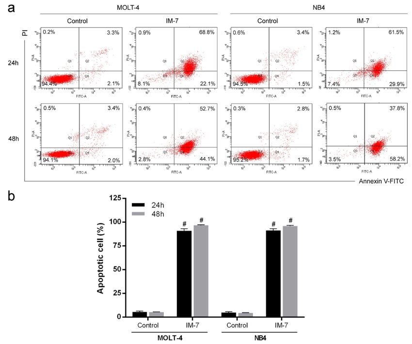
IM-7 induces apoptosis in MOLT-4 and using Annexin V-FITC and PI staining fol-
NB4 cells lowed by flow cytometry. The results showed
To evaluate apoptosis induced by IM-7, that IM-7 induced apoptosis in MOLT-4 and
MOLT-4 and NB4 cells were treated with IM- NB4 by increasing the number of Annexin V-
7 at the IC50 value. Following 24 and 48 h of FITC-positive cells (Figure 4).
incubation, the apoptotic cells were analyzed
Figure 2: Screening for the cytotoxic effects of identified peptides in MOLT-4 and NB4 cells. MOLT-4
and NB4 cells were treated with eleven synthetic peptides obtained from DeCyder and Mascot analysis
at 1 mg/ml for 24 h. The cell viability was determined by MTT assay. #PEXCLI Journal 2021;20:709-721 – ISSN 1611-2156
Received: January 18, 2021, accepted: March 17, 2021, published: March 23, 2021
Figure 4: Effect of IM-7 on apoptosis in leukemic cells. (a) MOLT-4 and NB4 cells were treated with IM-
7 at the IC50 value for 24 and 48 h. The apoptotic cells were analyzed by flow cytometry. (b) Quantified
results of apoptosis assay. #PEXCLI Journal 2021;20:709-721 – ISSN 1611-2156
Received: January 18, 2021, accepted: March 17, 2021, published: March 23, 2021
Figure 5: Effect of IM-7 on autophagy in leukemic cells. (a) MOLT-4 and NB4 cells were treated with
IM-7 at the IC50 concentration for 24 and 48 h. The level of LC3-II autophagic marker was measured as
mean fluorescence intensity using flow cytometry. (b) Quantified results of autophagy assay. *PEXCLI Journal 2021;20:709-721 – ISSN 1611-2156
Received: January 18, 2021, accepted: March 17, 2021, published: March 23, 2021
IM-7 enhances the cytotoxicity of anti- Following 24 and 48 h of incubation, cell vi-
leukemic drug ability was determined by MTT assay. The re-
To evaluate the role of IM-7 in comple- sults revealed that IM-7 enhanced the cyto-
mentary treatment, leukemic cells and toxicity of daunorubicin in MOLT-4 and NB4
PBMCs were treated with the combination compared to the single treatment of IM-7 or
between IC50 value of IM-7 (1.0 mg/ml for daunorubicin. However, this combination
MOLT-4 and 1.1 mg/ml for NB4) and the containing 1.0 mg/ml of IM-7 and 1.6 μM of
well-known anti-leukemic drug daunorubicin daunorubicin appeared not to affect the cell
(1.6 μM for MOLT-4 and 1.3 μM for NB4). viability of healthy PBMCs (Figure 7).
Figure 7: Effect of
IM-7 in combination
with daunorubicin.
MOLT-4 (a) and NB4
(b) leukemic cells
and healthy PBMCs
(c) were treated with
IM-7 at IC50 value
with or without dau-
norubicin for 24 and
48 h. The cell viability
was determined by
MTT assay. *PEXCLI Journal 2021;20:709-721 – ISSN 1611-2156
Received: January 18, 2021, accepted: March 17, 2021, published: March 23, 2021
DISCUSSION (Luna Vital et al., 2014). Lactoferricin, milk-
derived peptide, has been shown to exert cy-
Over the past years, proteins and peptides
totoxic effect and increase apoptotic level in
have been developed for treatment in several
Jurkat leukemic cells (Mader et al., 2005).
diseases. More than 30 % of the therapeutic
Moreover, melittin, a polypeptide derived
proteins and peptides database have been en-
from the bee venom, has been found to induce
tered in clinical studies and 12 % of them
apoptosis through the upregulation of caspase
have been approved as drugs by FDA (Lau
3 and downregulation of Bcl-2 in U-937 leu-
and Dunn, 2018). Among the FDA approved
kemic cells (Moon et al., 2008).
protein and peptide-based drugs, 12 % are
Autophagy is a cellular process character-
classified as anticancer drugs (Usmani et al.,
ized by the formation of autophagosomes and
2017) such as bortezomib for multiple mye-
fusion with the lysosomes to degrade cellular
loma (Chen et al., 2011) and enfortumab ve-
components. It is regulated by several factors
dotin-ejfv for bladder cancer (de la Torre and
including autophagy-related genes (Atg),
Albericio, 2020). The present study discov-
mammalian target of rapamycin (mTOR) and
ered a bioactive peptide, namely IM-7
class III phosphatidylinositol 3-kinase
(IGTLILM), which was isolated from pepsin-
(PtdIns3K) (Glick et al., 2010). UNC-51-like
treated protein hydrolysate of sesame seeds. It
kinase 1 (ULK1) and Beclin1 that play a vital
was found to possess anti-leukemic activities
role at initiated step of autophagy have been
by inhibiting the proliferation of acute lym-
reported in the treatment of cancer. The acti-
phocytic leukemia MOLT-4 and acute pro-
vation of autophagy through AMPK/ULK1
myelocytic leukemia NB4 in dose-dependent
by natural flavone, baicalein, was effective
manner. However, it exhibited less effect on
for the treatment of prostate and breast cancer
healthy PBMCs. IM-7 majorly contained the
(Aryal et al., 2014). LYN-1604, a designed
hydrophobic amino acids that might be the
ULK1 agonist, has been shown to induce au-
important composition for cytotoxicity of leu-
tophagic cell death through the activation of
kemic cells. Previous studies have reported
ULK1 in breast cancer cells (Zhang et al.,
the positive correlation between anticancer
2017). Moreover, Beclin1 has been reported
activities and peptide hydrophobicity. The
as an insufficient tumor suppressor gene in
highly hydrophobic peptides can deeply pen-
several types of cancer and the disruption or
etrate into the hydrophobic core of cancer cell
deletion of Beclin1 can promote tumorigene-
membrane followed by pore formation and
sis (Qu et al., 2003; Yue et al., 2003). The re-
cancer cell lysis (Huang et al., 2011).
sults of the present study demonstrated that
Apoptosis is the biological process of cell
IM-7 activated autophagy by increasing the
death characterized by cell shrinkage, mem-
level of LC3-II in leukemic cells in time-de-
brane blebbing, phosphatidylserine exposure,
pendent manner. The effect was supported by
chromatin condensation and DNA fragmenta-
the upregulation of ULK1 and BECN1 ex-
tion. The mechanism is regulated by stimula-
pression.
tion of pro-apoptotic factors and suppression
In addition, the natural compounds have
of anti-apoptotic factors that are the promis-
been proposed to improve the effects of anti-
ing strategies for development of the anti-
cancer drugs by synergizing the anti-prolifer-
cancer drugs (Pistritto et al., 2016). It was
ation activity, reducing the chemotherapy-in-
found that IM-7 induced the apoptosis of
duced toxicity and suppressing the develop-
MOLT-4 and NB4 via the regulation of
CASP3 and BCL2. Several studies have in- ment of drug resistance (Fu et al., 2018;
vestigated the effect of natural-derived pep- Kojima-Yuasa et al., 2015; Rejhová et al.,
tides on apoptosis in cancer cells. The pep- 2018). In the present study, IM-7 enhanced
tides extracted from non-digestible fraction of the effect of anti-leukemic drug daunorubicin.
the common beans have been shown to pro- However, this combination had a minimal ef-
mote the apoptosis of colon cancer cells fect on healthy PBMCs. This suggested that
719EXCLI Journal 2021;20:709-721 – ISSN 1611-2156
Received: January 18, 2021, accepted: March 17, 2021, published: March 23, 2021
co-treatment of IM-7 and daunorubicin has a Chen YQ, Min C, Sang M, Han YY, Ma X, Xue XQ,
synergistic effect on anti-leukemic activities et al. A cationic amphiphilic peptide ABP-CM4 exhib-
its selective cytotoxicity against leukemia cells. Pep-
in MOLT-4 and NB4 cells. tides. 2010;31:1504-10.
Taken together, the present study demon-
strated the anti-leukemic activities of IM-7 Das R, Dutta A, Bhattacharjee C. Preparation of ses-
peptide isolated from the sesame hydrolysate. ame peptide and evaluation of antibacterial activity on
typical pathogens. Food Chem. 2012;131:1504-9.
It was potent to inhibit the proliferation of leu-
kemic cells, activate apoptosis and autophagy de la Torre BG, Albericio F. Peptide Therapeutics 2.0.
pathway, and synergize the sensitivity of anti- Molecules. 2020;25:2293.
leukemic drug. The findings may be benefi-
Fan Y, Chiu J-F, Liu J, Deng Y, Xu C, Zhang J, et al.
cial for the development of therapeutic pep- Resveratrol induces autophagy-dependent apoptosis in
tides in human leukemia. The further investi- HL-60 cells. BMC Cancer. 2018;18:581.
gations are essential to develop this peptide as
an anti-leukemic agent. Fu B, Wang N, Tan H-Y, Li S, Cheung F, Feng Y.
Multi-component herbal products in the prevention and
treatment of chemotherapy-associated toxicity and side
Acknowledgment effects: A review on experimental and clinical evi-
This work was supported by the Royal dences. Front Pharmacol. 2018;9:1394.
Golden Jubilee PhD scholarship
(PHD/0051/2558 and PHD/0016/2560) from Gerlach SL, Burman R, Bohlin L, Mondal D, Görans-
son U. Isolation, characterization, and bioactivity of
the Thailand Research Fund. cyclotides from the micronesian plant Psychotria lep-
tothyrsa. J Nat Prod. 2010;73:1207-13.
Conflict of interest
No conflict of interest was reported by the Glick D, Barth S, Macleod KF. Autophagy: Cellular
and molecular mechanisms. J Pathol. 2010;221:3-12.
authors.
Heidari N, Hicks MA, Harada H. GX15-070 (obato-
clax) overcomes glucocorticoid resistance in acute
REFERENCES lymphoblastic leukemia through induction of apoptosis
Aryal P, Kim K, Park P-H, Ham S, Cho J, Song K. Bai- and autophagy. Cell Death Dis. 2010;1:e76.
calein induces autophagic cell death through
AMPK/ULK1 activation and downregulation of Herrmann A, Burman R, Mylne JS, Karlsson G, Gullbo
mTORC1 complex components in human cancer cells. J, Craik DJ, et al. The alpine violet, Viola biflora, is a
FEBS J. 2014;281:4644-58. rich source of cyclotides with potent cytotoxicity. Phy-
tochemistry. 2008;69:939-52.
Bradford MM. A rapid and sensitive method for the
quantitation of microgram quantities of protein utiliz- Huang Y-B, Wang X-F, Wang H-Y, Liu Y, Chen Y.
ing the principle of protein-dye binding. Anal Bio- Studies on mechanism of action of anticancer peptides
chem. 1976;7:248-54. by modulation of hydrophobicity within a defined
structural framework. Mol Cancer Ther. 2011;10:416.
Bray F, Ferlay J, Soerjomataram I, Siegel RL, Torre
LA, Jemal A. Global cancer statistics 2018: GLO- Jarfelt M, Andersen NH, Hasle H. Is it possible to cure
BOCAN estimates of incidence and mortality world- childhood acute myeloid leukaemia without significant
wide for 36 cancers in 185 countries. CA Cancer J. cardiotoxicity? Br J Haematol. 2016;175:577-87.
Clin. 2018;68:394-424.
Kojima-Yuasa A, Huang X, Matsui-Yuasa I. Synergis-
Cassier PA, Castets M, Belhabri A, Vey N. Targeting tic anticancer activities of natural substances in human
apoptosis in acute myeloid leukaemia. Br J Cancer. hepatocellular carcinoma. Diseases. 2015;3:260-81.
2017;117:1089-98.
Lau JL, Dunn MK. Therapeutic peptides: Historical
Chen D, Frezza M, Schmitt S, Kanwar J, Dou QP. perspectives, current development trends, and future
Bortezomib as the first proteasome inhibitor anticancer directions. Bioorg Med Chem. 2018;26:2700-7.
drug: current status and future perspectives. Curr Can-
cer Drug Targets. 2011;11:239-53. Liu B-L, Chiang P-S. Production of hydrolysate with
antioxidative activity and functional properties by en-
zymatic hydrolysis of defatted sesame (Sesamum indi-
cum L.). Int J Appl Sci Eng. 2008;6:83-73.
720EXCLI Journal 2021;20:709-721 – ISSN 1611-2156
Received: January 18, 2021, accepted: March 17, 2021, published: March 23, 2021
Lu X, Zhang L, Sun Q, Song G, Huang J. Extraction, Svangård E, Burman R, Gunasekera S, Lövborg H,
identification and structure-activity relationship of an- Gullbo J, Göransson U. Mechanism of action of cyto-
tioxidant peptides from sesame (Sesamum indicum L.) toxic cyclotides: Cycloviolacin O2 disrupts lipid
protein hydrolysate. Food Res Int. 2019;116:707-16. membranes. J Nat Prod. 2007;70:643-7.
Luna Vital DA, González de Mejía E, Dia VP, Loarca- Thundimadathil J. Cancer treatment using peptides:
Piña G. Peptides in common bean fractions inhibit hu- Current therapies and future prospects. J Amino Acids.
man colorectal cancer cells. Food Chem. 2014;157: 2012;2012:967347.
347-55.
Usmani SS, Bedi G, Samuel JS, Singh S, Kalra S, Ku-
Mader JS, Salsman J, Conrad DM, Hoskin DW. Bo- mar P, et al. THPdb: Database of FDA-approved pep-
vine lactoferricin selectively induces apoptosis in hu- tide and protein therapeutics. PLoS One. 2017;12:
man leukemia and carcinoma cell lines. Mol Cancer e0181748.
Ther. 2005;4:612.
Yeshak MY, Burman R, Asres K, Göransson U. Cyclo-
Moon D-O, Park S-Y, Choi YH, Kim ND, Lee C, Kim tides from an extreme habitat: Characterization of cy-
G-Y. Melittin induces Bcl-2 and caspase-3-dependent clic peptides from Viola abyssinica of the Ethiopian
apoptosis through downregulation of Akt phosphoryla- highlands. J Nat Prod. 2011;74:727-31.
tion in human leukemic U937 cells. Toxicon. 2008;51:
112-20. Yilmaz M, Wang F, Loghavi S, Bueso-Ramos C,
Gumbs C, Little L, et al. Late relapse in acute myeloid
Nakano D, Ogura K, Miyakoshi M, Ishii F, Kawanishi leukemia (AML): Clonal evolution or therapy-related
H, Kurumazuka D, et al. Antihypertensive effect of an- leukemia? Blood Cancer J. 2019;9:7.
giotensin I-converting enzyme inhibitory peptides
from a sesame protein hydrolysate in spontaneously Yue Z, Jin S, Yang C, Levine AJ, Heintz N. Beclin 1,
hypertensive rats. Biosci Biotechnol Biochem. 2006; an autophagy gene essential for early embryonic devel-
70:1118-26. opment, is a haploinsufficient tumor suppressor. Proc
Natl Acad Sci U S A. 2003;100:15077-82.
Pathak N, Bhaduri A, Rai AK. Sesame: Bioactive com-
pounds and health benefits. In: Mérillon J-M, Ramawat Zhang L, Fu L, Zhang S, Zhang J, Zhao Y, Zheng Y, et
KG: Bioactive molecules in food (pp 1-20). Cham: al. Discovery of a small molecule targeting ULK1-
Springer International Publishing, 2017. modulated cell death of triple negative breast cancer in
vitro and in vivo. Chem Sci. 2017;8:2687-701.
Pistritto G, Trisciuoglio D, Ceci C, Garufi A, D'Orazi
G. Apoptosis as anticancer mechanism: Function and Zhang S-P, Niu Y-N, Yuan N, Zhang A-H, Chao D, Xu
dysfunction of its modulators and targeted therapeutic Q-P, et al. Role of autophagy in acute myeloid leuke-
strategies. Aging (Albany NY). 2016;8:603-19. mia therapy. Chin J Cancer. 2013;32:130-5.
Qu X, Yu J, Bhagat G, Furuya N, Hibshoosh H, Troxel Zhang Z, Liu Z, Chen J, Yi J, Cheng J, Dun W, et al.
A, et al. Promotion of tumorigenesis by heterozygous Resveratrol induces autophagic apoptosis via the lyso-
disruption of the beclin 1 autophagy gene. J Clin In- somal cathepsin D pathway in human drug-resistant
vest. 2003;112:1809-20. K562/ADM leukemia cells. Exp Ther Med. 2018;15:
3012-9.
Rejhová A, Opattová A, Čumová A, Slíva D, Vodička
P. Natural compounds and combination therapy in col-
orectal cancer treatment. Eur J Med Chem. 2018;144:
582-94.
721You can also read