NOVEL MONOCLONAL ANTIBODIES AND RECOMBINED ANTIBODIES AGAINST VARIANT SARS-COV-2 - FRONTIERS
←
→
Page content transcription
If your browser does not render page correctly, please read the page content below
ORIGINAL RESEARCH
                                                                                                                                            published: 30 August 2021
                                                                                                                                     doi: 10.3389/fimmu.2021.715464

                                          Novel Monoclonal Antibodies and
                                          Recombined Antibodies Against
                                          Variant SARS-CoV-2
                                          Jiajia Xie 1†, Chengchao Ding 1†, Jun He 2, Yuqing Zhang 1, Shuangshuang Ni 1,
                                          Xiangyu Zhang 1, Qingqing Chen 2, Jing Wang 1, Lina Huang 1, Hongliang He 1, Wenting Li 1,
                                          Huan Ma 3, Tengchuan Jin 3*, Siping Zhang 1* and Yong Gao 1*
                                          1 The First Affiliated Hospital of USTC, Division of Life Sciences and Medicine, University of Science and Technology of
                        Edited by:        China, Hefei, China, 2 Department of Microbiology, Anhui Provincial Center for Disease Control and Prevention, Hefei, China,
                                          3 Division of Life Sciences and Medicine, Laboratory of Structural Immunology, University of Science and Technology of
                   Sofia A. Casares,
     Naval Medical Research Center,       China (USTC), Hefei, China
                      United States

                       Reviewed by:       The mutants resulted from the ongoing SARS-CoV-2 epidemic have showed resistance
                    Owen Kavanagh,
             York St John University,
                                          to antibody neutralization and vaccine-induced immune response. The present study
                     United Kingdom       isolated and identified two novel SARS-CoV-2 neutralizing antibodies (nAbs) from
         Ferenc Alexander Scheeren,
                                          convalescent COVID-19 patients. These two nAbs (XG81 and XG83) were then
    Leiden University Medical Center,
                         Netherlands      systemically compared with nine nAbs that were reconstructed by using published
                 *Correspondence:         data, and revealed that, even though these two nAbs shared targeting epitopes on
                       Tengchuan Jin      spike protein, they were different from any of the nine nAbs. Compared with XG81, XG83
                    jint@ustc.edu.cn
                        Siping Zhang
                                          exhibited a higher RBD binding affinity and neutralization potency against wild-typed
                 zh_siping@126.com        pseudovirus, variant pseudoviruses with mutated spike proteins, such as D614G, E484Q,
                           Yong Gao
                                          and A475V, as well as the authentic SARS-CoV-2 virus. To explore potential broadly
               ygao387@ustc.edu.cn
     †
                                          neutralizing antibodies, heavy and light chains from all 18 nAbs (16 published nAbs, XG81
      These authors have contributed
       equally to this work and share     and XG83) were cross-recombined, and some of the functional antibodies were screened
                       first authorship    and studied for RBD binding affinity, and neutralizing activity against pseudovirus and the
                                          authentic SARS-CoV-2 virus. The results demonstrated that several recombined
                  Specialty section:
        This article was submitted to     antibodies had a more potent neutralization activity against variant pseudoviruses
Vaccines and Molecular Therapeutics,      compared with the originally paired Abs. Taken together, the novel neutralizing
              a section of the journal
             Frontiers in Immunology
                                          antibodies identified in this study are a likely valuable addition to candidate antibody
           Received: 27 May 2021
                                          drugs for the development of clinical therapeutic agents against SARS-CoV-2 to minimize
         Accepted: 09 August 2021         mutational escape.
         Published: 30 August 2021
                                          Keywords: SARS-CoV-2, neutralizing antibody, recombined antibody, mutation resistance, RBD binding affinity
                            Citation:
        Xie J, Ding C, He J, Zhang Y,
    Ni S, Zhang X, Chen Q, Wang J,
   Huang L, He H, Li W, Ma H, Jin T,
                                          INTRODUCTION
    Zhang S and Gao Y (2021) Novel
          Monoclonal Antibodies and
                                          The coronavirus disease 2019 (COVID-19) epidemic caused by the novel Severe Acute Respiratory
              Recombined Antibodies
        Against Variant SARS-CoV-2.
                                          Syndrome coronavirus type 2 (SARS-CoV-2) continues to spread on a global scale and has caused
         Front. Immunol. 12:715464.       great damage to public health (1). SARS-CoV-2 spike protein (S) is a key factor in determining the
   doi: 10.3389/fimmu.2021.715464         viral invasion of susceptible cells through the receptor binding domain (RBD) that mediates the

Frontiers in Immunology | www.frontiersin.org                                     1                                        August 2021 | Volume 12 | Article 715464
Xie et al.                                                                                                  Novel Antibodies Against SARS-CoV-2

binding of the virus to host cell surface receptor, angiotensin-         (rAbs) with heavy and light chains from different nAbs
converting enzyme II (ACE2) (2). With the evolution of SARS-             produced some new antibodies with a higher neutralization
CoV-2, a portion of emergent S mutants exhibited an increased            potency against E484Q or A475V mutations which showed
viral replication or infectivity in individuals, which has posed a       immune escape from the original nAbs. These new antibodies
strong impact on the pathogenesis and host immune system (3).            identified in the present study show a potential value for the
SARS-CoV-2 D614G variant, which could produce higher                     development of clinical therapeutic agents against mutated
infectious titers, has become dominant during the COVID-19               SARS-CoV-2.
pandemic. Previous report suggested that D614G altered SARS-
CoV-2 fitness and neutralization susceptibility, and that might be
a challenge for therapeutic antibodies or vaccines (4). Recently,
the mutation E484Q found in India variant (B.1.617) could
                                                                         METHODS AND MATERIALS
escape the vaccine-induced humoral immune response (5).                  Human Memory B Cell Enrichment and
A475V variant at RBD has also been reported to become
                                                                         Plasmablast Activation
resistant to some nAbs (6). Currently, all of the COVID-19
                                                                         PBMCs from 25 convalescent COVID-19 patients were
vaccines in clinical trials were based on the original SARS-CoV-2
                                                                         combined to enrich CD27+ memory B cells. Human memory B
sequence (Wuhan-1); hence, to which degree virus mutations
                                                                         cell enrichment was carried out by the Human Memory B Cell
impact the effectiveness of COVID-19 vaccines remains unclear
                                                                         Isolation Kit (Miltenyi Biotec, 130-093-546). Briefly, PBMCs
(7). Since multiple SARS-CoV-2 variants could escape from
                                                                         were incubated with a cocktail of Biotin antibody (Miltenyi
vaccine-induced humoral immunity, developing broadly
                                                                         Biotec, 130-093-546, CD2, CD14, CD16, CD36, CD43, and
protective interventions against the evolving virus has become
                                                                         CD235a) and then mixed with anti-Biotin conjugated beads to
an urgent need (8).
                                                                         remove non-B cells. CD27, a member of the TNF-receptor family, is
   Neutralizing antibodies (nAbs) from humans are promising
                                                                         expressed on most memory B cells. The mixture was subsequently
therapeutic agents against emerging viruses such as HIV, SARS,
                                                                         passed through the MACS LD column and followed by the positive
and Middle Eastern respiratory syndrome (MERS) and still
                                                                         isolation of memory B cells using CD27+ beads. Memory B cells
remain a high priority in clinical studies (9–12). Since the
                                                                         were further cultured in an activation medium that contained
outbreak of SARS-CoV-2, many institutions and researchers
                                                                         CD40L-expressing feeder cells. The ratio of B cells to CD40L cells
have committed to identify nAbs from convalescent COVID-
                                                                         was 1:8,000 (20). Interleukin (IL)-2 (absin, abs04045) at 100 U/mL
19 patients and more than 100 neutralizing antibodies have been
                                                                         and IL-21 (absin, abs00826) at 50 mg/mL in 96-well plate for 3–5
generated (13, 14). It was reported that some SARS-CoV-2
                                                                         days to be activated into plasmablast cells and culture supernatant
variants could not only increase the infectivity and antigenicity
                                                                         was collected for anti-RBD IgG test. The study was approved by the
but also become resistant to some nAbs and anti-serum
                                                                         Ethics Committee of the First Affiliated Hospital of USTC and all
containing polyclonal antibodies (6, 15, 16). A combination or
                                                                         participants have provided a written informed consent.
cocktail therapy with multiple nAbs might be effective in clearing
various different virus mutants and preventing SARS-CoV-2
resistance (17, 18). Particularly, antibody therapy might also           RBD Specific B Cells Isolation
improve the passive immunity against viral infection in severe           SARS-CoV-2 spike protein specific antibodies in plasmablast cell
symptomatic patients or those whose immune system was weak               culture supernatant were determined using enzyme-linked
or not able to elicit an effective immune response after infection       immunosorbent assay (ELISA). Briefly, 50 ng of recombinant
or vaccination (19). However, the manufacturing costs to                 SARS-CoV-2 RBD protein in 100 ml of PBS per well was coated
produce neutralizing antibodies and the inconsistent half-life           on 96-well ELISA plates overnight at 4°C. The ELISA plates were
of different antibodies and rapid emergency of SARS-CoV-2                blocked for 1 h with 100 mL of blocking buffer (5% non-fat dry
variants might also limit the usage of an antibody cocktail              milk in PBS). Ten-fold diluted culture supernatant was then
therapy. Furthermore, escape mutants might be selected under             added and incubated at 37°C for another one hour. After
pressure of antibody existence. Antibody therapy has showed a            washing with PBST buffer (0.05% Tween 20 in PBS), the plates
remarkable effect on the control of the epidemic; however, it is         were incubated with anti-human IgG-HRP (Genscript) antibody
important to discover a more novel antibody against virus                for 30 min at 37°C. The plates were allowed to react for 10 min
variants, especially those escaped viral mutants.                        and stopped with 1 M HCl following PBST washing and adding
   To screen for high-affinity humanized nAbs against SARS-               of TMB buffer. The optical density was measured at OD450 nm.
CoV-2 in this study, RBD specific B cells screening, single cell          RBD-specific antibodies secreting cells with positive values were
cloning and sequencing were performed to obtain the antibody             further loaded on a chip as single cell (Beacon, Genscript). The
sequences. The affinity and neutralizing activity of the identified        assay was processed by imported SARS-CoV-2 RBD specific
antibodies were verified following the antibody preparation and           beads (Berkeley Lights) and species specific anti-human IgG
purification. Two nAbs, XG81 and XG83, were identified                     secondary antibody conjugated with Alexa flour (Invitrogen).
showing a high neutralization potency against different SARS-            After exposure to the Cy5 channel, the image was recorded and
CoV-2 variants, including the ones with D614G, E484Q, or                 analyzed by the “Image Analyzer” software (Berkeley Lights) for
A475V mutations. Moreover, the recombined antibodies                     positive hits.

Frontiers in Immunology | www.frontiersin.org                        2                                 August 2021 | Volume 12 | Article 715464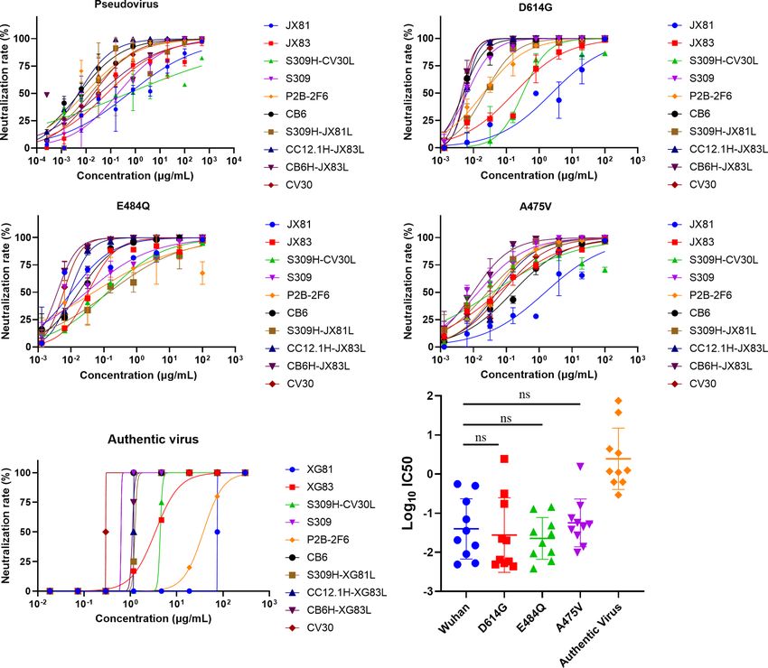
Xie et al.                                                                                                   Novel Antibodies Against SARS-CoV-2

Single B Cell Sequencing                                                 enzyme sites. To express these nAbs in small scale, the
RNA from single B cell was purified using the AMPure RNA                  synthesized sequences of paired heavy and light chain variable
Clean XP kit and eluted directly into a 9 µL RT (reverse                 regions were separately inserted into expression vectors
transcription) reaction. The RT reaction was performed by                containing the IgG1 constant regions of human and co-
incubation at 42°C for 90 min, followed by 10 cycles of 50°C             transfected into 293T cells. Meanwhile, different originated
for 2 min, 42°C for 2 min, 75°C for 15 min according to the              heavy and light chain expression vectors were cross-paired and
protocol of the manuscript (Berkeley Lights, 750-70002) and              co-transfected into 293T cells. After 48 h post-transfection, cell
then left at 4°C until the next step. The 9 µL RT reaction product       supernatant containing expressed antibodies were collected to
was added to a PCR mix using KAPA HiFi HotStart Readymix                 determine the RBD binding ability using ELISA assay.
(kapa, KK2602) in total of 30 µL to amplify the complementary                To explore the characters of antibodies, the corresponding
DNA (cDNA). The cDNA amplification was performed at 98°C                  antibodies with positive OD values (OD ratio > 2 relative to the
for 3 min, followed by 20 cycles of 98°C for 15 s, 65°C for 30 s,        negative control) detected by ELISA were expressed in a large
72°C for 6 min, with a final incubation at 72°C for 10 min, and           scale. Equal amounts of heavy and light chain expression
stored at 4°C. A 1 µL portion of the cDNA amplification product           plasmids were co-transfected into HEK293F cells at a density
was used to amplify the lambda and kappa chains in separate 15           of 2 × 10⁶ cells/mL at 37°C in a humidified 5% CO2 incubator
µL reactions using primers in the antibody constant regions and          rotating at 130 rpm. Briefly, to transfect 100 mL volume of
forward primer to obtain the specific product for sequencing.             HEK293F cells, 100 mg of heavy and 100 mg of light chain
The nucleotide sequences of the referenced primers were showed           expression plasmids were prepared and mixed with 0.8 mg of
in Table S3 (21). The product sequences were analyzed using the          polyethylenimine (PEI, Polyscience) and were added into cells.
international ImMunoGeneTics information system®.                        After 5 days, cell culture supernatants were collected and purified
                                                                         using Protein A column (GE Healthcare). Bound mAbs were
Construction of Plasmids and Cell Lines                                  washed with buffer containing 20 mM Na2HPO4, 150 mM NaCl
The SARS-CoV-2 specific antibody expression vectors were                  (pH 7.0), eluted using 0.1 M acetic acid (pH 3.0-4.5) on ÄKTA
derived from the human immunodeficiency virus (HIV)                       pure (GE Healthcare), and resuspended into PBS by
neutralizing antibody VRC01 heavy and light chain expression             centrifugation with 10 kDa MWCO membrane centrifugal filter
plasmids (22) that contain the IgG1 constant regions of human            units (Millipore). The illustration of RBD specific B cells isolation,
from NIH with CMV promoter and Kana resistance. To express               expression vectors construction, antibodies purification and
SARS-CoV-2-specific antibody, the sequences of heavy and light            identification are shown in Figure 1. The purity of antibodies was
chain variable regions were separately cloned into the                   verified using SDS-PAGE (Figure S1A).
abovementioned antibodies expression vectors. SARS-CoV-2                     Sequence of RBD fused with human IgG1-Fc fragment were
surface glycoprotein gene (GenBank: MN_908947) with C-                   cloned into the pTT5 vector. A TEV (Tobacco Etch Virus)
terminal 19 amino acids deletion was synthesized after codon-            protease cleavage site was added between the RBD and IgG1-
optimizing for Homo sapiens. Synthesized sequence added with             Fc sequence. Briefly, the Fc gene was amplified from antibody
restriction enzyme sites (Xho I and Xba I) was inserted into             heavy chain expression plasmid using the forward primer: 5’-
eukaryotic expression plasmid pcDNA3.1(+) to generate the                TCTAGAGGTTCTGAGAATCTTTATTTCCAAGGTTCTGGT
envelope expression plasmids pcDNA3.1(+)-Opt-S. The                      TCT CCCAAATCTTGTGACAAAAC-3’ and the reverse
lentiviral packaging plasmid pNL4-3 Luc+R-E- carrying an Env-            primer: 5’-GCGGCCGCTTATTTACCCGGA GACAGG
defective, luciferase-expressed HIV-1 genome was gifted by               GAGA-3’ (underlined sequences are Xba I and Not I
Binlian Sun, Jianghan University. pTT5 vector and HEK293F                restriction enzyme sites, respectively). The PCR product
cell line was generously gifted by Tengchuan Jin, University of          encoding Fc with N’-TEV protease cleavage site was cloned
Science and Technology of China. To construct the HEK293T-               into the pTT5 plasmid to generate pTT5-TEV-Fc vector. The
hACE2 cell line that stably expressed hACE2 for pseudovirus              gene encoding RBD (321–591) was amplified from spike gene of
neutralization assay, the lentivirus containing hACE2 gene was           SARS-CoV-2 (Gene ID: MN908947) using the forward primer:
transfected into 293T cells and were selected with 2 mg/mL               5’-GTCGACCAACCAACAGAATCTATTGT-3’ and the reverse
of Puromycin.                                                            primer: 5’- TCTAGA AGAACATGGTGTAATGTCAA-3’
                                                                         (underlined sequences are Sal I and Xba I restriction enzyme
Expression and Purification of mAbs and                                   sites, respectively). The PCR product was cloned into the pTT5-
SARS-CoV-2 RBD Protein                                                   TEV-Fc plasmid to generate the pTT5-RBD-TEV-Fc vector. The
The amino acid sequence of 14 published SARS-CoV-2 nAbs, 1               resultant vector was transfected into HEK293F cells using PEI
SARS nAb, and 1 MERS nAb were downloaded from the PDB                    and fusion protein RBD-Fc was purified from cell supernatants 3
(protein data bank) database. Each antibody DNA sequence                 days post-transfection using Protein A column as described
transformed from the amino acid sequence was codon-                      above. To remove the Fc tag, 20 mM TEV, 1 mM DTT, and 1
optimized for human cells. Heavy chain variable region                   M Tris were added into the purified fusion protein. The mixture
sequences were synthesized with Age I and Sal I restriction              was incubated at 4°C overnight and purified using Protein A
enzyme sites, while light chain (k or l) variable region                 column. Fc-free recombinant proteins were collected and the
sequences were synthesized with Age I and BsiW I restriction             purity of proteins was verified by SDS-PAGE (Figure S1B).

Frontiers in Immunology | www.frontiersin.org                        3                                  August 2021 | Volume 12 | Article 715464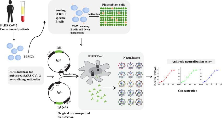
Xie et al.                                                                                                                              Novel Antibodies Against SARS-CoV-2

  FIGURE 1 | Illustration of antibodies production and neutralization assay. MAbs were produced by the co-transfection HEK293F cells with heavy and light chain
  expression vectors originated from a single sequence of RBD specific B cells. RAbs were produced by the co-transfection HEK293F cells with recombination of
  heavy and light chain from different single cells. Neutralization assay was performed by incubation of pseudovirus of live SARS-CoV-2 virus with serial diluted antibodies.

ELISA Quantification for RBD                                                                 separately. RBD at 2 ng/mL (200 ng of RBD protein in 100 uL of
Binding Ability                                                                             PBS per well) was coated on ELISA plates overnight at 4°C. The
SARS-CoV-2 RBD protein at 2 ng/mL was coated onto ELISA                                     plates were washed with PBST followed by blocked with 5%
plates overnight at 4°C. After washing and blocking, 100 mL of cell                         defatted milk for 2 h at 37°C. Meanwhile, nine previously
culture supernatant containing mAbs from 293T cells were added                              published SARS-CoV-2 nAbs with known targeted epitopes at 4-
to each well and incubated at 37°C for 2 h. Plates were washed and                          fold serially diluted in PBS with the initial dilution of 20 mg/mL
incubated with anti-human IgG (H+L)/HRP (Abcam) for 1 h at                                  (concentration range of 20 mg/mL to 0.078 mg/mL) were added into
37°C. TMB (Beyotime) substrate was added and reacted under                                  wells and followed by an equal volume of antibody-Biotin diluted at
dark and optical density (OD) was measured at 450 nm.                                       1 mg/mL. After 1 h of incubation and three times washing, the
                                                                                            plates were incubated with HRP conjugated Streptavidin (1:5,000,
Antibodies Binding Affinity Measurement                                                      Beyotime) for 30 min at 37°C. Next, the plates were washed with
The dissociation coefficient was detected using surface plasmon                              PBST and treated with the TMB buffer. After 10 min, 50 mL of stop
resonance (SPR). Briefly, RBD was coated on a CM5 SA sensor                                  buffer was added to stop the reaction, and the absorbance was read
chip (GE Healthcare) by covalent bonding at a level of 60                                   at OD450 nm. The inhibition rate was calculated by comparing to
response units (RU) using a Biacore T200 (GE Healthcare).                                   the negative control well with only antibody-Biotin added.
Running buffer was composed of PBS with the regeneration
solution composed of 50 mM NaOH. Purified mAbs were diluted                                  Pseudotyped Virus Neutralization Assay
in serial concentrations and injected at a flow rate of 30 mL/min.                           Pseudoviruses with spike mutation were produced by co-transfection
All injections were performed with an association time of 300 s                             HEK293T cells with pNL4-3 Luc+R-E- and plasmids encoding
and a dissociation time of 600 s. Data were fit to a 1:1 binding                             various spike mutation, while Wuhan reference pseudovirus was
model using the Biacore Evaluation Software. All curves were                                produced with pNL4-3 Luc+R-E- and pcDNA3.1(+)-Opt-S. The
plotted using GraphPad Prism 8.3.                                                           supernatant was harvested at 48 h post-transfection, passed through
                                                                                            a 0.45-mm filter, and centrifuged at 800 × g for 10 min to remove cell
Competitive ELISA Assay                                                                     debris. Cell supernatant was collected and stored at -80°C. The 293T
The antibody competition assay was performed to identify the                                cells stably expressing hACE2 were seeded in 96-well plates at a
targeted epitope of the two mAbs. The two mAbs were biotinylated                            density of 1.0 × 10⁴/well 24 hours prior to the assay. The various
using Sulfo-NHS-LC-LC-Biotin (Thermo) overnight at 4°C                                      diluted mAbs (5-fold serial dilution at an initial concentration of 100

Frontiers in Immunology | www.frontiersin.org                                           4                                          August 2021 | Volume 12 | Article 715464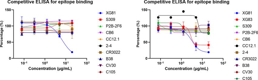
Xie et al.                                                                                                        Novel Antibodies Against SARS-CoV-2

mg/mL, 50 mL aliquots) were mixed with an equal volume of SARS-              searched the PBD (protein bank data) database and downloaded 14
CoV-2 pseudovirus with luciferase units of 1 × 10⁵ and incubated at          IgG heavy (IgH) and light (IgL, k or l) sequences of SARS-CoV-2
37°C for 1 h, which were added to the HEK293T-hACE2 cell wells.              nAbs (S309, CC12.1, CC12.3, C105, REGN10933, REGN10987,
Negative control wells were supplied with 100 mL of DMEM while               CV30, 2-4, EY6A, P2B-2F6, BD23, B38, CB6, and 4A8), one
positive control wells were supplied with 50 mL of DMEM and 50 mL            SARS-CoV nAb (CR3022), and one MERS nAb (LCA60). The
of pseudovirus. After 48 hours of incubation at 37°C with 5% CO2,            CDR3 region, length of nucleotides sequence, variable region
culture supernatants were removed and the values of RLU were                 identity, and germline feature of IgH and IgL of the total 18
measured by the Britelite plus Reporter Gene Assay System                    antibodies were analyzed using Igblast (Figures 2A, C). Figure 2C
(PerkinElmer). The inhibition rate was calculated by comparing               is the pie chart that represents the germline distribution of heavy and
the OD value to the positive control wells. Fifty percent inhibitory         light chains of the 18 mAbs. As shown in Figure 2C, about 33.33% of
concentration (IC50) was determined by a four-parameter logistic             the heavy chains belong to the IgHV3-53 group, and 11.11% belong
regression using GraphPad Prism 8.0.                                         to the IgH3-15 group, while others are distributed to 10 variable
                                                                             germlines evenly. As for the light chains, 16.67% belonged to the
Authentic Virus Neutralization Assay                                         IgKV1-39, IgKV3-20, and IgLV2-8 germlines, and 11.11% belonged
The neutralization assay of authentic SARS-CoV-2 was detected                to the IgKV1-9 and IgLV2-14 germlines. The average CDR3 length
using CPE (cytopathic effect) assay. The CPE was recorded when               for IgH was 44.67 and the average IgL CDR3 length was 29.33
virus caused morphological changes in the Vero E6 cells. Vero E6             (Figure 2D). The length of CDR3 region of XG81 was 54 and the
cells were seeded in monolayers in 96-well plates and incubated              CDR3 region length of XG83 was 51, which were both above the
at 37°C overnight. Four-fold dilution of mAbs with an initial                average CDR3 length. The length of CDR3 region of XG81 IgL was
concentration of 300 mg/mL were mixed with the same volume of                27, while the CDR3 length of XG83 IgL was 33 and the value was
SARS-CoV-2 authentic virus at 100 TCID50 and incubated for 1                 also above the average length. The heavy chain variable region of
h at 37°C. The mAbs and virus mixtures were then transferred to              XG81 and XG83 belongs to the VH 3-15 and VH1-69 families while
Vero E6 cell wells and incubated at 37°C for 1 h. One hour later,            the corresponding light chains belong to the Vk1-40 and VL1-39
the supernatant was removed and 200 mL of DMEM with 2% FBS                   families. The amino acid sequences variety and alignment for CDR3
was added to cells. After the incubation of the cells with the               region of IgH, Igk, and Igl are presented in Figure 2B. The size of
mixture for 5 days at 37°C, CPE caused by the infection was                  the letter represents the frequency of the occurrence of a certain
recorded. The authentic SARS-CoV-2 virus was isolated from a                 amino acid in the 18 mAbs at the same site, suggesting the
COVID-19 patient from Anhui Province. All experiments                        differences in the amino acid sequence among these antibodies
associated with the authentic virus were conducted in Biosafety              (Figure 2B). The alignment of the 18 mAbs for CDR1, CDR2,
Level 3 (BSL-3) laboratory in Anhui Provincial Center for Disease            and CDR3 regions is shown in the extended data (Figure S2).
Control and Prevention. All experiments were complied with the
biosecurity and institutional safety.                                        Binding Profiles of Originally Paired
                                                                             Monoclonal Antibodies and rAbs to
Statistical Analysis                                                         SARS-CoV-2 RBD Protein
IC50 that is defined as the dilution at which the RLU values were             Heavy and light chains from all 18 nAbs were recombined to
reduced by 50% compared with the pseudovirus control wells                   form recombined antibodies (rAbs) and subjected to studies for
was calculated in GraphPad Prism 8 (GraphPad Software). The                  their RBD binding characteristics. All 324 transfection
difference of the Log10 IC50 of two independent groups was tested            supernatants from 293T cells were assayed using ELISA for the
for statistical significance with a Mann-Whitney U test in                    RBD binding affinity. The ratio of the OD value to the negative
GraphPad Prism 8. The KD values were calculated using 1:1                    control over than 2 was considered positive binding to RBD
binding model in Origin 8.0 (OriginLab).                                     (extended data, Table S2). As shown in Figure 3, in total, 44 out
                                                                             of 324 antibodies were tested positive of RBD binding. Of the 18
                                                                             originally paired mAbs, 15 were measured positive for RBD and
                                                                             the remaining 3 mAbs were negative, including one against
RESULTS                                                                      MERS (LCA60), one against N-terminal of SARS-CoV-2 spike
Single B Cell Antibody Cloning and                                           protein (4A8), and another one binds the “down” RBD in
                                                                             protomer B (BD23). Interestingly, of the 306 rAbs, 29 antibodies
Sequence Analysis
                                                                             also showed positive for RBD binding test, suggesting that the
To isolate the monoclonal antibodies, we combined the PBMCs
                                                                             cross-paired expression form different origins might also be one
from 25 convalescent COVID-19 patients to enrich CD27+ memory
                                                                             potential tool to produce binding antibody.
B cells and activate memory B cells into plasmablast cells. We isolate
RBD specific B cells by detecting the SARS-CoV-2 spike protein
specific antibodies in the plasmablast cell culture supernatant using         Binding Activities of Antibodies to RBD
ELISA. Cells with positive OD values were used for further single cell       Through the initial screening of live SARS-CoV-2 virus
sequencing. We finally obtained two sequences of mAbs and they                neutralization test, we selected nine antibodies with a relatively
were detected positive for RBD binding after cloning and expression          better neutralizing ability, including XG81, XG83, 3 SARS-CoV-2
(sequences please see extended data, Table S1). Furthermore, we              nAbs (S309, P2B-2F6, and CB6), and four rAbs (S309H-CV30L,

Frontiers in Immunology | www.frontiersin.org                            5                                   August 2021 | Volume 12 | Article 715464
Xie et al. Novel Antibodies Against SARS-CoV-2
A
B
C D
FIGURE 2 | Sequencing analysis of germline and CDR3 region of antibodies. (A) Germline, CDR3 length of nucleotides sequence, and variable region identity of the
heavy and light chains of the 18 mAbs. (B) Amino acids sequence alignment of CDR3 region using THE MEGA 6.0 and Weblogo web server (http://weblogo.
berkeley.edu/logo.cgi). (C) Distribution of variable germline in heavy and light chains of the 18 mAbs. (D) CDR3 amino acid length of VH and VL of the 18 mAbs.
CDR3, complementary determining region 3.
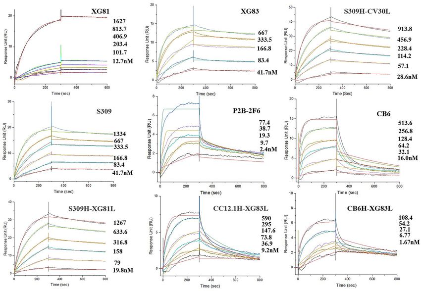
ranged from 10-8 to 10-10 M (Figures 4A–I). XG81 showed the KD
of 10.5 nM while XG83 showed the KD of 5.68 nM. S309, P2B-
2F6, and CB6 exhibited KD at 6.04, 0.81, and 43.8 nM,
respectively. The rAbs also exhibited RBD binding affinity with
the KD ranging from 0.58 to 21.1 nM. Interestingly, one of the
rAbs, CB6H-XG83L, showed the highest binding affinity to RBD
among the 9 antibodies with the KD at 0.58 nM (Table 1).
Neutralization Properties of RBD Specific
Antibodies
To better evaluate the neutralization properties of some original
mAbs and rAbs, we further selected 10 antibodies to measure the
inhibition rate of pseudovirus and live SARS-CoV-2 virus. For
the original Wuhan reference pseudovirus, the XG83 showed
IC50 of 0.069 mg/mL, and XG81 showed the IC50 of 0.556 mg/mL.
FIGURE 3 | Binding profiles of antibodies to RBD. Heatmap showed the
binding of antibodies to RBD using ELISA. Color in the boxes indicated the The S309, P2B-2F6, and CB6 showed the IC50 at 0.198, 0.009,
values of detecting antibodies binding to RBD relative to the negative control and 0.005 mg/mL and the CV30 showed the IC50 at 0.020 mg/mL,
and the ratio of an OD value > 2 was considered positive. Experiments were respectively (Figure 5A and Table 1). As we mentioned above,
repeated twice and data were shown as a representative experiment. the dominated variant D614G could increase infectivity while
E484Q and A475V exhibited resistance to some nAbs. Our study
S309H-XG81L, CC12.1H-XG83L, and CB6H-XG83L) to showed that all the original mAbs and rAbs could effectively
determine the binding affinity using surface plasmon resonance neutralize the three pseudovirus variants with a mutated S
(SPR). The dissociation constant (KD) for the nine antibodies protein containing D614G, E484Q, or A475V mutation
Frontiers in Immunology | www.frontiersin.org 6 August 2021 | Volume 12 | Article 715464Xie et al. Novel Antibodies Against SARS-CoV-2
A B C
D E F
G H I
FIGURE 4 | Affinity abilities of antibodies to SARS-CoV-2 RBD. Surface plasmon resonance (SPR) demonstrated the binding and dissociation kinetics of the 9
antibodies (A: XG81; B: XG83; C: S309H-CV30L; D: S309; E: P2B-2F6; F: CB6; B: S309H-XG81L; H: CC12.1-XG83L; I: CB6H-XG83L) against S.
TABLE 1 | Affinity and neutralization characteristics of 10 antibodies.
Antibody KD (RBD, nM) IC50 (mg/mL)
Wuhan D614G E484Q A475V Authentic Virus
XG81 10.5 0.556 2.431 0.010 1.531 75.000
XG83 5.68 0.069 0.174 0.033 0.057 3.443
S309H-CV30L 19.5 0.501 0.318 0.129 0.038 4.404
S309 6.04 0.198 0.006 0.029 0.014 0.628
P2B-2F6 0.81 0.009 0.020 0.049 0.044 37.500
CB6 43.8 0.005 0.005 0.017 0.155 0.628
S309H-XG81L 21.1 0.015 0.022 0.144 0.027 1.248
CC12.1H-XG83L 12.9 0.005 0.006 0.009 0.052 1.172
CB6H-XG83L 0.58 0.036 0.004 0.004 0.010 1.101
CV30 / 0.020 0.005 0.006 0.075 0.293
(Figures 5B–D and Table 1). We also compared the neutralization A475V residue of RBD all showed increased IC50 values on
activities between the mutant and Wuhan reference pseudovirus. mutant pseudovirus (ratio > 1, Table 2). Interestingly, some rAbs
The IC50 ratio higher than 1 indicated the decreased ability of antibodies such as CB6H-XG83L and S309H-CV30L were granted a
antibodies in neutralizing mutant pseudovirus relative to Wuhan significantly higher neutralizing ability to mutant pseudoviruses
reference pseudovirus. The antibodies that targeted E484Q or when compared with the original mAbs (ratio < 1, Table 2).
Frontiers in Immunology | www.frontiersin.org 7 August 2021 | Volume 12 | Article 715464Xie et al. Novel Antibodies Against SARS-CoV-2
A B
C D
E F
FIGURE 5 | Neutralizing capacities of antibodies for pseudovirus and authentic virus. (A) Neutralization of antibodies to SARS-CoV-2 pseudovirus in ACE2-293T
cells. (B) Neutralization to D614G mutant pseudovirus. (C) Neutralization to E484Q mutant pseudovirus. (D) Neutralization to A475V mutant pseudovirus. (E)
Neutralization of antibodies to authentic SARS-CoV-2 in Vero-E6 cells. (F) Log10 IC50 values of antibodies to four pseudoviruses and live virus. The difference of
Log10 IC50 values in two groups was tested for statistical significance with a Mann-Whitney U test in GraphPad Prism 8. ns p > 0.05. Experiments were repeated
twice and data were shown as Mean ± SD of a representative experiment. ns, no significant difference.
To confirm the neutralization activities of the antibodies, we Additionally, the authentic virus showed higher Log10 IC50 value,
further performed the cytopathic effect (CPE) inhibition assay exhibiting more difficult to be neutralized than the pseudovirus.
with the authentic SARS-CoV-2 virus. The XG81 and XG83
showed IC50 of 75.00 and 3.443 mg/mL, respectively, and CV30 Competitive Characteristics for Antibodies
showed the lowest IC50 of 0.293 mg/mL, followed by S309 and A competition ELISA was further performed for the two nAbs to
CB6 with IC50 of 0.628 mg/mL (Figure 5E). The rAbs all showed determine if there were overlapping epitopes between different
their neutralization abilities against both pseudovirus and live nAbs. Nine previously reported SARS-CoV-2 RBD nAbs (S309,
SARS-CoV-2 virus, and the neutralization potencies were mostly P2B-2F6, CB6, CC12.1, 2-4, CR3022, B38, CV30, and C105) were
between or close to the original antibodies. As shown in used as references while the two nAbs (XG81 and XG83)
Figure 5F, the Log10 IC50 values of the 10 antibodies against themselves were used as positive controls. With the decrease
Wuhan pseudotyped virus were higher than that of D614G and change of the concentration of the nine nAbs, the absorbance
E484Q pseudoviruses, but lower than A475V, suggesting that percentage showed no obvious change, suggesting that they
G614G and E484Q were more susceptible to neutralization while target new epitopes on the spike protein (Figures 6A, B). With
A475V showed a somewhat escape from neutralization. the decreasing change of the concentration of XG83, the
Frontiers in Immunology | www.frontiersin.org 8 August 2021 | Volume 12 | Article 715464Xie et al. Novel Antibodies Against SARS-CoV-2
TABLE 2 | Comparison of the neutralization ability of 10 antibodies for mutant these antibodies against the mutant pseudoviruses. Both XG81 and
and Wuhan pseudotyped virus.
XG83 could effectively neutralize against Wuhan, E484Q, A475V,
Antibody E484Q IC50 ratio A475V IC50 ratio and D614G pseudoviruses. Meanwhile, XG83 had a higher affinity
residue (E484Q/Wuhan) residue (A475V/Wuhan) to SARS-CoV-2 RBD, and a higher potency to neutralize Wuhan,
mutant pseudoviruses, and authentic SARS-CoV-2 virus.
VH VL VH VL
Competitive ELISA revealed that XG81 and XG83 shared similar
XG81 / / 0.018 / / 2.754 targets but different from other nAbs, demonstrating new binding
XG83 / / 0.478 / / 0.816 sites for either of the two antibodies. The result suggests that XG83
S309H-CV30L – – 0.257 – – 0.076
could be a potential broadly protective monoclonal antibody with
S309 – – 0.148 – – 0.070
P2B-2F6 – + 5.410 – – 4.920
new antigenic targets.
CB6 – – 3.306 + – 29.440 The rAbs also showed binding affinity to RBD and neutralizing
S309H-XG81L – / 9.639 – / 1.800 ability against both pseudovirus and authentic virus. One of the
CC12.1H-XG83L – / 1.918 – / 10.596 rAbs, CB6H-XG83L, showed the least KD value, which means the
CB6H-XG83L – / 0.108 + / 0.276
highest binding affinity to RBD. Since A475V is located within the
CV30 – – 0.290 + – 3.658
binding sites of CB6, the IC50 value of A475V pseudovirus is 29.4
The IC50 ratio higher than 1 indicated the decreased ability of antibodies in neutralizing the times relative to Wuhan pseudovirus. Similarly, the IC50 of CV30 in
mutant pseudovirus relative to Wuhan reference pseudovirus.
neutralizing A475V is 3.7 times relative to Wuhan pseudovirus,
while the IC50 of P2B-2F6 is 5.4 times targeting E484Q compared
absorbance percentage of XG81 increased obviously, which to Wuhan pseudovirus. Interestingly, when compared with the
suggests that they have a competitive effect with each other originally paired mAbs, the rAb, CB6H-XG83L, effectively
and shared similar epitopes of SARS-CoV-2 (Figure 6B). enhanced its neutralizing activity against A475V pseudoviruses so
as to avoid the immune escape of the mutation (Table 2). The other
rAb, S309H-CV30L, also showed a decreased IC50 ratio with a
DISCUSSION higher potency of neutralization compared with the original CV30
antibody (Table 2). Pseudoviruses test with single amino acid
The rapid spread of SARS-CoV-2 has caused great threat mutation could be helpful for evaluating the factors and exact
globally. Despite the fact that the vaccines and antiviral drugs mutation sites that influenced antibodies neutralizing activity, and
has been widely used on the market, the protective effectiveness highlights the key to modify and produce antibodies with higher
remains uncertain against the rapidly emerging variants. Therefore, neutralizing potency and breadth.
isolation of nAbs still remains an important strategy for the clinical SARS-CoV-2 variants with various S mutations possibly arise
treatment of SARS-CoV-2 infection. We have identified two SARS- over the global infected individuals along with virus evolution,
CoV-2 nAbs from plasmablast B cells of convalescent COVID-19 which leaves a challenge and pressure for antiviral therapy. Studies
patients that both recognize the RBD region and neutralize live proposed that the non-competing antibody cocktail therapy present
virus. One of the nAbs, XG83, showed a longer CDR3 length of a promising approach against SARS-CoV-2 variants with spike
IgH and IgL, and belonged to an independent germline. Since some protein mutations (23). However, the identification of neutralizing
variants, including A475V, E484Q, and D614G, have been reported mAbs with simultaneous effectiveness requires further study. Our
to show a decreased sensitivity to neutralizing nAbs or an increased study provided new insights to antibody therapy, that is
infectivity (6, 15), we next investigated the neutralizing activity of recombination of heavy and light chains from various nAbs
A B
FIGURE 6 | Competition characteristics of XG81 and XG83. (A) The competitive capacity of XG81 with nine SARS-CoV-2 nAbs that was indicated by the level of
response unit relative to the negative control with the increase of nAbs concentration. XG81 was used as a positive control. (B) The competitive capacity of XG83
with XG81 and another nine SARS-CoV-2 nAbs. XG83 itself was used as a positive control.
Frontiers in Immunology | www.frontiersin.org 9 August 2021 | Volume 12 | Article 715464Xie et al. Novel Antibodies Against SARS-CoV-2
targeting specific antigenic sites, which could contribute to the AUTHOR CONTRIBUTIONS
further designing of antibody-based COVID-19 therapeutics. The
underlying mechanisms of the rAbs in enhancing the neutralizing TJ, SZ, and YG designed the study. JX, CD, JH, YZ, SN, XZ, QC,
activity might be the overlapping of critical residues in RBD mutants. JW, LH, HH, WL, and HM performed the experiments. JX
Notably, identification of an ideal combination could be essential for analyzed the data and wrote the manuscript. All authors
the design of rAbs expression. Since the rAbs that showed contributed to the article and approved the submitted version.
neutralizing ability was a new and interesting discovery of our
study, the danger of these rAbs still remains unclear now. The
potential risks might include off target or cross immune response.
Therefore, the stability, exact targeting sites, and underlying FUNDING
mechanisms rAbs in enhancing neutralization potency remain an
interesting topic and warrant future study. Besides, more SARS- This work is supported by Postdoctoral Research Foundation of
CoV-2 variants with single or multiple mutations and currently China (2021M693076; 2020M670084ZX), Key Research and
circulating strains should be concerned in investigating broadly Development Project of Anhui Province (202104j07020042;
protective effectiveness of XG83 mAb and rAbs. 202104a07020032), Special Project for Emergency Scientific
In conclusion, our data reports a fully human neutralizing and Technological Research on New Coronavirus Infection
mAb, XG83, could be used as a potential therapeutic agent for (YD9110002001), Emergency Research Project of Novel
clinical treatment of SARS-CoV-2 infection. Combined expression Coronavirus Infection of Anhui Province (202004a07020002;
of rAbs from different origination may function as additional 202004a07020004), the Fundamental Research Funds for the
potent neutralizing antibodies and be used to fight against various Central Universities (WK9110000166; WK9110000167), and the
escaping mutants. Hefei Comprehensive National Science Center.
DATA AVAILABILITY STATEMENT
ACKNOWLEDGMENTS
The datasets presented in this study can be found in online
repositories. The names of the repository/repositories and We acknowledge the work and contribution of all the health-care
accession number(s) can be found below: NCBI Genbank, workers associated with this study.
accession numbers: MZ668598, MZ668599, MZ668600, MZ668601.
ETHICS STATEMENT SUPPLEMENTARY MATERIAL
The study was approved by the Ethics Committee of the First The Supplementary Material for this article can be found online
Affiliated Hospital of USTC and all participants provided their at: https://www.frontiersin.org/articles/10.3389/fimmu.2021.
written informed consent to participate in this study. 715464/full#supplementary-material
Antibody Cocktail. Science (2020) 369(6506):1010–4. doi: 10.1126/science.
REFERENCES abd0827
1. Callaway E, Cyranoski D, Mallapaty S, Stoye E, Tollefson J. The Coronavirus 8. Garcia-Beltran WF, Lam EC, St Denis K, Nitido AD, Garcia ZH, Hauser BM, et al.
Pandemic in Five Powerful Charts. Nature (2020) 579(7800):482–3. doi: Multiple SARS-CoV-2 Variants Escape Neutralization by Vaccine-Induced Humoral
10.1038/d41586-020-00758-2 Immunity. Cell (2021) 184(9):2372–83. doi: 10.1101/2021.02.14.21251704
2. Lu R, Zhao X, Li J, Niu P, Yang B, Wu H, et al. Genomic Characterisation and 9. Shi R, Shan C, Duan X, Chen Z, Liu P, Song J, et al. A Human Neutralizing
Epidemiology of 2019 Novel Coronavirus: Implications for Virus Origins and Antibody Targets the Receptor-Binding Site of SARS-CoV-2. Nature (2020)
Receptor Binding. Lancet (2020) 395(10224):565–74. doi: 10.1016/S0140- 584(7819):120–4. doi: 10.1038/s41586-020-2381-y
6736(20)30251-8 10. Zhou G, Zhao Q. Perspectives on Therapeutic Neutralizing Antibodies
3. Weisblum Y, Schmidt F, Zhang F, DaSilva J, Poston D, Lorenzi JC, et al. Against the Novel Coronavirus SARS-CoV-2. Int J Biol Sci (2020) 16
Escape From Neutralizing Antibodies by SARS-CoV-2 Spike Protein (10):1718–23. doi: 10.7150/ijbs.45123
Variants. Elife (2020) 9:e61312. doi: 10.7554/eLife.61312 11. Li Y, Wan Y, Liu P, Zhao J, Lu G, Qi J, et al. A Humanized Neutralizing
4. Plante JA, Liu Y, Liu J, Xia H, Johnson BA, Lokugamage KG, et al. Spike Antibody Against MERS-CoV Targeting the Receptor-Binding Domain of the
Mutation D614G Alters SARS-CoV-2 Fitness. Nature (2021) 592(7852):116– Spike Protein. Cell Res (2015) 25(11):1237–49. doi: 10.1038/cr.2015.113
21. doi: 10.1038/s41586-020-2895-3 12. Traggiai E, Becker S, Subbarao K, Kolesnikova L, Uematsu Y, Gismondo MR,
5. Wang R, Chen J, Gao K, Wei GW. Vaccine-Escape and Fast-Growing et al. An Efficient Method to Make Human Monoclonal Antibodies From
Mutations in the United Kingdom, the United States, Singapore, Spain, Memory B Cells: Potent Neutralization of SARS Coronavirus. Nat Med (2004)
India, and Other COVID-19-Devastated Countries. Genomics (2021) 113 10(8):871–5. doi: 10.1038/nm1080
(4):2158–70. doi: 10.1016/j.ygeno.2021.05.006 13. Wang C, Li W, Drabek D, Okba NMA, van Haperen R, Osterhaus A, et al. A
6. Li Q, Wu J, Nie J, Zhang L, Hao H, Liu S, et al. The Impact of Mutations in Human Monoclonal Antibody Blocking SARS-CoV-2 Infection. Nat
SARS-CoV-2 Spike on Viral Infectivity and Antigenicity. Cell (2020) 182 Commun (2020) 11(1):2251. doi: 10.1038/s41467-019-13907-7
(5):1284–94.e9. doi: 10.1016/j.cell.2020.07.012 14. Wu Y, Li C, Xia S, Tian X, Kong Y, Wang Z, et al. Identification of Human
7. Hansen J, Baum A, Pascal KE, Russo V, Giordano S, Wloga E, et al. Studies Single-Domain Antibodies Against SARS-CoV-2. Cell Host Microbe (2020) 27
in Humanized Mice and Convalescent Humans Yield a SARS-CoV-2 (6):891–8. doi: 10.1016/j.chom.2020.04.023
Frontiers in Immunology | www.frontiersin.org 10 August 2021 | Volume 12 | Article 715464Xie et al. Novel Antibodies Against SARS-CoV-2
15. Weissman D, Alameh MG, de Silva T, Collini P, Hornsby H, Brown R, et al. 22. Wu X, Yang ZY, Li Y, Hogerkorp CM, Schief WR, Seaman MS, et al. Rational Design
D614G Spike Mutation Increases SARS CoV-2 Susceptibility to of Envelope Identifies Broadly Neutralizing Human Monoclonal Antibodies to HIV-
Neutralization. Cell Host Microbe (2021) 29(1):23–31.e4. doi: 10.1016/ 1. Sci (New York NY) (2010) 329(5993):856–61. doi: 10.1126/science.1187659
j.chom.2020.11.012 23. Ku Z, Xie X, Davidson E, Ye X, Su H, Menachery VD, et al. Molecular
16. Greaney AJ, Starr TN, Gilchuk P, Zost SJ, Binshtein E, Loes AN, et al. Determinants and Mechanism for Antibody Cocktail Preventing SARS-CoV-
Complete Mapping of Mutations to the SARS-CoV-2 Spike Receptor-Binding 2 Escape. Nat Commun (2021) 12(1):469. doi: 10.1038/s41467-020-20789-7
Domain That Escape Antibody Recognition. Cell Host Microbe (2021) 29
(1):44–57 e9. doi: 10.1016/j.chom.2020.11.007 Conflict of Interest: The authors declare that the research was conducted in the
17. Poeschla E. Neutralizing SARS-CoV-2. Elife (2020) 9:e64496. doi: 10.7554/ absence of any commercial or financial relationships that could be construed as a
eLife.64496 potential conflict of interest.
18. Baum A, Fulton BO, Wloga E, Copin R, Pascal KE, Russo V, et al. Antibody
Cocktail to SARS-CoV-2 Spike Protein Prevents Rapid Mutational Escape Publisher’s Note: All claims expressed in this article are solely those of the authors
Seen With Individual Antibodies. Science (2020) 369(6506):1014–8. doi: and do not necessarily represent those of their affiliated organizations, or those of
10.1126/science.abd0831 the publisher, the editors and the reviewers. Any product that may be evaluated in
19. Weinreich DM, Sivapalasingam S, Norton T, Ali S, Gao H, Bhore R, et al. this article, or claim that may be made by its manufacturer, is not guaranteed or
REGN-COV2, a Neutralizing Antibody Cocktail, in Outpatients With endorsed by the publisher.
Covid-19. N Engl J Med (2021) 384(3):238–51. doi: 10.1056/NEJMoa
2035002 Copyright © 2021 Xie, Ding, He, Zhang, Ni, Zhang, Chen, Wang, Huang, He, Li, Ma,
20. Kershaw MH, Hsu C, Mondesire W, Parker LL, Wang G, Overwijk WW, et al. Jin, Zhang and Gao. This is an open-access article distributed under the terms of the
Immunization Against Endogenous Retroviral Tumor-Associated Antigens. Creative Commons Attribution License (CC BY). The use, distribution or
Cancer Res (2001) 61(21):7920–4. reproduction in other forums is permitted, provided the original author(s) and the
21. Smith K, Garman L, Wrammert J, Zheng NY, Capra JD, Ahmed R, et al. Rapid copyright owner(s) are credited and that the original publication in this journal is
Generation of Fully Human Monoclonal Antibodies Specific to a Vaccinating cited, in accordance with accepted academic practice. No use, distribution or
Antigen. Nat Protoc (2009) 4(3):372–84. doi: 10.1038/nprot.2009.3 reproduction is permitted which does not comply with these terms.
Frontiers in Immunology | www.frontiersin.org 11 August 2021 | Volume 12 | Article 715464You can also read