Real-Time RT-PCR Assay - For Use Under an Emergency Use Authorization Only Centers for Disease Control and Prevention
←
→
Page content transcription
If your browser does not render page correctly, please read the page content below
Enterovirus D68 2014
     Real-Time RT-PCR Assay
 Centers for Disease Control and Prevention

For Use Under an Emergency Use
      Authorization Only
            Instructions for Use
Table of Contents
Introduction.................................................................................................................................... 2
Specimens ....................................................................................................................................... 3
Equipment and Consumables ........................................................................................................ 4
Quality Control............................................................................................................................... 6
Nucleic Acid Extraction ................................................................................................................. 7
Testing Algorithm .......................................................................................................................... 7
rRT-PCR Assay .............................................................................................................................. 8
Interpreting Test Results.............................................................................................................. 14
Overall Test Interpretation and Reporting Instructions............................................................. 19
Assay Limitations, Warnings and Precautions ........................................................................... 20
Performance Characteristics ....................................................................................................... 21
Contact Information .................................................................................................................... 30
May 12, 2015 Page 1 of 30Introduction
Purpose
This document describes the use of a real-time (TaqMan®) RT-PCR (rRT-PCR) assay for
detection of the viral protein 1 (VP1) gene of Enterovirus D68 (EV-D68).
Intended Use
The Enterovirus D68 2014 Real-time RT-PCR Assay (EV-D68 2014 rRT-PCR) is intended for
the in vitro qualitative detection of RNA from the enterovirus D68 (EV-D68) strains detected in
North America in 2014. It is intended for use with upper respiratory specimens (such as
nasopharyngeal swabs (NP), oropharyngeal swabs (OP), dual NP/OP swabs, and/or nasal
washes) and sera in conjunction with patient-matched upper respiratory specimen(s) from
individuals with signs and symptoms of EV-D68 infection and/or epidemiologic risk factors,
tested in qualified laboratories designated by the Centers for Disease Control and Prevention
(CDC).
Positive results generated from direct specimen testing are presumptive for the detection of RNA
from the EV-D68 strains detected in North America in 2014. When the Enterovirus D68 2014
rRT-PCR is run in conjunction with a cleared commercial assay (as outlined in the EV-D68
Respiratory Disease Algorithm located at http://www.cdc.gov/non-polio-
enterovirus/downloads/ev-d68-2014-respiratory-algorithm.pdf), positive results are considered
confirmatory for EV-D68 strains detected in North America in 2014.
Testing with the EV-D68 2014 rRT-PCR should not be performed unless the patient meets
clinical and/or epidemiological criteria for testing suspect specimens. Current information on
EV-D68, including case definitions, is available at http://www.cdc.gov/non-polio-
enterovirus/about/EV-D68.html.
The EV-D68 2014 rRT-PCR is intended for use by trained laboratory personnel who are
proficient in performing real-time RT-PCR assays. Assay results are for the presumptive
identification of EV-D68 strains detected in North America in 2014. The definitive identification
of EV-D68 requires additional testing. Laboratories are required to report results to CDC.
Negative EV-D68 2014 rRT-PCR results do not preclude EV-D68 infection and should not be
used as the sole basis for patient management decisions. Expected diagnostic yield in sera is low;
however, EV-D68 has been detected in sera from a few patients aged 2 years to 20 years. Only a
limited evaluation of serum specimens has been performed due to the scarcity of serum
specimens from individuals with EV-D68 infection. Thus, sera may only be tested if a patient-
matched upper respiratory specimen is also tested with this assay.
The EV-D68 2014 rRT-PCR is only for use under the Food and Drug Administration’s
Emergency Use Authorization. Use is limited to qualified laboratories designated by CDC,
which includes qualified laboratories with training, facilities and equipment appropriate for
specimen handling, testing and interpretation of the results of this real-time RT-PCR assay.
May 12, 2015 Page 2 of 30Specimens
Biosafety information: Infection control precautions should include Standard, Contact, and
Droplet Precautions for the current outbreak of EV-D68. See Enterovirus D68 for Health Care
Professionals.
http://www.cdc.gov/non-polio-enterovirus/hcp/EV-D68-hcp.html
General Information about Enterovirus D68 2014
See: Non-Polio Enterovirus (Enterovirus D68 Disease) at http://www.cdc.gov/non-polio-
enterovirus/index.html
Acceptable Specimens
• Upper Respiratory Specimens (preferred)
o Nasal washes
o Swabs (dry or in VTM)
Nasopharyngeal (NP) swabs
Oropharyngeal (OP) swabs
Dual NP/OP swabs
Note: If testing a dry swab, elute the swab into 500 µL VTM prior to extraction.
• Serum: expected diagnostic yield is low; however, EV-D68 2014 has been detected in
sera from a few patients aged 2 years to 20 years.
NOTE: Sera may only be tested in conjunction with patient-matched upper respiratory
specimen(s). See Nucleic Acid Extraction section for use restrictions.
Specimen Collection
Refer to www.CDC.gov for guidance on specimen collection, storage and shipment. Information
can be found at http://www.cdc.gov/non-polio-enterovirus/lab-testing/specimen-collection.html.
Specimen Handling and Storage
• Specimens should be kept frozen at ≤ -20°C.
• If PCR cannot be performed the same day as specimen extraction, RNA should be stored
at -70ºC or lower.
• Extracted total nucleic acid should be stored at -70°C or lower.
May 12, 2015 Page 3 of 30Equipment and Consumables
Disclaimer: Names of vendors are provided as examples of suitable product sources. Use of
trade names is for identification purposes only and does not constitute endorsement by CDC or
the Department of Health and Human Services.
Materials provided by CDC
• EV-D68 2014 rRT-PCR Primer and Probe Set (CDC; Catalog #KT0158).
Refer to product insert for storage and expiration information. Set includes 1 set of
primers and 1FAM-labeled probe:
o VP1.2014 (forward primer [VP1.2014 -F], reverse primer [VP1.2014 -R] and probe
[VP1.2014 -P]
• RNase P Real-time PCR Primer and Probe Set (CDC; Catalog #KT0068)
Refer to product insert for storage and expiration information. Set includes 1 set of
primers and 1 FAM-labeled probe:
o Forward primer (RP-F), reverse primer (RP-R), and probe (RP-P)
Materials provided by CDC, but not included in kit
• EV-D68 2014 rRT-PCR Assay Positive Control (CDC; catalog #KT0159) (1.5 mL/vial)
Materials required but not provided
• SuperScript® III Platinum® One-Step qRT-PCR Kit (Invitrogen; catalog #11732-088)
• Molecular grade water, nuclease-free
• Extraction reagents:
o Manual
QIAamp Viral RNA Mini Kit (QIAGEN; catalog #52904 or 52906) or
QIAamp DSP Viral RNA Mini Kit (QIAGEN; catalog #61904)
o bioMérieux easyMAG
Refer to list of reagents and consumables under the easyMAG instrument
in Equipment and Consumables section (below).
• Human Genomic DNA: for use as a positive control for the RNase P Primer and Probe
Set. Either of the following products may be used:
o Human Genomic DNA from human blood (buffy coat) (Roche Applied Science;
Catalog #11691112001)
o Human Genomic DNA (Promega Corporation; Catalog #G3041)
May 12, 2015 Page 4 of 30Equipment and Consumables
• Acceptable surface decontaminants
o DNA Away™ (Fisher Scientific; catalog # 21-236-28)
o RNase Away™ (Fisher Scientific; catalog #21-236-21). This product eliminates
RNase and DNA.
o 10% bleach (1:10 dilution of commercial 5.25-6.0% hypochlorite bleach), made
fresh daily
• Disposable, powder-free gloves
• Laboratory marking pen
• P2/P10, P200, and P1000 aerosol barrier pipette tips
• 1.5 mL microcentrifuge tubes
• Vortex mixer
• Microcentrifuge
• Micropipettes (2 or 10 µL, 200 µL and 1000 µL)
• Racks for 1.5 mL microcentrifuge tubes
• 2 x 96-well -20°C cold blocks
• PCR Instrument System and Consumables
o 7500 Fast Dx Real-Time PCR Systems (Applied Biosystems; catalog #4406984)
0.1 mL PCR reaction strip tubes (Applied Biosystems; catalog #4358293)
0.1 mL PCR reaction plates (Applied Biosystems; catalog #4346907)
MicroAmp® Optical 8-cap Strips (Applied Biosystems; catalog #
4323032)
o 7500 Fast (Applied Biosystems; catalog #4351106)
0.1 mL PCR reaction strip tubes (Applied Biosystems; catalog #4358293)
0.1 mL PCR reaction plates (Applied Biosystems; catalog #4346907
MicroAmp® Optical 8-cap Strips (Applied Biosystems; catalog #
4323032)
o 7500 (Applied Biosystems; catalog #4351104)
0.2 mL PCR reaction strip tubes (Applied Biosystems; catalog #4316567 )
MicroAmp® Optical 8-cap Strips (Applied Biosystems; catalog #4323032)
MicroAmp® Optical 96-Well Reaction Plate with Barcode & Optical
Caps (Applied Biosystems catalog #403012)
• Automated Extraction Instrument
o NucliSENS easyMAG (bioMérieux; catalog #280140)
easyMAG Magnetic Silica (48 x 0.6 mL) ( bioMérieux; catalog #280133)
easyMAG Disposables (48 sets) (bioMérieux: catalog #280135)
easyMAG Lysis Buffer (bioMérieux; catalog #280134)
easyMAG Buffer 1 (bioMérieux: catalog #280130)
easyMAG Buffer 2 (bioMérieux; catalog #280131)
easyMAG Buffer 3 (bioMérieux: catalog #280132)
BioHit Pipette Tips (bioMérieux; catalog #280146)
Micro tubes w/caps (bioMérieux; catalog #200294)
May 12, 2015 Page 5 of 30Quality Control
rRT-PCR is an exquisitely sensitive test method and should be conducted following strict quality
control and quality assurance procedures. Following these guidelines will help minimize the
chance of false-positive and false-negative results.
General Considerations
At a minimum, good laboratory practices at the Biosafety Level 2 (BSL-2) should be
followed, as described in the Biosafety in Microbiological and Biomedical Laboratories
(BMBL) 5th Edition (http://www.cdc.gov/biosafety/publications/bmbl5/index.htm).
• Personnel must be familiar with the protocol and instruments used.
• Maintain separate areas and dedicated equipment (e.g., pipettes, microcentrifuges) and
supplies (e.g., microcentrifuge tubes, pipette tips, gowns and gloves) for
o assay reagent setup,
o handling of extracted nucleic acids and
o rRT-PCR amplification.
• Work flow must always be from the clean area to the dirty area.
• Wear clean, previously unworn, disposable gowns and new, powder-free gloves during
assay reagent setup and handling of extracted nucleic acids. Change gloves whenever
you suspect they may be contaminated.
• Store primer/probes and enzyme mastermix at appropriate temperatures (see product
inserts). Do not use reagents beyond their expiration dates.
• Keep reagent tubes and reactions capped as much as possible.
• Clean surfaces using an acceptable surface decontaminant (see above).
• To minimize cross-contamination, do not bring extracted nucleic acid or PCR amplicons
into the assay setup area. Do not wear any protective clothing that was worn in other
parts of the laboratory. Only clean PPE should be worn in this area.
• Use aerosol barrier (filter) pipette tips only.
• Use optical strip 8-cap strips only. Do not use PCR plate sealing film.
Assay Controls
Assay Controls should be run concurrently with all test samples.
• EV-D68 2014 rRT-PCR Positive Control –Used as a control for PCR reagent
function.EV-D68 2014 target RNA transcript; concentration adjusted to give a consistent
Ct value of 30-38. Two (2) positive controls must be run concurrently with all test
samples.
• NTC – A known negative template control (sterile, nuclease-free water) added during
rRT-PCR reaction set-up. Used as a control for PCR reagent function and cross
contamination. Two (2) NTC must be run concurrently with all test samples.
• RNase P – All clinical specimens should be tested for human RNase P gene to control for
specimen quality and extraction.
May 12, 2015 Page 6 of 30• RP Positive Control –Used as a control for RNase P primer and probe set function. The
material should be diluted to 50 ng/5 µL or 10 µg/mL with 10 mM Tris (pH 7.4-8.2) prior
to use. An acceptable Ct range is ≤30. Either of the following two products may be used:
(a) Human Genomic DNA from human blood (buffy coat); Roche Applied Science,
Catalog #11691112001
(b) Human Genomic DNA; Promega Corporation, Catalog #G3041
Table 1: Overview of positive and negative controls and expected results
Control Control VP1.2014 RP Expected
Used to Monitor
Type Name result result Ct Values
EV-D68 Performance of the
2014 VP1.2014 primer and 30-38 Ct for
Positive + -
rRT-PCR probe set. VP1.2014
Pos Ctrl
RNase P Performance of the
Ct ≤ 30 for
Positive positive RP primer and probe - +
RP
control set.
Reagent and/or
environmental None
Negative NTC - -
contamination during detected
PCR set-up
Nucleic Acid Extraction
• Upper respiratory and serum specimens should be extracted with either the QIAGEN
Viral RNA Mini Kit, following the manufacturer’s spin column protocol, or the
NucliSENS easyMAG (bioMérieux), following the manufacturer’s instructions.
• Sample extractions must yield RNA or total nucleic acid of sufficient volume to cover all
rRT-PCR assays (a minimum of 60 µL is recommended).
• Retain specimen extracts in cold block or on ice until testing. If testing will be delayed,
freeze immediately (preferably -70oC). Thaw only the number of extracts that will be
tested in a single day. Do not freeze or thaw extracts more than once before testing.
Testing Algorithm
Use VP1.2014 and RP for specimen testing.
• If the VP1.2014 is positive (CtrRT-PCR Assay
The EV-D68 rRT-PCR primer and probe set targets the coding region for the viral protein 1
(VP1) gene of the Enterovirus D68 and shows ≥91% nucleotide identity with strains circulating
within the United States in 2014.
Stock Reagent Preparation
Precautions: These reagents should be handled only in a clean area and stored at appropriate
temperatures (see below) in the dark. Freeze-thaw cycles should be avoided. Maintain cold
when thawed.
1. Real-time Primers/Probes
• EV-D68 2014 rRT-PCR Primer and Probe Set
o Precautions:
These reagents should only be handled in a clean area and stored at
appropriate temperatures (see below) in the dark. Freeze-thaw cycles should
be avoided. Maintain cold when thawed.
o Concentrated primer and probe stocks must be diluted to the working
concentrations in nuclease free H2O.
Primers VP1.2014-F and VP1.2014-R should be diluted to 10 μM (10
pmol/μL). Probe VP1.2014-P should be diluted to 5 μM (5 pmol/μL).
o Sterilely suspend lyophilized reagents in 0.40 mL PCR grade nuclease-free
water (50X working concentration) and allow to rehydrate for 15 min at room
temperature in the dark.
o Mix diluted working stocks and aliquot primers/probe in 100 μL (enough
volume for a single 96 well reaction plate) or smaller volumes, depending on
the specimen load in your laboratory. Store a single working aliquot of
primers/probe at 2-8°C in the dark. Do not refreeze.
o Each EV-D68 2014 rRT-PCR primer and probe kit will contain material to
perform 500 reactions. Store rehydrated aliquots of primers and probes at
≤ -20°C. Do not store in frost-free freezers. Rehydrated primers and probes
may be stored frozen for up to 12 months. For complete information on
storage conditions, see package insert.
• RNase P Primer and Probe
o Sterilely suspend lyophilized reagents in
0.25 mL nuclease-free water and allow to rehydrate for 15 minutes at room
temperature.
o Store rehydrated aliquots of primers and probes at ≤ -20°C. Do not store in
frost free freezers. Rehydrated primers and probes may be stored frozen for
up to 12 months. For complete information on storage conditions, see
package insert.
May 12, 2015 Page 8 of 30®
2. Invitrogen SuperScript™ III Platinum One-Step Quantitative RT-PCR System
• Place Invitrogen 2X PCR Master Mix and Superscript III RT/Platinum Taq
enzyme mix in a cold rack at 2-8°C.
• Completely thaw the 2X PCR Master Mix vial.
• Mix the 2X PCR Master Mix by inversion 10 times.
• Pulse centrifuge 2X PCR Master Mix and Superscript III RT/Platinum Taq
enzyme mix then place in cold rack.
4. No Template Control (NTCs) (not provided)
• Sterile, nuclease-free water
• Aliquot in small volumes
• Use to check for contamination during plate set-up
5. EV-D68 2014 rRT-PCR Positive Control
• Precautions: This reagent should be handled with caution in a dedicated nucleic
acid handling area to prevent possible contamination. Freeze-thaw cycles should
be avoided. Maintain on ice when thawed.
• Used to assess performance of VP1.2014 primer and probe set.
• Centrifuge tube in microcentrifuge at maximum speed. Ensure pellet is at the
bottom (pellet will be a bright pink color).
• Add 1500 µL of cold nuclease-free water and mix gently. Centrifuge tube. Pellet
is in solution when no pink precipitate is visible.
• To ensure complete rehydration, hold tube on ice for 20 minutes before handling
further.
• Aliquot in 15 µL volumes and store at ≤ -70ºC. These aliquots are the working
concentrations.
• Thaw a single working dilution aliquot for each experiment. Discard any unused
portion of the aliquot. Do not refreeze.
• Add 5 µL of positive control to each specific EV-D68 positive control reaction.
• Expected Ct value 30-38.
• For complete use and storage conditions, see package insert.
6. RNase P Positive Control (Human Genomic DNA)
• Precautions: This reagent should be handled with caution in a dedicated nucleic
acid handling area to prevent possible contamination. Freeze-thaw cycles should
be avoided. Maintain on ice when thawed.
• Used to assess performance of RP primer and probe set.
• Dilute material to 50 ng/5 µL or 10 µg/mL with 10 mM Tris, pH 7.4-8.2 prior to
use.
• Add 5 µL of human genomic DNA to each specific RP positive control reaction.
• Expected Ct value ≤30.
Equipment Preparation
1. Turn on AB 7500 Fast Dx (or AB7500 or AB7500 Fast) and allow block to reach
optimal temperature.
May 12, 2015 Page 9 of 302. Perform plate set up and select cycling protocol on the instrument
Cycling Conditions
Table 2: rRT-PCR cycling conditions
AB 7500 Fast Dx
Step Cycles Temp Time
Reverse transcription 1 50°C 30 min
Taq inhibitor inactivation 1 95°C 2 min
95°C 15 sec
PCR Amplification 45
55°C 1 min
72°C 5 sec
Fluorescent detection at the 55°C annealing step.
Instrument Settings
Table 3: Instrument Settings
7500 Fast Dx 7500 Fast 7500
Reporter: FAM Reporter: FAM Reporter: FAM
Quencher: None Quencher: None Quencher: None
Passive Reference Dye: Passive Reference Dye: Passive Reference Dye:
None None None
Run Mode: Standard Run Mode: Standard Run Mode: Standard
Sample Volume: 25 µL Sample Volume: 25 µL Sample Volume: 25 µL
Master Mix and Plate Set-Up
Note: Plate set-up or strip tube configuration can vary with the number of specimens and work
day organization. NTCs, RP positive control, and EV-D68 2014 rRT-PCR positive control must
be included in each run.
1. In the reagent set-up room clean hood, place rRT-PCR buffer, enzyme, and
primer/probes on ice or cold-block. Keep cold during preparation and use.
2. Thaw 2X Reaction Mix prior to use. Once thawed, the 2X reaction buffer can be
aliquoted in volumes appropriate for the laboratory testing volume or work flow.
3. Mix buffer, enzyme, and primer/probes by inversion 5 times.
4. Pulse centrifuge buffer and primers/probes and return to ice.
5. Label one 1.5 mL microcentrifuge tube for the master mix.
6. Determine the number of reactions (N) to set up per assay. It is necessary to make
excess reaction mix for the NTC, EV-D68-PC, and RP reactions and for pipetting
error. Use the following guide to determine N:
• If the number of samples (n) including controls equals 1 through 14,
then N = n + 1
• If the number of samples (n) including controls is greater than 15-40,
then N = n + 3
• If the number of samples (n) including controls is >40, then N = n + 5
May 12, 2015 Page 10 of 30rRT-PCR Reaction Mix:
For each primer and probe set, calculate the amount of each reagent to be added for
each reaction mixture (N = number of reactions).
NOTE: Reactions are singleplex, thus reaction mixtures for VP1.2014 primer/probe
set and RP primer/probe set must be prepared separately.
Table 4: rRT-PCR Reaction Master Mix
SuperScript® III Platinum® One-Step qRT-PCR Kit
Component VP1.2014
2X Reaction Mix = N x 12.50 µL
SS III RT/Platinum Taq Mix = N x 0.50 µL
Forward primer VP1.2014-F (10µM = N x 0.80 µL
stock )
Reverse primer VP1.2014-R (10 µM = N x 0.80 µL
stock)
Probe VP1.2014-P (5 µM) = N x 0.80 µL
MgSO4 (50 mM) = N x 0.50 µL
Water, nuclease-free = N x 4.10 µL
Total volume = N x 20.00 µL
Sample RNA 5 µL
Component RP
2X Reaction Mix = N x 12.50 µL
SS III RT/Platinum Taq Mix = N x 0.50 µL
Forward primer RP-F = N x 0.25 µL
Reverse primer RP-R = N x 0.25 µL
Probe RP-P = N x 0.25 µL
MgSO4 (50 mM) = N x 0.50 µL
Water, nuclease-free = N x 5.75 µL
Total volume = N x 20.00 µL
Sample RNA 5 µL
Note: The reaction mixture is the same for the AB 7500, AB 7500 Fast, and AB 7500
Fast Dx.
7. Mix reaction components by pipetting slowly up and down (avoid bubbles).
8. Add 20 µL of master mix into each well of a chilled optical plate as shown in
examples below.
NOTE: The plate set-ups are provided for example only. Laboratories may configure their
plate as best meets the number of samples and standard operations of the laboratory.
May 12, 2015 Page 11 of 30Figure 1: Example Plate Set-Up for primers/probes
1 2 3 4 5 6 7 8 9 10 11 12
VP1 VP1
A VP1 VP1 VP1
NTC NTC
RP RP
B RP RP RP NTC NTC
C
D
E
F
EV-D68 EV-D68
G POS POS
RP RP
H POS POS
VP1.2014 (VP1) primer/probe; RNase P (RP) primer/probe; No template reaction mix controls (NTC)
Figure 2: Example Plate Set-Up for testing 3 samples
1 2 3 4 5 6 7 8 9 10 11 12
VP1 VP1
A A B C NTC NTC
RP RP
B A B C
NTC NTC
C
D
E
F
G EV-D68 EV-D68
POS POS
RP RP
H POS POS
RNase positive control (RP POS); No template reaction mix controls (NTC);
EV-D68 2014 positive control (EV-D68 POS); A, B and C (samples)
May 12, 2015 Page 12 of 3010. Before moving the plate to the nucleic acid handling area, add 5 µL of nuclease-free
water to the NTC wells.
11. Loosely apply the optical strip caps to the tops of the reaction wells and move
plate/strip tubes to the nucleic acid handling area on cold block.
12. Gently mix specimen RNA extracts and positive controls and pulse centrifuge.
13. Set up the extracted RNA specimen reactions.
(a) Remove the optical strip caps.
(b) Pipette 5 µL of the first sample into all the wells designated for that sample. For
example, dispense 5 µL of sample A into the wells labeled “A” in Fig. 2.
Follow in sequential order to your plate/strip tube template.
14. Change tips after each specimen addition.
15. Cap the column/strip to which the RNA has been added. This will enable you to
keep track of where you are on the plate/strip tubes.
16. Continue with the remaining samples. Change gloves between samples if you
suspect they have become contaminated.
17. Pipette 5 µL of the positive controls (EV-D68 2014 and RNase P positive control)
into designated well(s) and cap. Secure all strip caps with capping tool.
18. Transport the plate to the amplification area on cold block.
19. Centrifuge the plate at 500 x g for 1 minute at room temperature to remove bubbles
or drops that may be present in the wells. Strip tubes may be spun 10-30 seconds in
a strip microcentrifuge. Be sure to use a balance plate, if necessary.
20. Place plate/strip tubes on pre-programed AB 7500 Fast Dx (AB 7500 or 7500 Fast)
and start run.
22. For detailed instructions on launching and programming the Applied Biosystems
7500 Fast Dx System software, refer to the Programming of the AB 7500 Fast Dx
located under Documents/Instrument Programming and Maintenance on the LRN
secure website.
Data Analysis
After completion of the run, save and analyze the data following the instrument manufacturer’s
instructions. Analyses should be performed using a manual threshold setting. The threshold
should be adjusted to fall within the exponential phase of the fluorescence curves and above any
background signal. The procedure chosen for the setting the threshold should be used
consistently.
May 12, 2015 Page 13 of 30Interpreting Test Results
Accurate interpretation of rRT-PCR results requires careful consideration of several assay
parameters. The following are general guidelines:
Interpret Run Controls:
Are
No INVALIDATE RUN.
all NTC’s
REPEAT TESTING.
negative?
Yes
Are all
No INVALIDATE RUN.
positive control
REPEAT TESTING.
reactions
positive?
Yes
Run is valid. Proceed with
specimen result interpretation.
For each specimen:
Is VP1.2014 No Is RP positive No
reaction for this INCONCLUSIVE
positive? specimen?
Yes Yes
No Is VP1.2014 NEGATIVE
EQUIVOCAL Ct value
< 43?
Yes
PRESUMPTIVE
POSITIVE
Figure 3: Order of Interpretation of Assay Results
May 12, 2015 Page 14 of 30a. Interpretation of Controls
1. The Positive Controls must be positive and with Ct values within the values listed in
Table 1 for all primer and probe sets.
a. If positive controls are negative, the testing results for that plate are invalid.
i. Repeat rRT-PCR test.
ii. If repeat testing generates negative EV-D68 2014 Positive Control results,
send the experiment run file (extension .eds) to LRN@cdc.gov for
consultation.
2. NTCs must be negative.
a. If NTCs are positive, the testing results for that plate are invalid.
i. Clean potential DNA contamination from bench surfaces and pipettes in
the reagent setup and template addition work areas.
ii. Discard working reagent dilutions and remake from fresh stocks.
iii. Repeat extraction and test multiple NTCs during rRT-PCR run.
iv. Repeat rRT-PCR test.
3. RP Assay for each specimen must be positive (CtIf all controls have been performed appropriately, proceed to analyze each target.
• True EV-D68 2014 rRT-PCR positives should produce exponential curves with
logarithmic, linear, and plateau phases (Figure 4).
• Note: Weak positives will produce high Ct values that are sometimes devoid of a plateau
phase; however the exponential plot will be seen.
Linear View Log View
Plateau
phase Linear Plateau
phase phase
Exponential
Linear Log phase
phase
Exponential
Log phase
Figure 4: Linear and log views of PCR curves noting each stage of the amplification plots
• For a sample to be a true positive, the curve must cross the threshold in a similar fashion
as shown in Figure 4. It must NOT cross the threshold and then dive back below the
threshold.
• Figure 5 shows examples of false positives that do not amplify exponentially.
Figure 5: Examples of false positive curves
• To better understand and evaluate challenging curves more effectively, use the
background fluorescence view (Rn versus Cycle with AB software) to determine if the
curve is actually positive. In this view, a sharp increase in fluorescence indicates a true
positive while a flat line (or wandering line) indicates no amplification.
May 12, 2015 Page 16 of 30• Figure 6 shows a curve with a Ct value of 29.2 though it is evident that the sample is
negative by looking at the background fluorescence view.
• Figure 7 shows an amplification plot with 3 curves: a moderately weak positive (black), a
very weak positive (red), and a negative control (blue). The weak positive is verified to
be positive by the sharp increase in fluorescence seen in the background fluorescence
view.
Figure 6: Amplification plot of a sample with a “wandering” curve (left) and the corresponding background
fluorescence view (right)
Figure 7: Amplification plot of three samples in the linear view (left) and the corresponding background
fluorescence view (right)
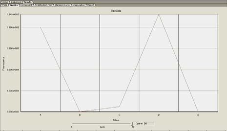
May 12, 2015 Page 17 of 30• AB software has a spectra component that also can help evaluate challenging curves
more efficiently. The spectra component shows the difference in total fluorescence at
every cycle. If there is an obvious difference in the fluorescence from cycle 1 to cycle
43, the sample is a true positive. Figure 8 shows the spectra view of a positive sample.
Filter A is the FAM filter and indicates if there is an accumulation of fluorescence during
the reaction. Filter D is the ROX filter and should remain constant.
Figure 8: Spectra component of a positive sample. Left screenshot shows fluorescence at cycle 1 and right
screenshot shows fluorescence at cycle 40.
• As described above, close examination of the amplification curves can help determine if a
sample is truly positive or not and eliminates the need to rely solely on Ct values.
However, this does not answer the question of the source of the sample positivity: Is the
sample truly positive for the pathogen or did contamination occur during or after sample
collection? It is important to be very careful during sample collection, extraction, and
rRT-PCR setup to avoid contamination.
• A note on weak positive samples (Ct ≥38)
Weak positives should always be interpreted with caution. Look carefully at the
fluorescence curves associated with these results. If curves are true exponential curves,
the reaction should be interpreted as positive.
• If repeat testing of a weak specimen is necessary, it is important to repeat the sample in
replicates as a single repeat test run has a high likelihood of generating a discrepant
result.
• If re-extracting and re-testing the specimen, it may be helpful to elute in a lower volume
to concentrate the sample.
• Contact CDC for guidance, to help determine if repeat testing may be warranted and to
discuss additional testing strategies as appropriate.
May 12, 2015 Page 18 of 30Overall Test Interpretation and Reporting Instructions
Table 5: EV-D68 2014 rRT-PCR Test Interpretation and Reporting Instructions
D68 RP Interpretation Reporting Actions
Refer to the CDC EV-D68
EV-D68 2014 EV-D68 North America Respiratory Disease Algorithm.
- +
Negative RNA not detected by rRT-PCR
Report results to CDC
If there are no additional
Inconclusive for 2014 EV-D68
specimens available for the patient,
North America RNA by rRT-
request collection of additional
- - Inconclusive PCR. An inconclusive result may
specimens.
occur in the case of an inadequate
specimen.
Report results to CDC.
2014 EV-D68 North America
RNA detected by rRT-PCR.
Refer to the CDC EV-D68
EV-D68 Additional analysis required.
+ Respiratory Disease Algorithm.
+ or - Presumptive Refer to CDC EV-D68
CtAssay Limitations, Warnings and Precautions
Interpretation of rRT-PCR test results must account for the possibility of false-negative and
false-positive results. False-negative results can arise from:
• poor sample collection or
• degradation of the viral RNA during shipping or storage or
• specimen collection conducted prior to symptom onset or late in illness
• failure to follow the authorized assay procedures
• failure to use authorized extraction kit and platform
Application of appropriate assay controls that identify poor-quality specimens (such as RNase P)
and adherence to CDC guidelines for EV-D68 testing (http://www.cdc.gov/non-polio-
enterovirus/index.html) can help avoid most false-negative results.
A high Ct observed for a specimen taken during early onset of disease should be followed up
with another specimen taken 24-48 hours later. Refer to CDC EV-D68 laboratory guidance for
current advice (http://www.cdc.gov/non-polio-enterovirus/index.html) and consultation
instructions.
The most common cause of false-positive results is contamination with previously amplified
DNA. Liberal use of negative control samples in each assay can help ensure that laboratory
contamination is detected and that false positive test results are not reported.
Negative results do not preclude infection with Enterovirus D68 and should not be used as the
sole basis of a patient treatment/management decision. All results should be interpreted by a
trained professional in conjunction with review of the patient’s history and clinical signs and
symptoms. Only a limited evaluation of serum specimens has been performed due to the scarcity
of serum specimens from individuals with EV-D68 infection. Thus, sera may only be tested in
conjunction with patient-matched upper respiratory specimen(s) with the EV-D68 2014 rRT-
PCR.
This assay is for in vitro diagnostic use under FDA Emergency Use Authorization only and is
limited to qualified laboratories designated by CDC.
All specimens should be handled as if infectious. Proper biosafety precautions, including
personal protective equipment, must be used when handling specimen materials.
Proper collection, storage and transport of specimens are essential for correct results.
Extraction of nucleic acid from clinical specimens must be performed with either the bioMérieux
NucliSENS easyMAG instrument with its associated reagents, the QIAamp Viral RNA Mini Kit,
or the QIAamp DSP Viral RNA Mini Kit from QIAGEN. Other extraction kits have not been
evaluated for use with this assay.
May 12, 2015 Page 20 of 30Performance has only been established with the specimen types listed in the Intended Use. Other
specimen types have not been evaluated.
Performance Characteristics
The EV-D68 2014 rRT-PCR has been developed and evaluated by the CDC Picornavirus
Laboratory from mid-September to mid-October 2014. The assay and protocol are primarily
focused on evaluating respiratory disease due to EV-D68 strains detected in North America in
2014.
Unless otherwise noted, studies presented below were performed following the EV-D68 2014
rRT-PCR test procedure using the Applied Biosystems 7500 Real-time PCR system and the
QIAGEN QIAamp Viral RNA Mini Kit for specimen extraction. The comparator method
employed in several of the evaluations listed below, the enterovirus VP1 reverse transcription,
semi-nested PCR followed by sequencing (EV VP1 Sequencing Assay), is described in the
following publication:
Nix, W.A., M. S. Oberste, and M. A. Pallansch. Sensitive, semi-nested PCR
amplification of VP1 sequences for direct identification of all enterovirus serotypes from
original clinical specimens. Journal of Clinical Microbiology 2006, 44(8):2698-2704.
1. VP1.2014 Primers and Probe Set Development
The EV-D68 2014 specific assay primers and probe were designed from an alignment of
576 EV-D68 partial VP1 sequences from the 2014 US outbreak and recent EV-D68
phylogenetic ancestors from the United States (2013 viruses; CDC database), Spain (2012
virus; KF254918), Italy (2012 viruses; KC763167, KC763162), China (2012 viruses;
JX898785, JQ924865), and Thailand (2011 virus; JQ411807).
The Public Health Agency of Canada employs the CDC VP1 Sequencing method and
shared five representative EV-D68 Canadian outbreak sequences with CDC in December
2014. These sequences were first analyzed phylogenetically and clustered with US 2014 co-
circulating EV-D68 strains. In silico inspection of the VP1.2014 primer sites and probe site
showed 100% nucleotide identity with all five Canadian EV-D68 strains.
2. Analytical Sensitivity
a) Preliminary Limit of Detection (LoD) in Minimum Essential Medium (MEM)
Serial ten-fold dilutions of RNA extracted from EV-D68 US/MO/14-18949 strain (108
CCID50/mL) were prepared in MEM. The serial dilutions from 10-1 to 10-10 were tested in
triplicate using the EV-D68 2014 rRT-PCR. The lowest dilution at which all replicates were
positive was at 10-7 CCID50/mL or 10-1.1 CCID50/5µL with the EV-D68 2014 rRT-PCR.
Results of testing with the EV VP1 Sequencing Assay at each concentration are also
included to aid in interpretation of comparative study data presented in the clinical
performance sections.
May 12, 2015 Page 21 of 30Table 6: EV-D68 2014 rRT-PCR Assay Limits of Detection Estimation with EV-D68 Isolate RNAa
Diluted in Minimum Essential Mediumb and Comparison to Limits of Detection Estimation with the EV
VP1 Sequencing Assay.
Result EV
Result
Concentration Replicates VP1
RNA Dilutionc EV-D68 2014
(CCID50 / 5 µL) (Ct Values) Sequencing
rRT-PCR
Assay
10-1 104.9 15.8 16.0 16.3 3/3 Positive 3/3 Positive
10-2 103.9 19.3 19.7 19.6 3/3 Positive 3/3 Positive
10-3 102.9 24.5 24.7 25.4 3/3 Positive 3/3 Positive
10-4 101.9 29.1 29.6 30.1 3/3 Positive 3/3 Positive
10-5 100.9 33.7 33.6 34.2 3/3 Positive 3/3 Positive
10-6 10-0.1 37.7 36.9 36.8 3/3 Positive 3/3 Positive
10-7 10-1.1 40.6 41.9 39.9 3/3 Positive 3/3 Positive
1/3 Equivocal 2/3 Positive
10-8 10-2.1 43.5 Negative Negative
2/3 Negative 1/3 Negative
10-9 10-3.1 Negative Negative Negative 3/3 Negative 3/3 Negative
10-10 10-4.1 Negative Negative Negative 3/3 Negative 3/3 Negative
a
Serial dilutions were prepared from frozen stock.
b
Minimum essential medium is a cell culture medium that can be substituted for the several commercial formulations
of viral transport medium, used for NP swabs
c
1:10 dilution series of EV-D68 US/MO/14-18949 control RNA (lot 10/16/14) from titered virus control stock
b) LoD Confirmation in Clinical Matrix – Pooled Dual NP/OP Swabs (PCR Instrument and
Extraction Method Bridging Study)
A study was performed to compare the limits of detection for VP1.2014 in pooled leftover
dual NP/OP swab specimens in viral transport media across PCR instruments and across
extraction methods. Prior to use in the study, the leftover dual NP/OP swab eluents had
tested negative for the presence of enterovirus, including D68. The pooled matrix was
divided and spiked with EV-D68 US/MO/14-18949 at three concentrations (100.9 CCID50/5
µL, 10-0.1 CCID50/5 µL, and 10-1.1 CCID50/5 µL). Each concentration was extracted 20
times by each extraction method. The resulting nucleic acid samples were each tested once
on each PCR instrument system. The limits of detection observed for each PCR instrument
and extraction method combination are presented in Table 17.
All three PCR instrument systems yielded the same limit of detection, 10-0.1 CCID50/5 µL,
with material extracted manually using the QIAGEN QIAamp Viral RNA Mini Kit. With
this extraction method, the three instruments achieved equivalent limits of detection for the
EV-D68 2014 rRT-PCR and are all considered to be acceptable for use with this assay.
Material extracted on the easyMAG generated the same limit of detection for the EV-D68
2014 rRT-PCR as the QIAamp extracts when tested on the AB 7500. However, the other
May 12, 2015 Page 22 of 30two instrument systems (7500 Fast and 7500 Fast Dx) missed the 95% cutoff for this
concentration by 1 and 2 reactions, respectively. Thus, the limit of detection for these two
instruments with the easyMAG is set at the next highest concentration (100.9 CCID50/5 µL).
Though the 7500 Fast and Fast Dx instruments, when used with the easyMAG, did not
achieve the same LoD as the 7500 Fast, the performance was similar enough to consider
these instrument/extraction method combinations acceptable for use with the EV-D68 2014
rRT-PCR.
The RNase P primer and probe set was tested against each nucleic acid sample. Testing was
performed exclusively on the AB 7500 instrument and yielded 100% positive results in this
evaluation.
Table 7: EV-D68 2014 rRT-PCR Limits of Detection - Spiked Pooled Dual NP/OP Swab Specimen
Matrix
PCR Platforms
Extraction Methods
7500 7500 Fast 7500 Fast Dx
-0.1 -0.1 -0.1
Qiagen QIAamp 10 CCID50/5 µL 10 CCID50/5 µL 10 CCID50/5 µL
easyMAG 10-0.1 CCID50/5 µL 100.9 CCID50/5 µL 100.9 CCID50/5 µL
c) LoD Confirmation in Clinical Matrix – Pooled Serum (Extraction Method Bridging
Study)
A study was performed to evaluate the QIAamp, easyMAG and MagNA Pure Compact
extraction methods for use in preparing nucleic acid from human sera for subsequent
testing with the EV-D68 2014 rRT-PCR. The study was performed using pooled leftover
sera. Prior to use in the study, the leftover serum specimens had tested negative for the
presence of enterovirus nucleic acid, including EV-D68. The pooled matrix was divided
and spiked with EV-D68 US/MO/14-18949 at three concentrations (100.9 CCID50/5 µL,
10-0.1 CCID50/5 µL, and 10-1.1 CCID50/5 µL). Each concentration was extracted 20 times
by each extraction method. The resulting nucleic acid samples were each tested once by
EV-D68 2014 rRT-PCR on the AB 7500 Fast Dx instrument system. The limits of
detection observed for each extraction method are presented in Table 19.
Material extracted manually using the QIAGEN QIAamp Viral RNA Mini Kit generated
a limit of detection of 10-0.1 CCID50/5 µL. Material extracted on the easyMAG generated
a lower limit of detection, 10-1.1 CCID50/5 µL. The MagNA Pure Compact was also
included in this evaluation, but data has been excluded as the instrument is not
recommended for use with the assay.
As the limits of detection for sera extracted with the QIAamp method and the easyMAG
method were the same or lower than the limits of detection achieved in the evaluation of
pooled dual NP/OP swabs with the EV-D68 2014 rRT-PCR, both are acceptable for use
with serum in preparation of nucleic acid for subsequent testing by this assay.
The RNase P primer and probe set was tested against each nucleic acid sample. RP
testing was performed exclusively on the AB 7500 instrument and yielded 100% positive
results in this evaluation.
May 12, 2015 Page 23 of 30Additional information and line listing data may be found in Attachment E.
Table 7: EV-D68 2014 rRT-PCR Limits of Detection - Spiked Pooled Sera
PCR Platform
Extraction Methods
7500 Fast Dx
Qiagen QIAamp 10-0.1 CCID50/5 µL
easyMAG 10-1.1 CCID50/5 µL
d) Performance of RNase P Primer and Probe Set with Recommended Human DNA
Controls
The RNase P Primer and Probe Set (RP) was tested against serial dilutions of two
commercial human DNA preparations. A concentration of 50 ng/5 µL (10 ng/µL) should
be added to each reaction. The two commercial human DNA preparations have been
used as positive controls for RP in other Laboratory Response Network PCR assays, but
not with the combination of master mix and cycling conditions used with this assay. The
evaluation successfully demonstrated that the human DNA preparations would generate
expected results with RP in this assay, and were suitable for use as RP positive control
material when used at a concentration of 50 ng/5 µL. Results are presented in Table 20.
Table 9: EV-D68 2014 rRT-PCR Performance of Human DNA Controls with the RNase P Primer
and Probe Set
Platform AB 7500 AB 7500 Fast Dx
[DNA] Promega Roche Promega Roche
50 ng / 5 µL 28.4 27.6 26.0 24.8
5.0 ng / 5 µL 31.9 30.9 29.3 28.2
0.5 ng / 5 µL 36.4 34.8 33.2 32.2
0.05 ng / 5 µL 40.7 38.6 37.3 36.4
0.005 ng / 5 µL Negative Negative 40.4 39.9
Note: This study was not conducted on the AB 7500 Fast; however, the performance is expected to be
similar.
3. Analytical Specificity
a) Cross-Reactivity With Other Enteroviruses
Enterovirus Species D
To evaluate the analytical specificity of the EV-D68 2014 rRT-PCR, we used a panel of
enterovirus species D (EV-D) cell culture isolates (n=7) that included the EV-D68, 1962
Fermon prototype strain. Respiratory clinical specimens positive for EV-D68 Fermon,
EV-D70, EV-D94, EV-D111 were not available for testing, therefore RNA from these
viruses was extracted from frozen undiluted cell culture supernatants and tested. The
EV-D68 2014 rRT-PCR did not cross-react with other enterovirus species D viruses
including the Fermon prototype strain.
May 12, 2015 Page 24 of 30Table 10: EV-D68 2014 rRT-PCR Analytical Specificity Confirmed Using EV-D Cell Culture
Isolates
EV-D68 2014 rRT-
a
EV-D Cell Culture Isolate PCR Result Assay
Result
EV-D70, Prototype J670/1971 Negative
EV-D94, Nigeria 2010, a Negative
EV-D94, Nigeria 2010, b Negative
EV-D94, Angola 2012 Negative
EV-D111, Angola 2012, a Negative
EV-D111, Angola 2012, b Negative
EV-D68, Prototype Fermon 1962 Negative
a
Purified RNA in elution buffer
EV-D120, isolated from gorillas and a chimpanzee, in Cameroon and the Democratic
Republic of Congo, respectively, was not available for testing. In silico testing was not
performed on this strain. To date this virus has not been detected in humans.
Rhinovirus Species A and B
Other genetically related common Rhinoviruses circulating during the EV season were
tested using undiluted cell culture supernatants. A total of 95 rhinoviruses (RV),
including 70 species A (RV-A) and 25 species B (RV-B) were tested (Table 11).
Results demonstrated no cross-reactivity with the Enterovirus species tested.
Table 11: EV-D68 2014 rRT-PCR Analytical Specificity Confirmed Using Undiluted RNA
Extracted from Rhinovirus Cell Culture Supernatants
EV-D68
Enterovirus 2014 rRT- EV-D68
Enterovirus Types
Speciesb PCR (neg/total)
Result
RV-A1, A2, A7, A8, A9, A10, A11, A12,
A13, A15, A16, A18, A19, A20, A21,
A22, A23, A24, A25, A28, A29, A30,
A31, A32, A33, A34, A36, A38, A39,
A40, A41, A43, A45, A46, A47, A49, All
RV-Aa 70/70
A50, A51, A53, A54, A55, A56, A57, Negative
A58, A59, A60, A61, A62, A63, A64,
A65, A66, A67, A68, A71, A73, A74,
A75, A76, A77, A78, A80, A82, A85,
A88, A89, A90, A94, A96, A100
RV-B3, B4, B5, B6, B14, B17, B26, B27,
B35, B37, B42, B48, B52, B69, B70, B72, All
RV-B 25/25
B79, B83, B84, B86, B91, B92, B93, B97, Negative
B99
a
RV = rhinovirus; Species A is the most abundant in humans
b
Undiluted RV RNA extracted from cell culture supernatants was tested.
May 12, 2015 Page 25 of 30b) Cross-reactivity with Other Common Respiratory Viruses
RNA extracted from fourteen common respiratory viruses and pooled nasal wash
(containing Streptococcus pneumoniae, rhinovirus and adenovirus) were tested using the
EV-D68 2014 rRT-PCR on both the AB 7500 and the AB 7500 Fast Dx. RNA was
extracted from viral cell culture supernatants (except for the pooled nasal wash clinical
specimen) and tested undiluted. All results matched expected results.
Table 8: EV-D68 2014 rRT-PCR Cross-Reactivity with Common Respiratory Viruses
Number Virus AB 7500 AB 7500 Fast Dx
1 Adenovirus C1 (Ad Negative Negative
71)
2 CoV 229E Negative Negative
3 CoV OC43 Negative Negative
4 CoV MERS Negative Negative
5 HMPV (CAN99-81) Negative Negative
6 Influenza A H1N1 Negative Negative
7 Influenza A H3N1 Negative Negative
8 Influenza B Negative Negative
9 PIV1 (C35) Negative Negative
10 PIV2 (Greer) Negative Negative
11 PIV3 (C-43) Negative Negative
12 PIV4a (CH 19503) Negative Negative
13 RSV Long A Negative Negative
14 Rhinovirus 1A Negative Negative
15 Pooled nasal wash* Negative Negative
16 EV-D68 14-18949 Positive (Ct 32.4) Positive (Ct 35.7)
* Streptococcus pneumoniae; rhinovirus; adenovirus
c) Cross-Reactivity with Other Clinically Relevant Organisms
A panel of nucleic acid samples extracted from clinically relevant organisms was tested
using the EV-D68 2014 rRT-PCR. Nucleic acid samples for viral panel members were
prepared from undiluted tissue culture grown virus. For bacterial and fungal panel
members, nucleic acid was extracted from bacterial and fungal colonies and tested
undiluted. Testing was performed by CDC laboratories in Atlanta, Georgia, and Ft.
Collins, Colorado. Results are presented in Table 13.
Table 93: EV-D68 2014 rRT-PCR Cross-reactivity with other clinically relevant organisms
Virus/Bacteria/Fungus VP1.2014 Result
Dengue Virus 1* Negative
Dengue Virus 2* Negative
Dengue Virus 3* Negative
Dengue Virus 4 Negative
May 12, 2015 Page 26 of 30Eastern Equine Encephalitis Virus* Negative
Western Equine Encephalitis Virus* Negative
Colorado Tick Fever Virus Negative
Powassan Virus* Negative
Japanese Encephalitis Virus* Negative
West Nile Virus* Negative
Yellow Fever Virus* Negative
Lacrosse Encephalitis Virus* Negative
St. Louis Encephalitis Virus* Negative
Chikungunya Virus* Negative
Herpes Simplex Virus-1 Negative
Herpes Simplex Virus-2 Negative
Varicella Zoster Virus Negative
Measles Virus Negative
Mumps Virus Negative
Neisseria meningitides Negative
Listeria monocytogenes Negative
Escherichia coli Negative
Bacillus cereus Negative
Lactobacillus acidophilus Negative
Crytococcus neoformans Negative
*Results for these agents were generated using a PCR instrument not validated for use with the EV-D68
2014 rRT-PCR.
d) In silico Analysis of Common Respiratory Illness-Causing Flora and a Selection of
Other Flora Found in Respiratory Specimens and Sera
Additional evaluation of the analytical specificity of the EV-D68 rRT-PCR was
performed through in silico analysis of the VP1.2014 primer and probe sequences
against common causes of respiratory illness and other clinically relevant organisms.
BLASTn analysis queries of the VP1.2014 primers and probe were performed against
the GenBank public domain nucleotide sequences and showed no significant combined
homologies (primer target and probe target) with other conditions that would predict
potential false positive rRT-PCR results. Conditions and associated causative agents
covered in the in silico specificity analysis are presented in Table 14.
Table 104: VP1.2014 Primer and Probe Set In silico Specificity Analysis
Disease/condition Tax ID Agent
Human parainfluenza 11226 Parainfluenza virus 4b
Human parechovirus 12063 Parechovirus 1b
Common cold, bronchiolitis, 208895 Respiratory syncytial virus B
pneumonia
Sinusitis 1280 Staphylococcus aureus
Pertussis (whooping cough) 520 Bordetella pertussis
Mycoplasma pneumonia 2104 Mycoplasma pneumoniae
Pharyngitis, bronchitis, atypical 83558 Chlamydia pneumoniae
pneumonia (or Chlamydophila
pneumonia)
May 12, 2015 Page 27 of 30Bacteremia, pneumonia, epiglottitis 727 Haemophilus influenza
and acute bacterial meningitis
Tuberculosis 1773 Mycobacterium tuberculosis
Lyme disease 139 Borrelia burgdorferi
Respiratory infection 550 Enterobacter cloacae
Septicaemia 1354 Enterococcus hirae
Foodborne illness 1502 Clostridium perfringens
Abdominal infection (rare) 46503 Parabacteroides merdae
Human gastrointestinal bacterium 1680 Bifidobacterium adolescentis
Human gastrointestinal bacterium 216816 Bifidobacterium longum
Human gastrointestinal bacterium 40518 Ruminococcus bromii
4. Precision
Time Period: October 16, 2014- October 19, 2014
Number of Days Tested: 1 assay run (5 replicates/sample) per day for 3 non-consecutive
days with two separate PCR instruments (one on Day 1, another on Days 2 and 3).
Number of Operators: 1 operator
Number of Samples Types in Each Run:
• Two concentrations of EV-D68 US/MO/14-18949 control RNA from frozen stock
(lot: 10/8/14).
o A low cell culture 50% infectious dose (CCID50) dilution of EV-D68 US/MO/14-
18949 control RNA contained 0.79 CCID50/ 5µL input RNA
o A high CCID50 dilution contained 102.9 CCID50 / 5µL input RNA.
• Two clinical NP swab specimens in VTM (one high Ct Positive and one low Ct
positive). Swabs were collected from two different patients.
• NTCs
Cumulative results are presented in Table 15 (below).
Table 11: EV-D68 2014 rRT-PCR Precision Testing
Positive
Cumulative Results
Sample Detection
Replicates (N) (Positive)
Rate (%)
1: Low Concentration EV-
15 15/15 100
D68 control RNAa
2: High Concentration EV-
15 15/15 100
D68 control RNAa
3: NTC control 15 0/15 100
4: EV-D68 Positive Clinical
15 15/15 100
Specimen (low Ct)b
5: EV-D68 Positive Clinical
15 15/15 100
Specimen (high Ct)b
a
purified RNA from virus cell culture isolate
b
RNA extracted from clinical specimens
May 12, 2015 Page 28 of 305. Clinical Evaluation
The clinical study was conducted from September to October 2014. A total of 128
specimens (116 respiratory and 12 sera) were tested using the EV-D68 2014 rRT-PCR and
the enterovirus VP1 reverse transcription semi-nested PCR assay followed by sequencing
(EV VP1 Sequencing Assay). All controls were run in duplicate. The same lot of non-
infectious control RNA was run concurrently with all test samples. The following standard
controls were used during the clinical study.
• Positive control: 2014 outbreak strain EV-D68 US/MO/14-18949 viral RNA,
concentration adjusted to yield Ct value of 30-32 (low but consistent positive);
• Negative control: no-template controls (NTC) of nuclease free water
• RNase P (RP) Primer and Probe Set: Included as a control for specimen quality and for
the presence of nucleic acid in the sample following extraction. RP was performed on
all but 16 specimens (11 NP swab specimens, 5 nasal washes). For specimens without
RP results, specimen results were interpreted on the basis of VP1.2014 alone.
Positive percent agreement (PPA) and negative percent agreement (NPA) against the
comparator (EV VP1 Sequencing Assay) is presented for upper respiratory specimens in
Table 16 and serum in Table 17. A breakdown of the negative respiratory specimens is
presented in Table 18.
Table 12: EV-D68 2014 rRT-PCR Clinical Performance Summary for Upper Respiratory Specimens
EV-D68 Positive by EV VP1 EV-D68 Negative by EV
Specimen Sequencing Assay VP1 Sequencing Assay
Type PCR PCR PPA PCR NPA
Total Total
+ Ea (95% CI) - (95% CI)
98.3%
NP and OP 58 1 59 (91.0% - 40b 42
95.2%
Swabsd 99.7%)
(84.2% - 98.7%)
Nasal 3 0 3
100%
11c 12
91.6%
Wash (43.9%- 100%) (64.6% - 98.5%)
a
PCR E refers to an equivocal result (Ct value ≥ 43 andTable 14: EV-D68 2014 rRT-PCR Breakdown of Clinical Respiratory Specimens Negative for EV-D68
by EV VP1 Sequencing Assay
EV-D68 2014 rRT-PCR
Speciesa Type(s) Results
# Tested # Negative
RV-A2, A10, A16, A24, A31,
b
RV-A A34, A40, A49, A58, A59, 22 21c
A67, A63, A73, A85
RV-B RV-B4, B6, B27, B83, B48 5 5
RV-C Types not determined 2 2
EV-B E9, E11, CVB4 3 3
EV-A CV-A10 1 1
Negative for Enterovirus species 21 19d
Total 54 51
a
RNA extracted from clinical respiratory specimens
b
RV = rhinovirus; A species is the most abundant in humans
c
One specimen gave a positive result with the EV-D68 2014 rRT PCR. Further analyses indicated that this specimen was a
co-infection with a clear mixture of virus sequences on the sequence chromatogram. The virus identified by GenBank NT
BLAST of the readable portion of the sequence was RV-A10. The CT value for the EV-D68 2014 rRT-PCR was 31.2.
Data suggests that the EV-D68 2014 rRT-PCR can detect a specific target in a co-infection.
d
Two “false positive” specimens had high EV-D68 2014 rRT-PCR Ct values (40.1 and 40.9). The fluorescent signal from
these specimens demonstrated a clear sigmoid curve.
Contact Information
When questions arise in the real-time testing process, consultation is available via email. Send
the AB 7500 experiment file as an attachment to LRN@cdc.gov with an explanation of what the
issue is and we will get back to you with comments and suggestions.
For questions or additional information, please contact:
Laboratory Response Network Helpdesk
LRN@cdc.gov
May 12, 2015 Page 30 of 30You can also read