Meta-Control in Pigeons (Columba livia) and the Role of the Commissura Anterior - MDPI
←
→
Page content transcription
If your browser does not render page correctly, please read the page content below
SS symmetry
Article
Meta-Control in Pigeons (Columba livia) and the Role
of the Commissura Anterior
Emre Ünver 1, *, Qian Xiao 1,2 and Onur Güntürkün 1
 1   Department of Biopsychology, Institute of Cognitive Neuroscience, Faculty of Psychology,
     Ruhr University Bochum, 44801 Bochum, Germany; qianxiao@moon.ibp.ac.cn (Q.X.);
     Onur.Guentuerkuen@ruhr-uni-bochum.de (O.G.)
 2   Key Laboratory of Interdisciplinary Science, Institute of Biophysics, Chinese Academy of Sciences,
     Beijing 100101, China
 *   Correspondence: emre.uenver@rub.de; Tel.: +49-234-32-26213

 Received: 30 November 2018; Accepted: 16 January 2019; Published: 22 January 2019

 Abstract: Meta-control describes an interhemispheric response conflict that results from the
 perception of stimuli that elicit a different reaction in each hemisphere. The dominant hemisphere for
 the perceived stimulus class often wins this competition. There is evidence from pigeons that
 meta-control results from interhemispheric response conflicts that prolong reaction time when
 the animal is confronted with conflicting information. However, recent evidence in pigeons
 also makes it likely that the dominant hemisphere can slow down the subdominant hemisphere,
 such that meta-control could instead result from the interhemispheric speed differences. Since both
 explanations make different predictions for the effect of commissurotomy, we tested pigeons in a
 meta-control task both before and after transection of the commissura anterior. This fiber pathway
 is the largest pallial commissura of the avian brain. The results revealed a transient phase in which
 meta-control possibly resulted from interhemispheric response conflicts. In subsequent sessions
 and after commissurotomy, however, the results suggest interhemispheric speed differences as a
 basis for meta-control. Furthermore, they reveal that meta-control is modified by interhemispheric
 transmission via the commissura anterior, although it does not seem to depend on it.

 Keywords: birds; hemispheric interactions; brain asymmetry; reaction time; color discrimination

1. Introduction
      Meta-control refers to the one hemisphere taking charge of response selection when the
two hemispheres are brought into conflict [1–3]. This phenomenon was first demonstrated in
split-brain patients and healthy people [1,4], but was also later revealed in monkeys [5], chicken [6],
and pigeons [2,3,7]. It is often assumed that meta-control results from one hemisphere inhibiting
the other via the various commissures that connect the two halves of the brain at the midbrain and
telencephalic level [8,9].
      Meta-control becomes especially visible in species with pronounced brain asymmetries.
Depending on the type of stimulus, one or the other hemisphere regularly gains control. Birds are
ideal subjects for these studies [10]. Their left hemisphere is superior in discrimination, categorization,
and memorization of visual patterns (chicks: [11]; quail: [12]; pigeons: [13,14]) and visuomagnetic
cues (pigeons: [15]; chicks: [16]), while their right hemisphere is superior in visually guided
interactions with emotionally charged stimuli (chicks: [17]), attentional shifts (chicks and pigeons: [18]),
social interactions (chicks: [19]), as well as in relational and spatial analyses of visual information
(chicks: [20]; pigeons: [14,21]).

Symmetry 2019, 11, 124; doi:10.3390/sym11020124                               www.mdpi.com/journal/symmetry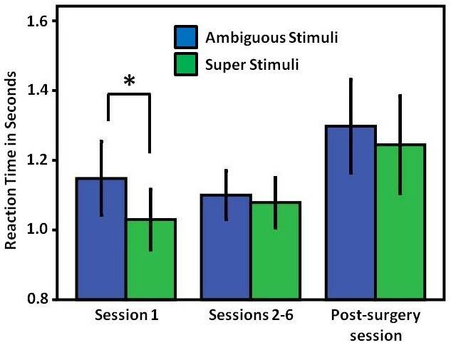
Symmetry 2019, 11, 124                                                                             2 of 11

      Meta-control could result from either inter-hemispheric response conflict or differences in
hemisphere-specific speed. If inter-hemispheric response conflict was the cause, situations in which
each half-brain competes to present a different response should produce longer reaction times than
non-conflicting situations [2,8]. This is because decision making with two incompatible options
usually requires a longer processing time [10]. If, however, meta-control simply results from
hemisphere-specific processing speed, the outcome would be different. The decision time would
be determined solely by the faster hemisphere, which would always win. Two competing hemispheres
would then be as fast as the faster hemisphere.
      A recent study conducted by Ünver & Güntürkün [2] in pigeons collected evidence for the
inter-hemispheric response conflict model. In their study, pigeons were trained by a forced-choice
color discrimination task monocularly, and each hemisphere learned to discriminate between its
own stimulus pair. Then, under binocular conditions, the birds were exposed to two types of test
stimuli. These test stimuli were created by combining positive and negative patterns learned by each
hemisphere. If the animal had to discriminate between a stimulus pair that consisted of two positive
(left- and right-hemispheric) patterns on one pecking key and two negative patterns on the other,
the choice was easy. Both hemispheres agreed to peck the pattern combination that was positive for both
half-brains. Consequently, the animals responded quickly to this “super stimulus”. The situation was
different when each stimulus was composed of the positive pattern of one hemisphere and the negative
pattern of the other hemisphere. In the case of such an “ambiguous stimulus”, the overall pattern
signaled an interhemispheric reward history conflict. As it turned out, the ambiguous stimulus caused
a significant response delay. This makes it likely that meta-control rests mainly on an inter-hemispheric
response conflict and not on hemisphere-specific speed.
      A recent study, however, proposed a different mechanism. Qian & Güntürkün [22] recorded
signals from the sensorimotor arcopallium of pigeons while the birds were conducting a color
discrimination task under monocular conditions. All birds in their study learned faster and responded
more quickly with their right eye/left hemisphere. The arcopallium not only harbors descending
premotor neurons but also commissural neurons that constitute the commissura anterior—the largest
avian interhemispheric connection at the pallial level. As shown by Letzner et al. [23], the commissura
anterior originates from the telencephalic arcopallium/amygdala-complex and contains a small cluster
of non-GABAergic sensorimotor and amygdaloid fibers that project onto a wide range of contralateral
structures such as the posterior amygdala, the sensorimotor arcopallium, as well as further sensory
and motor components of the nidopallium. We chose this commissure for our study due to these
widespread projections onto the contralateral hemisphere. Qian & Güntürkün [22] transiently blocked
the arcopallial activity of one hemisphere and recorded from the contralateral arcopallium during
color discrimination to determine the effect of left-to-right and right-to-left information transfer.
They discovered that the left hemisphere was able to modify the timing of individual activity patterns
of the neurons in the right hemisphere via asymmetrical commissural interactions. In contrast to that,
right arcopallial neurons were hardly able to alter the activity pattern of left arcopallial cells. Thus,
under conditions of interhemispheric competition, left arcopallial neurons could delay the contralateral
spike time of those in the right hemisphere. As a result, the neurons of the right hemisphere would
come too late to control a response and the left hemisphere would govern decisions. This finding
could imply that hemispheric dominance in birds is realized at least in part by time shifts of the neural
activity of one or the other hemisphere.
      The studies by Ünver & Güntürkün [2] and Qian & Güntürkün [22] make contradictory predictions
of the mechanisms of meta-control. Both would assume that the commissura anterior plays a decisive
role in inter-hemispheric response conflicts but would predict different choice patterns from birds
in a meta-control task after commissurotomy. Ünver & Güntürkün [2] would infer that the loss of
the commissura anterior should reduce reaction times when presented with an ambiguous stimulus
because an inter-hemispheric response conflict could no longer result in an inter-hemispheric delay
in processing time. In contrast, Qian & Güntürkün [22] would not expect a change in reaction
Symmetry 2019, 11, 124 3 of 11
Symmetry
times 2019, 11,
under thex FOR PEER REVIEW
ambiguous stimulus because the dominant hemisphere already determines3 ofthe
11
response. They would, however, expect that the dominance of the left hemisphere would weaken
would weaken after commissurotomy because the left-to-right control of the neuronal spike times
after commissurotomy because the left-to-right control of the neuronal spike times could no longer
could no longer be executed. To test these predictions, we conducted a meta-control study as
be executed. To test these predictions, we conducted a meta-control study as published by Ünver &
published by Ünver & Güntürkün [2], and subsequently transected the commissura anterior to
Güntürkün [2], and subsequently transected the commissura anterior to re-test the animals with the
re-test the animals with the same task.
same task.
2. Materials
2. and Method
Materials and Method
2.1. Subjects
Nine naïve pigeons of unknown sex were used in the study. study. All pigeons were housed in single
cages with other conspecifics and maintained on a 12:12 hh light–dark
light–dark cycle.
cycle. Their body weight was
maintained at
maintained at 80–90%
80–90%of oftheir
theirfree-feeding
free-feedingweight
weightbybyfeeding
feedingdiet
dietfood
food
onon weekdays
weekdays andand a mixture
a mixture of
of peas,
peas, corn,
corn, and sunflower
and sunflower seeds seeds
on theon the weekends.
weekends. Water
Water was was provided
provided ad libitum.adForlibitum. For the
the monocular
monocular
sessions, sessions,
velcro ringsvelcro ringsaround
were fixed were fixed around
the eyes thepigeons
of the eyes ofusing
the pigeons
glue that using
was glue that was
non-irritating
non-irritating
to to the skin. Cone-shaped
the skin. Cone-shaped eye caps that eye
werecaps that were
attached to theattached to the
other sides of other sides rings
the velcro of theatvelcro
their
rings at their bases and were created using cardboard. These eye caps could be easily
bases and were created using cardboard. These eye caps could be easily attached and removed from attached and
removed
the from the ringsthe
rings surrounding surrounding the eyes for
eyes for monocular monocular
testing (Figuretesting
1). All (Figure 1). All
procedures procedures
were conducted were
in
conducted in
compliance withcompliance withfor
the guidelines the
theguidelines
care and useforofthe care and
laboratory use ofand
animals laboratory
approvedanimals and
by the local
approved by
committee the local committee (LANUV).
(LANUV).
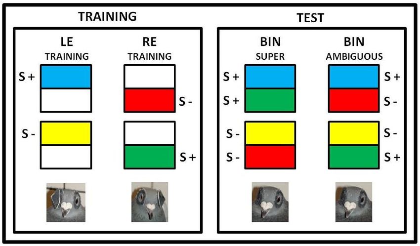
Figure 1.1.The
Thestimuli used
stimuli usedin the experiment.
in the Super
experiment. stimuli
Super consisted
stimuli of a combination
consisted of two positive
of a combination of two
and two and
positive negative stimuli presented
two negative to the left
stimuli presented to eye (LE)eye
the left and right
(LE) eye
and (RE)
right during
eye the training
(RE) during phase.
the training
Ambiguous stimuli were created by combining a negative stimulus for one hemisphere
phase. Ambiguous stimuli were created by combining a negative stimulus for one hemisphere and a and a positive
stimulus for the other.
positive stimulus for theBoth eyes
other. (BIN
Both eyes= (BIN
binocular) were open
= binocular) wereduring the testthe
open during phase. The color
test phase. The
combinations shownshown
color combinations in the figure
in theare merely
figure areexamples of the various
merely examples of thecombinations used. Below
various combinations are
used.
photographs showing theshowing
Below are photographs animals the
withanimals
a cap onwith
one aeye
cap(left) or both
on one eye eyes
(left)uncovered (right).
or both eyes uncovered
(right).
2.2. Apparatus
A custom-made operant chamber measuring 40 × 35 × 35 cm (W × D × H) in size was used
2.2. Apparatus
for the experiment. The chamber was equipped with a feeder and illuminated using a house light.
A custom-made operant chamber measuring 40 × 35 × 35 cm (W × D × H) in size was used for
The feeder was immediately illuminated when food was presented. The stimuli (5 × 5 cm in size) were
the experiment. The chamber was equipped with a feeder and illuminated using a house light. The
introduced on a TFT LCD touchscreen monitor with 1024 × 768 resolution. The monitor was placed
feeder was immediately illuminated when food was presented. The stimuli (5 × 5 cm in size) were
introduced on a TFT LCD touchscreen monitor with 1024 × 768 resolution. The monitor was placed
on the same side of the chamber as the feeder to ensure that the pigeons could easily reach the feederSymmetry 2019, 11, 124 4 of 11
on the same side of the chamber as the feeder to ensure that the pigeons could easily reach the feeder
immediately after pecking at the stimuli on the screen. The experimental sessions were controlled by a
custom-written MATLAB program (MathWorks, Natick, MA, USA) using the Biopsy Toolbox [24].
2.3. Procedure
Before learning the color discrimination task, all pigeons were trained in autoshaping sessions
consisting of 40 trials. In these sessions, the pigeons were made to peck on a white square presented
on the screen under monocular conditions. The white square was presented for 4 s, and food was
delivered immediately following a single peck on the white square. These sessions were conducted
according to a fixed ratio (FR1) schedule. The birds were trained in a counterbalanced manner—on one
day, only the left eye (LE) was blocked, whereas on the next day, only the right eye (RE) was blocked.
Response to the white square in >85% of the trials in two consecutive sessions per eye condition
was set as the criterion for progress to the subsequent schedules. Once the birds met this criterion,
their training progressed to a variable ratio (VR) schedule wherein they were progressively trained
with variable ratios VR2, VR4, and VR8 under monocular conditions again, with the same criterion.
All the sessions in the VR schedule consisted of 40 trials.
Once the birds met the response criterion for the VR, we commenced the color discrimination
training. Rectangles of four different colors (red, yellow, green, or blue) were used as stimuli. The color
discrimination sessions were conducted under monocular conditions, and the color combinations were
balanced among pigeons to prevent color preferences. As shown in Figure 1, they were always placed
in a compound at the upper or lower position of a larger white rectangle. Each eye of the pigeons was
exposed to a different pair of stimuli (e.g., red and yellow for the LE; blue and green for the RE). One of
these colors served as S+ and the other as S− for each eye. The pigeons had to choose between an
upper and a lower compound stimulus that each consisted of a colored and a white rectangle. Pecks on
the S+ compound were rewarded regardless of whether the peck location was on the colored or on
the white part of the compound. The same rule was applied for the S− compound. The monocular
sessions were conducted in a counterbalanced manner, similar to the autoshaping sessions.
The stimuli were presented for 4 s. A single peck on the S+ compound immediately activated
the feeder for 2 s, whereas a peck on the S− compound resulted in switching off the house lights
for 5 s and playing a loud noise for 1 s. Once the birds responded to the S+ compound in >85% of
the trials in two consecutive sessions for each eye condition, the number of trials per session was
increased to 200 in steps of 20. The criteria that was applied in each step was that the pigeons had to
make at least 85% correct choices (responses to the S+ compound) for each eye condition in a single
session. As the number of trials in each session was increased, the reward ratio (responses to S+) was
decreased in steps of 10% until reaching 40%. This procedure was employed to prevent extinction
learning in subsequent catch trials. As a final step, a new stimulus pair, a white (S+) square and a
gray (S−) square, were introduced. Because the birds had already been trained to respond to the
white square during the autoshaping sessions, we expected them to be able to rapidly discriminate
between this new stimulus pair. This white/gray “dummy” discrimination procedure was necessary
to maintain the birds’ responses during the critical test sessions that included catch trials. In the catch
trials, the colored stimuli were re-arranged to create “super” and “ambiguous” stimuli that were not
rewarded. Each of the final sessions consisted of 200 trials, with 80% of the stimuli being presented
as white (S+) and gray (S−) dummy stimuli. As outlined above, both S+ (the S+ of the LE and the S+
of the RE) on one pecking key and both S− on the other key were termed super stimuli. Unlike the
other sessions, the critical test sessions were performed under binocular conditions. The gray/white
stimuli represented a common associative background for both stimuli. This was not applied to the
ambiguous stimuli. On each key, the S+ of one hemisphere was always combined with the S− of
the other hemisphere. The proportion of catch trials in the final session was 20% (i.e., the number
of catch trials was 40, with 20 being ambiguous and 20 being super stimuli). The remaining trials
consisted of the white/gray stimuli pair (the number of white/gray stimuli was 160). No feedbackSymmetry 2019, 11, 124 5 of 11
for the catch trials was available, whereas the white/gray stimuli discrimination had a 40% reward
probability. Following the first critical test session that included catch trials, the pigeons were further
trained using the well-known training stimuli under monocular conditions. These sessions using
the well-known training stimuli between each critical test session were conducted because it was
necessary to maintain the pigeons’ response at a stable level during the subsequent critical test sessions.
Therefore, this sequence was repeated until enough catch trial responses were collected.
After six sessions at most of testing for meta-control, pigeons underwent a commissurotomy
operation. After a two-week recovery period, the same task and procedure were applied, and data
were collected.
2.4. Surgery
Before surgery, nine birds participating in the experiment were given a mixture of ketamine
(ketamine hydrochloride, 100 mg/mL; Zoetis, Berlin, Germany) and xylazine (xylazine hydrochloride,
23.32 mg/mL, methyl-4-hydroxybenzoate, 1.5 mg/mL; Bayer Vital, Leverkusen, Germany) by
intramuscular injection (7:3 ratios, 0.12 mL/100 g body weight). The anesthetized birds were placed
on a warming pad in a stereotaxic device. Their heads were fixed at a 45◦ angle in the head holder
according to the coordinates of the pigeon brain atlas [25]. Prior to the commissurotomy, the scalp was
opened and a window was opened in the skull with a drill, centered at the anterior 7.75 and lateral
0.0 coordinates. Then, the dura mater was removed. The main vessel in the gap between the two
hemispheres was delicately pulled aside with a hand-made hook. Finally, a 2-mm-wide, 0.3 mm thick
blade was slowly lowered into the region with the following coordinates: Anterior 7.75, lateral 0.0 at
a depth of 9.0 mm from the surface of the brain [25]. The blade was lowered in increments of 1 mm,
with a 2 min pause between each increment. Thus, the risk of damage to the brain due to the pressure
caused by the blade was minimized. At the end of the operation, the knife was removed in the same
manner, i.e., by lifting 1 mm every 2 min. The skin was stitched after a medical sponge was placed
on the operation area. Finally, a painkiller was sprayed over the operation area and an antibacterial
powder (Tyrasor; Engelhard Arzneimittel, Niederdorfleben, Germany) was applied. In addition,
an intramuscular painkiller (Rimadyl, 0.04 mL/100 g body weight; Pfizer, GmbH, Münster, Germany)
was administered. The pigeons were kept in their individual cages for one week to allow them to
overcome the effects of the operation. Then, the tests were conducted.
2.5. Histology
The pigeons were deeply anesthetized with equithesin (0.55 mL/100 g body weight) and
perfused with 4% paraformaldehyde (VWR Prolabo Chemicals, Leuven, Belgium) after the last
post-operation tests. The brain was removed, immersed in gelatin (Merck, Darmstadt, Germany) and
sectioned into 40-µm frontal slices using a freezing microtome (Leica Microsystems Nussloch GmbH,
Nussloch, Germany). Sections were mounted, nissl and klüver-barrera stained, and the success of the
commissurotomy was verified microscopically. In all nine birds, the commissura anterior was verified
to be completely sectioned (Figure 2). In some animals the blade had been successfully lowered along
the midline (Figure 2b), in others it was slightly off the midline and had damaged the medial most
parts of the hemispheres in the medial meso- and nidopallium, as well the area above the commissura
anterior (Figure 2a). These are not areas associated with the visual system and we could not see any
correlation between our histological verifications and our behavioral results.Symmetry 2019, 11, 124 6 of 11
Symmetry 2019, 11, x FOR PEER REVIEW 6 of 11
Figure
Figure 2. 2.AAnisslnissl(a)
(a)and
andaanissl/klüver-barrera
nissl/klüver-barrera (b)(b) stained
stainedfrontal
frontalsection
sectionofoftwo twopigeons
pigeonswith
with
transections of the commissura anterior. The straight arrows point to the tissue
transections of the commissura anterior. The straight arrows point to the tissue rupture resulting rupture resulting
from
from thethe passingofofthe
passing theblade,
blade,while
while the
the broken
broken arrows
arrows indicate
indicateremaining
remainingfibers
fibers ofof
thethe
commissura.
commissura.
Note
Note that
that inin(a)(a)the
theblade
bladehas
hasdamaged
damaged thethe area
area above
above thethe commissure
commissuresincesinceit itwas
wasslightly offoff
slightly thethe
midline.
midline. This
This isisnot
notthe
thecase
caseinin(b).
(b).Scale
Scale bar
bar in
in (b)
(b) also
also applies
appliesto
to(a).
(a).
3. Results
3. Results
TwoTwo variables
variables were
were important
important in studying
in studying thethe effect
effect of the
of the commissurotomy
commissurotomy on on meta-control.
meta-control. First,
First, how many individuals display significant meta-control before vs.
how many individuals display significant meta-control before vs. after commissurotomy? Meta-control after commissurotomy?
Meta-control
in our in ouras
task is defined task is defined ashigher
a significantly a significantly
numberhigher number
of choices that of
arechoices
dominatedthat are
by dominated by
one hemisphere
being faced with an ambiguous pattern. Second, how did the reaction times to ambiguous- to
one hemisphere being faced with an ambiguous pattern. Second, how did the reaction times and
ambiguous- change
super-stimuli and super-stimuli change after the commissurotomy?
after the commissurotomy?
Meta-control: A
Meta-control: A meta-control
meta-control effect effect was
was observed
observed inin threethreeout outofofnineninebirdsbirdsbefore
before
commissurotomy (for each individual: chi square test, pSymmetry 2019, 11, 124 7 of 11
Symmetry 2019, 11, x FOR PEER REVIEW 7 of 11
the super
to the superstimulus
stimulus between
between session 1 and
session sessions
1 and 2–62–6
sessions (paired sample
(paired samplet-test; t = 0.755,
t-test; p = p0.475,
t = 0.755, n =n8).
= 0.475, =
The same applied to the ambiguous stimulus (paired-sample t-test; t = 0.033, p
8). The same applied to the ambiguous stimulus (paired-sample t-test; t = 0.033, p = 0.975, n = 8). Note= 0.975, n = 8). Note that
the
thataverage values
the average of sessions
values 2–6 were
of sessions 2–6 derived from 8from
were derived birds, since one
8 birds, pigeon
since stopped
one pigeon working
stopped on the
working
task after session 1 (and then restarted after surgery). Similarly, in the post-surgery
on the task after session 1 (and then restarted after surgery). Similarly, in the post-surgery tests, no tests, no significant
differences
significant in the reaction
differences in times betweentimes
the reaction superbetween
and ambiguous
super and signals were observed
ambiguous signals(super stimulus:
were observed
1.24 s; ambiguous
(super stimulus: 1.24 stimulus:
s; ambiguous stimulus: 1.29 s;t-test,
1.29 s; (paired-sample t = 0.614, p t-test,
(paired-sample = 0.556, t =n0.614,
= 9)). pMoreover,
= 0.556, n there
= 9)).
was no significant difference between the response times to the two stimulus
Moreover, there was no significant difference between the response times to the two stimulus types types in the pre-surgery
sessions (excludingsessions
in the pre-surgery session 1) and post-surgery
(excluding session 1) sessions (mean of super
and post-surgery stimulus
sessions (mean sessions
of super 2–6: 1.07 s;
stimulus
post-surgery
sessions 2–6: 1.07session:
s; post-surgery session: 1.15 t-test,
1.15 s; (paired-sample t = 0.680, p t-test,
s; (paired-sample = 0.518,t = n0.680,
= 8);pmean= 0.518,of nambiguous
= 8); mean
stimulus
of ambiguous sessions 2–6: 1.1
stimulus s; post-surgery
sessions 2–6: 1.1 session: session: 1.22 s; t-test,
1.22 s; (paired-sample
s; post-surgery t = 1.097, pt-test,
(paired-sample = 0.309,
t =
n1.097,
= 8)) p(Figure
= 0.309,3).n = 8)) (Figure 3).
Figure 3. Average
Figure 3. Average reaction
reactiontimes
times of
of subjects
subjects to
to ambiguous
ambiguous and
and super
super stimuli
stimuli during
during sessions
sessions prior
prior to
to
the commissurotomy and in the first session after the commissurotomy. Significant differences
the commissurotomy and in the first session after the commissurotomy. Significant differences are are
indicated
indicated by
by an
an asterisk
asterisk(p
(pSymmetry 2019, 11, 124 8 of 11
meta-control. The mechanism based on the interhemispheric conflict would imply that a section of the
commissura anterior should reduce reaction times to ambiguous stimuli (no commissural exchange
→ no interhemispheric conflict), whereas the model based on hemisphere specific speed would not
predict post-surgery changes in reaction time to ambiguous stimuli (no commissural exchange → no
change in hemisphere-specific speed). At the same time, the results of Qian & Güntürkün [22] suggest
that the advantage of the left hemisphere would be smaller after commissurotomy (no commissural
exchange → no possibility to further delay response execution of the right hemisphere). Our findings
suggest that the birds only experience interhemispheric conflict on the first session with ambiguous
stimuli, and the effect disappears in the following sessions. A subsequent commissurotomy does not
alter reaction times to ambiguous stimuli but does modify meta-control. Overall, our data would be
compatible with a model according to which interhemispheric conflict occurs in a short, initial period,
but then gives way to lateralized reaction patterns determined by hemisphere-specific speed.
As visible in Figure 3, reaction times to super and ambiguous stimuli were the most different in
the first session in which the animals were first presented these two stimulus types under binocular
conditions. However, in subsequent sessions reaction times became increasingly similar. Ünver &
Güntürkün [2] had based their conclusion of interhemispheric conflict on the first session after
introducing ambiguous stimuli. This conclusion may remain valid but is obviously restricted to
this initial session. In subsequent sessions, a different mechanism seems to prevail. It is indeed
conceivable that the animals quickly learned about the absence of negative or positive feedback
when responding to the ambiguous stimuli. It is known that pigeons are extremely sensitive to
reward alterations in operant categorization tasks, and subsequently tend to bias their choices towards
initially favored alternatives [26]. Similar findings were also observed in studies with monkeys [27,28].
This makes it likely that our commissurotomy was performed at a point in time in which the pigeons
were no longer pondering response conflicts but instead biased their choices according to mechanisms
based on hemisphere-specific speed. Consequently, response times to ambiguous stimuli were not
altered by commissurotomy.
This scenario is compatible with the explanation that each hemisphere rushes with its own
hemisphere-specific speed to motor areas. During color discrimination, the left hemisphere usually
produces faster reaction times. This has been observed in various studies with pigeons [29] and
other birds [17,30]. This was also observed by Qian & Güntürkün [22] when recording from the
pigeon arcopallium during color discrimination. This study also offers a mechanistic explanation
of this observation by revealing that the left hemisphere can modify the spike time of the right
hemisphere. Thus, under conditions of conflict, the left hemisphere could delay the right hemispheric
response speed, thereby accelerating its own advantage. From this point of view, a transection of the
commissura anterior should reduce, but not completely terminate the left hemispheric superiority.
Indeed, we observed major alterations of meta-control after surgery. Usually, an individually significant
extent of meta-control is observed in only a fraction of pigeons [2,3,7]. With the procedure used in this
study, it was mostly the left hemisphere that evinced meta-control [2,7]. In the current experiment,
three out of nine birds demonstrated meta-control before commissurotomy (two left hemispheric,
one right hemispheric). This is a typical result pattern [2,7]. After transecting the commissura anterior,
however, all three birds lost their hemisphere-specific advantage. Instead, two other birds displayed
significant meta-control (one left, one right). Although this is certainly not a strong proof of the
conclusion of Qian & Güntürkün [22], it is conceivable that the changes observed in meta-control
in our nine pigeons resulted from the loss of a left hemispheric advantage that resulted in biased
interhemispheric interactions. If indeed neuronal speed differences cause the bias towards the right
eye in metacontrol studies, the large individual differences may result from the fact that neurons show
within the pigeon’s visual system substantial latency differences between individual birds [22,31–33].
It is known that the commissura anterior connects with the anterior and intermediate arcopallium.
These structures project onto a wide cluster of visual and sensorimotor areas. Our study focused on
the contribution of the commissura anterior to visual asymmetries. However, further commissuralSymmetry 2019, 11, 124 9 of 11
systems may also play a role in metacontrol since studies of both chicks [34] and pigeons [35–37]
suggested that subpallial commissures also play key roles in visually-guided lateralized behavior.
The supraoptic decussation (DSO) is one such subpallial connection, and is known to be responsible
for interocular transfer during visual discrimination [38]. This may be due to the indirect connection
of the DSO to telencephalic visual structures such as Wulst. More recently, it has been shown that the
nucleus of the lateral ponto-mesencephalic tectum (nLPT), a midbrain structure, contains GABAergic
neurons and its projections terminate in the contralateral optic tectum (TeO) via the commissura
tectalis [39]. Therefore, this midbrain commissure may also play a crucial role during meta-control.
Thus, the present study must be complemented by further experiments to reveal the full scenario of
interhemispheric interactions of lop-sided bird brains.
Although our study was centered on the mechanisms of meta-control, it might also offer some
more general insights on the behavior of organisms with lateralized brains. A key problem of these
species is the production of a single response from two asymmetrically specialized hemispheres.
Our results suggest that the default option in such situations could be to let both hemispheres compete
based on hemisphere-specific processing speed. Because the dominant hemisphere for a certain
stimulus class usually produces faster responses [22], the most competent half-brain would primarily
determine the response. The commissural slowing mechanism discovered by Qian & Güntürkün [22]
would amplify this interhemispheric speed difference to ensure that the dominant hemisphere controls
the overall response.
Author Contributions: Conceptualization: Q.X. and O.G.; designed experiment: Q.X. and O.G.; performed
experiment: E.Ü.; statistical analysis: E.Ü. and O.G.; manuscript preparation: E.Ü. and O.G.; funding acquisition
and project supervision: O.G. All authors revised and approved the paper.
Acknowledgments: We are grateful for the support of Annika Simon during surgery and the conduct of the
histological procedure. We also thank Felix Ströckens and Sarah von Eugen for help during documentation of
histological results. Supported by the Deutsche Forschungsgemeinschaft through SFB 874.
Conflicts of Interest: The authors declare no conflict of interest.
References
1. Levy, J.; Trevarthen, C. Metacontrol of hemispheric function in human split-brain patients. J. Exp.
Psychol. Hum. 1976, 2, 299–312. [CrossRef]
2. Ünver, E.; Güntürkün, O. Evidence for interhemispheric conflict during meta-control in pigeons.
Behav. Brain Res. 2014, 270, 146–150. [CrossRef] [PubMed]
3. Adam, R.; Güntürkün, O. When one hemisphere takes control: Metacontrol in pigeons (Columba livia).
PLoS ONE 2009, 4, e5307. [CrossRef] [PubMed]
4. Urgesi, C.; Bricolo, E.; Aglioti, S.M. Hemispheric metacontrol and cerebral dominance in healthy individuals
investigated by means of chimeric faces. Cogn. Brain Res. 2005, 24, 513–525. [CrossRef] [PubMed]
5. Kavcic, V.; Fei, R.; Hu, S.; Doty, R.W. Hemispheric interaction, meta-control, and mnemonic processing in
split-brain macaques. Behav. Brain Res. 2000, 111, 71–82. [CrossRef]
6. Vallortigara, G. Comparative neuropsychology of the dual brain: A stroll through animals’ left and right
perceptual worlds. Brain Lang. 2000, 73, 189–219. [CrossRef]
7. Freund, N.; Valencia-Alfonso, C.E.; Kirsch, J.; Brodmann, K.; Manns, M.; Güntürkün, O. Asymmetric
top-down modulation of ascending visual pathways in pigeons. Neuropsychologia 2016, 83, 37–47. [CrossRef]
[PubMed]
8. Chiarello, C.; Maxfield, L. Varieties of interhemispheric inhibition, or how to keep a good hemisphere down.
Brain Cogn. 1996, 30, 81–108. [CrossRef]
9. Zeier, H.J.; Karten, H.J. Connections of the anterior commissure in the pigeon (Columba livia). J. Comp. Neurol.
1973, 150, 201–216. [CrossRef]
10. Rogers, L.J.; Vallortigara, G.; Andrew, R.J. Divided Brains: The Biology and Behaviour of Brain Asymmetries;
Cambridge University Press: Cambridge, UK, 2013.
11. Rogers, L.J. Asymmetry of brain and behavior in animals: Its development, function, and human relevance.
Genesis 2014, 52, 555–571. [CrossRef]Symmetry 2019, 11, 124 10 of 11
12. Valenti, A.; Sovrano, V.A.; Zucca, P.; Vallortigara, G. Visual lateralisation in quails (Coturnix coturnix japonica).
Laterality 2003, 8, 67–78. [CrossRef] [PubMed]
13. Güntürkün, O.; Kesch, S. Visual lateralization during feeding in pigeons. Behav. Neurosci. 1987, 101, 433–435.
[CrossRef] [PubMed]
14. Yamazaki, Y.; Aust, U.; Huber, L.; Hausmann, M.; Güntürkün, O. Lateralized cognition: Asymmetrical
and complementary strategies of pigeons during discrimination of “human concept”. Cognition 2007, 104,
315–344. [CrossRef] [PubMed]
15. Prior, H.; Wiltschko, R.; Stapput, K.; Güntürkün, O.; Wiltschko, W. Visual lateralization and homing in
pigeons. Behav. Brain Res. 2004, 154, 301–310. [CrossRef]
16. Rogers, L.J.; Munro, U.; Freire, R.; Wiltschko, R.; Wiltschko, W. Lateralized response of chicks to magnetic
cues. Behav. Brain Res. 2008, 186, 66–71. [CrossRef]
17. Rogers, L.J. Development and function of lateralization in the avian brain. Brain Res. Bull. 2008, 76, 235–244.
[CrossRef]
18. Diekamp, B.; Regolin, L.; Güntürkün, O.; Vallortigara, G. A left-sided visuospatial bias in birds. Curr. Biol.
2005, 15, R372–R373. [CrossRef]
19. Vallortigara, G.; Andrew, R.J. Differential involvement of right and left hemisphere in individual recognition
in the domestic chick. Behav. Proc. 1994, 33, 41–58. [CrossRef]
20. Vallortigara, G.; Pagni, P.; Sovrano, V.A. Separate geometric and non-geometric modules for spatial
reorientation: Evidence from a lopsided animal brain. J. Cogn. Neurosci. 2004, 16, 390–400. [CrossRef]
21. Pollonara, E.; Guilford, T.; Rossi, M.; Bingman, V.P.; Gagliardo, A. Right hemisphere advantage in the
development of route fidelity in homing pigeons. Anim. Behav. 2017, 123, 395–409. [CrossRef]
22. Xiao, Q.; Güntürkün, O. Asymmetrical commissural control of the subdominant hemisphere in pigeons.
Cell Rep. 2018, 25, 1171–1180. [CrossRef] [PubMed]
23. Letzner, S.; Simon, A.; Güntürkün, O. Connectivity and neurochemistry of the commissura anterior of the
pigeon (Columba livia). J. Comp. Neurol. 2016, 524, 343–361. [CrossRef] [PubMed]
24. Rose, J.; Otto, T.; Dittrich, L. The Biopsychology-Toolbox: A free, open-source Matlab-toolbox for the control
of behavioral experiments. J. Neurosci. Meth. 2008, 175, 104–107. [CrossRef] [PubMed]
25. Karten, H.J.; Hodos, W. A Stereotaxic Atlas of the Brain of the Pigeon: Columba Livia; Johns Hopkins Press:
Baltimore, MD, USA, 1967.
26. Stüttgen, M.; Yildiz, A.; Güntürkün, O. Adaptive criterion setting in perceptual decision making. J. Exp.
Anal. Behav. 2011, 96, 155–176. [CrossRef] [PubMed]
27. Feng, S.; Holmes, P.; Rorie, A.; Newsome, W.T. Can monkeys choose optimally when faced with noisy stimuli
and unequal rewards? PLoS Comput. Biol. 2009, 5, e1000284. [CrossRef] [PubMed]
28. Teichert, T.; Ferrara, V.P. Suboptimal integration of reward magnitude and prior reward likelihood in
categorical decisions by monkeys. Front. Neurosci. 2010, 4, 1–13. [CrossRef] [PubMed]
29. Güntürkün, O. Lateralization of visually controlled behavior in pigeons. Physiol. Behav. 1985, 34, 575–577.
[CrossRef]
30. Güntürkün, O. Avian visual lateralization: A review. Neuroreport 1997, 8, 3–11.
31. Verhaal, J.; Kirsch, J.A.; Vlachos, I.; Manns, M.; Güntürkün, O. Lateralized reward-associated visual
discrimination in the avian entopallium. Eur. J. Neurosci. 2012, 35, 1337–1343. [CrossRef]
32. Folta, K.; Troje, N.; Güntürkün, O. Timing of ascending and descending visual signals predicts the response
mode of single cells in the thalamic nucleus rotundus of the pigeon (Columba livia). Brain Res. 2007, 1132,
100–109. [CrossRef]
33. Folta, K.; Diekamp, B.; Güntürkün, O. Asymmetrical modes of visual bottom-up and top-down integration
in the thalamic nucleus rotundus of pigeons. J. Neurosci. 2004, 24, 9475–9485. [CrossRef] [PubMed]
34. Parsons, C.H.; Rogers, L.J. Role of the tectal and posterior commissures in lateralization of the avian brain.
Behav. Brain Res. 1993, 54, 153–164. [CrossRef]
35. Güntürkün, O.; Böhringer, P.G. Lateralization reversal after intertectal commissurotomy in the pigeon.
Brain Res. 1987, 408, 1–5. [CrossRef]
36. Skiba, M.; Diekamp, B.; Prior, H.; Güntürkün, O. Lateralized interhemispheric transfer of color cues: Evidence
for dynamic coding principles of visual lateralization in pigeons. Brain Lang. 2000, 73, 254–273. [CrossRef]
[PubMed]Symmetry 2019, 11, 124 11 of 11
37. Keysers, C.; Diekamp, B.; Güntürkün, O. Evidence for physiological asymmetres in the phasic intertectal
interactions in the pigeon (Columba livia) and their potential role in brain lateralisation. Brain. Res. 2000, 852,
406–413. [CrossRef]
38. Watanabe, S. Interhemispheric transfer of visual discrimination in pigeons with supraoptic decussation
(DSO) lesions before and after monocular learning. Behav. Brain Res. 1985, 17, 163–170. [CrossRef]
39. Stacho, M.; Letzner, S.; Theiss, C.; Manns, M.; Güntürkün, O. A GABAergic tecto-tegmento-tectal pathway
in pigeons. J. Comp. Neurol. 2016, 524, 2886–2913. [CrossRef]
© 2019 by the authors. Licensee MDPI, Basel, Switzerland. This article is an open access
article distributed under the terms and conditions of the Creative Commons Attribution
(CC BY) license (http://creativecommons.org/licenses/by/4.0/).You can also read