IN VITRO EPIDERM SKIN CORROSION TEST (EPI-200-SCT) - MATTEK CORPORATION
←
→
Page content transcription
If your browser does not render page correctly, please read the page content below
Protocol

     In Vitro EpiDerm™ Skin Corrosion Test (EPI-200-SCT)
   For use with MatTek Corporation’s Reconstructed Human Epidermal Model EpiDerm™

Note: This protocol is based on ZEBET’s SOP dated May 7, 2006 (drafted by Manfred Liebsch and Dieter
Traue of ZEBET at the BfR and approved by Helena Kandarova).

Performing the EpiDerm SCT as outlined fulfills criteria set forth in OECD TG 431.

MatTek Corporation                                                         MatTek In Vitro Life Science Laboratories
200 Homer Avenue, Ashland, MA - USA             www.mattek.com                  Mlynské Nivy 73, Bratislava - Slovakia
+1-508-881-6771                               support@mattek.com                                    +421-2-3260-7401
MK-24-007-0024                                   Page 1 of 32                                             1/26/2021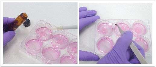
Protocol: In Vitro EpiDerm™ Skin Corrosion Test (EPI-200-SCT)

                                                                    Table of Contents

1       Rationale And Background .....................................................................................................................3
2       Specific Purpose Of The Method............................................................................................................3
3       Basic Procedure .......................................................................................................................................3
4       Limitations Of The Method......................................................................................................................4

5       Materials....................................................................................................................................................4
    5.1 Materials Required For The Experiments ..............................................................................................4
    5.2 Epiderm Skin Corrosion Kit Components ..............................................................................................5

6       Methods ....................................................................................................................................................6
    6.1    Tissue And Medium Storage .................................................................................................................6
    6.2    Test For Interference Of Chemicals With MTT Endpoint And Correction Procedures ..........................6
    6.3    Test For Mesh Compatibility (Liquid Test Substances Only) ................................................................8
    6.4    Preparations ...........................................................................................................................................9
        6.4.1       MTT Medium .................................................................................................................................9
        6.4.2       Dulbecco's PBS ............................................................................................................................9
        6.4.3       Test Substances ........................................................................................................................ 10
    6.5 Prediction Model ................................................................................................................................. 12
    6.6 Assay Quality Controls ....................................................................................................................... 13
        6.6.1       Assay Acceptance Criterion 1: Negative Control (NC) .............................................................. 13
        6.6.2       Assay Acceptance Criterion 2: Positive Control (PC) ................................................................ 13
        6.6.3       Assay Acceptance Criterion 3: Coefficient Of Variation (CV) .................................................... 13
    6.7 Experimental Procedure ..................................................................................................................... 13
    6.8 Documentation .................................................................................................................................... 23
        6.8.1       Method Documentation Sheet, MDS (See Annex) .................................................................... 23
        6.8.2       Ms Excel Data Spreadsheets .................................................................................................... 23

7 References ................................................................................................................................................. 24
Annex A: Methods Documentation Sheet (MDS) .………………………..................................……………… 25
Annex B: Test for Interference of Chemicals with MTT Endpoint and
          Correction Procedures……………………………………...…………………………………………...32

MatTek Corporation                                                                                                      MatTek In Vitro Life Science Laboratories
200 Homer Avenue, Ashland, MA - USA                                         www.mattek.com                                   Mlynské Nivy 73, Bratislava - Slovakia
+1-508-881-6771                                                           support@mattek.com                                                     +421-2-3260-7401

MK-24-007-0024                                                                Page 2 of 32                                                                     1/26/2021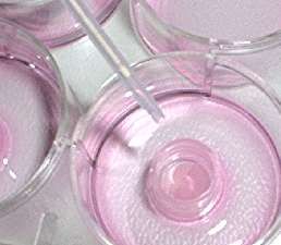
Protocol: In Vitro EpiDerm™ Skin Corrosion Test (EPI-200-SCT)

1.   Rationale and Background

     Skin corrosion refers to the production of irreversible tissue damage in the skin following the application of a
     test material, as defined by the Globally Harmonized System (GHS) for Classification and Labeling of
     Chemical Substances and Mixtures (1).

     The potential for chemical induced skin corrosion is an important consideration in establishing procedures for
     the safe handling, packing and transport of chemicals. Various systems for classification of corrosive
     potential are included in international regulatory requirements.

     The present test is based on the experience that corrosive chemicals are cytotoxic after a short-term
     exposure to the stratum corneum of the epidermis, if cytotoxicity is immediately observed after chemical
     exposure. It is designed to predict and classify skin corrosivity potential of a chemical by using a three-
     dimensional human epidermis model.

     In the year 1998 the EPISKIN and TER in vitro corrosivity test were successfully validated and met the
     acceptance criteria previously defined by the Management Team of the ECVAM International Validation
     Study (2). Because EPISKIN was not available after the Study, a Catch-up (validation study was performed
     with the epidermis model EpiDerm (3).

     In 2002 National Coordinators of OECD Test Guideline Programme (WNT) endorsed New Draft Test
     Guidelines TG430 (TER) and TG431 (Human Skin Model) for In Vitro Skin Corrosion Testing which were
     finally adopted in 2004. In Guideline TG 431 (paragraphs 9 – 11) general functional and performance criteria
     were defined if other (or new) skin or epidermis models are used in the context of this guideline (4, 6).

2. Specific Purpose of the Method

     This test can reliably discriminate chemicals that are corrosive to skin from non-corrosive chemicals and can
     therefore be used for the classification of skin corrosion hazard according to the GHS System adopted by the
     OECD (1). This test allows sub-categorisation of corrosive substances and mixtures into optional Sub-
     category 1A, in accordance with the UN GHS (1), as well as a combination of Sub-categories 1B and 1C (7)
     (8) (9). This test does not distinguish between Sub-categories 1B and 1C and it is not designed to predict
     skin irritation potential.

3. Basic Procedure
     EpiDerm tissues are conditioned by pre-incubation (1 hour or overnight) for release of transport stress
     related compounds and debris. After pre-incubation tissues are transferred to fresh Maintenance Medium
     and topically exposed with the test chemicals for 3 min and 1 hr, respectively. Two (alternatively three)
     tissues each are used per treatment, negative control (NC) and positive control (PC). After exposure tissues
     are rinsed and blotted and assay medium is replaced by MTT-medium. After 3 hr incubation, tissues are
     washed with PBS, blotted, and the blue formazan salt is extracted with MatTek extractant solution. The
     optical density (OD) of the formazan extract is determined spectrophotometrically at 540 -570 nm, and cell
     viability is calculated for each tissue as % of the mean of the negative control tissues. Skin corrosivity
     potential of the test materials is classified according to the remaining cell viability obtained after 3 minutes or
     1 hour exposure with the test chemical.

MatTek Corporation                                                                       MatTek In Vitro Life Science Laboratories
200 Homer Avenue, Ashland, MA - USA                      www.mattek.com                       Mlynské Nivy 73, Bratislava - Slovakia
+1-508-881-6771                                        support@mattek.com                                         +421-2-3260-7401

MK-24-007-0024                                            Page 3 of 32                                                  1/26/2021
Protocol: In Vitro EpiDerm™ Skin Corrosion Test (EPI-200-SCT)
4. Limitations of the Method
One limitation is possible interference of the test substance with the endpoint MTT: A test substance may
directly reduce MTT, thus mimicking dehydrogenase activity of the cellular mitochondria. This property of the
test substance is only a problem, if at the time of the MTT test (after the chemical has been rinsed off) there
is still sufficient amounts of the test substance present on (or in) the tissues. In this case the (true) metabolic
MTT reduction and the (false) direct MTT reduction can be differentiated and quantified by a procedure
described in section 6.2.
The method is not designed to be compatible with highly volatile test substances. However, possible toxic
interference across plate wells can be avoided by sealing the wells with an adhesive cover sheet or testing
volatile chemicals on separate plates.
5. Materials
5.1 Materials Required for the Experiments
Sterile, blunt-edged forceps For transferring tissues from agarose
500 mL wash bottle For rinsing tissue after test material exposure
200 mL beaker For collecting PBS washes
For diluting, adding, and removing media and test
Sterile disposable pipettes, pipette tips and
materials. For topically applying test materials to
pipetters
tissues
37°C incubator 5% CO2 For incubating tissues prior to and during assays
Vacuum source/trap (optional) For aspirating solutions
Laminar flow hood (optional) For transferring tissues under sterile conditions
Cotton swabs For drying the tissue surface
Mortar and Pestle For grinding solids
Adjustable Pipette 1 mL For pipetting assay medium under inserts (1 mL)
Pipette 300 µL For pipetting MTT medium into 24-well plates
For pipetting MTT extraction solution into 24-well
Pipette 2 mL
plate
For pipetting extracted formazan from 24-well plate
Pipette 200 µL
into 96 well plate to be used in a plate photometer
Pipette 50 µL For application of liquid test materials
Positive displacement pipette 50 µL For application of semi-solid test materials
For application of solids
Aesculap, Purchase No.: FK 623
MEDKA KG
Sharp spoon (NaCl weight: 25 1 mg)
Arzt- und Krankenhausbedarf
Bismarrckstr. 101
D –10625 Berlin
bulb headed Pasteur pipette To aid leveling the spoon and spreading the chemical
Laboratory balance For pipette verification and checking spoonful weight
96-well plate photometer 570 or 540 nm For reading OD
Shaker for microtiter / MILLICELL® plates For extraction of formazan
Stop-watches To be used during application of test materials
Potassium Hydroxyde, 8 N (Sigma P4494)
To be used as positive control with each kit
Dulbecco’s Phosphate Buffered Saline (TC- Used for rinsing tissues
PBS)
MTT (MTT-100-CON) For preparing MTT-medium
HCl For pH adjustment of PBS (if applicable)
MatTek Corporation MatTek In Vitro Life Science Laboratories
200 Homer Avenue, Ashland, MA - USA www.mattek.com Mlynské Nivy 73, Bratislava - Slovakia
+1-508-881-6771 support@mattek.com +421-2-3260-7401
MK-24-007-0024 Page 4 of 32 1/26/2021Protocol: In Vitro EpiDerm™ Skin Corrosion Test (EPI-200-SCT)
NaOH For pH adjustment of PBS (if applicable)
Nylon Mesh 200 µm (EPI-MESH) Use as a spreading aid for liquid test materials,
provided a pre-test shows the compatibility of test
material and nylon mesh and only for chemicals
which are not mechanically spread.
H2O, pure (distilled or aqua-pure) To be used as negative control with each kit
Extractant solution (MTT-100-EXT) For MTT extraction
5.2 EpiDerm™ Skin Corrosion Test Kit Components
EPI-200-SCT kits are shipped from Ashland, MA, USA or from Bratislava, Slovakia on Monday (an
alternative shipping procedure – over weekend - is possible). Upon receipt of the EpiDerm tissues, place the
sealed 24-well plates and the assay medium into the refrigerator (2-8°C). Place the MTT concentrate
containing vial in the freezer (-20°C) and the MTT diluent in the refrigerator (2-8°C).
Record lot numbers of all kit components in the Methods Documentation Sheet (MDS) (see ANNEX A).
EPI-200-SCT Kit Components
Amount Item Contains/Used for:
1 Sealed 24-well plate (EPI-200-SCT) Contains 24 tissues on agarose
2 24-well plates (sterile) For MTT viability assay
4 6-well plates (sterile) For corrosivity assay
1 bottle, 100 mL Assay Medium (EPI-100-ASY) For corrosivity assay
2 bottle, 100 mL PBS Rinse Solution (TC-PBS) For rinsing tissues
Nylon Mesh circles 8 mm diameter,
25 pieces For spreading test chemicals
200 µm pore (EPI-MESH)
1 MK-24-007-0024 Skin corrosivity test (SCT) protocol
Skin irritant reference chemical
1 vial, 10 mL 1% Triton X-100 Solution (TC-TRI)
Do not use in present method
MTT-100 Assay Kit Components (ready-to-use kit)
1 vial, 2 mL MTT concentrate (MTT-100-CON) Frozen MTT concentrate (5 mg/mL)
For diluting MTT concentrate prior to
1 vial, 8 mL MTT diluent (MTT-100-DIL)
use in the MTT assay
1 bottle, 60 mL Extractant Solution (MTT-100-EXT) For extraction of formazan
MatTek Corporation MatTek In Vitro Life Science Laboratories
200 Homer Avenue, Ashland, MA - USA www.mattek.com Mlynské Nivy 73, Bratislava - Slovakia
+1-508-881-6771 support@mattek.com +421-2-3260-7401
MK-24-007-0024 Page 5 of 32 1/26/2021Protocol: In Vitro EpiDerm™ Skin Corrosion Test (EPI-200-SCT)
Expiration and Kit Storage
Part # Description Conditions Shelf life*
EPI-200-SCT* EpiDerm cultures refrigerator (2-8°C) 96 hours
EPI-100-ASY Assay medium refrigerator (2-8°C) 14 days
MTT-100-DIL** MTT diluent refrigerator (2-8°C) 2 months
MTT-100-CON** MTT concentrate freezer (-20 ± 5°C) 2 months
*Refers to storage time @ 2-8°C in unopened package.
** MTT-100 kits must be ordered separately.
Note: Examine all kit components for integrity. If there is a concern call MatTek immediately.
Contact persons:
Yulia Kaluzhny (US) Bella Gershenovich (US) Silvia Letasiova (EU)
Phone: +1-508-881-6771, ext. 229 Phone: +1-508-881-6771, ext. 114 Phone: +421-2-3260-7401
Email: ykaluzhny@mattek.com Email: bgershenovich@mattek.com Email: sletasiova@mattek.com
6. Methods
6.1 Tissue and Medium Storage
For the EpiDerm reconstructed epidermal model and Medium refer to the relevant Technical Data and Safety
Sheet, located in the plastic file inside the shipping box.
If the test is not performed on the day of receipt, store the EpiDerm™ tissues in the refrigerator at 4°C until
next day. If you plan to determine any additional endpoints to MTT viability measurements, place the tissues
immediately upon arrival into the EpiDerm Maintenance Medium and pre-incubate 1 hour (37°C, 5%. CO2,
humidified atmosphere). Afterwards replace the medium and continue with overnight pre-incubation.
Store Maintenance Medium at 4°C in the dark. The shelf life is limited (see Technical Data file in the shipping
box). Use EpiDerm Maintenance Medium at room temperature, do not pre-heat.
Record lot numbers of all components in the Methods Documentation Sheet (see ANNEX A)
6.2 Test for Interference of Chemicals with MTT Endpoint and Correction
Procedures
As specified in Section 4, a test substance may interfere with the MTT endpoint if: a) it is colored and/or b)
able to directly reduce MTT (for possible combination of interactions, see Annex B). The MTT assay is
affected only if the test material is present in the tissues when the MTT viability test is performed.
Some non-colored test materials may change into colored materials in wet or aqueous conditions and thus
stain tissues during the 60 min exposure. Therefore, before exposure, a functional check for this possibility
should be performed (Step 1).
MatTek Corporation MatTek In Vitro Life Science Laboratories
200 Homer Avenue, Ashland, MA - USA www.mattek.com Mlynské Nivy 73, Bratislava - Slovakia
+1-508-881-6771 support@mattek.com +421-2-3260-7401
MK-24-007-0024 Page 6 of 32 1/26/2021Protocol: In Vitro EpiDerm™ Skin Corrosion Test (EPI-200-SCT)
Step 1
Add 50 µL (liquid) or 25 mg (solid - using a sharp spoon as per Section 7.2) of the test substance into 0.3
mL of deionized water. Perform the test in a transparent, preferably glass test-tube since plastic test tubes
may react with the test articles during the incubation time. Incubate the mixture in the incubator (37±1°C,
5±1 % CO2, 95% RH) for 60 min. At the end of the exposure time, shake the mixture and evaluate the
presence and intensity of the staining (if any). If the solution changes color significantly, the test substance
is presumed to have the potential to stain the tissue. A functional check on viable tissues should be
performed (Step 2).
Step 2
To check the tissue-binding of a colored test article (or a chemical that changes into a colored substance),
expose one viable tissue to 50 µL of liquid or 25 mg of solid test substance. In parallel, expose a tissue to
DPBS (negative control). Follow all procedures as described in this SOP Section 6.7 except incubate the
tissue for 3-h incubation in culture media without MTT (37±1°C, 5±1% CO 2, 95% RH) instead of incubating in
media containing MTT. After the 3 hour incubation, rinse the tissues and extract the tissues using 2.0 mL of
isopropanol and measure the optical density (OD) at 570 nm.
Note: If the colored test substance does not completely rinse off, pipette 1.0 mL of the extracting agent into each
well so that the MTT is extracted through the bottom of the tissue culture insert. After extraction is complete,
remove the insert and add an additional 1.0 mL of extractant to bring the total volume to 2.0 mL.
Data correction procedure – colored substances
If the extract from tissues treated by colored substance (or substance detected in step 1) has an OD
between 5% and 30% of the negative control tissue (treated with PBS), the chemical should be further tested
on more tissues using the procedure described above. The real MTT OD (unaffected by interference with
the colored test materials) is calculated using following formula:
OD = OD colored tissue (MTT assay) – OD colored tissue (no MTT assay)
Note: If the extract from tissues treated by colored substance (or substance detected in step 1) has an OD 30% of
the PBS treated control tissue, additional steps and expert judgment must be performed to determine if the
test substance must be considered as incompatible with the test.
Step 3
All test materials (including those already evaluated in Step 1 and Step 2) should be further evaluated for
their potential to interfere with MTT assay. To test if a material directly reduces MTT, add 50 µL (liquid) or
25 mg (solid - using sharp spoon 7.2) of the test substance to 1 mL of the MTT medium and incubate in the
incubator (37±1°C, 5±1 CO2, 95% RH) for 60 min. Untreated MTT medium is used as control. If the MTT
solution turns blue/purple, the test substance reduces MTT and additional functional check (Step 4) must be
performed.
MatTek Corporation MatTek In Vitro Life Science Laboratories
200 Homer Avenue, Ashland, MA - USA www.mattek.com Mlynské Nivy 73, Bratislava - Slovakia
+1-508-881-6771 support@mattek.com +421-2-3260-7401
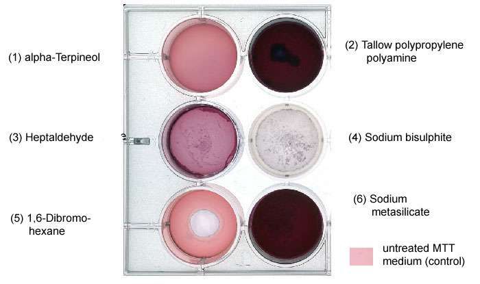
MK-24-007-0024 Page 7 of 32 1/26/2021Protocol: In Vitro EpiDerm™ Skin Corrosion Test (EPI-200-SCT)
Figure 1: Example of test for
direct MTT reduction ability
(Step 3). Test substances
(2) (3) and (6) have directly
reduced MTT. In these
cases, Step 4 (below) must
be performed.
Step 4:
The procedure employs freeze-killed tissues that possess no metabolic activity but absorb and bind the test
substance similar to viable tissues.
Freeze-killed tissues (part # EPI-200-FRZN-EA) can be ordered separately from MatTek Corporation (US)
or MatTek In Vitro Life Science Laboratories (Slovakia). The frozen tissues may be stored indefinitely in
the freezer (-20 ± 5°C).
Each MTT reducing chemical is applied to two freeze-killed tissues. In addition, two freeze killed tissues
are left untreated (Note: The untreated killed controls will show a small amount of MTT reduction due to
residual reducing enzymes within the killed tissue). The entire assay protocol is performed on the frozen
tissues in parallel to the assay performed with the live EpiDerm tissues. Data are then corrected as
follows:
Data correction procedure – MTT reducers
True viability = Viability of treated tissue – Interference from test chemical = OD tvt – OD kt
where OD kt = (mean OD tkt – mean OD ukt)
tvt = treated viable tissue kt = killed tissues
tkt = treated killed tissue ukt = untreated killed tissue (NC treated tissue)
If the interference by the test substance is greater than 30% of the negative control value, additional steps
must be taken into account or the test substance may be considered incompatible with this test system
(expert judgment).
If the interference by the test substance is < 30% of the negative control value, the net OD of the test
substance treated killed control may be subtracted from the mean OD of the test substance treated viable
tissues to obtain the true amount of MTT reduction that reflects metabolic conversion only.
Note 1: If the colored test material or the MTT reducing chemical is classified as irritant (tissue viabilityProtocol: In Vitro EpiDerm™ Skin Corrosion Test (EPI-200-SCT)
6.3 Test for Mesh Compatibility (Liquid Test Substances Only)
Since the surface of the EpiDerm tissues is slightly hydrophilic, spreading of highly lipophilic substances
might sometimes present a problem due to the surface tension effects. Therefore, a nylon mesh (see 5.1)
can be used as a spreading support if mechanical re-spreading is not efficient. Some chemicals, however,
may react with the mesh and therefore the compatibility of test chemical with nylon mesh must be performed.
To test if a test chemical interacts with the mesh, place the mesh on a glass slide and apply 50 µL of test
substance. After 60 minutes exposure, check using a microscope. If an interaction between test substance
and the mesh is noticed (Figure 2), the test substance has to be applied without the mesh.
Figure 2: The mesh compatibility test
Note: The mesh (25 pieces) is included in the EPI-200-SCT kit from MatTek Corporation and MatTek In Vitro Life
Science Laboratories.
6.4 Preparations
6.4.1 MTT Medium (Prepare Fresh on Day of Testing!)
If you buy MTT-100 kit (ready-to-use kit) from MatTek, thaw the MTT concentrate (MTT-100-CON) and dilute
with the MTT diluent (MTT-100-DIL). Store the remaining MTT solution in the dark at 4°C for later use on the
same day (do not store until next day since MTT will degrade with time).
If you are not using the MTT-100 kit provided by MatTek, prepare the stock solution (5 mg/mL) of MTT in
DPBS. Stock solution can be stored frozen (-20 ±5°C) for up to 2 months. Before use, filter the stock solution
and dilute the filtrate with the assay medium to final concentration (1 mg/mL). Record the preparation in the
MDS. Do not store the diluted MTT solution overnight.
Safety precaution: MTT is toxic (Risk phrases: H315, H319, H335, H341). Wear protective gloves during
manipulation with MTT solution!
Note: MTT is light sensitive. Protect all solutions from light
6.4.2 Dulbecco’s PBS
Sterile ready-to-use DPBS should be used. About two liters are sufficient for all rinsing performed with one
kit. If PBS is prepared from powder or 10x concentrated PBS is used, prepare according to supplier
instructions and adjust to pH 7.0 with either NaOH or HCl. Record the pH adjustment in the MDS.
MatTek Corporation MatTek In Vitro Life Science Laboratories
200 Homer Avenue, Ashland, MA - USA www.mattek.com Mlynské Nivy 73, Bratislava - Slovakia
+1-508-881-6771 support@mattek.com +421-2-3260-7401
MK-24-007-0024 Page 9 of 32 1/26/2021Protocol: In Vitro EpiDerm™ Skin Corrosion Test (EPI-200-SCT)
6.4.3 Test Substances
Safety Instruction
a) For handling of non-coded test substances follow instructions given in the Material Safety Data Sheet.
b) If coded chemicals are supplied, no (or possibly incomplete) information regarding the safe handling will
be provided. Therefore, all test materials must be treated as if they were corrosive and toxic and work must
be performed in accordance with chemical safety guidelines (use ventilated cabinet, wear gloves, protect
eyes and face).
c) Store all test substances according to recommendations. Respect special store conditions (special
temperature, protection from light, protection from oxidization by nitrogen, etc.)
Liquids: Dispense 50 µL directly atop the EpiDerm tissue and spread with bulb headed Pasteur pipette. If
necessary, gently place a nylon mesh (8 mm diameter) on the surface (see Figure 3, picture b).
Record the use of mesh as spreading tool in the MDS.
A B
Figure 3: Application of liquids.
MatTek Corporation MatTek In Vitro Life Science Laboratories
200 Homer Avenue, Ashland, MA - USA www.mattek.com Mlynské Nivy 73, Bratislava - Slovakia
+1-508-881-6771 support@mattek.com +421-2-3260-7401
MK-24-007-0024 Page 10 of 32 1/26/2021Protocol: In Vitro EpiDerm™ Skin Corrosion Test (EPI-200-SCT)
Semisolids: Dispense 50 µL using a positive displacement pipette directly atop the tissue. If necessary,
spread to match size of tissue. Record the use of spreading in the MDS.
Figure 4: Application of semisolids.
Solids: Crush and grind test material with a mortar with pestle wherever this improves the
consistency. Fill 25 mg sharp application spoon* (see 5.1 and Figure 5) with fine ground
test material. Level the spoon by gently scratching the excess material away with an
appropriate aid, avoiding compression ("packing") of the test material. "Packing" can be
avoided by using a rod-shaped spoon instead of a flat spatula. If a bulb headed sound is
used the bulb can be used to empty the spoon completely. Add the 25 μL of H20 necessary
for wetting of the test material to increase tissue surface contact. Increase the volume of H 20
in case of materials where this is not enough for wetting. If necessary, spread material to
match the size of the tissue. Record in the MDS if grinding was not used and the H20 volume
necessary to wet the chemical.
Figure 5: Application of solids.
MatTek Corporation MatTek In Vitro Life Science Laboratories
200 Homer Avenue, Ashland, MA - USA www.mattek.com Mlynské Nivy 73, Bratislava - Slovakia
+1-508-881-6771 support@mattek.com +421-2-3260-7401
MK-24-007-0024 Page 11 of 32 1/26/2021Protocol: In Vitro EpiDerm™ Skin Corrosion Test (EPI-200-SCT)
Waxes: For test substances with waxy consistence the spoon application does not work. In these
cases, try to form a flat “disc like” piece of about 8 mm diameter and place it atop the tissue,
wetted with 15 μL H2O. To improve the contact between test substance and tissue weigh
down the “disc” with a stainless steel aid like that shown in Figure 6.
Figure 6: Stainless steel aid.
*Note: Since the surface of the solid covering the tissues is more important than the weight, the “leveled spoon
technique” is an accepted dosing procedure. The spoon used here has been calibrated to equal 25 mg of fine
grinded NaCl. The weight will be different if other materials are used.
**Note: determine in a pre-test a volume of H2O necessary to wet test chemical.
6.5 Prediction Model
Corrosivity potential of the test materials is predicted from the relative mean tissue viabilities obtained after a
3 min treatment compared to the negative control tissues concurrently treated with H20. A chemical is
classified "corrosive" if the relative tissue viability after 3 min treatment with a test material is decreased
below 50%. In addition, materials are classified "corrosive" if the relative tissue viability after 1 hr treatment
with a test material is decreased below 15%.
Mean tissue viability
Prediction
(expressed as % of negative control)
3 min < 50% corrosive
3 min 50% and 1 hour: < 15% corrosive
3 min 50% and 1 hour: 15% non-corrosive
For (optional) sub-categorisation of chemicals which are initially classified as “corrosive”, a chemical is classified
as Sub-category 1A if the tissue viability was < 25% after the 3 min treatment and is classified as a combination of
sub-categories 1B/1C if the tissue viability was ≥ 25%.
Mean tissue viability
Prediction
(expressed as % of negative control)
3 min < 25% Sub-category 1A
3 min ≥ 25% A combination of Sub-categories 1B/1C
MatTek Corporation MatTek In Vitro Life Science Laboratories
200 Homer Avenue, Ashland, MA - USA www.mattek.com Mlynské Nivy 73, Bratislava - Slovakia
+1-508-881-6771 support@mattek.com +421-2-3260-7401
MK-24-007-0024 Page 12 of 32 1/26/2021Protocol: In Vitro EpiDerm™ Skin Corrosion Test (EPI-200-SCT)
6.6 Assay Quality Controls
6.6.1 Assay Acceptance Criterion 1: Negative Control (NC)
The absolute OD of the H2O treated NC tissues in the MTT-test is an indicator of tissue viability obtained in
the testing laboratory after the shipping and storing procedure and under specific conditions of the assay.
Tissue viability is meeting the acceptance criterion if the mean OD of the mean of NC is OD 0.8.
6.6.2 Assay Acceptance Criterion 2: Positive Control (PC)
8N KOH (Sigma P4494) is used as PC and must be tested once on each testing day. The mean tissue
viability following 1 hour exposure of the PC must be ≤ 15%.
6.6.3 Assay Acceptance Criterion 3: Difference (Diff) and/or Coefficient of
Variation (CV)
Based on the test design (explained in 6.7), the experiments are performed on two or three tissue replicates
per exposure time. In the range 20 - 100% viability, the Difference (Diff) of viability between the two tissue
replicates should not exceed 30%, or if using three tissue replicates per time point, the Coefficient of
Variation (CV) between the tissue replicates must be ≤ 30%.
6.7 Experimental Procedure
Introductory Note:
The experiment can be performed using either: a) two tissues per timepoint (as in the validation study), or b)
three tissues per timepoint. Alternative b) is recommended mainly for laboratories establishing the test. Examples
of both test design are given below.
ALTERNATIVE A) 24 tissues (1 EPI-200-SCT kit) are used for testing 4 test chemicals, negative control and
positive control, each of them applied both for 3 min and 1 hr to two tissue replicates per treatment.
ALTERNATIVE B) 36 tissues (1 EPI-200-SCT and 1 EPI-212-SCT kit) are used for testing 4 test chemicals,
negative control and positive control, each of them applied both for 3 min and 1 hr to three tissue replicates per
treatment..
Day Prior To Testing
If the EpiDerm tissues are not used in the day of receipt, keep the originally sealed plates until the
next day in the refrigerator at 4°C.
If necessary, prepare sufficient amount of rinsing PBS for the next day according to 6.4.1
Note: Since the present test is a short-term test which makes use of the EpiDerm model over a period of only 5
hours, sterility is not as important as is in other applications of the EpiDerm model. Nevertheless, it is important to
keep assay media sterile and to keep risk of contamination at a low level.
MatTek Corporation MatTek In Vitro Life Science Laboratories
200 Homer Avenue, Ashland, MA - USA www.mattek.com Mlynské Nivy 73, Bratislava - Slovakia
+1-508-881-6771 support@mattek.com +421-2-3260-7401
MK-24-007-0024 Page 13 of 32 1/26/2021Protocol: In Vitro EpiDerm™ Skin Corrosion Test (EPI-200-SCT)
ALTERNATIVE A) – Example With 4 Chemicals, Two Tissues Per Timepoint
Day of Testing
Tissue Conditioning (Pre-incubation)
a) Prepare two 6-well plates for four chemicals and the negative (NC) and positive control (PC) for
the 3 min application. Pipette 0.9 mL Maintenance Medium in each well.
b) Prepare two 6-well plates for four chemicals and the negative (NC) and positive control (PC) for
the 1 hr application. Pipette 0.9 mL Maintenance Medium in each well.
c) Remove the shipped multiwell plate from the plastic bag. Open the 24-well plate under a sterile
airflow and remove the sterile gauze. Carefully take out each insert containing the epidermal tissue,
rapidly remove any remaining agarose that adheres to the outer sides of the insert by gentle blotting
on the sterile filter paper, and immediately place it in a well of the prepared 6-well plate. Act quickly
as the epidermal cultures dry out rapidly when not in contact with medium. Make sure that no air
bubbles are formed underneath the insert!
d) Mark the 6-well plates as shown in Figure 7.
e) Place the 6-well plates containing the tissues into a humidified (37°C, 5% CO 2) incubator for 1
hour.
3 minutes exposure 1 hour exposure
NC Chemical 1 Chemical 2 NC Chemical 1 Chemical 2
tissue 1 tissue 1 tissue 1 tissue 1 tissue 1 tissue 1
NC Chemical 1 Chemical 2 NC Chemical 1 Chemical 2
tissue 2 tissue 2 tissue 2 tissue 2 tissue 2 tissue 2
Chemical 3 Chemical 4 PC 1 Chemical 3 Chemical 4 PC 1
tissue 1 tissue 1 tissue 1 tissue 1 tissue 1 tissue 1
Chemical 3 Chemical 4 PC 2 Chemical 3 Chemical 4 PC 2
tissue 2 tissue 2 tissue 2 tissue 2 tissue 2 tissue 2
3 minutes exposure 1 hour exposure
Figure 7: 6-well plate design
Preparations for the Main Test (Make During the 1 Hr Pre-incubation Time)
a) Prepare the MTT medium according to 6.4.1.
b) Prepare two 24-well plates to be used as “holding and “MTT plates” one for the 3 min
experiment, the other for the 1 hr experiment.
c) Use the plate design shown in Figure 8. Pipette 300 µL of either maintenance medium or MTT
medium in each well.
d) Place the plates in the incubator.
MatTek Corporation MatTek In Vitro Life Science Laboratories
200 Homer Avenue, Ashland, MA - USA www.mattek.com Mlynské Nivy 73, Bratislava - Slovakia
+1-508-881-6771 support@mattek.com +421-2-3260-7401
MK-24-007-0024 Page 14 of 32 1/26/2021Protocol: In Vitro EpiDerm™ Skin Corrosion Test (EPI-200-SCT)
3 minutes
NC C1 C2 C3 C4 PC holding plate
pre-filled with 300 µL of assay medium
NC C1 C2 C3 C4 PC
MTT assay plate
pre-filled with 300 µL of MTT medium
1 hour
NC C1 C2 C3 C4 PC holding plate
pre-filled with 300 µL of assay medium
NC C1 C2 C3 C4 PC
MTT assay plate
pre-filled with 300 µL of MTT medium
Figure 8: 24-well plate design. "Holding and MTT plate", both for the 1 hr experiment and the 3 min
experiment. NC = negative control; C1 – C4 = test chemicals 1-4; PC = positive control.
MatTek Corporation MatTek In Vitro Life Science Laboratories
200 Homer Avenue, Ashland, MA - USA www.mattek.com Mlynské Nivy 73, Bratislava - Slovakia
+1-508-881-6771 support@mattek.com +421-2-3260-7401
MK-24-007-0024 Page 15 of 32 1/26/2021Protocol: In Vitro EpiDerm™ Skin Corrosion Test (EPI-200-SCT)
1-Hour Application
Note: Dosing time interval is dictated by rinsing procedure. If the technician has performed the test already, 45
seconds intervals is sufficient for both application and washing procedures. However, if the test is performed for
the first time, 1 minute dosing interval is recommended.
a) After 1 hour of pre-incubation, transfer each insert to new 6-well plates with fresh medium (0.9 mL
per well). Alternatively, aspirate the pre-incubation medium from the 6-well plates and pipet 0.9 mL
of fresh medium into each well.
b) Set the timer to 1 hr and start it. Add 50 µL of H2O (negative control) into the first insert atop the
tissue. After 45 sec repeat the procedure with the second tissue. Proceed with test material 1 - 4 (50
µL: liquids, 25 mg + 25 µL H2O: solids) and the positive control in the same manner until all tissues
are dosed.
c) Dosing interval scheme for the 1 hour experiment:
0.00-0.45 – tissue 1 (NC) 4.30-5.15 – tissue 7 (C3)
0.45-1.30 – tissue 2 (NC) 5.15-6.00 – tissue 8 (C3)
1.30-2.15 – tissue 3 (C1) 6.00-6.45 – tissue 9 (C4)
2.15-3.00 – tissue 4 (C1) 6.45-7.30 – tissue 10 (C4)
3.00-3.45 – tissue 5 (C2) 7.30-8.15 – tissue 11 (PC)
3.45-4.30 – tissue 6 (C2) 8.15-9.00 – tissue 12 (PC)
d) Place the 6-well plates into the incubator (37°C, 5% CO2) for the rest of the exposure time until 1
hour exposure is reched for first tissue dosed. Record start time in the MDS.
e) After the 1 hour period of test material exposure is complete, with forceps remove the first insert
from the 6-well plate. As shown in Figure 9, using a wash bottle gently rinse the tissue with PBS (=
fill and empty insert 20 times in a constant soft stream of PBS) to remove any residual test material.
Remove excess PBS by gently shaking the insert and blot bottom on blotting paper. Place the insert
in the prepared holding plate. Proceed with test materials 1 - 4 and the positive control in the same
manner until all tissues are rinsed. Rinse all tissues in an interval of 45 sec.
Figure 9: Rinsing: fill and empty 20 x
f) Once all tissues have been rinsed and are in the holding plate, dry the surface with cotton swab,
remove inserts from the holding plate, blot bottom and transfer into the 24-well plate, prepared for
the MTT assay.
g) Place plate in the incubator, record start time of MTT incubation in the MDS and incubate for 3
hours (37°C, 5% CO2).
MatTek Corporation MatTek In Vitro Life Science Laboratories
200 Homer Avenue, Ashland, MA - USA www.mattek.com Mlynské Nivy 73, Bratislava - Slovakia
+1-508-881-6771 support@mattek.com +421-2-3260-7401
MK-24-007-0024 Page 16 of 32 1/26/2021Protocol: In Vitro EpiDerm™ Skin Corrosion Test (EPI-200-SCT)
3 Min Application
Note: Dosing time interval is dictated by rinsing procedure. If the technician performs test already routinely, 45
seconds interval is sufficient for both application and washing procedures. However, if the test is performed for
the first time, 1 minute dosing interval is recommended.
a) Start the timer for 3 min. Add 50 µL H2O (negative control) into the first insert atop the tissue.
After 45 seconds repeat the procedure with the second tissue. Following 45 seconds intervals
enable to dose 4 tissues. Afterwards, washing of the tissue 1 have to start.
b) After the 3 min period of exposure for the first four tissues is complete, start the timer for 3 min
and with forceps remove the first insert from the 6-well plate. As shown in Figure 9, using a wash
bottle gently rinse the tissue with PBS (= fill and empty insert 20 times in a constant soft stream of
PBS) to remove any residual test material. Remove excess PBS by gently shaking the insert and
blot bottom on blotting paper. Place the insert in the prepared holding plate. After 45 seconds repeat
the procedure with the second insert, after 1:30 minutes with the third tissue...etc.
Dosing interval scheme for the 3 min experiment:
SET 1 = NC and chemical 1 (6 minutes)
Dosing:
0.00-0.45 – tissue 1 (NC)
0.45-1.30 – tissue 2 (NC)
1.30-2.15 – tissue 3 (C1)
2.15-3.00 – tissue 4 (C1)
Rinsing
3.00-3.45 – tissue 1 (NC)
3.45-4.30 – tissue 2 (NC)
4.30-5.15 – tissue 3 (C1)
5.15-6.00 – tissue 4 (C1)
SET 2 = chemical 2, 3 (6 minutes)
SET 3 = chemical 4, PC (6 minutes)
c) Proceed with all test materials (50 µ: liquids, 25 mg + 25 µL H2O: solid) and the positive control
in the same manner until all tissues are dosed and rinsed.
d) Once all tissues have been rinsed and are in the holding plate, carefully dry the surface of the
tissue with a cotton swab. Afterwards remove inserts from the holding plate, blot bottom and transfer
into the 24-well plate, prepared for the MTT assay. Place plates in the incubator, record start time of
MTT incubation in the MDS and incubate for 3 hours (37°C, 5% CO2).
MatTek Corporation MatTek In Vitro Life Science Laboratories
200 Homer Avenue, Ashland, MA - USA www.mattek.com Mlynské Nivy 73, Bratislava - Slovakia
+1-508-881-6771 support@mattek.com +421-2-3260-7401
MK-24-007-0024 Page 17 of 32 1/26/2021Protocol: In Vitro EpiDerm™ Skin Corrosion Test (EPI-200-SCT)
ALTERNATIVE B) – Example With 4 Chemicals, Three Tissues Per Timepoint
Day of Testing
Tissue Conditioning (Pre-Incubation)
a) Prepare three 6-well plates for four chemicals and the negative (NC) and positive control (PC) for
the 3 min application. Pipette 0.9 mL Maintenance Medium in each well.
b) Prepare three 6-well plates for four chemicals and the negative (NC) and positive control (PC) for
the 1 hr application. Pipette 0.9 mL Maintenance Medium in each well.
c) Remove the shipped multiwell plate from the plastic bag. Open the 24-well plate under a sterile
airflow and remove the sterile gauze. Carefully take out each insert containing the epidermal tissue,
rapidly remove any remaining agarose that adheres to the outer sides of the insert by gentle blotting
on the sterile filter paper, and immediately place it in a well of the prepared 6-well plate. Act quickly
as the epidermal cultures dry out rapidly when not in contact with medium. Make sure that no air
bubbles are formed underneath the insert!
d) Mark the 6-well plates as shown in Figure 10.
e) Place the 6-well plates containing the tissues into a humidified (37°C, 5% CO 2) incubator for 1
hour.
3 minutes exposure 1 hour exposure
NC NC NC NC NC NC
tissue 1 tissue 2 tissue 3 tissue 1 tissue 2 tissue 3
Chemical 1 Chemical 1 Chemical 1 Chemical 1 Chemical 1 Chemical 1
tissue 1 tissue 2 tissue 3 tissue 1 tissue 2 tissue 3
Chemical 2 Chemical 2 Chemical 2 Chemical 2 Chemical 2 Chemical 2
tissue 1 tissue 2 tissue 3 tissue 1 tissue 2 tissue 3
Chemical 3 Chemical 3 Chemical 3 Chemical 3 Chemical 3 Chemical 3
tissue 1 tissue 2 tissue 3 tissue 1 tissue 2 tissue 3
Chemical 4 Chemical 4 Chemical 4 Chemical 4 Chemical 4 Chemical 4
tissue 1 tissue 2 tissue 3 tissue 1 tissue 2 tissue 3
PC PC PC PC PC PC
tissue 1 tissue 2 tissue 3 tissue 1 tissue 2 tissue 3
3 minutes exposure 1 hour exposure
Figure 10: 6-well plate design
MatTek Corporation MatTek In Vitro Life Science Laboratories
200 Homer Avenue, Ashland, MA - USA www.mattek.com Mlynské Nivy 73, Bratislava - Slovakia
+1-508-881-6771 support@mattek.com +421-2-3260-7401
MK-24-007-0024 Page 18 of 32 1/26/2021Protocol: In Vitro EpiDerm™ Skin Corrosion Test (EPI-200-SCT)
Preparations for the main test (make during the 1 hr pre-incubation time)
a) Prepare MTT medium according to 6.4.1.
b) Prepare two 24-well plates to be used as “holding plates.” One for the 3 min experiment, the
other for the 1 hr experiment. Pipette 300 µL of maintenance medium into each well of the
holding plates. In addition, prepare two 24-well plates for the MTT assay. Pipette 300 µL of MTT
medium in each well of the MTT assay plate.
c) Use the plate design shown in Figure 11.
d) Place the plates in the incubator.
3 minutes – holding plate 1 hour – holding plate
NC NC NC C4 C4 C4 NC NC NC C4 C4 C4
C1 C1 C1 PC PC PC C1 C1 C1 PC PC PC
C2 C2 C2 C2 C2 C2
C3 C3 C3 C3 C3 C3
3 minutes – MTT plate 1 hour – MTT plate
NC NC NC C4 C4 C4 NC NC NC C4 C4 C4
C1 C1 C1 PC PC PC C1 C1 C1 PC PC PC
C2 C2 C2 C2 C2 C2
C3 C3 C3 C3 C3 C3
Figure 11: 24-well plate design. "Holding and MTT plates", both for the 3 min and 1 hour experiment.
NC = negative control; C1 – C4 = test chemicals 1- 4; PC = positive control
MatTek Corporation MatTek In Vitro Life Science Laboratories
200 Homer Avenue, Ashland, MA - USA www.mattek.com Mlynské Nivy 73, Bratislava - Slovakia
+1-508-881-6771 support@mattek.com +421-2-3260-7401
MK-24-007-0024 Page 19 of 32 1/26/2021Protocol: In Vitro EpiDerm™ Skin Corrosion Test (EPI-200-SCT)
1-hour application
Note: Dosing time interval is dictated by rinsing procedure. In this test design, a 1 minute dosing interval is
recommended.
a) After 1 hour of pre-incubation, transfer each insert to new 6-well plates with fresh medium (0.9 mL
per well). Alternatively, aspirate the pre-incubation medium from the 6-well plates and pipet 0.9 mL
of fresh medium into each well.
b) Set a timer to 1 hr and start it. Add 50 µL of H2O (negative control) into the first insert atop the
tissue. After 60 sec repeat the procedure with the second tissue. Proceed with test material 1 - 4 (50
µL: liquids, 25 mg + 25 µL H2O: solids) and the positive control in the same manner until all tissues
are dosed.
Dosing interval scheme for the 1 hour experiment, alternative b):
0.00-1.00 – tissue 1 (NC) 9.00-10.00 – tissue 10(C3)
1.00-2.00 – tissue 2 (NC) 10.00-11.00 – tissue 11 (C3)
2.00-3.00 – tissue 3 (NC) 11.00-12.00 – tissue 12 (C3)
3.00-4.00 – tissue 4 (C1) 12.00-13.00 – tissue 13 (C4)
4.00-5.00 – tissue 5 (C1) 13.00-14.00 – tissue 14 (C4)
5.00-6.00 – tissue 6 (C1) 14.00-15.00 – tissue 15 (C4)
6.00-7.00 – tissue 7 (C2) 15.00-16.00 – tissue 16 (PC)
7.00-8.00 – tissue 8 (C2) 16.00-17.00 – tissue 17 (PC)
8.00-9.00 – tissue 9 (C2) 17.00-18.00 – tissue 18 (PC)
c) Place the 6-well plates into the incubator (37°C, 5% CO2) for the remaining exposure time until 1
hour exposure is reached for first tissue dosed. Record start time in the MDS.
d) After the 1 hour period of test material exposure is complete, use forceps to remove the first
insert from the 6-well plate. As shown in Figure 9, using a wash bottle gently rinse the tissue with
PBS (= fill and empty insert 20 times in a constant soft stream of PBS) to remove any residual test
material. Remove excess PBS by gently shaking the insert and blot the bottom on blotting paper.
Place the insert in the prepared holding plate. Proceed with test materials 1 - 4 and the positive
control in the same manner until all tissues are rinsed. Rinse all tissues in an interval of 60 sec.
e) Once all the tissues have been rinsed and are in the holding plate, dry the tissue surface with a
cotton swab. Remove inserts from the holding plate, blot bottom and transfer into the 24-well plate,
prepared for the MTT assay.
f) Place the plate in the incubator, record start time of MTT incubation in the MDS and incubate for
3 hours (37°C, 5% CO2).
MatTek Corporation MatTek In Vitro Life Science Laboratories
200 Homer Avenue, Ashland, MA - USA www.mattek.com Mlynské Nivy 73, Bratislava - Slovakia
+1-508-881-6771 support@mattek.com +421-2-3260-7401
MK-24-007-0024 Page 20 of 32 1/26/2021Protocol: In Vitro EpiDerm™ Skin Corrosion Test (EPI-200-SCT)
3 Min Application
Note: Dosing time interval is dictated by rinsing procedure. In this test design, 1 minute dosing interval is
recommended.
a) Start the timer for 3 min. Add 50 µL of H2O (negative control) into the first insert atop the tissue.
After 60 seconds repeat the procedure with the second tissue. Following 60 seconds interval enable
to dose 3 tissues (= 1 test substance).
b) After the 3 min period of exposure for the first three tissues is complete, remove the first insert
from the 6-well plate. As shown in Figure 10, using a wash bottle gently rinse the tissue with PBS (=
fill and empty insert 20 times in a constant soft stream of PBS) to remove any residual test material.
Remove excess PBS by gently shaking the insert and blot the bottom on blotting paper. Place the
insert into the prepared holding plate. After 60 seconds repeat the procedure with the second
insert....etc.
Dosing interval scheme for the 3 min experiment:
NC (6 minutes)
Dosing:
0.00-1.00 – tissue 1 (NC)
1.00-2.00 – tissue 2 (NC)
2.00-3.00 – tissue 3 (NC)
Rinsing
3.00-4.00 – tissue 1 (NC)
4.00-5.00 – tissue 2 (NC)
5.00-6.00 – tissue 3 (NC)
chemical 1 = 6 minutes
chemical 2 = 6 minutes
chemical 3 = 6 minutes
chemical 4 = 6 minutes
PC = 6 min
Total duration of the test: 36 min
c) Proceed with all test materials (50 µL: liquids, 25 mg + 25 µL H2O: solid) and the positive control
in the same manner until all tissues are dosed and rinsed.
d) Once all the tissues have been rinsed and are in the holding plate carefully dry the tissue surface
with a cotton swab. Afterwards remove inserts from the holding plate, blot the bottom and transfer
into the 24-well plate, prepared for the MTT assay. Place plate in the incubator, and incubate for 3
hours (37°C, 5% CO2). Record the start time of MTT incubation on the MDS.
MatTek Corporation MatTek In Vitro Life Science Laboratories
200 Homer Avenue, Ashland, MA - USA www.mattek.com Mlynské Nivy 73, Bratislava - Slovakia
+1-508-881-6771 support@mattek.com +421-2-3260-7401
MK-24-007-0024 Page 21 of 32 1/26/2021Protocol: In Vitro EpiDerm™ Skin Corrosion Test (EPI-200-SCT)
MTT Test and Reading – Both test designs
a) After the 3 hour MTT incubation period is complete, gently aspirate MTT from all wells (e.g. using
a suction pump), refill wells with PBS and aspirate. Repeat the rinsing twice and make sure that
tissues are dry after the last aspiration. Transfer inserts to new 24 well plates.
b) Immerse the inserts by gently pipetting 2 mL extractant solution (MTT-100-EXT) into each insert.
The level will rise above the upper edge of the insert, thus completely covering the tissue from both
sides.
c) Seal the 24 well plate (e.g. with a zip bag) to inhibit the extractant solution evaporation. Record
the start time of extraction in the MDS. Extract either over night without shaking at room temperature
or, alternatively, 2 hours with shaking (~120 rpm) at room temperature.
d) After the extraction period is complete, pierce the inserts with an injection needle and allow the
extract to run into the well from which the insert was taken. Afterwards the insert can be discarded.
Place the 24-well plates on a shaker for 15 minutes until solution is homogeneous in colour.
e) Per each tissue transfer 200 µL of the blue formazan solution into a 96-well flat bottom microtiter
plate, both from the 3 min exposure and from the 1 hr exposure. For the 96-well plate, use the exact
plate design given in Figure 12, as this configuration is used in the data spreadsheets. Read the OD
in a spectrophotometer at 570 nm, without reference filter. Alternatively, ODs can be read at 540 nm.
Note: Readings are performed without a reference filter, since the "classical" reference filter often used in the
MTT test (630 nm) is still within the absorption curve of formazan. Since filters may have a tolerance in some
cases the reference filter reduces the dynamics of the signal (OD) up to 40%.
Fixed 96 well-plate design for OD readings
1 2 3 4 5 6 7 8 9 10 11 12
A NC-T1 PC-T1 C1-T1 C2-T1 C3-T1 C4-T1 C5-T1 C6-T1 C7-T1 C8-T1 C9-T1 C10-T1
B NC-T1 PC-T1 C1-T1 C2-T1 C3-T1 C4-T1 C5-T1 C6-T1 C7-T1 C8-T1 C9-T1 C10-T1
3 min
C NC-T2 PC-T2 C1-T2 C2-T2 C3-T2 C4-T2 C5-T2 C6-T2 C7-T2 C8-T2 C9-T2 C10-T2
D NC-T2 PC-T2 C1-T2 C2-T2 C3-T2 C4-T2 C5-T2 C6-T2 C7-T2 C8-T2 C9-T2 C10-T2
E NC-T1 PC-T1 C1-T1 C2-T1 C3-T1 C4-T1 C5-T1 C6-T1 C7-T1 C8-T1 C9-T1 C10-T1
F NC-T1 PC-T1 C1-T1 C2-T1 C3-T1 C4-T1 C5-T1 C6-T1 C7-T1 C8-T1 C9-T1 C10-T1 1
G NC-T2 PC-T2 C1-T2 C2-T2 C3-T2 C4-T2 C5-T2 C6-T2 C7-T2 C8-T2 C9-T2 C10-T2 hour
H NC-T2 PC-T2 C1-T2 C2-T2 C3-T2 C4-T2 C5-T2 C6-T2 C7-T2 C8-T2 C9-T2 C10-T2
Figure 12. Alternative A): Transfer 3 aliquots (200 µL) from each tissue into the 96-well plates using the
plate design above. Use one 96-well plate for both time points. T1 and T2 refer to the duplicate (N=2)
tissues used for each exposure time and test article.
MatTek Corporation MatTek In Vitro Life Science Laboratories
200 Homer Avenue, Ashland, MA - USA www.mattek.com Mlynské Nivy 73, Bratislava - Slovakia
+1-508-881-6771 support@mattek.com +421-2-3260-7401
MK-24-007-0024 Page 22 of 32 1/26/2021Protocol: In Vitro EpiDerm™ Skin Corrosion Test (EPI-200-SCT)
6.8 Documentation
6.8.1 Method Documentation Sheet, MDS (see ANNEX A)
The MDS allows to check the correct set up, calibration and function of the equipment as well as correct
weights, applications etc. The MDS is designed as a paper document "in the spirit of GLP". Per each test,
make a hardcopy of the MDS, fill in and sign the requested information, starting the day prior to testing and
ending after the test has been conducted.
Note (1): If several tests are performed per week, pipette verification (weighing H20 on a balance) is only
necessary once at the beginning of each week. Nevertheless, if adjustable pipettes are used the correct
adjustment shall be checked and recorded in the MDS before each test.
Note (2): If solids cannot be sufficiently ground to a fine powder, it is recommended to check the weight of the
levelled application spoon and record this weight in the MDS.
6.8.2 MS EXCEL Data Spreadsheets
The MS EXCEL workbook can be obtained from MatTek Corporation (USA) or MatTek In Vitro Life Science
Laboratories (Slovakia). Data of optical densities (ODs) generated by the microplate reader and already
corrected by substraction of the blanks mean from all OD values are copied from the Reader software
or manually imputted to the Windows Clipboard and then pasted into the first spreadsheet (Import) of the
EXCEL workbook in the 96-well format given above in Figure 12.
The workbook consists of three spreadsheets, named Import, Results and Classification. The first sheet
(Import) is used for pasting or manually transposing the OD values. In addition, fill in required information
on test substances, tissue lots, etc. in the first sheet (Import). The second sheet (Results) makes
calculations and provides a column graph of the results. Entries on test materials made in the first sheet will
be copied from there automatically to the correct positions in the Results sheet. A summary of the
classification for each test article and whether the test was qualified (Q) or not (NQ) is presented on the
Classification sheet. Add any special comment(s) related to the experiment or measurement in the last
(Remarks) column on the third sheet.
MatTek Corporation MatTek In Vitro Life Science Laboratories
200 Homer Avenue, Ashland, MA - USA www.mattek.com Mlynské Nivy 73, Bratislava - Slovakia
+1-508-881-6771 support@mattek.com +421-2-3260-7401
MK-24-007-0024 Page 23 of 32 1/26/2021Protocol: In Vitro EpiDerm™ Skin Corrosion Test (EPI-200-SCT)
7. REFERENCES
1. UN. (2013). United Nations Globally Harmonized System of Classification and Labelling of Chemicals
(GHS), Fifth Revised Edition, UN New York and Geneva.
[http://www.unece.org/trans/danger/publi/ghs/ghs_rev05/05files_e.html]
2. Fentem, J.H., Archer, G.E.B., Balls, M., Botham, P.A., Curren, R.D., Earl, L.K., Esdaile, D.J., Holzhutter,
H.-G., and Liebsch, M. (1998). The ECVAM international validation study on in vitro tests for skin
corrosivity. 2. Results and evaluation by the Management Team. Toxicol. in Vitro 12, 483-524
3. Liebsch, M., Traue, D., Barrabas, C., Spielmann, H., Uphill, P., Wilkins, S., McPherson, J.P., Wiemann,
C., Kaufmann, T., Remmele, M. and Holzhütter, H.-G (2000). The ECVAM prevalidation study on the use
of EpiDerm for skin corrosivity testing. ATLA 28, pp. 371-401.
4. OECD (2004). Guidelines for the Testing of Chemicals, No. 431: In Vitro Skin Corrosion: Human
SkinModel Test. Paris, France: OECD, 8 p.
5. Botham, P., Chamberlain, M., Barrat, M. D., Curren, R.D., Esdaile, D.J., Gardner, J.R., Gordon, V.C.,
Hildebrand, B., Lewis, R.D., Liebsch, M., Logemann, P., Osborne, R.,
Ponec, M., Régnier, J.-F., Steiling, W., Walker, A.P. & Balls, M. (1995): A prevalidation study on in vitro
skin corrosivity testing. The Report and Recommendations of ECVAM Workshop 6. ATLA 23: 219-255.
6. OECD, 2019. OECD Guideline for the Testing of Chemicals, No. 431: In Vitro Skin Corrosion:
Reconstructed Human Epidermis (RhE) Test Method. Organisation for Economic Cooperation and
Development, Paris, 18 June 2019
7. OECD. (2013). Summary Document on the Statistical Performance of Methods in OECD Test Guideline
431 for Sub-categorisation. Environment, Health, and Safety Publications, Series on Testing and
Assessment (No. 190.). Organisation for Economic Cooperation and Development, Paris.
8. Alépée N., Grandidier M.H., and Cotovio J. (2014). Sub-Categorisation of Skin Corrosive Chemicals by
the EpiSkin™ Reconstructed Human Epidermis Skin Corrosion Test Method According to UN GHS:
Revision of OECD Test Guideline 431. Toxicol.In Vitro 28, 131-145.
9. Desprez B., Barroso J., Griesinger C., Kandárová H., Alépée N., and Fuchs, H. (2015). Two Novel
Prediction Models Improve Predictions of Skin Corrosive Subcategories by Test Methods of OECD Test
Guideline No. 431. Toxicol. In Vitro 29, 2055 2080.
MatTek Corporation MatTek In Vitro Life Science Laboratories
200 Homer Avenue, Ashland, MA - USA www.mattek.com Mlynské Nivy 73, Bratislava - Slovakia
+1-508-881-6771 support@mattek.com +421-2-3260-7401
MK-24-007-0024 Page 24 of 32 1/26/2021You can also read