Iguratimod: Novel Molecular Insights and a New csDMARD for Rheumatoid Arthritis, from Japan to the World
←
→
Page content transcription
If your browser does not render page correctly, please read the page content below
life

Review
Iguratimod: Novel Molecular Insights and a New csDMARD
for Rheumatoid Arthritis, from Japan to the World
Yuji Nozaki

                                          Department of Hematology and Rheumatology, Faculty of Medicine, Kindai University, Osaka 589-8511, Japan;
                                          yuji0516@med.kindai.ac.jp, Tel.: +81-72-366-0221

                                          Abstract: Iguratimod (IGU) is a conventional synthetic disease-modifying anti-rheumatic drug
                                          (csDMARD) routinely prescribed in Japan since 2012 to patients with rheumatoid arthritis (RA).
                                          Iguratimod acts directly on B cells by inhibiting the production of inflammatory cytokines (tumor
                                          necrosis factor-α, interleukin (IL)-1β, IL-6, IL-8, IL-17), thereby suppressing the production of
                                          immunoglobulin and inhibiting the activity of nuclear factor kappa-light chain enhancer of activated
                                          B cells. In Japan, it is one of the most used csDMARDs in daily practice, but it is not recommended
                                          as a treatment for RA due to the lack of large-scale evidence established overseas. However, recent
                                          reports on the novel pharmacological effects of IGU on lymphocytes and synovial fibroblasts, as
                                          well as its efficacy in daily practice, have increased its importance as a drug for the treatment of RA.
                                          In this review, we highlighted the basic and clinical studies in IGU and discuss its potential as a new
                                          therapeutic agent for the treatment of RA.

                                          Keywords: iguratimod; csDMARD; rheumatoid arthritis


Citation: Nozaki, Y. Iguratimod:
                                          1. Introduction
Novel Molecular Insights and a New
csDMARD for Rheumatoid Arthritis,
                                                The indication for methotrexate (MTX) in the treatment of heumatoid arthritis (RA)
from Japan to the World. Life 2021, 11,   is maintained as part of the initial therapeutic strategy in the latest (2019) revised recom-
457. https://doi.org/10.3390/             mendations from the European League Against Rheumatism (EULAR) [1]. According
life11050457                              to these recommendations, the MTX dose should be increased to approx. 0.3 mg/kg
                                          per week [2] within 4–6 weeks in order to reach the optimal therapeutic dose, i.e., 20–25
Academic Editor: Erik Lubberts            mg/kg per week [3]. However, patients in East Asia generally have lower body weights
                                          compared to those in Europe and other western regions, and they may thus have different
Received: 17 April 2021                   pharmacogenetics, perhaps requiring lower maximum doses, such as 16 mg/kg per week
Accepted: 18 May 2021                     for Japanese [4]. The updated 2019 EULAR recommendation for patients with contraindi-
Published: 20 May 2021                    cations (or early intolerance) to MTX is to consider leflunomide or sulfasalazine (SASP)
                                          as part of combination therapy [1]. The novel disease-modifying antirheumatic drug
Publisher’s Note: MDPI stays neutral      (DMARD) iguratimod (IGU) is approved for RA in Japan, but to date, there are insufficient
with regard to jurisdictional claims in   data about its efficacy for controlling disease activity and regarding bone structure damage.
published maps and institutional affil-   Real-world clinical benefits of combination therapy for RA with the first conventional
iations.
                                          synthetic (1st cs) DMARDs together with biological (b) DMARDs and abstinence from the
                                          use of steroids have been reported [5,6], whereas only bench research and in vitro studies
                                          have been reported concerning IGU’s mechanisms of action in the inflammatory cytokine
                                          network [7], the nuclear translocation of NF-κB (nuclear factor kappa light chain enhancer
Copyright: © 2021 by the author.          of activated B cells) [8], the production of immunoglobulin [9], the differentiation of B
Licensee MDPI, Basel, Switzerland.        cells [9], and bone and cartilage metabolism [10].
This article is an open access article          Patients with RA who cannot take or tolerate sufficient doses of MTX are often encoun-
distributed under the terms and           tered in daily practice and present a treatment challenge. For such patients, a combination
conditions of the Creative Commons        of csDMARDs (which have a mechanism of action that differs from that of MTX) is the
Attribution (CC BY) license (https://
                                          therapy of choice. Iguratimod has attracted attention as a therapeutic agent for RA because
creativecommons.org/licenses/by/
                                          it exerts various immune effects and affects bone metabolism by influencing inflammatory
4.0/).

Life 2021, 11, 457. https://doi.org/10.3390/life11050457                                                       https://www.mdpi.com/journal/life
Life 2021, 11, 457 2 of 9
Life 2021, 11, x 2 of 9
cytokines and nuclear transcription factors. This review summarizes the evidence obtained
from basic research and clinical trials of new csDMARDs developed in Japan, and the
potential usefulness
because of IGU
it exerts various as a new
immune effectstherapeutic strategy
and affects bone for RA
metabolism is discussed.
by influencing in-
flammatory cytokines and nuclear transcription factors. This review summarizes the evi-
dence obtained from
2. Pharmacologic basic of
Actions research
IGU and clinical trials of new csDMARDs developed in
Japan, and the potential usefulness of IGU as a new therapeutic strategy for RA is dis-
Iguratimod’s
cussed. mechanism of action appears to differ from that of classical nonsteroidal
anti-inflammatory drugs (NSAIDs) [11,12]; it was described by Tanaka et al. as inhibitions
of (1)2.the
Pharmacologic
metabolism Actions of IGU
of prostaglandin E2, a metabolite of arachidonic acid, (2) bradykinin
Iguratimod’s mechanism of action appears
release, and (3) the productions of interleukin to differ
(IL)-1from
andthat
-6 of classical
[11,12] nonsteroi-
(Figure 1).
dal anti-inflammatory drugs (NSAIDs) [11,12]; it was described by Tanaka et al. as inhi-
bitions of (1) the metabolism of prostaglandin E2, a metabolite of arachidonic acid, (2)
bradykinin release, and (3) the productions of interleukin (IL)-1 and -6 [11,12] (Figure 1).
Figure 1. The chemical structure of iguratimod (IGU). N-[3-(formylamino)-4-oxo-6-phenoxy-4H-
chromen-7-yl]methanesulfonamide.
Figure 1. The chemical structure of iguratimod (IGU). N-[3-(formylamino)-4-oxo-6-phenoxy-4H-
chromen-7-yl]methanesulfonamide.
The Action of IGU on the Immune Response
T
Thecells have
Action been
of IGU suggested
on the to be important in the autoimmune response in RA, based
Immune Response
on the high content
T cells of T suggested
have been cells in the mononuclear
to be cellautoimmune
important in the infiltrate of the thickened
response in RA, synovium
based
and the on the
local high content
production ofofT Tcell-derived
cells in the mononuclear
cytokines cell
[13]infiltrate
(Figureof2).the thickened
synovium and the local production of T cell-derived cytokines [13] (Figure 2).
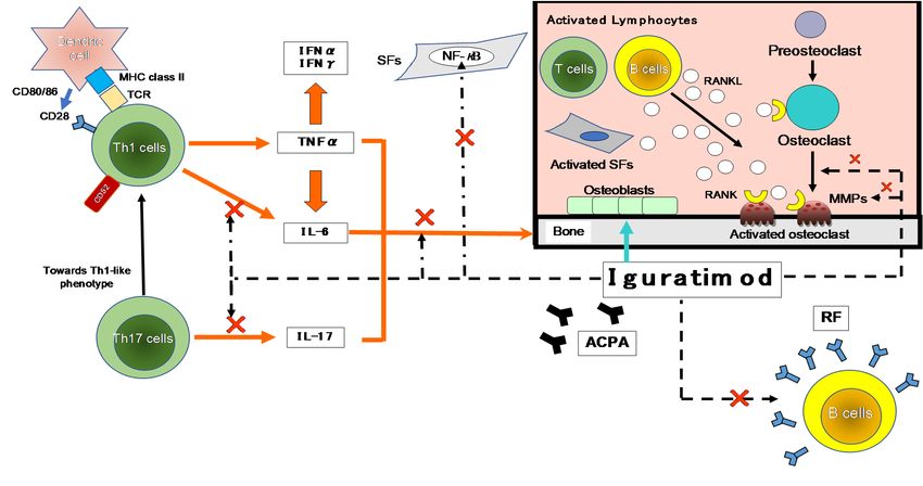
Figure 2. The inhibitory
Figure effectseffects
2. The inhibitory of IGU on the
of IGU immune
on the immune response. The
response. The firstfirst contact
contact betweenbetween
Th1 cellsTh1 cells and antigen-presenting
and antigen-presenting
cells (APCs;cells
e.g.,(APCs; e.g., dendritic cells) is made by T-cell receptors (TCRs) and major histocompatibility complex (MHC). A vari-
dendritic cells) is made by T-cell receptors (TCRs) and major histocompatibility complex (MHC). A variety
ety of environmental factors influence the production of autoantibodies such as anti-citrullinated protein antibodies
of environmental
(ACPA) and factors influence
rheumatoid factorthe
(RF).production
These immune ofcomplexes
autoantibodies such as
activate synovial anti-citrullinated
fibroblasts protein antibodies
(SFs) and macrophages, which (ACPA)
produce
and rheumatoid pro-inflammatory
factor (RF). Thesecytokines
immunesuch as tumor necrosis
complexes activatefactor-alpha (TNF-α) and (SFs)
synovial fibroblasts IL-6. As
andanother axis, they also
macrophages, which produce
affect Th17 cells that produce IL-17. These pro-inflammatory cytokines activate SFs and osteoclasts, leading to progressive
pro-inflammatory cytokines such as tumor necrosis factor-alpha (TNF-α) and IL-6. As another axis, they also affect Th17 cells
joint destruction. Iguratimod has effects on the production of pro-inflammatory cytokines in Th1 and Th17 cells, and on
that produce IL-17. These pro-inflammatory cytokines activate SFs and osteoclasts, leading to progressive joint destruction.
Iguratimod has effects on the production of pro-inflammatory cytokines in Th1 and Th17 cells, and on the production
of immunoglobulins and antibodies in B cells. It also has effects on bone metabolism by inhibiting osteoclast activation
and inducing osteoblast differentiation. NF-κB is activated in the pathogenesis of RA and is central to the chronic cycle of
inflammation that underlies its pathology. The inflammatory mediators, particularly TNF-α, activate cells in the synovium
in macrophages and SFs, and this is also largely NF-κB-dependent. SFs synthesize many NF-κB-induced genes in response
to TNF-α or IL-1, including chemokines that lead to further inflammatory infiltrates and matrix metalloproteinases (MMPs)
that promote joint destruction. →: stimulation, ×: inhibition. IFN-γ: interferon-gamma, NF-κB: nuclear factor-kappa B,
RANKL: receptor activator of nuclear factor-kappa B ligand.
1Life 2021, 11, 457 3 of 9
As shown in Figure 2, many cytokines are involved in RA, including TNF-α, IL-6, -1,
-17, and granulocyte-macrophage colony-stimulating factor (GM-CSF). Many important
biological processes involve cytokines: cell growth, proliferation, and differentiation;
inflammation, tissue repair, and the regulation of immune responses [14]. Cytokines are
responsible for the inflammation and joint destruction that occur in RA. Both T cells and B
cells have important roles in RA’s pathogenesis based on the coordinated interaction of
inflammatory cytokines [15,16]. In synovial tissue, CD4+ T cells differentiate mainly into
Th1-like effector cells that produce pro-inflammatory cytokines such as interferon-gamma
(IFN-γ) and TNF-α, with a distinct lack of differentiation into Th2-like effector cells that
produce anti-inflammatory cytokines such as IL-4, -10, and -13 [17]. In human monocytes
stimulated with lipopolysaccharide (LPS), IGU showed a low inhibitory effect on the pro-
inflammatory cytokines TNF-α, monocyte chemoattractant protein-1 (MCP-1), and IL-8,
confirming the anti-inflammatory effects of IGU as an inhibitor of macrophage migration
inhibitory factor (MIF) [18].
The main cytokine secreted by Th17 cells is IL-17, which comprises a cytokine family
with six members: IL-17A–F [19]. Th17 cells produce cytokines with pro-inflammatory
effects (including IL-17, -6, -21, -22 and TNF-α) that are suspected to play roles in the
immunopathogenesis of RA. In a clinical investigation of IGU for RA patients, after IGU
treatment Th1 and Th17 were downregulated whereas regulatory T cells (Tregs) were up-
regulated; these changes were accompanied by decreased levels of Th1, Th17, and follicular
helper T cell-associated transcription factors and inflammatory cytokines, plus increased
levels of Treg-associated transcription factors and cytokines [20]. In a murine model of
colitis, IGU reduced intestinal tissue damage and relieved colitis symptoms; the investiga-
tors speculated that these effects were due to the down-regulation of Th17 cells and the
up-regulation of Treg cells [21].
In a rat model of collagen-induced arthritis, IGU was also demonstrated to exert
a significant protective effect on cartilage and bone erosion by distorting a Th17-driven
response and inhibiting the production of anti-type II collagen antibodies [22]. Iguratimod
inhibited an IL-17 signal pathway by reducing both the stability of mRNA and the phos-
phorylation of mitogen-activated protein kinase (MAPK), targeting Act1 (adaptor for IL-17
receptors) as the adapter molecule and disrupting Act1’s interaction with tumor necrosis
receptor-associated factor 5 (Traf5) and inducible IκB kinase (Ikki) [23].
The pathogenesis of autoimmune diseases such as RA and systemic lupus erythemato-
sus (SLE) involves the disruption of B cell tolerance and the generation of high-affinity
autoantibodies [24,25], and thus therapies that target B cells have been examined. B cell
depletion therapy with the chimeric monoclonal antibody (mAb) rituximab for RA has been
successful [26–28], but its efficacy in SLE has been mixed [29,30]. Clinical trials targeting B
cell activators with the mAb belimumab and atacicept have been conducted in SLE, but only
belimumab achieved a positive endpoint [31]. In their in vitro study, Ye et al. observed that
IGU did not affect B cell activation or proliferation in the human antibody-secreting cell
differentiation system, but it did target the protein kinase C and early growth response 1
(EGR1) axes and inhibit the differentiation of human antibody-secreting cells [32].
Another important effect of IGU is the action that it exerts on NF-κB, as NF-κB and
other transcription factors regulate the expression of many genes that are involved in the
body’s immune and inflammatory responses [33,34]. In addition, when LPS interacts with
receptors on monocytes, the NF-κB complex is activated and specific gene groups such as
those underlying TNF-α and IL-1β, -6, and -8 are rapidly and transiently expressed [35,36].
The promoter regions of IL-6 and -8 have binding sites for NF-κB CCAAT/enhancer-
binding protein and activator protein (C/EBP)1, and their products are regulated at the
transcriptional level through the activation of these transcription factors [37–39]. However,
the NF-κB site was observed to be important for LPS-induced IL-6 gene expression in
THP-1 cells [33]. Regarding the IL-8 gene, NF-κB binding sites were demonstrated to
be essential for gene expression in all types of cells examined [37–39]. IGU suppressedLife 2021, 11, 457 4 of 9
TNF-α-induced production of IL-6, -8, and MCP-1 and reduced the accumulation of IL-6
and -8 mRNA in a concentration-dependent manner [37–39].
3. The Effects of IGU on Bone Metabolism and Cartilage Erosion
3.1. Promoting Bone Formation
Iguratimod exerts a protective effect under inflammatory conditions by (1) increasing
the expressions of osterix and Dlx5, (2) promoting osteoblast differentiation by increasing
the activation of P38, and (3) suppressing the level of phosphorylated NF-κB [40].
3.2. Inhibiting Osteoclast Differentiation and Bone Absorption
In vitro, IGU dose-dependently inhibited the osteoclast differentiation, migration, and
bone resorption that were induced by RANKL (receptor activator of NF-κB ligand) in
RAW264.7 mouse macrophage cells [41]. The RANKL-induced expressions of the three
chemokines CCL7, CCL4, and CCL12 and those of the osteoclast-related transcription
factors c-Jun, c-Fos, and NFATc1 were also suppressed by IGU dose-dependently.
3.3. Preventing Cartilage Erosion
As depicted in Figure 2, MMPs are secreted by both chondrocytes and synovial
membrane cells that are activated by inflammatory cytokines, and several MMPs are
closely involved in cartilage destruction. The activation of MMPs (including MMP-2 and
-9) by MMP-3 is the major cause of cartilage degradation [42]. The MMP-3 content in RA
patients’ synovial fluid is high, and the core proteins of proteoglycans are cleaved in MMP-3-
sensitive regions in the synovial fluid [43]. MMP-3 is also overexpressed in the synovium of
RA patients, which suggests that MMP-3 is a key protease for articular cartilage destruction
in RA. The serum concentration of MMP-3 is a direct indicator of synovitis associated with
RA disease activity [43], but this concentration is also influenced by factors such as gender,
renal dysfunction, and corticosteroid treatment [44]. The productions of MMP-1 and -3 by
rheumatoid synovial fibroblasts can be inhibited by treatment with IGU, thus inhibiting
the inflammatory cytokine-stimulated invasion of fibroblast-like synoviocytes [22]. These
findings indicated that IGU has properties that could make it an effective agent in multi-
targeted therapy for RA via the immune response and bone metabolism.
4. Clinical Findings Regarding the Efficacy of IGU
4.1. Phase III Clinical Study
A 20% improvement in the American College of Rheumatology Criteria (ACR20) was
observed in Japanese patients with active RA treated with IGU at 50 mg/day in a Phase III
study by Hara et al., and this was comparable to the improvement obtained with SASP
(IGU vs. SASP: 63.1% vs. 57.7%) [45]. Iguratimod treatment also reduced the patients’ RF
titers and the productions of IgG and IgM. These findings demonstrated that the efficacy of
IGU in RA patients was not inferior to that of SASP.
4.2. The Efficacy of IGU Treatment in Daily Practice
4.2.1. IGU as a First-Line csDMARD for RA
A recent retrospective analysis by our research group revealed the clinical efficacy
and adverse events (AEs) of IGU or SASP as the first-line csDMARD for 197 older RA
patients (IGU group’s age: 65.0 ± 13.2 years vs. SASP 62.2 ± 14.9 years) [5]. The retention
rate 36 months later was 52.4% in the IGU group and 32.1% in the SASP group, and the
response rate (good or moderate response) after 36 months was 85.8% in the IGU group and
65.2% in the SASP group. The IGU treatment reduced the patients’ RF titers; at 36 months,
prednisolone (PSL) use was 16.7% and 46.7% in the IGU and SASP groups, and the PSL
doses were 0.3 and 2.0 mg/day, respectively. The cumulative incidence of any AEs at
36 months was 19.8% and 29.2% in the IGU and SASP groups. The results also showed
that as a first-line csDMARD, compared to SASP, IGU was not significantly effective in
reducing RA patients’ DAS28-CRP (Disease Activity Score-28 for rheumatoid arthritis withLife 2021, 11, 457 5 of 9
C-reactive protein), but IGU did increase the treatment response rate and retention rate and
decrease steroid use. In addition, the AEs of the IGU-treated patients were not significantly
different from those of the SASP-treated patients, indicating that IGU is as effective as a
first-line csDMARD in patients who cannot tolerate an effective dose of MTX and have
difficulty reducing their steroid dosage.
4.2.2. IGU Treatment for RA Patients with an Inadequate Response to MTX
For elderly patients, it may be difficult to increase the dose of MTX or continue the
same dose due to hepatic or renal dysfunction. There are two reports describing the effect
of adding IGU to the treatment of RA patients in Japan with an inadequate response to
MTX [46,47]. Ishiguro et al. randomized 253 patients to IGU and placebo groups in a
double-blind study, and they reported that in the IGU group the 20% improvement in
ACR20 at 24 weeks was 69.5% (vs. 30.7% in the placebo group). Significant improvements
in the ACR50 and ACR70, RF titer, HAQ-DI (Health Assessment Questionnaire-Disability
Index), and DAS28 < 3.2 were also obtained. Hara et al. reported a randomized, double-
blind trial of IGU or placebo added to stable MTX therapy for RA, and they enrolled
patients in a 24-week extension study in which the patients who had been treated with
placebo+MTX were switched to IGU+MTX (switch group) [46]. In the IGU+MTX group,
the 20% improvement in ACR20 at 52 weeks (71.3%) was similar to the 20% improvement
in ACR20 at 24 weeks (69.5%). After switching to IGU therapy, the ACR20 improved
significantly from 30.7% at 24 weeks to 72.1% at 52 weeks. In patients with active RA who
showed an inadequate response to MTX, the efficacy and tolerability of IGU+MTX therapy
were maintained through 52 weeks.
We also conducted a retrospective study that evaluated the clinical efficacy of IGU in
RA patients treated with or without MTX for 54 weeks [48]: we divided RA patients into
those treated with MTX+IGU (n = 35) and those treated with IGU (n = 71). The between-
group difference in the change in the DAS28-CRP was −0.2. The DAS28-CRP decreased
significantly from baseline in both the MTX+IGU and IGU groups (−1.43 and −1.20 from
baseline, respectively). The retention rates were 71.4% and 59.2% and AEs were observed
in 17.1% and 28.2% in the MTX+IGU and IGU groups, respectively. Together these findings
indicated that treatment with IGU can be effective for patients with RA for whom MTX is
not an option.
4.2.3. IGU for Patients with an Inadequate Response to csDMARDs or bDMARDs
In a multicenter study, the addition of IGU for RA patients (n = 31) with an inadequate
response to intravenous and subcutaneous tocilizumab or other csDMARDs (SASP, MTX,
tacrolimus) improved outcome measures including the DAS28-CRP (from 2.9 to 1.7),
the Clinical Disease Activity Index for RA (CDAI; from 15.0 to 6.0), the modified HAQ-DI
(from 0.8 to 0.6), and the RF titer (from 382.1 to 240.3) [6]. The addition of IGU may thus be
an effective complementary treatment.
In another retrospective study, the use of IGU for RA patients with an inadequate
response to bDMARDs (n = 50) for >24 weeks significantly decreased the patients’ DAS28-
ESR (erythrocyte sedimentation rate) from 3.45 ± 0.92 at baseline to 2.85 ± 1.13 after
24 weeks [49]. Clinical remission was achieved by 38.3% of the patients, and inflammatory
synovitis as shown by ultrasound power Doppler was also improved.
4.3. Post-Marketing Clinical Study
A 52-week post-marketing study of Japanese RA patients was conducted by Mimori
et al. to determine the safety (n = 2666) and efficacy (n = 1614) in a final report [50].
The patients’ mean age was 64.1 years, and 51.8% were ≥ 65 years old. The mean duration
of RA in the patients was 9.9 years (median 7.0 years). The overall retention rate for IGU at
52 weeks was 56.3%. The discontinuation of IGU was due to AEs in 23.6% of the patients,
because of no change or worsening in 12.8%, site change or loss to follow-up in 8.7%, and
following improvement in 2.1%. The overall incidence of AEs, adverse drug reactionsLife 2021, 11, 457 6 of 9
(ADRs), serious AEs, and serious ADRs in the safety population was 46.92%, 38.26%,
7.35%, and 4.58%, respectively. The major ADRs were hepatic function abnormalities
(5.06%) and stomatitis (2.59%). Serious ADRs included pneumonia or bacterial pneumonia
(0.83%), interstitial lung disease (0.60%), and Pneumocystis jiroveci pneumonia (0.30%).
The incidence of ADRs peaked at approx. 4 weeks after the initiation of IGU treatment,
but the incidence of all ADRs decreased with time. Gastrointestinal disorders, hepatic
dysfunction, and renal dysfunction were more common at the start of IGU treatment,
whereas hematologic disorders and interstitial lung disease were reported less frequently
after 32 weeks. No specific trend was observed for peptic ulcer and infectious diseases in
relation to the time of onset.
In the study’s interim report at 24 weeks, a multivariate logistic regression was used
to evaluate risk factors for ADRs [51]. It revealed that the following were associated with a
lower risk of ADRs: age ≥ 65 years, low body weight, hepatic or renal dysfunction at base-
line, comorbidities, history of allergies, use of a concomitant glucocorticoid ≥ 5 mg/day
(vs. no use), MTX ≤ 8 mg/week (vs. no use), and concomitant bDMARD use (vs. no use).
In patients treated with warfarin + IGU, IGU interacted with the warfarin, resulting in
serious AEs including alveolar hemorrhage and an increased international normalized pro-
thrombin time ratio, suggesting that IGU enhances the anticoagulant effect of warfarin [51].
The incidence of side effects peaked at week 4 of treatment and then decreased without
increasing again at 28 weeks. No clinically important findings have been obtained since
the interim report regarding the combination of IGU and warfarin.
The clinical studies of IGU treatment for RA are summarized below in Table 1.
Table 1. Clinical trials of iguratimod (IGU) for rheumatoid arthritis patients in Japan.
Authors [Reference] Design No. of Patients Endpoint
IGU, n = 101
Nozaki et al. 2019 [5] Retrospective 36 months
SASP, n = 96
Ebina et al. 2019 [6] Retrospective Total, n = 31 24 weeks
Total, n = 376
IGU, n = 147
Hara et al. 2007 [45] RCT 28 weeks
SASP, n = 156
Placebo, n = 73
Total, n = 253 52 weeks
Hara et al. 2014 [46] RCT IGU+MTX, n = 165
Placebo+MTX, n = 88
Total, n = 253 24 weeks
Ishiguro et al. 2013 [47] RCT IGU+MTX, n = 165
Placebo+MTX, n = 88
Total, n = 106
Inoe et al. 2020 [48] Retrospective IGU+MTX, n = 35 54 weeks
MTX, n = 71
Yoshikawa et al. 2018 [49] Retrospective Total, n = 50 24 weeks
Okamura et al. 2015 [52] Retrospective Total, n = 41 52 weeks
RCT: randomized controlled trial, SASP: sulfasalazine, MTX: methotrexate.
5. Conclusions
About 10 years have passed since the csDMARD iguratimod was approved in Japan
for the treatment of RA. There have been several studies and trials for IGU conducted
in Japan and elsewhere regarding its immunological mechanism of action, its effects
on bone and cartilage metabolism, and its efficacy in daily practice. Unfortunately, the
evidence regarding IGU and its use for RA patients is limited in Western countries outside
of Asia, but as the number of patients with difficult-to-treat RA continues to increase,
the importance of basic research and clinical trials of the effectiveness of IGU as a new
treatment option for RA patients who cannot tolerate MTX has been highlighted. In Japan,Life 2021, 11, 457 7 of 9
IGU has become one of the most important drugs used in the routine treatment of RA, and
the clinical efficacy of IGU has been shown to be non-inferior to that of SASP. Moreover,
IGU treatment is steroid-sparing compared to SASP. Further evidence regarding IGU’s
safety and efficacy will be obtained in Japan and other countries.
Funding: This research received no external funding.
Conflicts of Interest: Y.N. has received honoraria or a research grant from AbbVie GK, Astellas
Pharma, Asahi Kasei, AYUMI Pharmaceutical, Chugai Pharmaceutical Co., Eisai Co., Daiichi-Sankyo,
MSD, Mitsubishi Tanabe Pharma Corp., Takeda, Ono, Otsuka Co., Pfizer, Janssen, and UCB Japan.
The funders had no role in the writing of this review.
References
1. Aringer, M.; Costenbader, K.; Daikh, D.; Brinks, R.; Mosca, M.; Ramsey-Goldman, R.; Smolen, J.S.; Wofsy, D.; Boumpas, D.T.;
Kamen, D.L.; et al. 2019 European league against rheumatism/American college of rheumatology classification criteria for
systemic lupus erythematosus. Ann. Rheum. Dis. 2020, 78, 1151–1159. [CrossRef]
2. Gaujoux-Viala, C.; Rincheval, N.; Dougados, M.; Combe, B.; Fautrel, B. Optimal methotrexate dose is associated with better
clinical outcomes than non-optimal dose in daily practice: Results from the ESPOIR early arthritis cohort. Ann. Rheum. Dis. 2017,
76, 2054–2060. [CrossRef] [PubMed]
3. Visser, K.; van der Heijde, D. Optimal dosage and route of administration of methotrexate in rheumatoid arthritis: A systematic
review of the literature. Ann. Rheum. Dis. 2009, 68, 1094–1099. [CrossRef] [PubMed]
4. Kameda, H.; Fujii, T.; Nakajima, A.; Koike, R.; Sagawa, A.; Kanbe, K.; Tomita, T.; Harigai, M.; Suzuki, Y.; Japan College of
Rheumatology Subcommittee on the Guideline for the Use of Methotrexate in Patients with Rheumatoid Arthritis. Japan college
of rheumatology guideline for the use of methotrexate in patients with rheumatoid arthritis. Mod. Rheumatol. 2019, 29, 31–40.
[CrossRef] [PubMed]
5. Nozaki, Y.; Inoue, A.; Kinoshita, K.; Funauchi, M.; Matsumura, I. Efficacy of iguratimod vs. salazosulfapyridine as the first-line
csDMARD for rheumatoid arthritis. Mod. Rheumatol. 2020, 30, 249–258. [CrossRef] [PubMed]
6. Ebina, K.; Miyama, A.; Tsuboi, H.; Kaneshiro, S.; Nishikawa, M.; Owaki, H.; Tsuji, S.; Hirao, M.; Etani, Y.; Goshima, A.; et al.
The add-on effectiveness and safety of iguratimod in patients with rheumatoid arthritis who showed an inadequate response to
tocilizumab. Mod. Rheumatol. 2019, 29, 581–588. [CrossRef]
7. Xie, S.; Li, S.; Tian, J.; Li, F. Iguratimod as a new drug for rheumatoid arthritis: Current landscape. Front. Pharmacol. 2020, 11, 73.
[CrossRef]
8. Aikawa, Y.; Yamamoto, M.; Yamamoto, T.; Morimoto, K.; Tanaka, K. An anti-rheumatic agent T-614 inhibits NF-kappaB
activation in LPS- and TNF-alpha-stimulated THP-1 cells without interfering with IkappaBalpha degradation. Inflamm. Res. 2002,
51, 188–194. [CrossRef]
9. Tanaka, K.; Yamamoto, T.; Aikawa, Y.; Kizawa, K.; Muramoto, K.; Matsuno, H.; Muraguchi, A. Inhibitory effects of an anti-
rheumatic agent T-614 on immunoglobulin production by cultured B cells and rheumatoid synovial tissues engrafted into SCID
mice. Rheumatology 2003, 42, 1365–1371. [CrossRef]
10. Du, F.; Lü, L.J.; Fu, Q.; Dai, M.; Teng, J.L.; Fan, W.; Chen, S.L.; Ye, P.; Shen, N.; Huang, X.F.; et al. T-614, a novel immunomodulator,
attenuates joint inflammation and articular damage in collagen-induced arthritis. Arthritis Res. Ther. 2008, 10, R136. [CrossRef]
11. Tanaka, K.; Shimotori, T.; Makino, S.; Eguchi, M.; Asaoka, K.; Kitamura, R.; Yoshida, C. Pharmacological studies on 3-formylamino-
7-methylsulfonylamino-6-phenoxy-4H-1-benzopyran-4-one (T-614), a novel antiinflammatory agent. 3rd communication: The in-
volvement of bradykinin in its analgesic actions. J. Pharmacobio Dyn. 1992, 15, 641–647. [CrossRef] [PubMed]
12. Tanaka, K.; Kawasaki, H.; Kurata, K.; Aikawa, Y.; Tsukamoto, Y.; Inaba, T. T-614, a novel antirheumatic drug, inhibits both the
activity and induction of cyclooxygenase-2 (COX-2) in cultured fibroblasts. Jpn. J. Pharmacol. 1995, 67, 305–314. [CrossRef]
[PubMed]
13. Mitamura, M.; Nakano, N.; Yonekawa, T.; Shan, L.; Kaise, T.; Kobayashi, T.; Yamashita, K.; Kikkawa, H.; Kinoshita, M. T cells are
involved in the development of arthritis induced by anti-type II collagen antibody. Int. Immunopharmacol. 2007, 7, 1360–1368.
[CrossRef]
14. Lina, C.; Conghua, W.; Nan, L.; Ping, Z. Combined treatment of etanercept and MTX reverses Th1/Th2, Th17/Treg imbalance in
patients with rheumatoid arthritis. J. Clin. Immunol. 2011, 31, 596–605. [CrossRef]
15. Smolen, J.S.; Steiner, G. Therapeutic strategies for rheumatoid arthritis. Nat. Rev. Drug. Discov. 2003, 2, 473–488. [CrossRef]
16. Smolen, J.S.; Aletaha, D.; Koeller, M.; Weisman, M.H.; Emery, P. New therapies for treatment of rheumatoid arthritis. Lancet 2007,
370, 1861–1874. [CrossRef]
17. Unutmaz, D.; Pileri, P.; Abrignani, S. Antigen-independent activation of naive and memory resting T cells by a cytokine
combination. J. Exp. Med. 1994, 180, 1159–1164. [CrossRef]
18. Bloom, J.; Metz, C.; Nalawade, S.; Casabar, J.; Cheng, K.F.; He, M.; Sherry, B.; Coleman, T.; Forsthuber, T.; Al-Abed, Y. Identification
of iguratimod as an inhibitor of macrophage migration inhibitory factor (MIF) with steroid-sparing potential. J. Biol. Chem. 2016,
291, 26502–26514. [CrossRef]Life 2021, 11, 457 8 of 9
19. Yao, Z.; Painter, S.L.; Fanslow, W.C.; Ulrich, D.; Macduff, B.M.; Spriggs, M.K.; Armitage, R.J. Human IL-17: A novel cytokine
derived from T cells. J. Immunol. 1995, 155, 5483–5486.
20. Xu, Y.; Zhu, Q.; Song, J.; Liu, H.; Miao, Y.; Yang, F.; Wang, F.; Cheng, W.; Xi, Y.; Niu, X.; et al. Regulatory effect of iguratimod on
the balance of Th subsets and inhibition of inflammatory cytokines in patients with rheumatoid arthritis. Mediat. Inflamm. 2015,
2015, 356040. [CrossRef]
21. Jiang, X.P.; Huang, X.L.; Yang, Z.P.; Wang, S.C.; Xie, W.; Miao, L.; Tang, L.; Huang, Z.M. Iguratimod ameliorates inflammatory
responses by modulating the Th17/Treg paradigm in dextran sulphate sodium-induced murine colitis. Mol. Immunol. 2018,
93, 9–19. [CrossRef]
22. Du, F.; Lü, L.J.; Teng, J.L.; Shen, N.; Ye, P.; Bao, C.D. T-614 alters the production of matrix metalloproteinases (MMP-1 andMMP-
3) and inhibits the migratory expansion of rheumatoid synovial fibroblasts, in vitro. Int. Immunopharmacol. 2012, 13, 54–60.
[CrossRef]
23. Luo, Q.; Sun, Y.; Liu, W.; Qian, C.; Jin, B.; Tao, F.; Gu, Y.; Wu, X.; Shen, Y.; Xu, Q. A novel disease-modifying antirheumatic drug,
iguratimod, ameliorates murine arthritis by blocking IL-17 signaling, distinct from methotrexate and leflunomide. J. Immunol.
2013, 191, 4969–4978. [CrossRef]
24. Sabahi, R.; Anolik, J.H. B-cell-targeted therapy for systemic lupus erythematosus. Drugs 2006, 66, 1933–1948. [CrossRef]
25. Keystone, E. B cell targeted therapies. Arthritis Res. Ther. 2005, 7, S13–S18. [CrossRef]
26. Cohen, S.B.; Emery, P.; Greenwald, M.W.; Dougados, M.; Furie, R.A.; Genovese, M.C.; Keystone, E.C.; Loveless, J.E.; Burmester,
G.-R.; Cravets, M.W.; et al. Rituximab for rheumatoid arthritis refractory to anti-tumor necrosis factor therapy: Results of a
multicenter, randomized, double-blind, placebo-controlled, phase III trial evaluating primary efficacy and safety at twenty-four
weeks. Arthritis Rheum. 2006, 54, 2793–2806. [CrossRef]
27. Emery, P.; Deodhar, A.; Rigby, W.F.; Isaacs, J.D.; Combe, B.; Racewicz, A.J.; Latinis, K.; Mendoza, C.A.; Szczepański, L.J.;
Roschmann, R.A.; et al. Efficacy and safety of different doses and retreatment of rituximab: A randomised, placebo-controlled
trial in patients who are biological naive with active rheumatoid arthritis and an inadequate response to methotrexate (Study
Evaluating Rituximab’s Efficacy in MTX iNadequate rEsponders (SERENE)). Ann. Rheum. Dis. 2010, 69, 1629–1635.
28. Mariette, X.; Rouanet, S.; Sibilia, J.; Combe, B.; Le Loët, X.; Tebib, J.; Jourdan, R.; Dougados, M. Evaluation of low-dose rituximab
for the retreatment of patients with active rheumatoid arthritis: A non-inferiority randomised controlled trial. Ann. Rheum. Dis.
2014, 73, 1508–1514. [CrossRef] [PubMed]
29. Merrill, J.T.; Neuwelt, C.M.; Wallace, D.J.; Shanahan, J.C.; Latinis, K.M.; Oates, J.C.; Utset, T.O.; Gordon, C.; Isenberg, D.A.;
Hsieh, H.-J.; et al. Efficacy and safety of rituximab in moderately-to-severely active systemic lupus erythematosus: The random-
ized, double-blind, phase II/III systemic lupus erythematosus evaluation of rituximab trial. Arthritis Rheum. 2010, 62, 222–233.
[CrossRef] [PubMed]
30. Rovin, B.H.; Furie, R.; Latinis, K.; Looney, R.J.; Fervenza, F.C.; Sanchez-Guerrero, J.; Maciuca, R.; Zhang, D.; Garg, J.P.; Brunetta, P.;
et al. Efficacy and safety of rituximab in patients with active proliferative lupus nephritis: The lupus nephritis assessment with
rituximab study. Arthritis Rheum. 2012, 64, 1215–1226. [CrossRef]
31. Stohl, W.; Hiepe, F.; Latinis, K.M.; Thomas, M.; Scheinberg, M.A.; Clarke, A.; Aranow, C.; Wellborne, F.R.; Abud-Mendoza, C.;
Hough, D.R.; et al. Belimumab reduces autoantibodies, normalizes low complement levels, and reduces select B cell populations
in patients with systemic lupus erythematosus. Arthritis Rheum. 2012, 64, 2328–2337. [CrossRef] [PubMed]
32. Ye, Y.; Liu, M.; Tang, L.; Du, F.; Liu, Y.; Hao, P.; Fu, Q.; Guo, Q.; Yan, Q.; Zhang, X.; et al. Iguratimod represses B cell terminal
differentiation linked with the inhibition of PKC/EGR1 axis. Arthritis Res. Ther. 2019, 21, 92. [CrossRef] [PubMed]
33. Libermann, T.A.; Baltimore, D. Activation of interleukin-6 gene expression through the NF-kappa B transcription factor. Mol. Cell.
Biol. 1990, 10, 2327–2334. [CrossRef] [PubMed]
34. Ishikawa, Y.; Mukaida, N.; Kuno, K.; Rice, N.; Okamoto, S.; Matsushima, K. Establishment of lipopolysaccharide-dependent
nuclear factor kappa B activation in a cell-free system. J. Biol. Chem. 1995, 270, 4158–4164. [CrossRef] [PubMed]
35. Heumann, D.; Gallay, P.; Barras, C.; Zaech, P.; Ulevitch, R.J.; Tobias, P.S.; Glauser, M.P.; Baumgartner, J.D. Control of lipopolysac-
charide (LPS) binding and LPS-induced tumor necrosis factor secretion in human peripheral blood monocytes. J. Immunol. 1992,
148, 3505–3512. [PubMed]
36. Matsusaka, T.; Fujikawa, K.; Nishio, Y.; Mukaida, N.; Matsushima, K.; Kishimoto, T.; Akira, S. Transcription factors NF-IL6 and
NF-kappa B synergistically activate transcription of the inflammatory cytokines, interleukin 6 and interleukin 8. Proc. Natl. Acad.
Sci. USA 1993, 90, 10193–10197. [CrossRef] [PubMed]
37. Geng, Y.; Zhang, B.; Lotz, M. Protein tyrosine kinase activation is required for lipopolysaccharide induction of cytokines in
human blood monocytes. J. Immunol. 1993, 151, 6692–6700. [PubMed]
38. Mukaida, N.; Shiroo, M.; Matsushima, K. Genomic structure of the human monocyte-derived neutrophil chemotactic factor IL-8.
J. Immunol. 1989, 143, 1366–1371.
39. Mukaida, N.; Mahe, Y.; Matsushima, K. Cooperative interaction of nuclear factor-kappa B- and cis-regulatory enhancer binding
protein-like factor binding elements in activating the interleukin-8 gene by pro-inflammatory cytokines. J. Biol. Chem. 1990,
265, 21128–21133. [CrossRef]
40. Song, J.; Liu, H.; Zhu, Q.; Miao, Y.; Wang, F.; Yang, F.; Cheng, W.; Xi, Y.; Niu, X.; He, D.; et al. T-614 promotes osteoblastic cell
differentiation by increasing Dlx5 expression and regulating the activation of p38 and NF-κB. Biomed. Res. Int. 2018, 2018, 4901591.
[CrossRef]Life 2021, 11, 457 9 of 9
41. Gan, K.; Yang, L.; Xu, L.; Feng, X.; Zhang, Q.; Wang, F.; Tan, W.; Zhang, M. Iguratimod (T-614) suppresses RANKL-induced
osteoclast differentiation and migration in RAW264.7 cells via NF-κB and MAPK pathways. Int. Immunopharmacol. 2016,
35, 294–300. [CrossRef]
42. Nagase, H.; Woessner, J.F., Jr. Matrix metalloproteinases. J. Biol. Chem. 1999, 274, 21491–21494. [CrossRef] [PubMed]
43. Ishiguro, N.; Ito, T.; Oguchi, T.; Kojima, T.; Iwata, H.; Ionescu, M.; Poole, A.R. Relationships of matrix metalloproteinases and
their inhibitors to cartilage proteoglycan and collagen turnover and inflammation as revealed by analyses of synovial fluids from
patients with rheumatoid arthritis. Arthritis Rheum. 2001, 44, 2503–2511. [CrossRef]
44. Yamanaka, H.; Matsuda, Y.; Tanaka, M.; Sendo, W.; Nakajima, H.; Taniguchi, A.; Kamatani, N. Serum matrix metalloproteinase 3
as a predictor of the degree of joint destruction during the six months after measurement, in patients with early rheumatoid
arthritis. Arthritis Rheum. 2000, 43, 852–858. [CrossRef]
45. Hara, M.; Abe, T.; Sugawara, S.; Mizushima, Y.; Hoshi, K.; Irimajiri, S.; Hashimoto, H.; Yoshino, S.; Matsui, N.; Nobunaga, M.; et al.
Efficacy and safety of iguratimod compared with placebo and salazosulfapyridine in active rheumatoid arthritis: A controlled,
multicenter, double-blind, parallel-group study. Mod. Rheumatol. 2007, 17, 1–9. [CrossRef]
46. Hara, M.; Ishiguro, N.; Katayama, K.; Kondo, M.; Sumida, T.; Mimori, T.; Soen, S.; Nagai, K.; Yamaguchi, T.; Yamamoto, K.;
et al. Safety and efficacy of combination therapy of iguratimod with methotrexate for patients with active rheumatoid arthritis
with an inadequate response to methotrexate: An open-label extension of a randomized, double-blind, placebo-controlled trial.
Mod. Rheumatol. 2014, 24, 410–418. [CrossRef]
47. Ishiguro, N.; Yamamoto, K.; Katayama, K.; Kondo, M.; Sumida, T.; Mimori, T.; Soen, S.; Nagai, K.; Yamaguchi, T.; Hara, M.; et al.
Concomitant iguratimod therapy in patients with active rheumatoid arthritis despite stable doses of methotrexate: A randomized,
double-blind, placebo-controlled trial. Mod. Rheumatol. 2013, 23, 430–439. [CrossRef]
48. Inoue, A.; Nozaki, Y.; Hirooka, Y.; Kinoshita, K.; Chiba, Y.; Funauchi, M.; Matsumura, I. The effectiveness and retention rate
of iguratimod in Japanese rheumatoid arthritis patients with/without methotrexate in daily medical care. Life 2020, 10, 261.
[CrossRef]
49. Yoshikawa, A.; Yoshida, S.; Kimura, Y.; Tokai, N.; Fujiki, Y.; Kotani, T.; Matsumura, Y.; Takeuchi, T.; Makino, S. Add-on iguratimod
as a therapeutic strategy to achieve remission in patients with rheumatoid arthritis inadequately responding to biological
DMARDs: A retrospective study. Mod. Rheumatol. 2018, 28, 227–234. [CrossRef]
50. Mimori, T.; Harigai, M.; Atsumi, T.; Fujii, T.; Kuwana, M.; Matsuno, H.; Momohara, S.; Takei, S.; Tamura, N.; Takasaki, Y.; et al.
Safety and effectiveness of iguratimod in patients with rheumatoid arthritis: Final report of a 52-week, multicenter postmarketing
surveillance study. Mod. Rheumatol. 2019, 29, 314–323. [CrossRef]
51. Mimori, T.; Harigai, M.; Atsumi, T.; Fujii, T.; Kuwana, M.; Matsuno, H.; Momohara, S.; Takei, S.; Tamura, N.; Takasaki, Y.; et al.
Safety and effectiveness of 24-week treatment with iguratimod, a new oral disease-modifying antirheumatic drug, for patients
with rheumatoid arthritis: Interim analysis of a post-marketing surveillance study of 2679 patients in Japan. Mod. Rheumatol.
2017, 27, 755–765. [CrossRef]
52. Okamura, K.; Yonemoto, Y.; Suto, T.; Okura, C.; Takagishi, K. Efficacy at 52 weeks of daily clinical use of iguratimod in patients
with rheumatoid arthritis. Mod. Rheumatol. 2015, 25, 534–539. [CrossRef]You can also read