COVID-19-associated mucormycosis presenting to the Emergency Department-an observational study of 70 patients - Oxford Academic Journals
←
→
Page content transcription
If your browser does not render page correctly, please read the page content below
QJM: An International Journal of Medicine, 2021, 1–7

                                                                                 doi: 10.1093/qjmed/hcab190
                                                                                 Original paper

                                                                                                                                                    Downloaded from https://academic.oup.com/qjmed/advance-article/doi/10.1093/qjmed/hcab190/6319726 by guest on 30 September 2021
ORIGINAL PAPER

COVID-19-associated mucormycosis presenting to
the Emergency Department—an observational study
of 70 patients
A. Ramaswami1,*, A.K. Sahu1,*, A. Kumar 1, S. Suresh1, A. Nair1, D. Gupta1,
R. Chouhan1, R. Bhat1, R. Mathew1, J.A. Majeed1, P. Aggarwal1, J. Nayer1,
M. Ekka1, A. Thakar2, G. Singh 3, I. Xess3 and N. Wig4
From the 1Department of Emergency Medicine, All India Institute of Medical Sciences, Ansari Nagar,
New Delhi 110029, India, 2Department of Otorhinolaryngology, All India Institute of Medical Sciences, Ansari
Nagar, New Delhi 110029, India, 3Department of Microbiology, All India Institute of Medical Sciences, Ansari
Nagar, New Delhi 110029, India and 4Department of Medicine, All India Institute of Medical Sciences,
Ansari Nagar, New Delhi 110029, India

*These authors contributed equally to this work.
Address correspondence to Dr A. Kumar, Department of Emergency Medicine, All India Institute of Medical Sciences, Ansari Nagar, New Delhi 110029,
India. email: akshay2111@gmail.com

Summary
Background: Mucormycosis (MM) is a deadly opportunistic fungal infection and a large surge in COVID-19-associated
mucormycosis (CAM) is occurring in India.
Aim: Our aim was to delineate the clinico-epidemiological profile and identify risk factors of CAM patients presenting to the
Emergency Department (ED).
Design: This was a retrospective, single-centre, observational study.
Methods: We included patients who presented with clinical features or diagnosed MM and who were previously treated for
COVID-19 in last 3 months of presentation (recent COVID-19) or currently being treated for COVID-19 (active COVID-19).
Information regarding clinical features of CAM, possible risk factors, examination findings, diagnostic workup including
imaging and treatment details were collected.
Results: Seventy CAM patients (median age: 44.5 years, 60% males) with active (75.7%) or recent COVID-19 (24.3%) who
presented to the ED in between 6 May 2021 and 1 June 2021, were included. A median duration of 20 days (interquartile
range: 13.5–25) was present between the onset of COVID-19 symptoms and the onset of CAM symptoms. Ninety-three
percent patients had at least one risk factor. Most common risk factors were diabetes mellitus (70%) and steroid use for
COVID-19 disease (70%). After clinical, microbiological and radiological workup, final diagnosis of rhino-orbital CAM was
made in most patients (68.6%). Systemic antifungals were started in the ED and urgent surgical debridement was planned.
Conclusion: COVID-19 infection along with its medical management have increased patient susceptibility to MM.

Received: 2 July 2021
C The Author(s) 2021. Published by Oxford University Press on behalf of the Association of Physicians. All rights reserved.
V
For permissions, please email: journals.permissions@oup.com

                                                                                                                                            1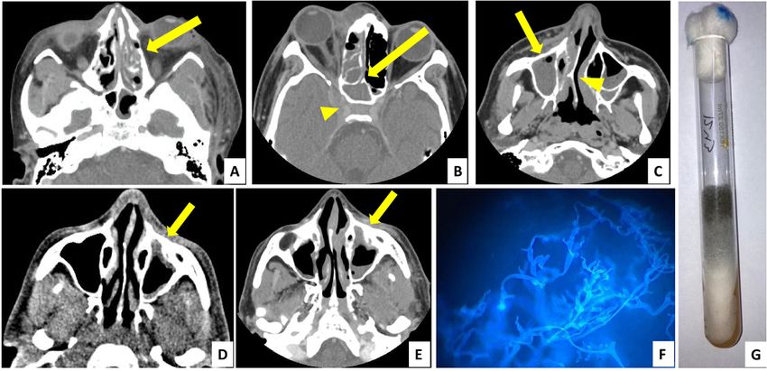
2   | QJM: An International Journal of Medicine, 2021, Vol. 00, No. 0

Introduction                                                               from the hospital records with an emphasis on the demograph-
                                                                           ic profile, date of arrival, date of onset of CAM symptoms and
Mucormycosis (MM) are syndromes in humans caused by the
                                                                           COVID-19 symptoms, clinical features of CAM, clinical features
mucorale group of fungi. These fungi are ubiquitous and pre-
                                                                           of current COVID-19, detailed comorbidities and risk factors of
sent in any environment including hospitals. Inhalation of fun-
                                                                           CAM, steroid usage details for COVID-19, COVID-19 treatment
gal spores is harmless in immunocompetent individuals but
                                                                           received prior to CAM symptoms, arrival vitals, diagnostic eval-
can cause life-threatening disease in those who are immuno-
                                                                           uations in ED (radiological and microbiological), medical treat-
compromised.1 The immune system is weak in those with un-
                                                                           ment given in ED and final disposition with surgical plan.
controlled diabetes mellitus, prolonged intake of steroids or
                                                                           Details of the recent COVID-19 presentation, severity and its

                                                                                                                                                Downloaded from https://academic.oup.com/qjmed/advance-article/doi/10.1093/qjmed/hcab190/6319726 by guest on 30 September 2021
immunosuppressant medications, malignancies and other
                                                                           treatment (e.g. steroid use, oxygen supplementation) were
debilitating conditions like chronic liver disease and chronic
                                                                           retrieved from the available documents.
malnutrition state.2 It is notable that these conditions can also
indicate risk of severe COVID-19 infection.3
    COVID-19 pandemic left the world reeling over the past year.           Statistical analysis
The second wave has been particularly devastating in India.                Counts and percentages were used to summarize categorical
During the months of April and early May 2021, millions were               data. Mean and standard deviation were used to summarize
affected and thousands were seeking hospital care.4                        normally distributed data, whereas median, range and inter-
Unprecedented numbers needed oxygen therapy and admission                  quartile range (IQR) were used to summarize non-normal con-
putting tremendous pressure on health infrastructure.5                     tinuous data. Normality of data was tested by Kolmogorov–
Overlapping with the rise in COVID-19 cases, there was a surge             Smirnov test. As this was a descriptive study, no analytical tests
of rhino-orbito-cerebral mucormycosis in those with active or              were applied on any subgroups. All the analyses were per-
recent COVID-19. MM has a high mortality even with the best of             formed with IBM SPSS Statistics for Windows, Version 26.0.
treatment.6 Thus, we designed a retrospective observational                Armonk, NY: IBM Corp.
study in our department with an objective to document the clin-
ical features, radiological extent and possible risk factors which
might be contributing to this illness in the context of the                Results
COVID-19 pandemic.                                                         Demographic profile

                                                                           A total of 70 diagnosed CAM were included for the analysis
Methodology                                                                (Table 1). These patients presented to our ED in between 6 May
                                                                           2021 and 1 June 2021. Fifty-three out of 70 patients (75.7%) were
This was a retrospective, single-centre, observational study fol-          active COVID-19, whereas 17 patients (24.3%) were with recent
lowing the Strengthening the Reporting of Observational                    COVID-19 infection (COVID-19 negative during ED presentation).
Studies in Epidemiology guidelines and the recommendations                 Among the active COVID-19 cases, 7 patients (10%) presented
by Kaji et al.7 The study was conducted in the Emergency                   primarily with CAM symptoms, but incidentally detected to
Department (ED), at a tertiary care teaching institute of India.           have COVID-19, i.e. they were asymptomatic for COVID-19. The
Between 1 April and 1 June 2021, our ED catered to 1647 COVID-             median age of the included patients was 44.5 years, with an IQR
19 confirmed admission requiring patients. We observed a                   of 38–55.5 years, with 60% (n ¼ 42) males. Overall, a lag period
surge in MM cases in the later half, i.e. 6 May 2021 to 1 June             was observed between the onset of COVID-19 symptoms and
2021. We planned to conduct this study to delineate the clinico-           the onset of CAM symptoms, with median duration being
epidemiological profile of MM in active or recent COVID-19                 20 days (IQR: 13.5–25).
patients. Active COVID-19 cases were defined as patients who
were laboratory confirmed for SARS-CoV-2 in the ED (by rapid
antigen or nucleic acid amplification test). Recent COVID-19               Risk factors for MM in COVID-19 patients
cases were defined as patients who had suffered from COVID-19              Only 5 out of 70 patients (7.1%) had no comorbidities, immuno-
in the past 3 months of presentation, but currently SARS-CoV-2             suppressant use (including steroids) or recent blood glucose ele-
negative in the ED. The time limit of 3 months was taken                   vation. Majority of patients had underlying diabetes mellitus
according to commonly accepted definition of post-acute                    (n ¼ 49, 70%), of which five patients were recently diagnosed
COVID-19 syndrome.8 COVID-19-associated mucormycosis                       during their COVID-19 illness. Nine patients (12.8%) had con-
(CAM) was defined as patients with MM along with acute or re-              comitant diabetic ketoacidosis. Following diabetes, the second
cent COVID-19 illness. The ethical approval was obtained from              most common comorbidity was hypertension (24.3%). Other
the Institute Ethics Committee before the commencement of                  comorbidities were present in
A. Ramaswami et al. | 3
Table 1. Demographic profile and risk factors
Characteristics Characteristics Total, n ¼ 70 (%)
Age Median years (IQR) 44.5 (38–55.5)
Gender Male 42 (60)
Female 28 (40)
Duration between COVID-19 onset and mucormyco- Median days (IQR) (n ¼ 63) 20 (13.5–25)
sis onset
COVID-19 status on arrival Positive at presentation 53 (75.7)
Downloaded from https://academic.oup.com/qjmed/advance-article/doi/10.1093/qjmed/hcab190/6319726 by guest on 30 September 2021
Post-COVID, negative 17 (24.3)
Comorbid illness Diabetes 49 (70)
On oral antidiabetic agents 33
On insulin 9
Recently diagnosed 5
Hypertension 17 (24.3)
Coronary artery diseases 4 (5.7)
Organ transplant 2 (2.9)
Chronic kidney diseases 6 (8.6)
Long term immunosuppressive therapy Prior steroid use 3 (4.3)
Any other immunosuppressant 1 (1.4)
Steroid use in the recent COVID-19 Received systemic steroids 49 (70)
Route of systemic steroids Intravenous 27 (38.6)
Oral 22 (31.4)
Type of steroids Methylprednisolone 15 (21.4)
Dexamethasone 14 (20)
Prednisolone 7 (10)
Hydrocortisone 1 (1.4)
Budesonide 13 (18.8)
Duration of steroids used Median days (IQR) (n ¼ 50) 7.5 (7–10.5)
Complication of steroid use Hyperglycaemia during steroid use which required 33
in-hospital insulin
Inhalational steroid use Inhalational 13 (18.8)
IQR, interquartile range.
antibiotics (azithromycin, doxycycline, amoxycillin, piperacil- mount with calcofluor stain was positive for aseptate hyphae in
lin-tazobactam) prior to ED arrival with CAM symptoms. 56 patients (80%). For 32 patients, fungal culture was sent,
which turned out to be positive in 28 patients (87.5%).
Signs and symptoms of MM Radiological diagnostic modalities included contrast-enhanced
computed tomography of brain, orbit and paranasal sinuses in
Details of the signs and symptoms of CAM are presented in the ED. Most common radiological diagnosis was rhinosinusitis,
Table 2 (Figure 1). Most common symptom reported in CAM was followed by orbital extension and intracranial invasion. Six
related to the eye and its adnexal tissues. Nearly 80% patients patients (8.6%) had cerebral infarct and two patients (2.6%) had
had eye pain, swollen eyes and significant lid oedema on exam- intracranial bleed. After clinical, microbiological and radiologic-
ination. Other ophthalmic symptoms were diminution of vision, al workups, final diagnosis was made (Figure 2). Rhino-orbital
proptosis, ptosis and double vision. Sino-nasal symptoms like CAM was the most common variety (n ¼ 48, 68.6%), followed by
nasal stuffiness, nasal discharge and epistaxis were present in rhino-orbito-cerebral CAM (n ¼ 17, 24.3%).
38.6%, 25.7% and 18.6% of patients, respectively. On examin-
ation of nasal cavity, crusting and ulceration were present in
24.3% patients. Another common symptom was facial pain,
which was the presenting complaint in 34.3% patients. Nearly Management of MM in the ED
10% of patients presented with hemiplegia.
Along with the stabilization of haemodynamic parameters, all
the CAM cases were managed with initiation of systemic anti-
Diagnosis of MM in the ED fungals as soon as possible. Intravenous liposomal amphoteri-
Sixteen out of 70 patients (22.9%) presented to our ED with un- cin—B (LAMB) with an initial dose of 5 mg/kg/day was initiated
stable vital signs and triaged as ‘Red’ category (highest priority) in majority of the cases (n ¼ 68, 97.1%). For patients with intra-
as per institution protocol. Details of the presenting vitals and cranial extension, high dose of LAMB, i.e. 10 mg/kg was initiated
the point-of-care laboratory values are described in Table 3. The at the earliest. Oral posaconazole (300 mg twice a day on Day 1
value of C-reactive protein was available for only 10 patients, all followed by once daily) was initiated in two cases (2.9%). Control
of them had values higher than 10 mg/l, indicating severe in- of underlying comorbid illness including insulin therapy for
flammation due to COVID-19. For definitive diagnosis of CAM, hyperglycaemia was initiated for all. Urgent otorhinolaryngol-
microbiological samples were taken from the active lesions ogy, ophthalmology and neurosurgery consultations were taken
(with or without nasal endoscopy). Potassium hydroxide (KOH) for shared decision on the surgical debridement pathway.4 | QJM: An International Journal of Medicine, 2021, Vol. 00, No. 0
Table 2. Presenting symptoms and clinical examination findings in mucormycosis patients
Symptoms Total, n ¼ 70 (%) Signs Total, n ¼ 70 (%)
Eyes and adnexa Eye pain 57 (81.4) Lid oedema 52 (74.3)
Swollen eyes 56 (80) Visual acuity 29 (41.4)
Diminution of vision 26 (37.1) Proptosis 28 (40)
Protrusion of eyeball 24 (34.3) Chemosis 24 (34.3)
Ptosis 14 (20) Ptosis 22 (31.4)
Double vision 1 (1.4) Ophthalmoplegia 21 (30)
Downloaded from https://academic.oup.com/qjmed/advance-article/doi/10.1093/qjmed/hcab190/6319726 by guest on 30 September 2021
Nasal cavity Nasal stuffiness 27 (38.6) Crusting and ulceration 17 (24.3)
Nasal discharge 18 (25.7) Discharge 7 (10)
Epistaxis 13 (18.6) Active epistaxis 6 (8.8)
Face Facial pain 24 (34.3) Ulceration 3 (4.3)
Headache 20 (29)
Facial ulcers 3 (4.3)
Neurological problems One sided weakness 8 (11.4) Hemiplegia 8 (11.4)
Altered sensorium 5 (7) Facial palsy 3 (2.9)
Facial deviation 2 (2.9) Altered sensorium 5 (7)
Decreased facial sensation 2 (2.8) Decreased facial sensation 2 (2.8)
Oral cavity Oral ulcers 4 (5.7) Discoloration 7 (10)
Crusting and ulceration 5 (7.1)
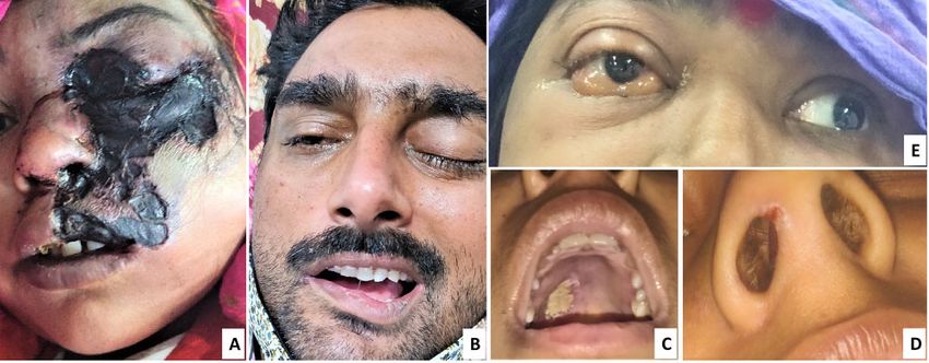
Figure 1. (A–E) Signs and symptoms of mucormycosis. (A) Black eschar lesion over face. (B) Periorbital oedema along with facial palsy. (C) Palatine ulcer with white base
over right posterior region. (D) Congestion and small ulcer in right nares. (E) Chemosis and proptosis of right eye and restriction of eye movement on lateral gaze (left
eye normal movement).
Discussion Many experts believe that the combination of high dose ste-
roids and uncontrolled diabetes has led to this epidemic of MM
We conducted a single-centre, retrospective study of 70 patients in COVID-19 patients.11–13 In pre-COVID era, Prakash and
with CAM who presented to the ED in the setting of acute or re- Chakrabarti14 found diabetes mellitus as a predisposing factor
cent COVID-19. To the best of our knowledge, this study is one in 17–88% cases globally and in India it was a risk factor in over
of the largest ED-based case series on deadly combinations of 50% cases. In the setting of COVID-19, case series by Sharma et
MM and COVID-19. As the number of CAM cases were increas- al.15 described diabetes as a risk factor in 90% cases of which
ing in India during the second wave of COVID-19 pandemic, we 52% had uncontrolled disease. A systematic review of 101 cases
have tried to delineate the clinico-epidemiological profile of of MM in COVID-19 by Singh et al.12 noted that more than 80%
these patients. cases had either pre-existing or new onset hyperglycaemia as a
Majority of the patients in our study were middle-aged (age: risk factor. Our study has reflected their findings in that 70% of
38–55 years), of which nearly two-thirds were male. This demo- included patients were diabetic.
graphic profile was similar to the population of 82 MM patients Prolonged use of corticosteroids increasing risk of MM has
studied by Chander et al.,9 of which two-third were male and been reported in patients.16 Ribes et al.17 described that acute or
aged between the ages 31–60 years. It has been hypothesized that chronic use of steroids in such patients predisposed them to
the effect of oestrogen might be protective in systemic fungal in- fungal infection. Steroid use during the pandemic has been sup-
fection, which could have led to lower incidence in females.10 ported by the Randomized Evaluation of COVID-19 TherapyA. Ramaswami et al. | 5
Table 3. Presenting vitals, lab parameters and diagnosis of mucormycosis in the study population
ED parameters N (%) or median (IQR)
Vitals at presentation Heart rate (bpm) 98 (81–110)
Respiratory rate (per min) 20 (18–22)
Oxygen saturation (%) 98 (96–98)
Lab values Random blood sugar at arrival (mg/dl) 235 (186–300)
Total leucocyte count (1000 s per mm3) 12.8 (8.8–16.1)
Platelets (lakhs per mm3) 2.46 (1.68–3.10)
Downloaded from https://academic.oup.com/qjmed/advance-article/doi/10.1093/qjmed/hcab190/6319726 by guest on 30 September 2021
D—dimer 311 (92–537)
Ferritin 898 (346–1580)
C-reactive protein 34 (26–64)
Microbiological diagnosis KOH-calcofluor positive 56 (80)
Culture positive (out of 32) 28 out of 32 (87.5)
Radiological diagnosis Rhinosinusitis 55 (78.6)
Orbital extension 31 (44.3)
Intracranial extension 12 (17.1)
Brain infarct 6 (8.6)
Intracranial bleed 2 (2.9)
Final diagnosis Rhino-orbital CAM 48 (68.6)
Rhino-orbito-cerebral CAM 17 (24.3)
Treatment started in the ED Liposomal amphotericin—B 68 (97.1)
Posaconazole 2 (2.9)
bpm, beats per minute; CAM, COVID-19-associated mucormycosis; ED, Emergency Department; IQR, interquartile range; KOH, potassium hydroxide mount for fungal
hyphae detection.
Figure 2. Diagnosis of mucormycosis—radiology (A–E axial section, contrast-enhanced computed tomography images) and microbiology (F and G). (A) Left ethmoidal
sinusitis with bony erosion (yellow arrow). (B) Right ethmoidal and sphenoid sinusitis (yellow arrow) with involvement of orbit and orbital apex, extension into cavern-
ous sinus (yellow arrowhead). (C) Right maxillary sinusitis (yellow arrow) with deviated nasal septum (yellow arrowhead). (D) Left maxillary sinus showing thickened
mucoperiosteal lining (yellow arrow). (E) Same patient from (D) showed increase in thickening and involvement of maxillary sinus after 1 week (yellow arrow). (F)
Mucorales with aseptate hyphae seen on potassium hydroxide mount with calcofluor white stain. (G) Mucorales growth as greyish white colonies on Sabouraud dex-
trose agar medium.
trial, only in those receiving supplemental oxygen therapy and counter steroids, shortage of hospital beds, social media, home-
has been endorsed by major international guidelines.18 made tutorials from unverified sources and inadequate moni-
Subsequently the WHO guidelines recommended against the toring of the patients taking steroids.21 The improper use of
use of it in non-oxygen requiring patients.19 In India, there were corticosteroids has been identified as an independent risk factor
many reports suggesting indiscriminate use of steroids even in for CAM by the MucoCovi network. In their retrospective ana-
mild COVID-19 patients.20 The underlying reasons included lysis of 287 Indian CAM patients during the first wave, they
non-evidence-based clinical practice, availability of over the found 32% with COVID-19 as the only underlying disease among6 | QJM: An International Journal of Medicine, 2021, Vol. 00, No. 0
which 78% had received steroid therapy.11 Our study had 62 antifungals, avoiding unnecessary antibiotics and systemic ste-
patients who received steroid therapy, 49 patients were on sys- roids, and prompting for early multidisciplinary surgical de-
temic steroids as part of COVID-19 management. Among this bridement including performing lateral canthotomy in ED are
group, 57% had non-hypoxic disease and steroids were not indi- the learning points for an emergency physician.35 It is also im-
cated as per guidelines. Other comorbid conditions identified perative for the ED physician to exercise caution during the
include hypertension, coronary artery disease, chronic kidney management of acute COVID-19. This includes strict control of
disease, chronic liver disease, organ transplant recipients and hyperglycaemia, titration of oxygen therapy only as per patient
immunosuppression. need and proper cleaning and maintenance of oxygen delivery
Airway epithelial damage and immune dysfunctions are devices within hospital settings.
Downloaded from https://academic.oup.com/qjmed/advance-article/doi/10.1093/qjmed/hcab190/6319726 by guest on 30 September 2021
known complications of COVID-19, which may provide an op-
portunity for fungus to invade lung tissues.22,23 Additional risk
factors were hypothesized for a resource-limited developing na- Limitations
tion that deserve attention. In this pandemic, acute shortage of One of the limitations of our study is that it was conducted in a
oxygen and hospital beds24 led to unhygienic delivery of oxygen single centre. As a result of insufficient data, we were unable to
including use of industrial oxygen, prolonged use of humidifiers demonstrate whether oxygen from non-healthcare sources and
without cleaning and unmonitored use of oxygen delivery devi- multivitamin supplementation were risk factors for CAM.
ces like nasal cannula (may lead to micro-injuries). This might Patient data up to recovery was not included in our study and
have added fuel to this fire of CAM surge.25 Some experts this limits our understanding of whether COVID-19 has an im-
believed that wearing face masks over a long time without pact on MM resolution.
washing them26 and multivitamins supplementation including
zinc and iron might have some role in CAM pathogenesis,
though extensive research is needed in this aspect.27 Micro- Conclusion
trauma due to multiple swab tests for diagnosis of COVID-19,
steam inhalation and burn injuries may have had a role in this The surge in MM after the peak of the second wave of COVID-19
substantial rise of CAM.28 in India highlights several issues in the healthcare system.
Patterns of MM in patients can differ based on their risk fac- Novel illness and the limited evidence in COVID-19 treatment
tors, e.g. sinus involvement is common among diabetics. It is fuelled the crisis. Uncontrolled diabetes and unchecked steroid
noteworthy that in the ED setting, early features of MM may be use were major risk factors for the development of CAM. Many
missed if not evaluated with a high level of suspicion. The pres- patients in our study had received steroids for mild COVID-19
entation may be overlapping with common sinusitis. Presence though not recommended. Other factors like unhygienic oxygen
of associated facial erythema, perinasal swelling, nasal ulcers or therapy, indiscriminate antibiotic use and COVID-19 itself may
eschar should serve as early pointers.29 Palatal necrosis is a have contributed to the CAM crisis. A large-scale multi-centric
hallmark sign which may be seen in 38% patients.30 The red flag prospective study would help gain useful data on this deadly
signs to look for are cranial nerve palsy, diplopia, periorbital disease.
swelling, proptosis, orbital apex syndrome, sinus pain and pal-
atine ulcer.31 Cutaneous involvement was reported in half the
Acknowledgements
patients with no underlying disease.10 In the background of
COVID-19, Satish et al.13 reported that 48% patients in their case We acknowledge Dr Ajith Antony for his support with the
series had rhino-orbital disease followed by rhino-orbito- radiological images.
cerebral form. Our study too had most common features related
to rhino-orbital CAM (69%) followed by rhino-orbito-cerebral
CAM (24%). Mishra et al.32 in their case series of rhino-orbito- Supplementary material
cerebral MM in COVID-19 reported sinusitis in 100% subjects.
Supplementary material is available at QJMED online.
MM with Central Nervous System involvement may present
with signs and symptoms of acute ischaemic stroke to ED.33 Conflict of interest. No conflicts of interest.
Careful elicitation of prior history of symptoms of MM and me-
ticulous ophthalmic examination to identify co-existing signs
with secondary stroke (due to internal carotid artery involve- References
ment) becomes crucial to prevent treading the wrong treatment 1. Farmakiotis D, Kontoyiannis DP. Mucormycoses. Infect Dis
pathway in ED. Microbiological diagnosis was confirmed by Clin North Am 2016; 30:143–63.
KOH-calcofluor mount showing aseptate hyphae and extent 2. McNulty JS. Rhinocerebral mucormycosis: predisposing fac-
was assessed with contrast-enhanced computed tomography tors. Laryngoscope 1982; 92(10 Pt1):1140–3.
scans as per guidelines.34 All patients in our study were started 3. CDC. Underlying Medical Conditions Associated with High Risk
on systemic antifungals and majority received LAMB. Our study for Severe COVID-19: Information for Healthcare Providers. https://
showed an overall 14-day mortality of 23%, whereas in the sys- www.cdc.gov/coronavirus/2019-ncov/hcp/clinical-care/under
tematic review of 101 CAM patients, the overall mortality was lyingconditions.html (2 July 2021, date last accessed).
30.7% (although time-specific mortality was not available).12 4. Ranjan R, Sharma A, Verma MK. Characterization of the
Identifying potential risk factors of CAM, its varied presenta- second wave of COVID-19 in India. medRxiv 2021; 1:1–10, doi:
tion, appropriate triaging in ED, assessment of vision, pupil, 10.1101/2021.04.17.21255665.
ocular motility and sinus tenderness as a part of routine physic- 5. Lancet E. India’s COVID-19 emergency. Lancet 2021; 397:1683.
al evaluation in ED are needed in the current situation.23 The 6. Petrikkos G, Skiada A, Sambatakou H, Toskas A, Vaiopoulos
overall prognosis of MM is poor and the outcome may drastical- G, Giannopoulou M, et al. Mucormycosis: ten-year experience
ly change based on the initial treatment trajectory. Sending ap- at a tertiary-care center in Greece. Eur J Clin Microbiol Infect Dis
propriate investigations, early administration of systemic 2003; 22:753–6.A. Ramaswami et al. | 7
7. Kaji AH, Schriger D, Green S. Looking through the retrospec- 22. Thompson GR, Cornely OA, Pappas PG, Patterson TF, Hoenigl
toscope: reducing bias in emergency medicine chart review M, Jenks JD, et al. Invasive aspergillosis as an under-recognized
studies. Ann Emerg Med 2014; 64:292–8. superinfection in COVID-19. Open Forum Infect Dis 2020; 7:4–6.
8. Nalbandian A, Sehgal K, Gupta A, Madhavan MV, McGroder C, 23. Revannavar SM, Supriya PS, Samaga L, Vineeth VK. COVID-19
Stevens JS, et al. Post-acute COVID-19 syndrome. Nat Med triggering mucormycosis in a susceptible patient: a new phe-
2021; 27:601–15. nomenon in the developing world? BMJ Case Rep 2021; 14:
9. Chander J, Kaur M, Singla N, Punia RPS, Singhal SK, Attri AK, e241663.
et al. Mucormycosis: battle with the deadly enemy over a five- 24. Bhuyan A. Experts criticise India’ s complacency over COVID-
year period in India. J Fungi 2018; 4:46. 19. Lancet 2021; 397:1611–2.
Downloaded from https://academic.oup.com/qjmed/advance-article/doi/10.1093/qjmed/hcab190/6319726 by guest on 30 September 2021
10. Roden MM, Zaoutis TE, Buchanan WL, Knudsen TA, 25. Tandon A. Black Fungus: Experts Flag Role of Industrial Oxygen.
Sarkisova TA, Schaufele RL, et al. Epidemiology and outcome Tribune News Service [Internet]. 2021. www.tribuneindia.com/
of zygomycosis: a review of 929 reported cases. Clin Infect Dis news/nation/black-fungus-experts-flag-role-of-industrial-
2005; 41:634–53. oxygen-256498 (2 July 2021, date last accessed).
11. Patel A, Agarwal R, Rudramurthy SM, Shevkani M, Xess I, 26. PTI. Experts Divided Over Unhygienic Masks Being Contributing
Sharma R, et al. Multicenter epidemiologic study of corona- Factor for Black Fungus. The Indian Express [Internet]. 2021.
virus disease-associated mucormycosis, India. Emerg Infect www.indianexpress.com/article/cities/delhi/experts-divided-
Dis 2021; 27:1–22, doi: 10.3201/eid2709.210934. over-unhygienic-masks-being-contributing-factor-for-black-
12. Singh AK, Singh R, Joshi SR, Misra A. Mucormycosis in fungus-mucormycosis-covid-patients-7323450/ (2 July 2021,
COVID-19: a systematic review of cases reported worldwide date last accessed).
and in India. Diabetes Metab Syndr 2021; 15:102146. 27. Gokulshankar S, Mohanty BK. COVID-19 and black fungus.
13. Satish D, Joy D, Ross A, Balasubramanya. Mucormycosis Asian J Med Heal Sci 2021; 4:2–5.
coinfection associated with global COVID-19: a case 28. Brewster CT, Choong J, Thomas C, Wilson D, Moiemen N.
series from India. Int J Otorhinolaryngol Head Neck Surg 2021; 7: Steam inhalation and paediatric burns during the COVID-19
815–20. pandemic. Lancet 2020; 395:1690–1.
14. Prakash H, Chakrabarti A. Global epidemiology of mucormy- 29. Long B, Koyfman A. Mucormycosis: what emergency physi-
cosis. J Fungi 2019; 5:26. cians need to know? Am J Emerg Med 2015; 33:1823–5.
15. Sharma S, Grover M, Bhargava S, Samdani S, Kataria T. Post 30. Ferguson BJ. Mucormycosis of the nose and paranasal
coronavirus disease mucormycosis: a deadly addition to the sinuses. Otolaryngol Clin North Am 2000; 33:349–65.
pandemic spectrum. J Laryngol Otol 2021; 135:442–7. 31. Corzo-León DE, Chora-Hernández LD, Rodrı́guez-Zulueta AP,
16. Torack RM. Fungus infections associated with antibiotic and Walsh TJ. Diabetes mellitus as the major risk factor for
steroid therapy. Am J Med 1957; 22:872–82. mucormycosis in Mexico: epidemiology, and identification
17. Ribes JA, Vanover-sams CL, Baker DJ. Zygomycetes in human diagnosis, and outcomes. Med Mycol 2018; 56:29–43.
disease. Clin Microbiol Rev 2000; 13:236–301. 32. Mishra N, Mutya VSS, Thomas A, Rai G, Reddy B, M AA, et al.
18. Bhimraj A, Morgan RL, Shumaker AH, Lavergne V, Baden L, A case series of invasive mucormycosis in patients with
Cheng VC. Infectious diseases society of America guidelines on COVID-19 infection. Int J Otorhinolaryngol Head Neck Surg 2021; 7:
the treatment and management of patients with COVID-19. 867–70.
Infect Dis Soc Am 2021; 0–152. Version 4.3.0. https://www.idsoci 33. Nagendra V, Thakkar KD, Hrishi AP, Prathapadas U. A rare
ety.org/practice-guideline/covid-19-guideline-treatment (2 July case of rhinocerebral mucormycosis presenting as Garcin
2021, date last accessed). syndrome and acute ischemic stroke. Indian J Crit Care Med
19. WHO. Corticosteroids for COVID-19—Living Guidance. 2020. www. 2020; 24:1137–8.
who.int/publications/i/item/WHO-2019-nCoV-Corticosteroids- 34. Cornely OA, Alastruey-izquierdo A, Arenz D, Chen SCA,
2020.1 (2 July 2021, date last accessed). Dannaoui E, Hochhegger B, et al. Global guideline for the diag-
20. Ray A, Goel A, Wig N. Corticosteroids for treating mild nosis and management of mucormycosis: an initiative of the
COVID-19: opening the floodgates of therapeutic misadven- European Confederation of Medical Mycology in cooperation
ture. QJM 2021. with the Mycoses Study Group Education and Research
21. Bhaumik S, John O, Jha V. Comment low-value medical Consortium. Lancet Infect Dis 2019; 19:e405–21.
care in the pandemic—is this what the doctor ordered? 35. Werthman-Ehrenreich A. Mucormycosis with orbital com-
Lancet Glob Heal [Internet] 2021; 1:10–1, doi: partment syndrome in a patient with COVID-19. Am J Emerg
10.1016/S2214-109X(21)00252-7. Med 2021; 42:264.e5–8.You can also read