Increased C reactive protein, cardiac troponin I and GLS are associated with myocardial inflammation in patients with non ischemic heart failure ...
←
→
Page content transcription
If your browser does not render page correctly, please read the page content below

www.nature.com/scientificreports
OPEN Increased C reactive protein,
cardiac troponin I and GLS are
associated with myocardial
inflammation in patients
with non‑ischemic heart failure
S. Schwuchow‑Thonke1,3, S. Göbel1,3, T. Emrich4, V. H. Schmitt1, F. Fueting1, C. Klank1,
F. Escher6,7, H. P. Schultheiss5, T. Münzel1,3, K. Keller1,2,8 & P. Wenzel1,2,3,8*
Inflammatory cardiomyopathy diagnosed by endomyocardial biopsy (EMB) is common in non-
ischemic heart failure (HF) and might be associated with adverse outcome. We aimed to identify
markers predicting myocardial inflammation in HF. We screened 517 patients with symptomatic
non-ischemic HF who underwent EMB; 397 patients (median age 54 [IQR 43/64], 28.7% females)
were included in this study. 230 patients were diagnosed with myocardial inflammation, defined
as ≥ 7.0 CD3+ lymphocytes/mm2 and/or ≥ 35.0 Mac1 macrophages/mm2 and were compared to 167
inflammation negative patients. Patients with myocardial inflammation were more often smokers
(52.4% vs. 39.8%, p = 0.013) and had higher C-reactive protein (CRP) levels (5.4 mg/dl vs. 3.7 mg/dl,
p = 0.003). In logistic regression models CRP ≥ 8.15 mg/dl (OR 1.985 [95%CI 1.160–3.397]; p = 0.012) and
Troponin I (TnI) ≥ 136.5 pg/ml (OR 3.011 [1.215–7.464]; p = 0.017) were independently associated with
myocardial inflammation, whereas no association was found for elevated brain natriuretic peptide (OR
1.811 [0.873–3.757]; p = 0.111). In prognostic performance calculation the highest positive predictive
value (90%) was detected for the combination of Global longitudinal strain (GLS) ≥ -13.95% and
TnI ≥ 136.5 pg/ml (0.90 (0.74–0.96)). Elevated CRP, TnI and GLS in combination with TnI can be useful to
detect myocardial inflammation. Smoking seems to predispose for myocardial inflammation.
Abbreviations
ACC American College of Cardiology
AHA American Heart Associaton
AUC Area under the curve
BMI Body Mass Index
BNP Brain natriuretic peptide
CI Confidence interval
CRP C-reactive Proteine
CVRF Cardiovascular risk factors
EMB Endomyocardial biopsy
ESC European Society of Cardiology
GLS Global longitudinal strain
Hb Haemoglobin
1
Center of Cardiology, Cardiology I, University Medical Center Mainz (Johannes Gutenberg-University Mainz),
Langenbeckstr. 1, 55131 Mainz, Germany. 2Center for Thrombosis and Hemostasis (CTH), University Medical Center
Mainz (Johannes Gutenberg-University Mainz), Mainz, Germany. 3German Center for Cardiovascular Research
(DZHK), Partner Site Rhine Main, Mainz, Germany. 4Department of Diagnostic and Interventional Radiology,
University Medical Center Mainz (Johannes Gutenberg University Mainz), Mainz, Germany. 5Institut Kardiale
Diagnostik Und Therapie (IKDT), Moltkestrasse 31, 12203 Berlin, Germany. 6Departement of Internal Medicine and
Cardiology, Charité – Universitätsmedizin Berlin, Campus Virchow Klinikum, Berlin, Germany. 7German Center for
Cardiovascular Research (DZHK), Partner Site, Berlin, Germany. 8These authors contributed equally: K. Keller and
P. Wenzel *email: wenzelp@uni‑mainz.de
Scientific Reports | (2021) 11:3008 | https://doi.org/10.1038/s41598-021-82592-8 1
Vol.:(0123456789)
www.nature.com/scientificreports/
HF Heart failure
HFmrEF Heart failure with mid-range reduced ejection fraction
HFrEF Heart failure with reduced ejection fraction
HFpEF Heart failure with preserved ejection fraction
IQR Interquartile range
LVEDD Left ventricular enddiastolic diameter
LVEDP Left ventricular enddiastolic pressure
LVEF Left ventricular ejection fraction
NYHA New York Heart Association
OR Odds ratio
RNA Ribonucleic acid
ROC Receiver operating characteristics
TnI Troponin I
VT Ventricular tachycardia
Heart failure (HF) is a global health problem which affects approximately over 37 million people world wide1.
According to data from the Framingham Heart Study, the lifetime risk of developing HF is estimated to be 20%
for the ages between 40 and 80 y ears2. It is caused by structural, but also functional cardiac abnormalities, which
result in loss of myocardial f unction3. However, in the absence of coronary artery disease, which is the leading
cause of HF4–6, inflammatory cardiomyopathy is common, particularly in HF with reduced ejection fraction
(HFrEF). Thereby, according to the report of the 1995 World Health Organization/International Society and
Federation of Cardiology Task Force on the Definition and Classification of Cardiomyopathies, inflammatory
cardiomyopathy is defined as cardiac dysfunction in co-prevalence with inflammatory disease of the myocardium
established by immunological, histological and immunohistochemical c riteria7. The exact incidence of inflam-
matory cardiomyopathy underlying HF is not known. However, according to postmortem analysis, inflamma-
tory cardiomyopathy or myocarditis seems to account for approximately 40% of the sudden cardiac deaths in
the young8. Regarding endomyocardial biopsy (EMB) studies myocardial inflammation can be diagnosed in 9
to 23% of the patients with non-ischemic cardiomyopathy9,10. According to the above mentioned definitions,
inflammatory cardiomyopathy can only be diagnosed on basis of EMB results11,12. Although the results of studies
addressing the immunosuppressive therapy of inflammatory cardiomyopathy remain c ontroversial13–15, results
of the TIMIC trial suggest that immunosuppressive therapy in patients with virus negative inflammatory car-
diomyopathy may be an effective and safe option for recovery of cardiac function in addition to optimal medical
therapy16. Inflammatory cardiomyopathy can progress rapidly and may require immediate immunosuppressive
therapy to prevent adverse outcomes. Thus, we aimed to identify predictors of myocardial inflammation in HF
patients in order to improve diagnostic and therapeutic management.
Methods
Study population. Patients presenting with symptoms of HF at the Heart Failure Outpatient clinic, the
emergency department or chest pain unit of the University Medical Centre Mainz between 14/10/2012 and
18/12/2018, who underwent EMB were enrolled in this retrospective monocentric analysis. Decision to obtain
EMB of the patients was based on guidelines published of the AHA, the ACC and the ESC in 2 00717. Ischemic
HF, valvular HF and systemic disease with known cardiac involvement were ruled out prior to EMB. The analysis
of the patients’ medical records involved the following data: personal medical history, clinical presentation, labo-
ratory values, echocardiography and the finding of the EMB, including viral activity. The exclusion criteria are
presented in Fig. 1 and comprise missing CD3+ and/or Mac1 cell counts or presence of relevant virus activity. All
data were obtained from individuals enrolled between 2013 and 2018 in the retrospective monocentric Mainz
Endomyocardial Biopsy in Heart Failure Study (My Biopsy-HF Study, DRKS #22178), which was approved by
the Ethics Committee of Rhineland Palatinate to be in accordance with the legal regulations and the declaration
of Helsinki. Informed consent was obtained from all included individuals to use EMB tissue samples for further
scientific purpose.
Echocardiography. Transthoracic echocardiography was performed in our echocardiography lab by
trained and certified specialists of our department using iE33 Philips, a General Electrics E9 or Siemens Acu-
son s2000 machines. All examinations were performed with the patient in the standard left lateral position
while apnea or quiet breathing. Routinely 2–4 cardiac cycles were obtained at frame rates of 50–100 fps and
digitally transferred into a picture archiving and communication system (Xcelera) for offline analysis. Left ven-
tricular (LV) ejection fraction (LVEF) was assessed by biplane Simpson’s method in the apical four and two
chamber view. For the assessment of cardiac structure, LV internal diameters were measured. LVEF was catego-
rized into classes LVEF ≥ 50% (HF with preserved LVEF = HFpEF), LVEF 40–49% (HF with mid-range reduced
LVEF = HFmrEF) and LVEF < 40% (HFrEF)3. Global longitudinal strain (GLS) was measured offline utilizing
QLab 13.0 (Philips Healthcare, Hamburg, Germany) automatic speckle-tracking software. The tracking result
was visually controlled and manually readjusted if necessary. In case of persistent poor tracking quality, datasets
were excluded from analysis. GLS was automatically calculated by average of GLS in apical four, three and two
chamber view.
Endomyocardial biopsy and myocardial inflammation. EMB was performed using a biopsy forceps
(Medwork bioptom, 180 cm, 1.8 mm, Cat.-No. BIO-C4-18-180) by taking up to eight biopsies either from the
right ventricular septum or from the lateral wall of the LV. In case of LV biopsy the procedure was carried out
Scientific Reports | (2021) 11:3008 | https://doi.org/10.1038/s41598-021-82592-8 2
Vol:.(1234567890)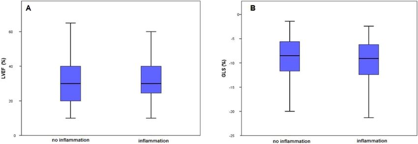
www.nature.com/scientificreports/
Figure 1. Study flow chart.
efore18. In case of right
via the right or left femoral artery or via trans-radial cardiac catheterization as reported b
heart biopsy, the femoral vein was used. Immediately after sampling the biopsy, the specimens were stabilized
in a solution to preserve ribonucleic acid (RNA) integrity (RNAlater, Ambion Inc., Austin, Texas) and were
sent for further examination to a specialized laboratory approved by Food and Drug Administration [Institut
Kardiale Diagnostik und Therapie (IKDT), Berlin, Germany]. For immunohistological evaluation, heart mus-
cle tissue probes fixated in RNAlater were embedded in Tissue Tec (SLEE, Mainz, Germany) and immediately
snap‐frozen in methyl butane which had been cooled in liquid nitrogen, and then stored at – 80 °C until pro-
cessing. Embedded specimens were cut serially into cryosections of 5 mm thickness and placed on 10% poly‐
l‐lysine‐pre‐coated slides. As part of a routinely performed work up in case of unclear heart failure histologi-
cal and immunohistochemical examination as well as virus detection was conducted. Using specific antibodies
inflammatory processes were detected by identifying immune cell infiltration and expression of cell adhesion
molecules. The specimens of the patients in this study were tested for CD3-positive lymphocytes (Dako; dilution
1:25), CD11a+/LFA-1+ lymphocytes (ImmunoTools; dilution 1:250), macrophages Mac-1 macrophages (Immu-
noTools; dilution 1:500), Perforin positive (cytotoxic T Cells) (clone dG9, BD Bioscience; dilution 1:150) and the
expression of adhesion molecules HLA-1 (Dako; dilution 1:2000) and ICAM-1 (ImmunoTools; dilution 1:800).
Myocardial inflammation was defined as ≥ 7.0 CD3+ lymphocytes/mm2 and/or ≥ 35.0 Mac1 macrophages/mm2
in accordance with previous published s tudies19. Diagnosis of relevant myocardial virus activity was performed
by polymerase chain reaction (PCR) detecting the genomic sequences of viruses that most commonly cause
myocarditis. This included: enterovirus, adenovirus, human cytomegalovirus, herpes simplex virus, Epstein–
Barr virus, human herpesvirus 6, parvovirus B19 and influenza A and B viruses. Virus load was calculated via
quantitative PCR methods. In case of relevant myocardial virus activity patients were excluded from analysis.
Statistical analysis. Descriptive statistics for relevant baseline comparisons of symptomatic non-valvular,
non-ischemic heart failure patients, who underwent EMB, stratified according inflammation (≥ 7.0 CD3+ lym-
phocytes/mm2 and/or ≥ 35.0 Mac1 macrophages/mm2) are provided as median and interquartile range (IQR) or
absolute numbers and corresponding percentages. We tested the continuous variables of the groups (inflamma-
tion vs. no inflammation) using the Mann–Whitney-U test and categorical variables with the Fisher’s exact or
the chi2 test, as appropriate.
Receiver operating characteristics (ROC) curves were calculated for TnI, BNP, CRP and GLS with regard
to myocardial inflammation and the area under the curve (AUC) is presented with the corresponding 95%
CI. Additionally, patient cohort-optimised cut-off values of these mentioned laboratory markers with regard
to myocardial inflammation were calculated based on ROC analyses using Youden index quantification. The
software SPSS (version 23.0; SPSS Inc., Chicago, Illinois) was used for computerized analysis. p values of < 0.05
(two-sided) were considered to be statistically significant.
Univariate and multivariate logistic regression models were analyzed to investigate predictors of myocardial
inflammation in heart failure patients, thereby using patient-optimised cut-off values derived from the ROC
analyses. Results are presented as odds ratio (OR) and 95%CI. The multivariate regression models were adjusted
for (1) age and sex, obesity and (2) cardiovascular risk factors (CVRF) including history of smoking, arterial
hypertension, diabetes mellitus and hyperlipoproteinaemia.
Results
Overall, 517 patients with symptomatic non-valvular, non-ischemic HF who underwent EMB between 2012 and
2018 were screened. Results from EMB with CD3+ and/or Mac1 cell count were available in 447 cases. 50 cases
were excluded due to relevant virus activity (Fig. 1). Thus, 397 patients remained in this study and were analysed.
Scientific Reports | (2021) 11:3008 | https://doi.org/10.1038/s41598-021-82592-8 3
Vol.:(0123456789)www.nature.com/scientificreports/
N 397
Age in years 54 [43/64]
Male 283 [71.3%, n = 397]
BMI in kg/m2 26.8 [24.0/30.0]
Comorbidities and risk factors
Diabetes mellitus 64 [16.1%, n = 397]
Hypertension 193 [48.6%, n = 397]
Alcohol 26 [6.6%, n = 394]
History of smoking 186 [47.1%, n = 395]
Hyperlipoproteinaemia 66 [16.7%; n = 396]
Symptoms at admission
NYHA I 88 [28.3%, n = 311]
NYHA II 87 [28.0%, n = 311]
NYHA III 83 [26.7%, n = 311]
NYHA IV 53 [17.0%, n = 311]
Angina pectoris 128 [32.4%; n = 395]
Cough 38 [9.6%; n = 395]
History of VTs 29 [7.3%; n = 392]
Common cold 86 [23.3%; n = 369]
Nausea 45 [11.4%, n = 395]
Oedema 76 [19.1%; n = 390]
Palpitation 55 [13.9%; n = 395]
Syncope 19 [4.8%; n = 395]
Time since onset
≤ 2 weeks 142 [36.4%; n = 390]
> 2 weeks, ≤ 3 months 118 [30.3%; n = 390}
> 3 months 127 [32.6%; n = 390]
Echocardiographic and parameters
LVEF in % 30.0 [20.0/40.0]
GLS in % -8.8 [-12.25/-6.0]
LVEDD in mm 5.9 [5.2/6.6]
LVEDP in mmHg 18 [12/26]
Laboratory measures
BNP in pg/ml 447 [137/1188.5]
CRP in mg/dl 4.5 [1.7/15.0]
TnI in pg/ml 24.9 [9.2/71.5]
Hb in g/dl 14.3 [13.1/15.4]
Immunhistochemistry
CD3+ Cells/mm2 6.470 [2.0/16.085]
Mac-1 Cells/mm2 33.6 [16.9/58.1]
CD11+ Cells/mm2 16.540 [8.8/32.105]
Perforin positive Cells/mm2 2.4 [1.01/5.2]
HLA1 in % AF 6.63 [5/8.70]
ICAM-1 Cells/mm2 2.4 [1.57/3.2]
Table 1. Baseline characteristics. Absolute and relative frequencies of echocardiographic markers,
cardiovascular risk factors and comorbiditis, symptoms at admission and laboratory parameters. BMI = Body
Mass Index kg/m2; LVEF = left ventricular ejection fraction in %; LVEDD = left ventricular enddiastolic
diameter in mm; LVEP = left ventricular enddiastolic pressure in mmHg; NYHA = New York Heart Associaton;
VTs = ventricular tachykardia; BNP = Brain natriuetic peptide in pg/ml; CRP = C-reactive protein in mg/l;
Hb = Haemoglobin in g/dl; TnI = Troponin I in pg/ml; AF = Area fraction
Patient characteristics are displayed in Table 1. In brief, median age was 54 years [IQR 43/64] and 71.3% of the
patients were males. Median left ventricular ejection fraction (LVEF) was calculated with 30.0% [20.0/40.0]
and median left ventricular enddiastolic diameter (LVEDD) with 5.9 mm [5.2/6.6] accompanied by an elevated
median BNP value of 447 pg/ml [137.0/1188.5]. GLS was reduced with − 8.8% [− 12.25/− 6.0]. Regarding symp-
toms, 71.7% patients were admitted with dyspnea (43.7% NYHA functional class III or IV), 32.4% complained
about angina pectoris at time of admission and in 19.1% edema were detectable. The duration of symptoms
Scientific Reports | (2021) 11:3008 | https://doi.org/10.1038/s41598-021-82592-8 4
Vol:.(1234567890)www.nature.com/scientificreports/
No Inflammation
N Inflammation (230) (167) p value
Age in years 54 [43/65] 54 [43/62] 0.795
Male 156 [67.8%; n = 230] 127 [76%; n = 167] 0.074
BMI in kg/m2 26.0 [23.0/30.0] 27.0 [24.0/30.70] 0.239
Comorbidities and risk factors
Diabetes mellitus 41 (17.8%; n = 230) 23 (13.8%; n = 167) 0.278
Hypertension 110 (47.8%; n = 230) 83 (49.7%; n = 167) 0.712
Alcohol 18 (7.9%; n = 228) 8 (4.8%; n = 166) 0.225
History of smoking 120 (52.4%; n = 229) 66 (39.8%; n = 166) 0.013
Hyperlipoproteinaemia 41 (17.8%; n = 230) 25 (15.1%; n = 166) 0.466
Symptoms at admission
NYHA > 2 79 (43.9%, n = 180) 57 (43.5%; n = 131) 0.947
Angina pectoris 73 (31.9%; n = 229) 55 (33.1%; n = 166) 0.793
Cough 25 (10.9%; n = 229) 13 (7.8%; n = 166) 0.305
History of VTs 19 (8.4%; n = 227 10 (6.1%; n = 165) 0.388
Common cold 51 (23.6%; n = 216) 35 (22.9%; n = 153) 0.869
Nausea 26 (11.4%; n = 229) 19 (11.4%; n = 166) 0.977
Oedema 46 (20.4%; n = 226) 30 (18.3%; n = 164) 0.612
Palpitations 30 (13.1%; n = 229) 25 (15.1%; n = 166) 0.579
Syncope 10 (4.4%; n = 229) 9 (5.4%; n = 166) 0.629
Echocardiographic measures
LVEF in % 30.0 [24.25/40.0] 30.0 [20.0/40.0] 0.712
GLS in % -9.1 [-12.4/-6.15] -8.5 [-11.8/-5.6] 0.239
LVEDD in mm 5.9 [5.025/6.575] 6.0 [5.3/6.625] 0.126
LVEDP in mmHg 19 [13/28] 17 [11.25/24.75] 0.230
Laboratory measures
BNP pg/ml 534.0 [155.0/1297.0] 364.5 [111.0/941.5] 0.116
TnI in pg/ml 29.0 [9.425/102.750] 22.8 [8.75/64.35] 0.265
CRP in mg/l 5.4 [2.1/19.0] 3.7 [1.5/10.0] 0.003
Hb in g/dl 14.1 [12.6/15.2] 14.7 [13.5/15.7] 0.001
Table 2. Comparison of patients with inflammation and no inflammation. Absolute and relative frequencies
of echocardiographic markers, cardiovascular risk factors and comorbiditis, symptoms at admission and
laboratory parameters. BMI = Body Mass Index kg/m2; LVEF = left ventricular ejection fraction in %;
GLS = global longitudinal strain in %; LVEDD = left ventricular enddiastolic diameter in mm; LVEDP = left
ventricular enddiastolic pressure in mmHg; NYHA = New York Heart Associaton; VTs = ventricular
tachykardia; BNP = Brain natriuretic peptide in pg/ml; CRP = C-reactive protein in mg/l; Hb = Hemoglobin in
g/dl; TnI = Troponin I in pg/ml. P-values are bold if they are below the significance level cut-off of 0.05.
differed: In 36.4% of the patients symptoms started not longer than 2 weeks, 30.3% had symptoms between
2 weeks and 3 months and 32.6% suffered from symptoms more than 3 months duration before admission. In
immunohistochemistry, median counts of C D3+ and Mac1 were 6.470/mm2 [IQR 2.0/16.085] and 33.6/mm2
[16.9/58.1), respectively, defining a cut-off of ≥ 7.0 CD3+ lymphocytes/mm2 and/or 35.0 Mac1 macrophages/mm2.
Comparison of symptomatic non‑valvular, non‑ischemic heart failure patients, who under‑
went EMB, stratified for presence of myocardial inflammation. In 230 patients (57.9%), myocar-
dial inflammation was diagnosed. Patients with and without inflammatory cardiomyopathy were of comparable
age and revealed no differences regarding presented symptoms at admission. History of smoking (52.4% vs.
39.8%, p = 0.013) was more prevalent in HF patients with myocardial inflammation. While the groups did not
differ regarding mean values of LVEF, GLS (Table 2, Fig. 2) and mean values of TnI and BNP (Table 2, Fig. 3),
mean values of CRP (5.4 [2.1/19] vs. 3.7 [1.5/10], p = 0.003) were higher and median hemoglobin levels (14.1
[12.6/15.2] vs. 14.7 (13.5/15.7] g/dl, p = 0.001) were lower in patients with myocardial inflammation (Table 2,
Fig. 3).
Predictors of myocardial inflammation. ROC curves demonstrated only a moderate prognostic per-
formance of CRP for prediction of myocardial inflammation with acceptable specificity and positive predictive
values (Table 3, supplementary Figure 1). The computed best cut-off (according Youden-Index calculation) for
CRP was 8.15 mg/l. In contrast, TnI, BNP and GLS were accompanied by a low prognostic performance to pre-
dict myocardial inflammation shown in the ROC curves. The computed best cut-off (according Youden-Index
calculation) for TnI was 136.5 pg/ml, for BNP 1030 pg/ml and for GLS -13.95% (Table 3). Nevertheless, when
Scientific Reports | (2021) 11:3008 | https://doi.org/10.1038/s41598-021-82592-8 5
Vol.:(0123456789)www.nature.com/scientificreports/
Figure 2. Boxplots showing 1st quartile, median and 3rd quartile values of: (A) LVEF = left ventricular ejection
fraction in % (not significant); (B) GLS = global longitudinal strain in % (not significant);
Figure 3. Boxplots showing 1st quartile, median and 3rd quartile values of: (A) Troponin I in pg/ml (not
significant); (B) Brain natriuretic peptide (BNP) in pg/ml (not significant); (C) C-reactive protein (CRP) in mg/l
(p = 0.003); (D) Hemoglobin (Hb) in g/dl (p = 0.001).
Scientific Reports | (2021) 11:3008 | https://doi.org/10.1038/s41598-021-82592-8 6
Vol:.(1234567890)www.nature.com/scientificreports/
Best cut-off Sensitivity (95% Specificity (95%
Parameter (Youden index) AUC (95% CI) p value CI) CI) PPV (95% CI) NPV (95% CI)
TnI 136.5 pg/ml 0.54 (0.47–0.60) 0.267 0.21 (0.16–0.27) 0.91 (0.85–0.95) 0.78 (0.66–0.87) 0.43 (0.37–0.48)
BNP 1030.5 pg/ml 0.56 (0.49–0.63) 0.116 0.34 (0.27–0.42) 0.79 (0.71–0.86) 0.70 (0.59–0.79) 0.45 (0.38–0.52)
CRP 8.15 mg/l 0.59 (0.53–0.65) 0.003 0.43 (0.37–0.50) 0.71 (0.64–0.78) 0.68 (0.60–0.75) 0.47 (0.41–0.53)
GLS − 13.95% 0.46 (0.38–0.53) 0.239 0.88 (0.81–0.92) 0.14 (0.08–0.22) 0.58 (0.51–0.64) 0.45 (0.29–0.62)
Table 3. Prognostic performance of TnI, BNP, CRP and GLS for prediction of myocardial inflammation.
P-values are bold if they are below the significance level cut-off of 0.05.
Adjustment for
crude + Age/Sex/Obesity + Age/Sex/Obesity/CVRF
OR [95% CI] p value OR [95% CI] p value OR [95% CI] p value
Age > 70 in years 1.2 [0.683–2.108] 0.525 1.042 [0.522–2.080] 0.907 1.127 [0.533–2.382] 0.754
Male 0.664 [0.423–1.042] 0.075 0.701 [0.408–1.205] 0.199 0.652 [0.373–1.140] 1.333
BMI > 30 kg/m2 0.943 [0.553–1.605] 0.828 0.940 [0.547–1.615] 0.823 0.930 [0.528–1.638] 0.802
Comorbidities and risk factors
Diabetes 1.358 [0.780–2.365] 0.279 1.709 [0.833–3.506] 0.144 1.843 [0.882–3.848] 0.104
Hypertension 0.928 [0.623–1.282] 0.712 0.763 [0.458–1.272] 0.300 0.685 [0.405–1.158] 0.157
Alcohol 1.693 [0.718–3.993] 0.229 1.353 [0.479–3.818] 0.568 1.532 [0.488–4.804] 0.465
History of Smoking 1.668 [1.113–2.500] 0.013 1.537 [0.938–2.520] 0.088 1.659 [1.000–2.753] 0.050
Hyperlipoproteinaemia 1.223 [0.711–2.106] 0.467 1.241 [0.646–2.386] 0.517 1.315 [0.667–2.590] 0.429
NYHA > 2 1.015 [0.645–1.599] 0.947 1.364 [0.811–2.294] 0.242 1.469 [0.857–2.518] 0.162
Echocardiographic measures
LVEF ≥ 50% 0.966 [0.545–1.711] 0.906 1.175 [0.604–2.285] 0.635 1.092 [0.555–2.149] 0.798
LVEF 40–49% 0.957 [0.466–1.963] 0.904 0.703 [0.302–1.638] 0.415 0.678 [0.271–1.700] 0.408
LVEF < 40% 1.081 [0.702–1.664] 0.724 1.077 [0.645–1.800] 0.776 1.116 [0.659–1.888] 0.683
GLS ≥ -13.95% 1.126 [0.527–2.407] 0.759 1.396 [0.569–3.423] 0.466 1.425 [0.573–3.542] 0.446
LVEDD > 47.6 mm 0.455 [0.222–0.934] 0.032 0.570 [0.246–1.317] 0.188 0.611 [0.262–1.424] 0.253
LVEDP > 15 mmHg 1.075 [0.681–1.696] 0.756 1.072 [0.608–1.888] 0.811 1.056 [0.588–1.895] 0.856
Laboratory measures
BNP ≥ 1030.5 pg/ml 1.928 [1.084–3.428] 0.025 1.837 [0.904–3.735] 0.093 1.811 [0.873–3.757] 0.111
Troponin I ≥ 136.5 pg/ml 2.660 [1.345–5.260] 0.005 3.121 [1.291–7.547] 0.012 3.011 [1.215–7.464] 0.017
CRP ≥ 8.15 mg/l 1.899 [1.238–2.911] 0.003 2.009 [1.191–3.386] 0.009 1.985 [1.160–3.397] 0.012
Table 4. Predictors of inflammation. OR (Odds Ratio), 95% CI (Confidence intervalls) and p values in a
crude and multivariate logistic regression analysis. BMI = Body Mass Index kg/m2; LVEF = left ventricular
ejection fraction in %; GLS = global longitudinal strain in %; LVEDD = left ventricular enddiastolic diameter
in mm; LVEDP = left ventricular enddiastolic pressure in mmHg; NYHA = New York Heart Associaton;
BNP = Brain natriuetic peptide in pg/ml; CRP = C-reactive protein in mg/l. P-values are bold if they are below
the significance level cut-off of 0.05.
calculating best cut-offs (according Youden-Index calculation), the cut-offs were able to differentiate between
myocardial inflammation vs. no cardiac inflammation.
In multivariate logistic regression analyses after adjustment for CVRF, elevated TnI values (≥ 136.5 pg/ml)
(OR 3.011 [95%CI 1.215–7.464]; p = 0.017) as well as elevated CRP values (≥ 8.15 mg/l) (OR 1.985 [95%CI
1.160–3.397], p = 0.012) were independently associated with myocardial inflammation. No association was
detected for elevated BNP (≥ 1030.5 pg/ml) values (OR 1.811 [95%CI 0.873–3.757]; p = 0.111). Regarding echo-
cardiographic parameters, LVEF categories and reduced GLS (≥ − 13.95%) were not associated with myocar-
dial inflammation (Table 4, Fig. 4). However when combining laboratory parameters with echocardiographic
parameters, a strong association with myocardial inflammation was found for the combined variable consisting
of reduced GLS and elevated TnI (OR 9.633 [95%CI 2.027–45.769]; p = 0.004). Additionally, but to a smaller
extent, the combination of TnI and CRP was also independently associated with myocardial inflammation (OR
5.761 [95% 1.240–26.771], p = 0.025) (Supplementary table 1). Furthermore, supplementary table 2 illustrates
the prognostic performance of combined parameters to predict myocardial inflammation. While all parameter-
combinations had only moderate sensitivity (21–39%), the specificity was better: 0.71 (0.62–0.79) for GLS and
elevated CRP, 0.95 (0.91–0.98) for positive TnI and elevated CRP and 0.96 (0.90–0.99) for the combination of
GLS and TnI. Remarkably, highest positive predictive value was also detected for the combination of GLS and
TnI 0.90 (0.74–0.96). The negative predictive values were only moderate in all of these combinations (41–46%).
Scientific Reports | (2021) 11:3008 | https://doi.org/10.1038/s41598-021-82592-8 7
Vol.:(0123456789)www.nature.com/scientificreports/
Figure 4. Predictors for inflammation (Forest plot). OR (Odds Ratio) and 95% CI (Confidence Intervall)
displayed in a Forest plot after adjustment for age/sex/CVRF. Absolute values are shown in Table 4.
Concerning the interrelation of cardiovascular risk factors with myocardial inflammation, only smoking was
associated with myocardial inflammation in crude logistic regression analysis (OR 1.6687 [95%CI 1.113–2.500];
p = 0.013), but this association showed only a trend towards significance in the fully adjusted model (OR 1.659
[95%CI 1.000–2.753]; p = 0.050) (Table 4, Fig. 4).
Discussion
The key findings of the present study are that (I) elevated CRP and TnI levels are associated with inflammatory
cardiomyopathy in patients with non-ischemic, non valvular heart failure; (II) TnI levels ≥ 136.5 pg/ml and CRP
levels ≥ 8.15 mg/l are potential predictors of myocardial inflammation; (III) common heart failure biomarkers
such as BNP and reduced LVEF were not predictive for myocardial inflammation and; (IV) GLS might add a
benefit to predict myocardial inflammation when combined with TnI.
A growing body of evidence indicates that myocardial inflammation may lead to severe cardiac damage,
deteriorate into dilated cardiomyopathy and be accompanied by poor prognosis. This implies that early diagnosis
using predictive markers may avert poor outcome20, since immunosuppressive therapy can significantly improve
the prognosis of HF patients with presence of myocardial inflammation16. Thus, readily available markers like
the ones identified in our study might help to select patients for EMB and for shorter control follow-up exami-
nations. Although cardiac MRI diagnostics has become increasingly important in the diagnosis of myocardial
inflammation21,22, sensitivity and specificity of MRI findings are not unequivocal. Since EMB can be performed
easily as part of coronary catheter examination18 and should be carried out as early as possible if there is clini-
cal or laboratory suspicion of inflammatory myocardial disease, our study results might help to accelerate the
diagnostic work-up of HF.
The association of elevated CRP and TnI with myocardial inflammation was independent of age and sex,
supporting earlier findings by Lauer et al., who reported that patients with elevated troponin T values were more
likely to have EMB-proven myocarditis23. Because cardiac troponins are detectable in the blood for approximately
one week24 and have a half-life of 2h25, elevated troponins are suitable to indicate permanent myocardial cell
injury26,27, which can be explained by inflammatory infiltration with consecutive cell damage documented in
the EMB. Contrary to our results, Sramko et al. demonstrated that troponin was not able to distinguish between
patients with idiopathic dilated cardiomyopathy or inflammatory dilated cardiomyopathy arguing that troponin
indicates myocardial injury without indicating weather its due to myocardial inflammation or simply caused
by myocardial wallstress related to heart f ailure28. However, the study sample size was small, and EMB was only
performed on the right ventricle making a sampling error more likely.
Scientific Reports | (2021) 11:3008 | https://doi.org/10.1038/s41598-021-82592-8 8
Vol:.(1234567890)www.nature.com/scientificreports/
Similar to positive troponin values, positive CRP values were associated with detection of inflammation in
EMB. These results go in line with observation by Liu et al., who demonstrated that highly sensitive CRP levels
were higher in patients with EMB-confirmed myocarditis than in patients without myocarditis29. However, it has
to be pointed out that the sensitivity for highly sensitive CRP in detecting myocarditis was only 50.1% whereas
the specificity was 80.7%. Since CRP is also increased in acute myocardial infarction30 or in rheumatic valve
disease31. CRP should not be interpreted alone, but in conjunction with cardiac troponin. Moreover, as the pre-
sent study indicates, cut-off values for TnI and CRP seem to be higher with regard to myocardial inflammation
compared to cut-off values used for diagnosis of acute coronary syndrom. Further studies are needed to prove
those cut-off values in patients with myocardial inflammation.
Interestingly GLS was not able to distinguish between EMB proven myocardial inflammation and no inflam-
mation. Escher et al. demonstrated before, that patients with acute myocarditis had reduced GLS values. After a
follow-up period of 6.2 months patients with persistent inflammation had worse GLS values than patients with-
out inflammation32. However in contrast to our study GLS values were compared when LVEF had already been
improved. Eventhough it has been shown that GLS is a superior predictor of adverse cardiac events compared
with LVEF33,34, studies demonstrate that in general and irrespectively of outcome prediction, GLS correlates
well with LVEF in heart failure patients35. Therefore the results of our study suggest that GLS alone is not able to
identify patients with myocardial inflammation in the presence of reduced LVEF. Nevertheless, the combined
evaluation of GLS and Troponin values were the second strongest predictor of myocardial inflammation in
multivariate regression analysis. Additionally, the best prognostic performance was found for the combination
of GLS and Troponin. In particular, the high positive prognostic value of 90% to predict myocardial inflamma-
tion is promising. Thus, the combination of GLS and Troponin may help to identify patients with myocardial
inflammation in the future and could add a benefit in the diagnosis and follow up treatment of those patients.
Interestingly, smoking was associated with myocardial inflammation in our study. In a very small study
sample, it was shown, that in patients hospitalized for acute myocarditis smoking was the most prevalent car-
diovascular risk f actor36. Recently, Detorakis et al. reported a significant correlation of smoking habits with late
gadolinium enhancement extent in cardiac MRI in patients with clinically suspected m yocarditis37. Prolonged
exposure to tobacco smoke may cause cardiovascular cell damage, suggesting that increased myocardial cell
necrosis and cell death would make myocardial inflammatory infiltration more l ikely38.
Another finding of the present study was that patients with myocardial inflammation had lower hemoglobin
levels. It is well known that anemia of inflammation or anemia of chronic disease is primarily a disorder of iron
distribution39 and even low grade inflammation is associated with lower hemoglobin levels40. As experimental
models have demonstrated that low cardiac iron levels promote heart f ailure41,42 the link between the observed
lower hemoglobin levels could be an effect of possibly local iron distribution disorders due to myocardial inflam-
mation. Since hemoglobin levels in the present study were generally not significantly decreased, more data are
needed to support this observation.
The primary stimulus for natriuretic peptide synthesis by myocardial cells is related to increased LV wall
stress induced for example by acute myocardial infarction43, arterial hypertension44, muscle hypertrophy45 or
increased pulmonary arterial p ressure46. We expected that inflammation would also cause elevated BNP levels as
reported earlier , particularly in patients with myocarditis and dilated cardiomyopathy48–50. Increase of BNP has
47
also been detected in systemic inflammation regardless of systolic f unction51. In the present study, no differences
in BNP levels and LVEF were found in patients with and without myocardial inflammation, reflecting a rather
compensated disease state of our study population with low rates of hydropic decompensation. Myocarditis and
inflammatory cardiomyopathy are a dynamic processes; after inflammation has subsided, a reduced LVEF is due
to secondary post-inflammatory fibrosis and myocardial cell death and scars52. As conditions with and without
inflammation lead to changes of LVEF and BNP, the present study demonstrates that increase of BNP and the
decrease of LVEF do not specifically reflect the presence of inflammation, but rather reflect a general loss of
myocardial tissue with consecutive changes in hemodynamic conditions regardless weather inflammation can
be detected or not.
Limitation
There are certain limitations of our study which need to be mentioned: Firstly, our study was conducted as a
monocentric retrospective data analysis. Therefore, it has to be emphasized that the results of this study can
only serve in terms of a hypothesis generating research and the results of the present study should be verified
by prospective trials. Secondly, there is no generally applicable definition of myocardial inflammation. Thus, it
might be difficult to adopt the results to other endomyocardial biopsy studies, but generating more information
about inflammation process was one of the main aims of our study. Finally, a potential limitation regarding
EMB interpretation might be the sampling error. Regarding this point, Hauck et al. demonstrated that sampling
error was prevalent 45% in Ieft and up to 37% in right E MB53. The authors conclude that only positive results of
EMB can be considered diagnostic. However, the cited study included a very small sample of only 36 patients.
Conclusion
Elevations of readily available markers like cardiac TnI and CRP as well as the combination of GLS and TnI may
predict inflammatory cardiomyopathy and might be useful to select patients for EMB and for shorter control
follow-up examinations. In the diagnostic approach to detect suspected myocardial inflammation, higher cut-
off values concerning cardiac and inflammatory biomarkers may have to be applied. Smoking is associated with
inflammatory cardiomyopathy in non-ischemic, non valvular HF patients and may indicate individuals at risk
to develop inflammatory cardiomyopathy.
Scientific Reports | (2021) 11:3008 | https://doi.org/10.1038/s41598-021-82592-8 9
Vol.:(0123456789)www.nature.com/scientificreports/
Ethics approval
All data were obtained from individuals enrolled between 2013 and 2018 in the retrospective monocentric Mainz
Endomyocardial Biopsy in Heart Failure Study (My Biopsy-HF Study, DRKS #22178), which was approved by
the Ethics Committee of Rhineland Palatinate to be in accordance with the legal regulations and the declaration
of Helsinki.
Received: 28 May 2020; Accepted: 18 January 2021
References
1. Vos, T. et al. Years lived with disability (YLDs) for 1160 sequelae of 289 diseases and injuries 1990–2010: a systematic analysis for
the Global Burden of Disease Study 2010. Lancet 380, 2163–2196 (2012).
2. Lloyd-Jones, D. M. et al. Lifetime risk for developing congestive heart failure: the Framingham Heart Study. Circulation 106,
3068–3072 (2002).
3. Ponikowski, P. et al. 2016 ESC Guidelines for the diagnosis and treatment of acute and chronic heart failure. Eur. Heart J. 37,
2129–2200 (2016).
4. Gheorghiade, M. & Bonow, R. O. Chronic heart failure in the United States. Circulation 97, 282–289 (1998).
5. Cowie, M. R. et al. Incidence and aetiology of heart failure; a population-based study. Eur. Heart J. 20, 421–428 (1999).
6. Uretsky, B. F. et al. Acute coronary findings at autopsy in heart failure patients with sudden death: results from the assessment of
treatment with lisinopril and survival (ATLAS) trial. Circulation 102, 611–616 (2000).
7. Richardson, P. et al. Report of the 1995 World Health Organization/International Society and Federation of Cardiology Task Force
on the Definition and Classification of Cardiomyopathies. Circulation 93, 841–842 (1996).
8. Basso, C., Calabrese, F., Corrado, D. & Thiene, G. Postmortem diagnosis in sudden cardiac death victims: macroscopic, microscopic
and molecular findings. Cardiovasc. Res. 50, 290–300 (2001).
9. Felker, G. M. et al. Idiopathic cardiomyopathy is less idiopathic than you think: An endomyocardial biopsy study of 1278 patients.
J. Card. Fail. 5, 63 (1999).
10. Sotiriou, E. et al. Therapeutic implications of a combined diagnostic workup including endomyocardial biopsy in an all-comer
population of patients with heart failure: a retrospective analysis. ESC Heart Fail 5, 630–641 (2018).
11. Holzmann, M. et al. Complication rate of right ventricular endomyocardial biopsy via the femoral approach. Circulation 118,
1722–1728 (2008).
12. Caforio, A. L. P. et al. A prospective study of biopsy-proven myocarditis: prognostic relevance of clinical and aetiopathogenetic
features at diagnosis. Eur. Heart J. 28, 1326–1333 (2007).
13. Cooper, L. T. et al. Usefulness of immunosuppression for giant cell myocarditis. Am. J. Cardiol. 102, 1535–1539 (2008).
14. Mason, J. W. et al. A clinical trial of immunosuppressive therapy for myocarditis. The Myocarditis Treatment Trial Investigators.
N. Engl. J. Med. 333, 269–275 (1995).
15. Wojnicz, R. et al. Randomized, placebo-controlled study for immunosuppressive treatment of inflammatory dilated cardiomyo-
pathy: two-year follow-up results. Circulation 104, 39–45 (2001).
16. Frustaci, A., Russo, M. A. & Chimenti, C. Randomized study on the efficacy of immunosuppressive therapy in patients with virus-
negative inflammatory cardiomyopathy: the TIMIC study. Eur. Heart J. 30, 1995–2002 (2009).
17. Cooper, L. T. et al. The role of endomyocardial biopsy in the management of cardiovascular disease: a Scientific Statement from the
American Heart Association, the American College of Cardiology, and the European Society of Cardiology Endorsed by the Heart
Failure Society of America and the Heart Failure Association of the European Society of Cardiology. Eur. Heart J. 28, 3076–3093
(2007).
18. Schulz, E. et al. Feasibility and safety of left ventricular endomyocardial biopsy via transradial access: technique and initial experi-
ence. Catheter. Cardiovasc. Interv. 86, 761–765 (2015).
19. Escher, F. et al. Presence of perforin in endomyocardial biopsies of patients with inflammatory cardiomyopathy predicts poor
outcome. Eur. J. Heart Fail. 16, 1066–1072 (2014).
20. Feldman, A. M. & McNamara, D. Myocarditis. N. Engl. J. Med. 343, 1388–1398 (2000).
21. Lurz, P. et al. Comprehensive cardiac magnetic resonance imaging in patients with suspected myocarditis: the MyoRacer-Trial. J.
Am. Coll. Cardiol. 67, 1800–1811 (2016).
22. Cheitlin, M. D. Diagnostic performance of cardiovascular magnetic resonance in patients with suspected acute myocarditis. Yearb.
Cardiol. 2006, 376–378 (2006).
23. Lauer, B. et al. Cardiac troponin T in patients with clinically suspected myocarditis. J. Am. Coll. Cardiol. 30, 1354–1359 (1997).
24. Cummins, B. & Cummins, P. Cardiac specific troponin-I release in canine experimental myocardial infarction: development of a
sensitive enzyme-linked immunoassay. J. Mol. Cell. Cardiol. 19, 999–1010 (1987).
25. Katus, H. A., Remppis, A., Scheffold, T., Diederich, K. W. & Kuebler, W. Intracellular compartmentation of cardiac troponin T and
its release kinetics in patients with reperfused and nonreperfused myocardial infarction. Am. J. Cardiol. 67, 1360–1367 (1991).
26. Park, K. C., Gaze, D. C., Collinson, P. O. & Marber, M. S. Cardiac troponins: from myocardial infarction to chronic disease. Car-
diovasc. Res. 113, 1708–1718 (2017).
27. Hoff, J., Wehner, W. & Nambi, V. Troponin in cardiovascular disease prevention: updates and future direction. Curr. Atheroscler.
Rep. 18, 12 (2016).
28. Šramko, M. et al. Utility of combination of cardiac magnetic resonance imaging and high-sensitivity cardiac troponin T assay in
diagnosis of inflammatory cardiomyopathy. Am. J. Cardiol. 111, 258–264 (2012).
29. Liu, Y. et al. Application value of hypersensitive C-reactive protein, lactic acid and myoglobin in the combined detection of myo-
carditis. Exp. Ther. Med. 17, 4471–4476 (2019).
30. Berton, G. et al. C-reactive protein in acute myocardial infarction: association with heart failure. Am. Heart. J. 145, 1094–1101
(2003).
31. Gölbasi, Z. et al. Increased levels of high sensitive C-reactive protein in patients with chronic rheumatic valve disease: evidence
of ongoing inflammation. Eur. J. Heart Fail. 4, 593–595 (2002).
32. Escher, F. et al. New echocardiographic findings correlate with intramyocardial inflammation in endomyocardial biopsies of
patients with acute myocarditis and inflammatory cardiomyopathy. Mediat. Inflamm. 2013, 875420 (2013).
33. Iacoviello, M. et al. Independent role of left ventricular global longitudinal strain in predicting prognosis of chronic heart failure
patients. Echocardiography 30, 803–811 (2013).
34. Stanton, T., Leano, R. & Marwick, T. H. Prediction of all-cause mortality from global longitudinal speckle strain: comparison with
ejection fraction and wall motion scoring. Circ. Cardiovasc. Imaging 2, 356–364 (2009).
35. Delgado, V. et al. Relation between global left ventricular longitudinal strain assessed with novel automated function imaging and
biplane left ventricular ejection fraction in patients with coronary artery disease. J. Am. Soc. Echocardiogr. 21, 1244–1250 (2008).
Scientific Reports | (2021) 11:3008 | https://doi.org/10.1038/s41598-021-82592-8 10
Vol:.(1234567890)www.nature.com/scientificreports/
36. Antit, S. et al. Diagnostic characteristics of acute myocarditis. Tunis. Med. 97, 789–794 (2019).
37. Detorakis, E., Illing, R., Lasithiotaki, I., Foukarakis, E. & Raissaki, M. Role of smoking in the evolution of cardiovascular magnetic
resonance and laboratory findings of acute myocarditis. Heart Views Off. J. Gulf Heart Assoc. 21, 22–30 (2020).
38. Leone, A. Jr., Biadi, O. & Balbarini, A. Smoking and cardiovascular system: cellular features of the damage. Curr. Pharm. Des. 14,
1771–1777 (2008).
39. Ganz, T. Anemia of inflammation. N. Engl. J. Med. 381, 1148–1157 (2019).
40. Kotzé, S. R. et al. Low-grade inflammation is associated with lower haemoglobin levels in healthy individuals: results from the
Danish blood donor study. Vox Sang 111, 144–150 (2016).
41. Lakhal-Littleton, S. et al. An essential cell-autonomous role for hepcidin in cardiac iron homeostasis. Elife 5, e19804 (2016).
42. Xu, W. et al. Lethal cardiomyopathy in mice lacking transferrin receptor in the heart. Cell Rep. 13, 533–545 (2015).
43. Morita, E. et al. Increased plasma levels of brain natriuretic peptide in patients with acute myocardial infarction. Circulation 88,
82–91 (1993).
44. Buckley, M. G., Markandu, N. D., Miller, M. A., Sagnella, G. A. & MacGregor, G. A. Plasma concentrations and comparisons of
brain and atrial natriuretic peptide in normal subjects and in patients with essential hypertension. J. Hum. Hypertens. 7, 245–250
(1993).
45. Lukowicz, T. V. et al. BNP as a marker of diastolic dysfunction in the general population: importance of left ventricular hypertrophy.
Eur. J. Heart Fail. 7, 525–531 (2005).
46. Nagaya, N. Plasma brain natriuretic peptide levels increase in proportion to the extent of right ventricular dysfunction in pulmo-
nary hypertension. J. Am. Coll. Cardiol. 31, 156A (1998).
47. George, J., Mackle, G., Manoharan, A., Khan, F. & Struthers, A. D. High BNP levels in rheumatoid arthritis are related to inflam-
mation but not to left ventricular abnormalities: a prospective case-control study. Int. J. Cardiol. 172, e116–e118 (2014).
48. Ogawa, T., Veinot, J. P., de Bold, M. L. K., Georgalis, T. & Bold, A. J. Angiotensin II receptor antagonism reverts the selective cardiac
BNP upregulation and secretion observed in myocarditis. Am. J. Physiol. Heart Circ. Physiol. 294, H2596–H2603 (2008).
49. Grabowski, M. et al. Diagnostic value of BNP in suspected perimyocarditis–a preliminary report. Kardiol Pol 61, 451–458 (2004).
50. Miller, W. L., Hartman, K. A., Burritt, M. F., Burnett, J. C. & Jaffe, A. S. Troponin, B-type natriuretic peptides and outcomes in
severe heart failure: differences between ischemic and dilated cardiomyopathies. Clin. Cardiol. 30, 245–250 (2007).
51. Shor, R. et al. BNP in septic patients without systolic myocardial dysfunction. Eur. J. Intern. Med. 17, 536–540 (2006).
52. Imanaka-Yoshida, K. Inflammation in myocardial disease: from myocarditis to dilated cardiomyopathy. Pathol. Int. https://doi.
org/10.1111/pin.12868(2019).
53. Hauck, A. J., Kearney, D. L. & Edwards, W. D. Evaluation of postmortem endomyocardial biopsy specimens from 38 patients with
lymphocytic myocarditis: implications for role of sampling error. Mayo Clin. Proc. 64, 1235–1245 (1989).
Acknowledgements
This manuscript contains results that are part of the medical doctoral theses work of Friederike Fueting and
Carolin Klank.
Author contributions
S.S.-T.: designed the study, analyzed and interpreted data, wrote the manuscript. P.W.: designed the study, ana-
lyzed and interpreted data, edited the manuscript. K.K.: designed the study, analyzed and interpreted data, edited
the manuscript. S.G.: designed the study. E.T.: analyzed and interpreted data. V.H.S.: critically commented and
edited the manuscript. F.F.: analyzed and interpreted data. C.K.: analyzed and interpreted data. F.E.: critically
commented and edited the manuscript. H.P.S.: critically commented and edited the manuscript. T.M.: critically
commented and edited the manuscript.
Funding
Open Access funding enabled and organized by Projekt DEAL. P.W. was supported by grants from the German
Federal Ministry for Education and Research (BMBF 01EO1503). T.M. and P.W. are Principal Investigators of
the German Center for Cardiovascular Research (DZHK).
Competing interests
The authors declare no competing interests.
Additional information
Supplementary Information The online version contains supplementary material available at https://doi.
org/10.1038/s41598-021-82592-8.
Correspondence and requests for materials should be addressed to P.W.
Reprints and permissions information is available at www.nature.com/reprints.
Publisher’s note Springer Nature remains neutral with regard to jurisdictional claims in published maps and
institutional affiliations.
Open Access This article is licensed under a Creative Commons Attribution 4.0 International
License, which permits use, sharing, adaptation, distribution and reproduction in any medium or
format, as long as you give appropriate credit to the original author(s) and the source, provide a link to the
Creative Commons licence, and indicate if changes were made. The images or other third party material in this
article are included in the article’s Creative Commons licence, unless indicated otherwise in a credit line to the
material. If material is not included in the article’s Creative Commons licence and your intended use is not
permitted by statutory regulation or exceeds the permitted use, you will need to obtain permission directly from
the copyright holder. To view a copy of this licence, visit http://creativecommons.org/licenses/by/4.0/.
© The Author(s) 2021
Scientific Reports | (2021) 11:3008 | https://doi.org/10.1038/s41598-021-82592-8 11
Vol.:(0123456789)You can also read