Myocardial Work Efficiency, A Novel Measure of Myocardial Dysfunction, Is Reduced in COVID-19 Patients and Associated With In-Hospital Mortality ...
←
→
Page content transcription
If your browser does not render page correctly, please read the page content below
ORIGINAL RESEARCH
                                                                                                                                              published: 14 June 2021
                                                                                                                                      doi: 10.3389/fcvm.2021.667721

                                            Myocardial Work Efficiency, A Novel
                                            Measure of Myocardial Dysfunction,
                                            Is Reduced in COVID-19 Patients and
                                            Associated With In-Hospital Mortality
                                            Anum S. Minhas 1† , Nisha A. Gilotra 1† , Erin Goerlich 1 , Thomas Metkus 1 , Brian T. Garibaldi 2 ,
                                            Garima Sharma 1 , Nicole Bavaro 1 , Susan Phillip 1 , Erin D. Michos 1 and Allison G. Hays 1*
                                            1
                                             Division of Cardiology, Department of Medicine, Johns Hopkins University School of Medicine, Baltimore, MD,
                                            United States, 2 Division of Pulmonary and Critical Care Medicine, Department of Medicine, Johns Hopkins University School
                          Edited by:
                                            of Medicine, Baltimore, MD, United States
                        Mingxing Xie,
  Huazhong University of Science and
                  Technology, China
                                            Background: Although troponin elevation is common in COVID-19, the extent of
                       Reviewed by:
                                            myocardial dysfunction and its contributors to dysfunction are less well-characterized.
                        Lígia Mendes,
     Hospital da Luz Setúbal, Portugal      We aimed to determine the prevalence of subclinical myocardial dysfunction and its
                   Alessandro Faragli,      association with mortality using speckle tracking echocardiography (STE), specifically
              Deutsches Herzzentrum
                      Berlin, Germany
                                            global longitudinal strain (GLS) and myocardial work efficiency (MWE). We also tested
                   *Correspondence:
                                            the hypothesis that reduced myocardial function was associated with increased systemic
                       Allison G. Hays      inflammation in COVID-19.
                     ahays2@jhmi.edu
                                            Methods and Results: We conducted a retrospective study of hospitalized COVID-19
     † These   authors have contributed     patients undergoing echocardiography (n = 136), of whom 83 and 75 had GLS (abnormal
                    equally to this work
                                            >−16%) and MWE (abnormal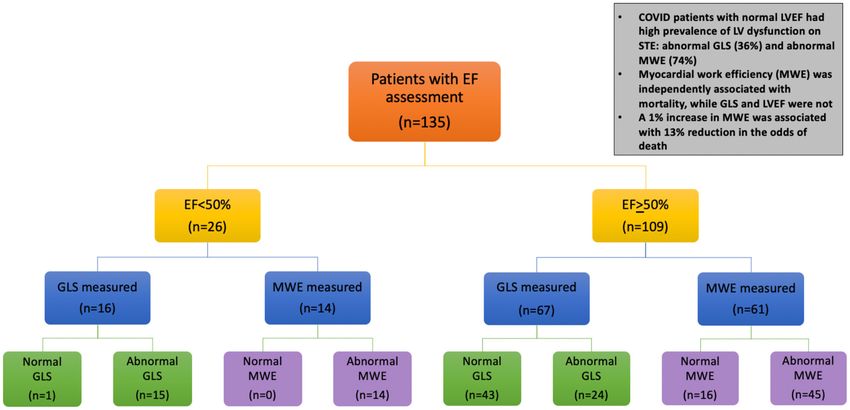
Minhas et al.                                                                                                         Subclinical Myocardial Dysfunction in COVID-19

INTRODUCTION                                                                               METHODS
COVID-19, the disease caused by the novel coronavirus                                      Study Design
SARS-CoV2, carries high acute cardiovascular morbidity and                                 This retrospective, single-center cohort study included 136
mortality (1, 2). The mechanisms for cardiac injury are not                                consecutive patients with confirmed COVID-19 who were
fully understood, with hypotheses ranging from systemic                                    hospitalized at Johns Hopkins Hospital and underwent clinically
inflammation due to cytokine release syndrome, angiotensin                                 indicated transthoracic echocardiogram between March 25, 2020
converting enzyme-2 mediated direct viral myocardial toxicity,                             and May 19, 2020, with follow-up completed by June 22,
autoimmune myocarditis, and sympathetic stress response                                    2020. All echocardiograms were ordered by the patient’s clinical
(1, 3). Over 25% of hospitalized COVID-19 patients have                                    care team. The study was approved by the Johns Hopkins
acute cardiac injury as detected by elevated cardiac troponin,                             Institutional Review Board and informed consent was waived per
associated with greater in-hospital mortality (1, 3, 4). However,                          IRB guidelines.
troponin alone has limited specificity and sensitivity in
myocarditis and can also rise in acute respiratory distress                                Clinical Data
syndrome (ARDS), another recognized complication of                                        Patient characteristics, including demographics, medical history,
COVID-19 (5–7). Additionally, although multiple inflammatory                               clinical presentation, laboratory testing, and clinical outcomes
pathways, such as interleukin-6 (IL-6), are implicated in                                  were extracted from the electronic medical record. Initial
myocardial injury in COVID-19, their effect on indices of                                  values after admission for the following serum biomarkers were
cardiac function is unknown and a better understanding                                     collected: cardiac troponin I, IL-6, C-reactive protein (CRP),
of the degree and determinants of myocardial function                                      ferritin, fibrinogen, and d-dimer. In- hospital all-cause mortality
may improve risk stratification and lead to new therapeutic                                during index hospitalization was ascertained from electronic
approaches (8–10).                                                                         medical records through the end of follow-up. Two separate
    Studies examining the degree of myocardial dysfunction                                 investigators independently reviewed the data.
in COVID-19 are limited, and assessment with cardiac
imaging has been challenging due to exposure concerns                                      Transthoracic Echocardiography
to echocardiography staff. Thus, it is likely that the true                                Conventional 2D Echocardiographic Analysis
prevalence of cardiac dysfunction is underreported (11).                                   Bedside transthoracic echocardiographic (TTE) examinations
Speckle tracking echocardiography (STE) can rapidly quantify                               were performed by experienced sonographers using VividTM E95
myocardial dysfunction (e.g., using global longitudinal strain                             ultrasound system (GE Vingmed Ultrasound; Horten, Norway).
[GLS]) with increased sensitivity compared with standard                                   Both standard 2D and Doppler echocardiography were acquired.
echocardiographic measures (12–14). More recently, a novel                                 Measurements including LV, right ventricular (RV) parameters
technique to measure LV function based on STE, global                                      and diastology were performed by a dedicated research
myocardial work (MW), was developed (15, 16). The advantage                                sonographer based on the American Society of Echocardiography
of MW [assessed by myocardial work index (MWI) and                                         (ASE) guidelines (18, 19). To limit exposure to patients and staff,
myocardial work efficiency (MWE)], is that it provides a more                              measurements that were not essential, including STE analyses,
load independent measure of LV function by accounting for                                  were performed offline, removed from the patient’s room, and
afterload; MW is also highly reproducible and adds incremental                             limited studies were performed according to COVID-19 specific
value to GLS in predicting adverse events (17).                                            imaging guidelines (20).
    Given the high mortality and severity of complications
with COVID-19, we conducted a clinical cardiac imaging                                     Speckle Tracking Echocardiography Analysis
study in hospitalized COVID-19 patients with echocardiograms                               STE analyses were conducted according to ASE
performed with the following aims: (1) to determine the                                    recommendations in a subset of TTEs that were (1) deemed to be
prevalence and extent of myocardial dysfunction using STE                                  of fair quality or greater for subendocardial image visualization
(GLS and MWE), (2) to examine the association of myocardial                                by two independent readers and (2) in a patient free of atrial
dysfunction with in-hospital mortality, and (3) to investigate                             or ventricular arrhythmias at the time of exam (n = 83) (18).
clinical and inflammatory biomarker risk factors associated with                           Two-dimensional images from the apical four-chamber, two-
worsened subclinical myocardial dysfunction.                                               chamber, and long-axis views were acquired with frame rates
                                                                                           between 50 and 80 frames/s to enable GLS. GLS was quantified
                                                                                           using semiautomated analysis software (EchoPAC version 202;
Abbreviations: COVID-19, SARS-CoV2; ARDS, acute respiratory distress
                                                                                           GE Vingmed Ultrasound). The automated algorithm traces
syndrome; GLS, global longitudinal strain; MW, myocardial work; MWI,                       and tracks the LV myocardium, with manual adjustments
myocardial work index; MWE, myocardial work efficiency; LV, left ventricular;              made when appropriate, and the software calculates GLS
RV, right ventricular; EF, ejection fraction; LVEDD, left ventricular end diastolic        from the weighted average of the peak systolic longitudinal
diameter; RVEDD, right ventricular end diastolic diameter; TAPSE, tricuspid                strain of all segments using the 17 segment model. GLS is
annular plane systolic excursion; TR, tricuspid regurgitation; STE, speckle tracking
echocardiography; ASE, American Society of Echocardiography; BMI, body mass
                                                                                           quantified as a negative number with cutoff as −18%, and
index; CRP, C-reactive protein; IL-6, interleukin 6; Pro-BNP, N-terminal pro-              more negative as normal for this system, but based on prior
hormone B-type natriuretic peptide.                                                        literature supporting use of a cutoff of −16% as the threshold

Frontiers in Cardiovascular Medicine | www.frontiersin.org                             2                                       June 2021 | Volume 8 | Article 667721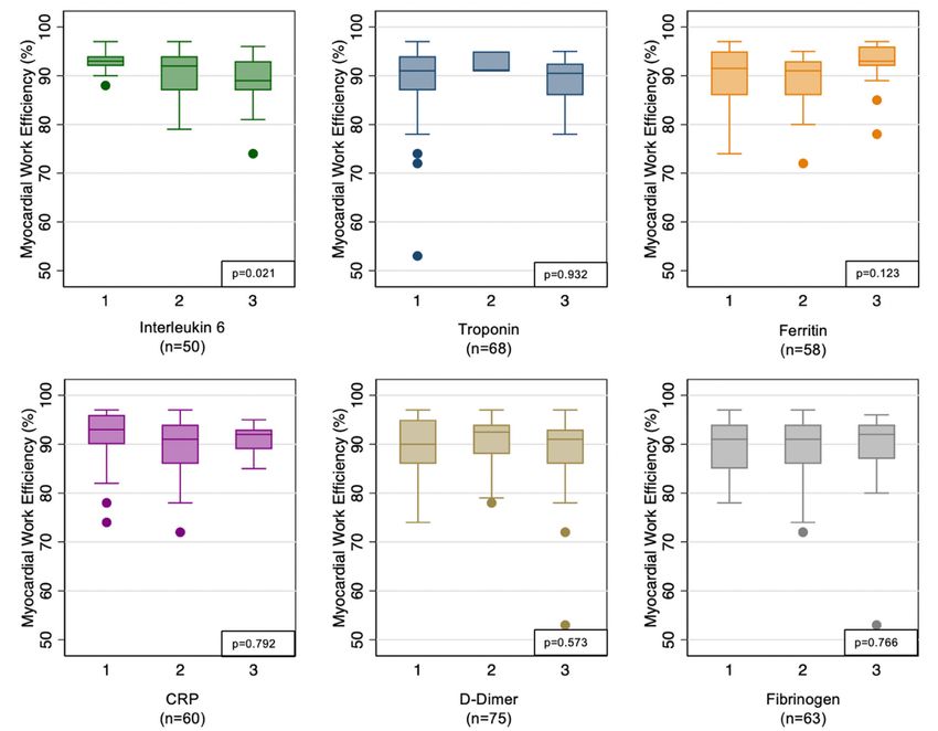
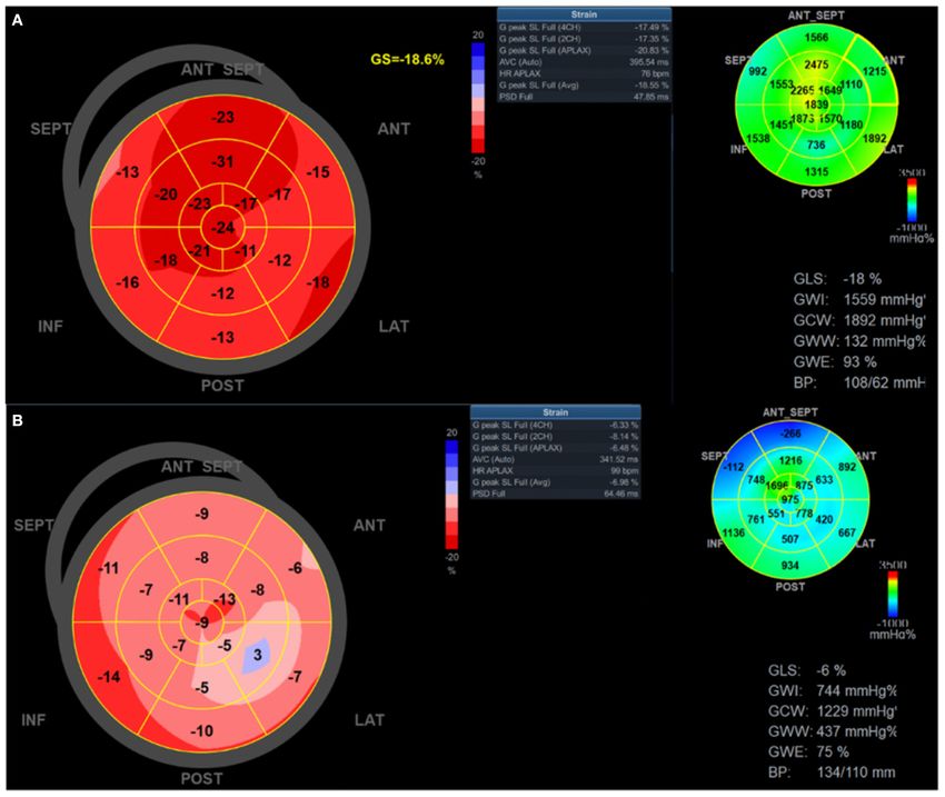
Minhas et al.                                                                                                             Subclinical Myocardial Dysfunction in COVID-19

for normal, analyses were conducted with > −16% as the                                  Myocardial Work
cutoff for normal (21–25). Tracking quality was assessed by                             Myocardial work (MW) was determined from non-invasive LV
the operator and over-ridden in segments with two or fewer                              pressure-strain analysis, which has previously been described
rejected regions where the operator deemed tracking quality                             and validated (26, 27). MW is calculated as the area of
to be acceptable. Images were analyzed by two independent                               the pressure-strain loop, similar in concept to deriving LV
observers blinded to clinical data on a dedicated offline research                      stroke work using pressure volume loops invasively. In this
workstation. Intraobserver and interobserver variability of                             technique, pressures are assessed using brachial systolic pressure
STE measures, specifically MWE, were assessed by intra- class                           and valvular event timing and strain measured with STE (15,
correlation coefficient (individual ICC of 0.994 and average ICC                        16). MW indices were calculated with the same software as
of 0.997 for intraobserver and 0.992 and 0.995 for interobserver,                       above to evaluate LV performance by incorporating afterload
respectively), and Bland-Altman analysis (all differences in                            determination using blood pressure; this provides a more load-
measurements within ±1 SD). The time between intraobserver                              independent measure compared with GLS (27). Blood pressure
measurements was 1 day.                                                                 was measured by sphygmomanometry at the time of the

 FIGURE 1 | Global longitudinal strain and myocardial work efficiency measurement in patients with COVID-19. Global longitudinal strain and myocardial work index
 bull’s eye mapping for two patients with COVID-19. (A) representative patient with relatively normal strain and myocardial work; (B) representative patient with severely
 reduced global longitudinal strain (apical predominant), myocardial work index, and work efficiency. ANT, anterior; ANT SEPT, anterospetal; APLAX, apical long axis;
 AVC, aortic valve closure; CH, chamber; GS, global strain; HR, heart rate; INF, inferior; LAT, lateral; POST, posterior; PSD, peak systolic dispersion; SEPT, septal; SL,
 strain length.

Frontiers in Cardiovascular Medicine | www.frontiersin.org                          3                                               June 2021 | Volume 8 | Article 667721
Minhas et al.                                                                                         Subclinical Myocardial Dysfunction in COVID-19

echocardiogram immediately before acquiring images for STE.                tertiles given non-normal distribution) as predictors of MWE.
The MW software then constructs a non-invasive LV pressure                 Values within each tertile are included in the supplement. These
curve adjusted according to the duration of isovolumic and                 markers included IL6, troponin, ferritin, C-reactive protein
ejection phases defined by the timing of aortic and mitral valve           (CRP), d-dimer, and fibrinogen. Missing data were considered to
opening and closing events (28). Global MW was quantified by               occur at random, and patients with missing inflammatory data
calculating the rate of regional shortening by differentiation of          were not included in this analysis.
the strain tracing and multiplying by instantaneous LV pressure
(estimated) integrated over time. During LV ejection time,                 RESULTS
segments were analyzed for wasted work and constructive work,
with global values determined as the averages of all segmental             Clinical Characteristics of Patients
values (see example Figure 1). The following parameters were               Undergoing Echocardiogram
acquired using EchoPAC software: Global MW index (MWI,                     Median time of symptom duration prior to admission was 6 days
mmHg%) defined as the area within the global LV pressure-                  (3–8 days). Median time to echocardiogram after admission was
strain loop and global MW efficiency (MWE, %), defined as                  4 days (2–8 days) and median overall time of admission was 16.5
constructive MW divided by the sum of constructed work                     days (9–31 days).
and wasted work, expressed as a percentage. Abnormal MWE                      Clinical characteristics of hospitalized patients with COVID-
was defined as 50%.
    We then performed unadjusted and adjusted logistic                     Longitudinal Strain Assessed
regression to estimate the odds of mortality with either GLS or            Among the patients with GLS performed (n = 83), 44 patients
MWE as the primary independent variable of interest, analyzed              had normal GLS and 39 (47%) had abnormal GLS (Table 1).
continuously. Covariates included were clinical characteristics            There were no significant differences in age, sex, race, or history
(age, sex, diabetes, and hypertension) and echocardiographic               of hypertension or CAD between patients with and without
measurements, selected one at a time for addition to the model             abnormal GLS. There was higher prevalence of diabetes mellitus
as the primary covariate of interest (LVEF, GLS, MWE, TAPSE,               in the abnormal compared with normal GLS group (51 vs. 27%,
RVSP, TR peak velocity, and E/E′ ). Clinical covariates selected           p = 0.025). Body mass index (BMI) was significantly higher in
for inclusion in the adjusted models were chosen based on prior            patients with abnormal compared with normal GLS (median 31.4
literature suggesting possible confounding, and included age, sex,         vs. 27.8 kg/m2 , p = 0.017). Patients with abnormal GLS had lower
history of hypertension, and diabetes (32–36). Model 1 included            LVEF (55 vs. 62%, p < 0.001), and lower TAPSE (1.7 vs. 2.0 cm,
the echocardiographic covariate of interest, adjusted for age and          p = 0.005) when compared with those with normal GLS.
sex. Model 2 included the echocardiographic covariate of interest,             Among the inflammatory markers, interleukin-6 was higher
adjusted for age, sex, diabetes, and hypertension. All variables for       among patients with abnormal GLS [median 164 (69–815)]
logistic regression were analyzed as continuous variables.                 compared with normal GLS [median 86 (32–167)], p = 0.034.
    To further understand the incremental value of STE analysis            All other inflammatory markers were not significantly different
over standard echocardiographic LVEF assessment for mortality              (Table 1). The value ranges of each inflammatory marker per
prediction, we performed subgroup analyses in patients with                tertile are presented in Supplementary Table 1.
normal (>50%) or abnormal (
Minhas et al. Subclinical Myocardial Dysfunction in COVID-19
TABLE 1 | Comparison of clinical characteristics and echocardiographic parameters in the cohort of hospitalized patients with COVID-19 and subgroups with normal vs.
abnormal global longitudinal strain (GLS) and myocardial work efficiency (MWE).
Variables Overall cohort Normal GLS Abnormal GLS p-value Normal MWE Abnormal MWE p-value
N = 136 N = 44 N = 39 N = 16 N = 59
Age, years 62.4 ± 13.9 61.9 ± 13.4 63.4 ± 14.4 0.614 55.2 ± 16.5 64.3 ± 13.1 0.023
Male 79 (58%) 27 (61%) 22 (56%) 0.647 13 (81%) 32 (53%) 0.039
Race 0.347 0.082
White 34 (25%) 10 (23%) 5 (13%) 3 (19%) 12 (21%)
African American 63 (47%) 20 (45%) 23 (61%) 5 (31%) 33 (57%)
Other 37 (27%) 14 (32%) 10 (26%) 8 (50%) 13 (22%)
Body mass index, kg/m2 30.0 (26.4–35.8) 27.8 (25.6–31.3) 31.4 (26.5–38.4) 0.017 27.7 (25.7–31.8) 28.7 (25.7–34.5) 0.544
Comorbidities
Hypertension 97 (72%) 29 (66%) 30 (77%) 0.269 7 (44%) 46 (78%) 0.008
Diabetes mellitus 55 (41%) 12 (27%) 20 (51%) 0.025 1 (6%) 29 (49%) 0.002
Coronary artery disease 20 (15%) 4 (9%) 8 (21%) 0.140 0 (0%) 10 (17%) 0.077
Heart failure 20 (15%) 2 (5%) 12 (31%) 0.001 0 (0%) 12 (20%) 0.049
Clinical presentation
Heart rate, beats per min 99 ± 20 97 ± 17 103 ± 21 0.151 95 ± 18 100 ± 20 0.392
Systolic blood pressure, mmHg 129 ± 25 129 ± 24 134 ± 24 0.368 126 ± 27 132 ± 23 0.343
Diastolic blood pressure, mmHg 71 ± 16 71 ± 16 74 ± 15 0.389 74 ± 16 71 ± 16 0.546
Laboratory measurements
White blood cell count, K/cu mm 6.7 (5.0–9.3) 6.4 (4.6–8.7) 6.0 (4.8–8.3) 0.773 6.4 (4.8–9.0) 6.4 (4.8–9.1) 0.946
Absolute lymphocyte count, K/cu mm 0.6 (0.1–1.1) 0.6 (0.1–1.0) 0.5 (0.0–1.3) 0.794 0.7 (0.0–1.2) 0.7 (0.03–1.2) 0.992
D-dimer, mg/L 2.0 (0.8–5.3) 2.0 (0.8–4.6) 2.2 (0.9–7.3) 0.433 2.0 (0.4–4.7) 2.2 (0.9–4.5) 0.213
Interleukin-6, pg/ml 130 (51–409) 86 (32–167) 164 (69–815) 0.034 114 (47–422) 125 (45–406) 0.695
CRP, mg/dl 15.3 (4.9–34.7) 11.7 (3.3–20.5) 13.7 (5.1–37.7) 0.410 4.9 (2.3–15.3) 15 (6.6–34.3) 0.009
Ferritin, ng/ml 735 (395–1,424) 737 (427–1,130) 800 (402–2,898) 0.525 830 (289–1,677) 719 (412–1,125) 0.897
Fibrinogen, mg/dl 596 (445–703) 737 (427–1,130) 800 (402–2,898) 0.695 568 (463–729) 597 (457–722) 0.694
Pro-BNP, pg/ml 422 (157–1,956) 242 (99–589) 564 (164–3,992) 0.044 176 (70–385) 392 (164–2,611) 0.032
Troponin I, ng/ml 0.03 (0.03–0.05) 0.03 (0.03–0.03) 0.03 (0.03–0.08) 0.454 0.03 (0.03–0.03) 0.03 (0.03–0.05) 0.305
Clinical events
Shock 72 (53%) 17 (39%) 23 (59%) 0.064 4 (25%) 30 (51%) 0.065
Mechanical ventilation 86 (63%) 22 (50%) 26 (67%) 0.125 5 (31%) 38 (64%) 0.017
ARDS 78 (57%) 19 (43%) 25 (64%) 0.057 5 (31%) 32 (54%) 0.103
DVT or PE 31 (23%) 8 (18%) 8 (21%) 0.788 3 (19%) 12 (20%) 0.888
Death 25 (19%) 7 (16%) 8 (21%) 0.620 2 (12%) 9 (16%) 0.764
Echocardiographic parameters
LA volume, ml 44 (35–71) 41 (29–45) 48 (39–95) 0.046 39.5 (28–42) 47 (39–55) 0.222
LVEDD, cm 4.2 (3.7–4.8) 4.1 (3.8–4.6) 4.3 (3.4–4.9) 0.378 4.4 (3.8–4.9) 4.1 (3.5–4.7) 0.276
LVEF, % 62 (52–62) 62 (57–64) 55 (40–62) 50%) 109 (81%) 43 (64%) 24 (36%)Minhas et al. Subclinical Myocardial Dysfunction in COVID-19
present in the majority (59/75, 79%). There were no significant in hospitalized patients with COVID-19. We report several
differences in demographics or clinical presentation between unique findings in our population: (1) Subclinical myocardial
patients with normal vs. abnormal MWE (Table 1). A history of dysfunction is prevalent among COVID-19 patients even
hypertension was more common among patients with abnormal in the setting of normal LVEF, especially in those with
MWE compared with normal MWE (78 vs. 44%, p = 0.008), as traditional cardiovascular risk factors, (2) lower, more abnormal
was a prior history of diabetes (29 vs. 1%, p = 0.002). Patients MWE, which is a sensitive measure of load independent
with abnormal MWE compared with those with normal MWE myocardial dysfunction, is associated with greater in-hospital
had lower LVEF (57 vs. 62%, p = 0.011), and lower TAPSE (1.8 mortality, and (3) higher level of the inflammatory marker,
vs. 2.1 cm, p = 0.003). IL-6, is predictive of lower MWE. Importantly, the finding
Among patients with normal LVEF (n = 67), a high of the association of MWE with mortality held true even
percentage had evidence of subclinical myocardial dysfunction after analyzing patients with normal LVEF, suggesting the
using STE: 36% had abnormal GLS (GLS>−16%) and 74% had prognostic benefit of MWE over LVEF and supporting use
abnormal MWE (MWEMinhas et al. Subclinical Myocardial Dysfunction in COVID-19
FIGURE 2 | Echocardiogram evaluation and main findings in hospitalized patients with COVID-19. Flow diagram of the study shows number of patients undergoing
echocardiogram, including with speckle tracking technique for strain measures. GLS, global longitudinal strain; LVEF, left ventricular ejection fraction; MWE, myocardial
work efficiency. Abnormal GLS is defined as ≤16% (the absolute value of −16%). Additional abbreviations in Figure 1.
TABLE 2 | Association of each echocardiographic parameter with mortality in hospitalized patients with COVID-19.
Unadjusted Model 1 Model 2
(age and sex) (age, sex, diabetes, hypertension)
odds ratio (95% CI) odds ratio (95% CI)
LVEF 1.00 (0.96–1.03) P = 0.248 1.00 (0.96–1.04) 1.00 (0.96–1.04) P = 0.918
P = 0.934
GLS 1.07 (0.94–1.22) P = 0.287 1.08 (0.94–1.23) 1.15 (0.98–1.35) P = 0.089
P = 0.287
MWE 0.92 (0.85–0.999) P = 0.048 0.90 (0.81–0.98) 0.87 (0.78–0.97) P = 0.009
P = 0.021
TAPSE 0.43 (0.11–1.71) P = 0.230 0.41 (0.10–1.74) 0.30 (0.06–1.45) P = 0.135
P = 0.228
RVSP 1.04 (1.00–1.09) P = 0.051 1.04 (1.00–1.09) 1.04 (1.0–1.09) P = 0.081
P = 0.073
TR peak velocity 1.03 (0.99–1.07) P = 0.182 1.03 (0.98–1.07) 1.03 (0.98–1.07) P = 0.235
P = 0.219
E/E’ 0.97 (0.91–1.05) P = 0.459 0.96 (0.87–1.06) 0.97 (0.90–1.05) P = 0.498
P = 0.392
The bold values represent significant p-values, with significant defined asMinhas et al. Subclinical Myocardial Dysfunction in COVID-19
FIGURE 3 | Association of myocardial work efficiency with inflammatory markers. Inflammatory markers are analyzed by tertile of each marker given non-normal
distribution.
cytokine release syndrome (45). Autopsy studies of severe in producing downstream effects resulting in organ damage,
COVID-19 disease suggest there can also be direct viral-induced including reduced myocardial contractility (49–51).
injury of multiple organs, including the heart (46). However, Additionally, IL-6 levels in the setting of COVID-19 have
the relative contribution and determinants of myocardial been reported to be elevated in several studies and have been
dysfunction have not been well-characterized, partially due to shown to correlate with mortality (52–54). Our study, along
limited ability to obtain widespread cardiac testing in these with these prior studies, supports a potential role of IL-6 and
patients. Given these limitations, the true prevalence of cardiac heightened inflammation in mediating myocardial dysfunction,
dysfunction has likely been underreported thus far, and is mainly thereby increasing risk of death. Of note, we did not find a similar
limited to case reports (6, 7). relationship with troponin and myocardial dysfunction, likely
In our study, patients underwent echocardiography at a related to the primarily normal-range troponin values for the
median 4 days after hospital admission and 6 days of symptom majority of patients.
onset, suggesting that impaired GLS and MWE occur early By characterizing subclinical myocardial dysfunction using
in COVID-19 during the systemic inflammatory response, and STE, the present study provides incremental knowledge,
cannot entirely be explained by a more chronic myocardial linking increased systemic inflammation (by IL-6 levels) to
process such as fibrosis. In addition, COVID-19 patients with the pathophysiology of myocardial injury and dysfunction
LV dysfunction on STE had more obesity, which is a pro- in COVID-19.
inflammatory state that initiates oxidative stress and adversely
affects immune function, leading to cardiac injury (40, 47, 48). Limitations
Finally, although inflammatory pathways have been implicated in The main limitation of this study is the relatively small
myocardial injury related to COVID-19, their effect on important sample size and retrospective cohort study design. Larger
indices of cardiac function has not been well-characterized. In the prospective studies are needed to further explore these novel
present study, we show that subclinical myocardial dysfunction echocardiographic parameters (GLS and MWE) with regard
is related to the degree of systemic inflammation measured by to cardiovascular mortality and other clinically meaningful
IL-6. IL-6 has previously been shown to act as a key cytokine outcomes in COVID-19 disease. Also, not all hospitalized
Frontiers in Cardiovascular Medicine | www.frontiersin.org 8 June 2021 | Volume 8 | Article 667721Minhas et al. Subclinical Myocardial Dysfunction in COVID-19
COVID-19 patients underwent echocardiogram and STE, which ETHICS STATEMENT
could result in selection bias and inability to detect true
prevalence of abnormal GLS or MWE among COVID-19 The studies involving human participants were reviewed
patients. Lastly, a minority of patients with GLS and MWE and approved by Johns Hopkins IRB. Written informed
performed did not have all inflammatory markers tested consent for participation was not required for this study
clinically, thus limiting the analyses. in accordance with the national legislation and the
institutional requirements.
CONCLUSIONS
AUTHOR CONTRIBUTIONS
In summary, sensitive indices of LV dysfunction, GLS and
MWE, measured with STE are abnormal in a substantial AM, NG, EG, and AH drafted the manuscript. BG, TM, GS, SP,
portion of hospitalized COVID-19 patients who underwent and NB edited the manuscript. AM, NG, EG, and NB collected
echocardiograms, even in those with normal LVEF. Impaired the data. AM analyzed the data and performed statistical
MWE is independently associated with in-hospital mortality analysis. All authors contributed to the article and approved the
in COVID-19 patients. Higher IL-6 levels are associated with submitted version.
reduced MWE, providing a possible pathophysiologic link
between increased inflammation and adverse outcomes in FUNDING
COVID-19. Based on these findings, it is possible that STE
measures of subclinical LV dysfunction may provide incremental AM was supported by the National Heart, Lung, and Blood
value to standard echocardiographic measures in patients with Institute training Grant T32HL007024. AH was supported
COVID-19. Given the acuity of presentation and cardiovascular by the National Heart, Lung, and Blood Institute Grant
complications of COVID-19, a better understanding of the 1R01HL147660. This study received device and funding
extent of myocardial injury and dysfunction early in the disease support from GE Healthcare. The funder was not involved
course may help triage at risk patients and implement early in the study design, collection, analysis, interpretation of
interventions aimed at reducing mortality. Further longitudinal data, the writing of this article or the decision to submit it
studies are needed to investigate persistence of impaired cardiac for publication.
function in the setting of COVID-19.
SUPPLEMENTARY MATERIAL
DATA AVAILABILITY STATEMENT
The Supplementary Material for this article can be found
The raw data supporting the conclusions of this article will be online at: https://www.frontiersin.org/articles/10.3389/fcvm.
made available by the authors, without undue reservation. 2021.667721/full#supplementary-material
REFERENCES 7. Inciardi RM, Lupi L, Zaccone G, Italia L, Raffo M, Tomasoni D, et al. Cardiac
involvement in a patient with coronavirus disease 2019 (COVID-19). JAMA
1. Guo T, Fan Y, Chen M, Wu X, Zhang L, He T, et al. Cardiovascular Cardiol. (2020) 5:819–24. doi: 10.1001/jamacardio.2020.1096
implications of fatal outcomes of patients with coronavirus 8. Zeng F, Huang Y, Guo Y, Yin M, Chen X, Xiao L, et al. Association of
disease 2019 (COVID-19). JAMA Cardiol. (2020) 5:811–8. inflammatory markers with the severity of COVID-19: A meta-analysis. Int
doi: 10.1001/jamacardio.2020.1017 J Infect Dis. (2020) 96:467–74. doi: 10.1016/j.ijid.2020.05.055
2. Shi S, Qin M, Shen B, Cai Y, Liu T, Yang F, et al. Association of 9. Rali AS, Ranka S, Shah Z, Sauer AJ. Mechanisms of myocardial
cardiac injury with mortality in hospitalized patients with COVID-19 in injury in coronavirus disease 2019. Card Fail Rev. (2020) 6:e15.
Wuhan, China. JAMA Cardiol. (2020) 5:802–10. doi: 10.1001/jamacardio.20 doi: 10.15420/cfr.2020.10
20.0950 10. Chen C, Zhou Y, Wang DW. SARS-CoV-2: a potential novel
3. Hoffmann M, Kleine-Weber H, Schroeder S, Krüger N, Herrler T, Erichsen etiology of fulminant myocarditis. Herz. (2020) 45:230–2.
S, et al. SARS-CoV-2 cell entry depends on ACE2 and TMPRSS2 and is doi: 10.1007/s00059-020-04909-z
blocked by a clinically proven protease inhibitor. Cell. (2020) 181:271–80.e8. 11. Cooper LT, Baughman KL, Feldman AM, Frustaci A, Jessup M, Kuhl U, et al.
doi: 10.1016/j.cell.2020.02.052 The role of endomyocardial biopsy in the management of cardiovascular
4. Richardson S, Hirsch JS, Narasimhan M, Crawford JM, McGinn T, disease. J Am Coll Cardiol. (2007) 50:1914–31. doi: 10.1016/j.jacc.2007.09.008
Davidson KW, et al. Presenting characteristics, comorbidities, and 12. Kostakou PM, Kostopoulos VS, Tryfou ES, Giannaris VD, Rodis IE, Olympios
outcomes among 5700 patients hospitalized with COVID-19 in the CD, et al. Subclinical left ventricular dysfunction and correlation with regional
New York City area. JAMA. (2020) 323:2052–9. doi: 10.1001/jama.20 strain analysis in myocarditis with normal ejection fraction. A new diagnostic
20.6775 criterion. Int J Cardiol. (2018) 259:116–21. doi: 10.1016/j.ijcard.2018.01.058
5. Gilotra NA, Minkove N, Bennett MK, Tedford RJ, Steenbergen C, Judge DP, 13. Han J, Mou Y, Yan D, Zhang Y-T, Jiang T-A, Zhang Y-Y, et al. Transient
et al. Lack of relationship between serum cardiac troponin I level and giant cardiac injury during H7N9 infection. Eur J Clin Invest. (2015) 45:117–25.
cell myocarditis diagnosis and outcomes. J Cardiac Fail. (2016) 22:583–5. doi: 10.1111/eci.12386
doi: 10.1016/j.cardfail.2015.12.022 14. Awadalla M, Mahmood SS, Groarke JD, Hassan MZO, Nohria A, Rokicki
6. Hu H, Ma F, Wei X, Fang Y. Coronavirus fulminant myocarditis treated A, et al. Global longitudinal strain and cardiac events in patients with
with glucocorticoid and human immunoglobulin. Eur Heart J. (2020) 4:260. immune checkpoint inhibitor-related myocarditis. J Am College Cardiol.
doi: 10.1093/eurheartj/ehaa190 (2020) 75:467–78. doi: 10.1016/j.jacc.2019.11.049
Frontiers in Cardiovascular Medicine | www.frontiersin.org 9 June 2021 | Volume 8 | Article 667721Minhas et al. Subclinical Myocardial Dysfunction in COVID-19
15. Sörensen J, Harms HJ, Aalen JM, Baron T, Smiseth OA, Flachskampf reduced ejection fraction treated by sacubitril/valsartan. Am J Cardiol. (2020)
FA. Myocardial efficiency. JACC: Cardiovasc Imag. (2019) 13:1564–76. 125:1856–62. doi: 10.1016/j.amjcard.2020.03.031
doi: 10.1016/j.jcmg.2019.08.030 32. Abou R, Leung M, Khidir MJH, Wolterbeek R, Schalij MJ, Ajmone Marsan N,
16. Chan J, Edwards NFA, Khandheria BK, Shiino K, Sabapathy S, Anderson et al. Influence of aging on level and layer-specific left ventricular longitudinal
B, et al. A new approach to assess myocardial work by non-invasive strain in subjects without structural heart disease. Am J Cardiol. (2017)
left ventricular pressure–strain relations in hypertension and dilated 120:2065–72. doi: 10.1016/j.amjcard.2017.08.027
cardiomyopathy. Eur Heart J Cardiovasc Imaging. (2019) 20:31–9. 33. Liu J-H, Chen Y, Yuen M, Zhen Z, Chan CW-S, Lam KS-L, et al. Incremental
doi: 10.1093/ehjci/jey131 prognostic value of global longitudinal strain in patients with type 2 diabetes
17. Edwards NFA, Scalia GM, Shiino K, Sabapathy S, Anderson B, Chamberlain mellitus. Cardiovasc Diabetol. (2016) 15:22. doi: 10.1186/s12933-016-0333-5
R, et al. Global myocardial work is superior to global longitudinal strain 34. Wierzbowska-Drabik K, Trzos E, Kurpesa M, Rechciński T, Miśkowiec D,
to predict significant coronary artery disease in patients with normal left Cieślik-Guerra U, et al. Diabetes as an independent predictor of left ventricular
ventricular function and wall motion. J Am Soc Echocardiogr. (2019) 32:947– longitudinal strain reduction at rest and during dobutamine stress test in
57. doi: 10.1016/j.echo.2019.02.014 patients with significant coronary artery disease. Eur Heart J Cardiovasc
18. Lang RM, Badano LP, Mor-Avi V, Afilalo J, Armstrong A, Ernande L, et al. Imaging. (2018) 19:1276–86. doi: 10.1093/ehjci/jex315
Recommendations for cardiac chamber quantification by echocardiography 35. Vrettos A, Dawson D, Grigoratos C, Nihoyannopoulos P. Correlation between
in adults: an update from the American society of echocardiography and global longitudinal peak systolic strain and coronary artery disease severity
the European association of cardiovascular imaging. J Am Soc Echocardiogr. as assessed by the angiographically derived SYNTAX score. Echo Res Pract.
(2015) 28:1–39.e14. doi: 10.1016/j.echo.2014.10.003 (2016) 3:29–34. doi: 10.1530/ERP-16-0005
19. Nagueh SF, Smiseth OA, Appleton CP, Byrd BF, Dokainish H, Edvardsen 36. Liou K, Negishi K, Ho S, Russell EA, Cranney G, Ooi S-Y. Detection of
T, et al. Recommendations for the evaluation of left ventricular diastolic obstructive coronary artery disease using peak systolic global longitudinal
function by echocardiography: an update from the American society of strain derived by two-dimensional speckle-tracking: a systematic review
echocardiography and the European association of cardiovascular imaging. and meta-analysis. J Am Soc Echocardiograp. (2016) 29:724–35.e4.
J Am Soc Echocardiograp. (2016) 29:277–314. doi: 10.1016/j.echo.2016.01.011 doi: 10.1016/j.echo.2016.03.002
20. Kirkpatrick JN, Mitchell C, Taub C, Kort S, Hung J, Swaminathan M. ASE 37. Smiseth OA, Torp H, Opdahl A, Haugaa KH, Urheim S. Myocardial strain
statement on protection of patients and echocardiography service providers imaging: how useful is it in clinical decision making? Eur Heart J. (2016)
during the 2019 novel coronavirus outbreak. J Am College Cardiol. (2020) 37:1196–207. doi: 10.1093/eurheartj/ehv529
75:3078–84. doi: 10.1016/j.jacc.2020.04.002 38. Thavendiranathan P, Poulin F, Lim K-D, Plana JC, Woo A, Marwick TH. Use
21. Farsalinos KE, Daraban AM, Ünlü S, Thomas JD, Badano LP, Voigt J-U. of myocardial strain imaging by echocardiography for the early detection of
Head-to-head comparison of global longitudinal strain measurements among cardiotoxicity in patients during and after cancer chemotherapy. J Am College
nine different vendors. J Am Soc Echocardiograp. (2015) 28:1171–81.e2. Cardiol. (2014) 63:2751–68. doi: 10.1016/j.jacc.2014.01.073
doi: 10.1016/j.echo.2015.06.011 39. Stokke TM, Hasselberg NE, Smedsrud MK, Sarvari SI, Haugaa
22. Haji K, Marwick TH. Clinical utility of echocardiographic strain KH, Smiseth OA, et al. Geometry as a confounder when assessing
and strain rate measurements. Curr Cardiol Rep. (2021) 23:18. ventricular systolic function. J Am College Cardiol. (2017) 70:942–54.
doi: 10.1007/s11886-021-01444-z doi: 10.1016/j.jacc.2017.06.046
23. D’Elia N, Caselli S, Kosmala W, Lancellotti P, Morris D, Muraru D, et al. 40. Honce R, Schultz-Cherry S. Impact of obesity on influenza A virus
Normal global longitudinal strain. JACC: Cardiovasc Imaging. (2020) 13:167– pathogenesis, immune response, and evolution. Front Immunol. (2019)
9. doi: 10.1016/j.jcmg.2019.07.020 10:1071. doi: 10.3389/fimmu.2019.01071
24. Pieske B, Tschöpe C, de Boer RA, Fraser AG, Anker SD, Donal E, et al. How 41. Estabragh ZR, Mamas MA. The cardiovascular manifestations of
to diagnose heart failure with preserved ejection fraction: the HFA–PEFF influenza: A systematic review. Int J Cardiol. (2013) 167:2397–403.
diagnostic algorithm: a consensus recommendation from the heart failure doi: 10.1016/j.ijcard.2013.01.274
association (HFA) of the European Society of Cardiology (ESC). Eur Heart 42. Cummings MJ, Baldwin MR, Abrams D, Jacobson SD, Meyer BJ, Balough
J. (2019) 40:3297–317. doi: 10.1093/eurheartj/ehz641 EM, et al. Epidemiology, clinical course, and outcomes of critically ill adults
25. Potter E, Marwick TH. Assessment of left ventricular function by with COVID-19 in New York City: a prospective cohort study. Lancet. (2020)
echocardiography. JACC: Cardiovascu Imaging. (2018) 11:260–74. 395:1763–70. doi: 10.1016/S0140-6736(20)31189-2
doi: 10.1016/j.jcmg.2017.11.017 43. Li Y, Li H, Zhu S, Xie Y, Wang B, He L, et al. Prognostic value of right
26. Russell K, Eriksen M, Aaberge L, Wilhelmsen N, Skulstad H, Gjesdal ventricular longitudinal strain in patients with COVID-19. JACC: Cardiovasc
O, et al. Assessment of wasted myocardial work: a novel method to Imaging. (2020) 13:2287–99. doi: 10.1016/j.jcmg.2020.04.014
quantify energy loss due to uncoordinated left ventricular contractions. Am J 44. Argulian E, Sud K, Vogel B, Bohra C, Garg VP, Talebi S, et al. Right ventricular
Physiol Heart Circul Physiol. (2013) 305:H996–1003. doi: 10.1152/ajpheart.001 dilation in hospitalized patients with COVID-19 infection. JACC: Cardiovasc
91.2013 Imaging. (2020) 13:2459–61. doi: 10.1016/j.jcmg.2020.05.010
27. Russell K, Eriksen M, Aaberge L, Wilhelmsen N, Skulstad H, Remme EW, et al. 45. Atri D, Siddiqi HK, Lang J, Nauffal V, Morrow DA, Bohula EA. COVID-19
A novel clinical method for quantification of regional left ventricular pressure- for the cardiologist: a current review of the virology, clinical epidemiology,
strain loop area: a non-invasive index of myocardial work. Eur Heart J. (2012) cardiac and other clinical manifestations and potential therapeutic strategies.
33:724–33. doi: 10.1093/eurheartj/ehs016 JACC: Basic Transl Sci. (2020) 5:518–36. doi: 10.1016/j.jacbts.2020.04.002
28. Hubert A, Le Rolle V, Leclercq C, Galli E, Samset E, Casset C, et al. 46. Buja LM, Wolf DA, Zhao B, Akkanti B, McDonald M, Lelenwa L, et al. The
Estimation of myocardial work from pressure-strain loops analysis: an emerging spectrum of cardiopulmonary pathology of the coronavirus disease
experimental evaluation. Eur Heart J Cardiovasc Imaging. (2018) 19:1372–9. 2019 (COVID-19): report of 3 autopsies from Houston, Texas, and review of
doi: 10.1093/ehjci/jey024 autopsy findings from other United States cities. Cardiovasc Pathol. (2020)
29. Manganaro R, Marchetta S, Dulgheru R, Ilardi F, Sugimoto T, Robinet S, 48:107233. doi: 10.1016/j.carpath.2020.107233
et al. Echocardiographic reference ranges for normal non-invasive myocardial 47. The GBD 2015 Obesity Collaborators. Health effects of overweight and
work indices: results from the EACVI NORRE study. Eur Heart J Cardiovasc obesity in 195 countries over 25 years. N Engl J Med. (2017) 377:13–27.
Imaging. (2019) 20:582–90. doi: 10.1093/ehjci/jey188 doi: 10.1056/NEJMoa1614362
30. El Mahdiui M, van der Bijl P, Abou R, Ajmone Marsan N, Delgado V, Bax 48. Kass DA, Duggal P, Cingolani O. Obesity could shift severe
JJ. Global left ventricular myocardial work efficiency in healthy individuals COVID-19 disease to younger ages. Lancet. (2020) 395:1544–5.
and patients with cardiovascular disease. J Am Soc Echocardiograp. (2019) doi: 10.1016/S0140-6736(20)31024-2
32:1120–7. doi: 10.1016/j.echo.2019.05.002 49. Pathan N, Hemingway CA, Alizadeh AA, Stephens AC, Boldrick JC, Oragui
31. Bouali Y, Donal E, Gallard A, Laurin C, Hubert A, Bidaut A, et al. EE, et al. Role of interleukin 6 in myocardial dysfunction of meningococcal
Prognostic usefulness of myocardial work in patients with heart failure and septic shock. Lancet. (2004) 363:203–9. doi: 10.1016/S0140-6736(03)15326-3
Frontiers in Cardiovascular Medicine | www.frontiersin.org 10 June 2021 | Volume 8 | Article 667721Minhas et al. Subclinical Myocardial Dysfunction in COVID-19
50. Johnson DE, O’Keefe RA, Grandis JR. Targeting the IL-6/JAK/STAT3 54. Gao Y, Li T, Han M, Li X, Wu D, Xu Y, et al. Diagnostic utility of clinical
signalling axis in cancer. Nat Rev Clin Oncol. (2018) 15:234–8. laboratory data determinations for patients with the severe COVID-19. J Med
doi: 10.1038/nrclinonc.2018.8 Virol. (2020) 92:791–6. doi: 10.1002/jmv.25770
51. Liu B, Li M, Zhou Z, Guan X, Xiang Y. Can we use interleukin-
6 (IL-6) blockade for coronavirus disease 2019 (COVID-19)-induced Conflict of Interest: The authors declare that the research was conducted in the
cytokine release syndrome (CRS)? J Autoimmun. (2020) 111:102452. absence of any commercial or financial relationships that could be construed as a
doi: 10.1016/j.jaut.2020.102452 potential conflict of interest.
52. Huang C, Wang Y, Li X, Ren L, Zhao J, Hu Y, et al. Clinical
features of patients infected with 2019 novel coronavirus in Wuhan, Copyright © 2021 Minhas, Gilotra, Goerlich, Metkus, Garibaldi, Sharma, Bavaro,
China. Lancet. (2020) 395:497–506. doi: 10.1016/S0140-6736(20) Phillip, Michos and Hays. This is an open-access article distributed under the terms
30183-5 of the Creative Commons Attribution License (CC BY). The use, distribution or
53. Chen N, Zhou M, Dong X, Qu J, Gong F, Han Y, et al. Epidemiological and reproduction in other forums is permitted, provided the original author(s) and the
clinical characteristics of 99 cases of 2019 novel coronavirus pneumonia copyright owner(s) are credited and that the original publication in this journal
in Wuhan, China: a descriptive study. Lancet. (2020) 395:507–13. is cited, in accordance with accepted academic practice. No use, distribution or
doi: 10.1016/S0140-6736(20)30211-7 reproduction is permitted which does not comply with these terms.
Frontiers in Cardiovascular Medicine | www.frontiersin.org 11 June 2021 | Volume 8 | Article 667721You can also read