Pronounced proliferation of non beta cells in response to beta cell mitogens in isolated human islets of Langerhans - Nature
←
→
Page content transcription
If your browser does not render page correctly, please read the page content below
www.nature.com/scientificreports
OPEN Pronounced proliferation
of non‑beta cells in response
to beta‑cell mitogens in isolated
human islets of Langerhans
Hasna Maachi1,2,4, Julien Ghislain1,4, Caroline Tremblay1 & Vincent Poitout1,3*
The potential to treat diabetes by increasing beta-cell mass is driving a major effort to identify beta-
cell mitogens. Demonstration of mitogen activity in human beta cells is frequently performed in
ex vivo assays. However, reported disparities in the efficacy of beta-cell mitogens led us to investigate
the sources of this variability. We studied 35 male (23) and female (12) human islet batches covering
a range of donor ages and BMI. Islets were kept intact or dispersed into single cells and cultured in
the presence of harmine, glucose, or heparin-binding epidermal growth factor-like growth factor
(HB-EGF), and subsequently analyzed by immunohistochemistry or flow cytometry. Proliferating
cells were identified by double labeling with EdU and Ki67 and glucagon, c-peptide or Nkx6.1, and
cytokeratin-19 to respectively label alpha, beta, and ductal cells. Harmine and HB-EGF stimulated
human beta-cell proliferation, but the effect of glucose was dependent on the assay and the donor.
Harmine potently stimulated alpha-cell proliferation and both harmine and HB-EGF increased
proliferation of insulin- and glucagon-negative cells, including cytokeratin 19-positive cells. Given the
abundance of non-beta cells in human islet preparations, our results suggest that assessment of beta-
cell mitogens requires complementary approaches and rigorous identification of cell identity using
multiple markers.
Type 2 diabetes is characterized by insufficient functional β-cell m ass1. In non-diabetic obese individuals and dur-
ing pregnancy, accretion of insulin secretory capacity per β cell combined with β-cell mass expansion maintains
glucose homeostasis by balancing levels of circulating insulin and insulin sensitivity. Deciphering the mechanisms
controlling β-cell proliferation has thus become a major research goal in the hope to identify therapeutic targets
to expand β-cell mass and prevent or delay the onset of diabetes.
Diverse factors including insulin2, GLP-13, serotonin4, epidermal growth factors5, osteoprotegerin6, serpinB17
and nutrients such as glucose and fatty acids8–12 promote replication of rodent β cells. Unfortunately, our knowl-
edge of the factors controlling human β-cell proliferation is much more limited13–15. Although human β cells
respond to glucose16–18, fatty acids12, serpinB17, dual-specificity tyrosine-regulated kinase 1A (DYRK1A) inhibi-
tors including h armine19–21, and heparin-binding epidermal growth factor-like growth factor (HB-EGF)22, pro-
liferation rates are very low compared to rodents and published data on the efficacy of human β-cell mitogens
are often highly v ariable23.
In this study we assessed the mitogenic properties of harmine, glucose and HB-EGF in both intact and dis-
persed human islets comparing immunochemistry to flow cytometry to differentiate proliferating β and non-β
cell types, including glucagon+ and cytokeratin-19 (CK19)+ cells.
Results
Proliferative effects of harmine, glucose, and HB‑EGF in dispersed human islets as assessed by
immunocytochemistry. As the majority of previous studies examined the effects of mitogens on human
β-cell proliferation in dispersed islet cultures17–19,21,24–27, we began by attempting to reproduce these findings. Fol-
lowing recovery, human islets were dispersed, plated and exposed to harmine (10 μM), high glucose (16.7 mM)
or HB-EGF (100 ng/ml) for 3 days in the presence of the proliferation marker 5-ethynyl-2′-deoxyuridine (EdU).
1
Montreal Diabetes Research Center, CRCHUM, 900 rue St Denis, Montreal, QC H2X 0A9, Canada. 2Department
of Pharmacology and Physiology, University of Montreal, Montreal, QC, Canada. 3Department of Medicine,
University of Montreal, Montreal, QC, Canada. 4These authors contributed equally: Hasna Maachi and Julien
Ghislain. *email: vincent.poitout@umontreal.ca
Scientific Reports | (2021) 11:11283 | https://doi.org/10.1038/s41598-021-90643-3 1
Vol.:(0123456789)
www.nature.com/scientificreports/
At the end of the treatment, cells were stained for EdU and the nuclear marker of mature β cells Nkx6.123 (Fig. 1).
Exposure to harmine in the presence of 5.5 mM glucose lead to a significant increase in β-cell proliferation
compared to vehicle in 5/6 donors (Fig. 1a–c) (fold-change 13.0 ± 4.0, b and c: P = 0.034). In contrast, 16.7 mM
glucose was a poor inducer of β-cell proliferation compared to 2.8 mM glucose with only 2/6 donors respond-
ing (Fig. 1d,e,g) (fold-change 1.2 ± 0.4, e and g: NS). Exposure to HB-EGF in the presence of 2.8 mM glucose
increased β-cell proliferation compared to vehicle in 5/6 donors (Fig. 1d,f,g) (fold-change 2.1 ± 0.5, f: P = 0.011, g:
NS). Interestingly, we frequently observed Nkx6.1−/EdU+ cells in the presence of harmine (Fig. 1a) and HB-EGF
(Fig. 1d), suggesting that non-β cells also respond to these compounds.
Proliferative effects of harmine, glucose, and HB‑EGF in intact human islets as assessed by
immunohistochemistry. Islet architecture and cell-to-cell contact may contribute to the capacity of β cells
to proliferate28. Therefore, we exposed intact human islets to harmine, glucose or HB-EGF and assessed β-cell
proliferation in cryosections by immunohistochemistry for Nkx6.1 and EdU (Fig. 2). As intact islets may be
subject to increased cell death during extended culture, we first confirmed that EdU was not staining apoptotic
cells. Double staining of islets from 1 donor exposed to harmine, glucose or HB-EGF for EdU and the apopto-
sis marker FragEL revealed no significant colocalization (Supplementary Fig. S1 online) suggesting that E dU+
cells are not apoptotic. Harmine increased proliferation of Nkx6.1+ cells in 5/8 donors (Fig. 2a,b,e) (fold-change
6.7 ± 2.2, b: P = 0.031, e: P = 0.031). In contrast to the results in dispersed islets, 7/9 donors responded to glucose
(Fig. 2a,c,e) (fold-change 3.8 ± 1.0, c: P = 0.022, e: NS), and whereas 6/9 donors responded to HB-EGF the aver-
age effect was not significant (Fig. 2a,d,e) (fold-change 4.5 ± 2.1, d and e: NS). As observed in dispersed islets,
many proliferating cells did not stain for Nkx6.1, which led us to quantify proliferation in this presumably non-β
cell population. In basal conditions (2.8 mM glucose) the mean percentage of E dU+/Nkx6.1− cells of the 9 donors
was 1.3 ± 0.3% (Fig. 2a,f–i). Harmine exposure led to a large increase in Nkx6.1− cell proliferation in 8/8 donors
(Fig. 2a,f,i) (fold-change 6.7 ± 2.0, f: P = 0.024, i: P = 0.0005). Glucose increased proliferation of Nkx6.1− cells in
4/9 donors and this effect was not significant (Fig. 2a,g,i) (fold-change 1.2 ± 0.3, g and i: NS), whereas HB-EGF
stimulated proliferation of Nkx6.1− cells in 7/9 donors (Fig. 2a,h,i) (fold-change 1.6 ± 0.4, h: P = 0.01, i: NS).
To confirm the results obtained with Nkx6.1 in intact islets, we used C-peptide (CPEP) as another β-cell
marker (Fig. 3). Similar to the results shown in Fig. 2, harmine, glucose and HB-EGF increased proliferation of
CPEP+ cells in 7/7 donors (Fig. 3a,b,e) (fold-change 6.4 ± 1.7, b: P = 0.025, e: P = 0.0013), 5/6 donors (Fig. 3a,c,e)
(fold-change 2.0 ± 0.5, c: P = 0.021, e: NS) and in 6/7 donors (Fig. 3a,d,e) (fold-change 2.6 ± 0.6, d: P = 0.014, e:
NS), respectively. Surprisingly, the fraction of E dU+/CPEP+ cells in both basal and stimulated conditions was
+ +
considerably larger compared to EdU /Nkx6.1 cells from several donors (compare Fig. 3b–e to Fig. 2b–e). To
confirm this observation, we stained donor islets (N = 3) simultaneously for CPEP, Nkx6.1 and EdU (Supplemen-
tary Fig. S2 online). On average of all conditions, Nkx6.1+ cells represented 42 ± 2% of the CPEP+ cells, whereas
proliferating Nkx6.1+ cells represented only 13 ± 4% of the proliferating C PEP+ cells. These data indicate that not
all β cells are detectable by Nkx6.1 staining and confirm our findings that more proliferating β cells are C PEP+
than Nkx6.1+. The proliferative response of C PEP− cells to harmine (Fig. 3a,f,i), (6/7 donors; fold-change 9.7 ± 2.3,
f: P = 0.012, i: P = 0.0007) glucose (Fig. 3a,g,i) (5/6 donors; fold-change 2.0 ± 0.4, g: P = 0.014, i: NS) and HB-EGF
(Fig. 3a,h,i) (5/7 donors; fold-change 3.2 ± 1.4, h and i: NS) was similar to the response of Nkx6.1− cells (Fig. 2).
To confirm the β-cell proliferative response to mitogens with a different proliferation marker we stained for
the cell cycle antigen Ki67 and CPEP to mark β cells (Fig. 4). Similar to the results using EdU (Fig. 3), harmine,
glucose and HB-EGF increased the percentage of Ki67+/CPEP+ cells in 5/5 donors (Fig. 4a,b,e) (fold-change
8.9 ± 2.2, b: P = 0.014, e: P = 0.002), in 4/5 donors (Fig. 4a,c,e) (fold-change 1.5 ± 0.4, c: P = 0.009, e: NS) and in
5/5 donors (Fig. 4a,d,e) (fold-change 4.2 ± 1.5, d and e: NS), respectively. Importantly, the percentage of Ki67+/
CPEP+ detected under the different conditions was comparable to the percentage of E dU+/CPEP+ when donor
islets were analyzed in parallel with both proliferation markers (compare Fig. 4 to Fig. 7 donors 30, 31 and 33).
The percentage of K i67+/CPEP− cells increased in response to harmine in 5/5 donors (Fig. 4a,f,i), (fold-change
23.8 ± 8.8, f: P = NS, i: P = 0.0012), but only in 2/5 donors in response to glucose (Fig. 4a,g,i) (fold-change 1.4 ± 0.7,
g and i: NS) and in 3/5 donors in response to HB-EGF (Fig. 4a,h,i) (fold-change 6.9 ± 4.2, h and i: NS) and was
similar to the results obtained using EdU (Fig. 3). Overall, comparable results were obtained when using EdU
or Ki67 as a proliferation marker of C PEP+ and CPEP− in all conditions tested.
Human islet preparations often contain abundant non-endocrine cell types including duct and acinar cells,
among others29. The abundance of proliferating non-β cells (Figs. 2, 3, 4) led us to ask whether a proportion of
these cells are positive for the ductal marker CK1930. Intact human islets were exposed to harmine, glucose or HB-
EGF and β-cell proliferation assessed by immunohistochemistry for CK19 and EdU (Fig. 5). Numerous clusters
of CK19+ cells were detected in all human islet preparations tested that were distinct from CPEP+ cells (Fig. 5a
and Supplementary Fig. S3 online). Although harmine increased proliferation of CK19+ cells in 4/4 donors, the
increase was not significant (Fig. 5a,b,e) (fold-change 2.5 ± 0.8, b and e: NS). In contrast glucose, and to a greater
extent HB-EGF, increased C K19+ cell proliferation in 5/6 (Fig. 5a,c,e) (fold-change 1.8 ± 0.2, c: P = 0.016, e: NS)
and 6/6 (Fig. 5a,d,e) (fold-change 5.4 ± 1.5, d: P = 0.011, e: P = 0.005) donors, respectively.
Scientific Reports | (2021) 11:11283 | https://doi.org/10.1038/s41598-021-90643-3 2
Vol:.(1234567890)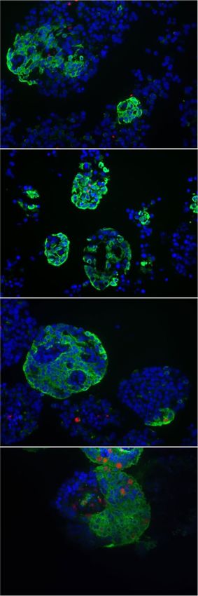
www.nature.com/scientificreports/
Vehicle
Harmine
0.5
0.4
% EdU+ of Nkx6.1+
0.3
0.2
0.1
0.0
Low Glucose
Harmine
Glucose, mM 5.5 5.5
- Har
2.8 Glu Vehicle
16.7 Glu HB-EGF 0.5
0.4
% EdU+ of Nkx6.1+
0.3
0.2
0.1
0.0
Low Glucose
High Glucose
HB-EGF
Glucose, mM 2.8 16.7 2.8
- - HB
Figure 1. Analysis of β-cell proliferation in dispersed human islets by immunocytochemistry for Nkx6.1. (a–g)
Dispersed human islets were exposed to 5.5 mM glucose (Vehicle; a–c), 2.8 mM glucose (Vehicle or 2.8 Glu;
d–g), harmine (10 μM) (Har; a–c), 16.7 mM glucose (16.7 Glu; d,e,g) and HB-EGF (100 ng/ml) (HB; d,f,g) for
72 h. EdU (10 μM) was added throughout. Proliferation was assessed by EdU staining and Nkx6.1 to mark β
cells. Representative images of Nkx6.1 (green), EdU (red) and nuclei (blue) staining are shown (a,d). Arrows
and arrowheads highlight E dU+/Nkx6.1+ and EdU+/Nkx6.1− cells, respectively. Scale bar, 100 μm. Images
were acquired using an Operetta CLS with Harmony 4.5 software (https://www.perkinelmer.com/en-ca/produ
ct/operetta-cls-system-hh16000000). Between 1000 and 1500 Nkx6.1+ cells were counted for each sample.
Proliferation is presented as the percentage of EdU+/Nkx6.1+ cells over the total Nkx6.1+ cells. Graphs showing
cell proliferation of individual (b,e,f) and combined (c,g) donors were generated using GraphPad Prism 9
software (https://www.graphpad.com/scientific-software/prism/). Donor identifiers (Supplementary Table S1
online) are indicated. Significance was tested using paired t tests (b,c,e,f) or one-way ANOVA (g). P < 0.05 was
considered significant. NS not significant.
Scientific Reports | (2021) 11:11283 | https://doi.org/10.1038/s41598-021-90643-3 3
Vol.:(0123456789)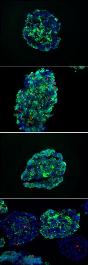
www.nature.com/scientificreports/
Vehicle
Harmine
2.8 Glu
16.7 Glu
Vehicle
HB-EGF
Figure 2. Analysis of β-cell proliferation in intact human islets by immunohistochemistry for Nkx6.1. (a–i) Intact human islets
were exposed to 2.8 mM glucose (Vehicle or 2.8 Glu; a–i), harmine (10 μM) (Har; a,b,e,f,i), 16.7 mM glucose (16.7 Glu; a,c,e,g,i) and
HB-EGF (100 ng/ml) (HB; a,d,e,h,i) for 72 h. EdU (10 μM) was added throughout. Proliferation was assessed by EdU staining and
Nkx6.1 to mark β cells. Representative images of Nkx6.1 (green), EdU (red) and nuclei (blue) staining are shown (a). Arrows and
arrowheads highlight EdU+/Nkx6.1+ and E dU+/Nkx6.1− cells, respectively. Scale bar, 50 μm. Images were acquired using an Axio
Imager with ZEN 2012 software (https://www.zeiss.com/microscopy/int/products/microscope-software/zen.html). A minimum of
1500 Nkx6.1+ and Nkx6.1− cells were counted for each sample. Proliferation is presented as the percentage of E dU+/Nkx6.1+ over
the total Nkx6.1+ cells (b–e) or EdU+/Nkx6.1− cells over the total Nkx6.1− cells (f–i). Graphs showing cell proliferation of individual
(b–d,f–h) and combined (e,i) donors were generated using GraphPad Prism 9 software (https://www.graphpad.com/scientific-softw
are/prism/). Donor identifiers (Supplementary Table S1 online) are indicated. Significance was tested using paired t tests (b–d,f–h) or
one-way ANOVA (e,i). P < 0.05 was considered significant. NS not significant.
Scientific Reports | (2021) 11:11283 | https://doi.org/10.1038/s41598-021-90643-3 4
Vol:.(1234567890)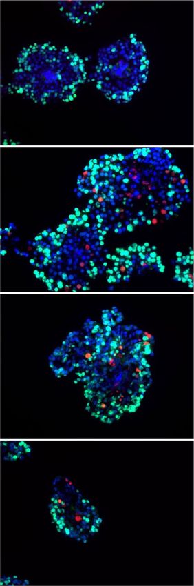
www.nature.com/scientificreports/
Proliferative effects of harmine, glucose, and HB‑EGF in intact human islets as assessed by
flow cytometry. The reliability of immunohistochemical assessment of β-cell proliferation is inherently
limited by the number of cells that can be manually counted and the operator’s visual appreciation of co-labeling.
We therefore exposed intact human islets to mitogens and assessed islet cell proliferation by flow cytometry
using EdU and immunostaining for CPEP (β cells), glucagon (GCG; α cells) and CK19 (ductal cells) (Fig. 6 and
Supplementary Fig. S4 online). On average, single cells represented 91 ± 3% of total islet cells of which 88 ± 2%
were live cells (Supplementary Table S1 online). The mean proportion of β and α cells in the donor islet batches
was 19 ± 3% and 42 ± 5%, respectively, whereas GCG−/CPEP− cells represented the remaining 39 ± 5% (Supple-
mentary Table S1 online). Although stringent gating of C PEP+ cells may have led to a slight underestimation of
the proportion of β cells, these data are comparable to non-diabetic donors in the Human Pancreas Analysis Pro-
gram (HPAP-RRID:SCR_016202) Database (https://hpap.pmacs.upenn.edu). Harmine induced β-cell prolifera-
tion in 5/6 donors (Fig. 6a,m) (fold-change 2.2 ± 0.6, a: P = 0.016, m: NS). Harmine was also a powerful mitogen
for α cells in 6/6 donors (Fig. 6b,n) (fold-change 7.7 ± 2.4, b: P = 0.022, n: P = 0.0015) and increased proliferation
of GCG−/CPEP− cells in 6/6 donors (Fig. 6c,o) (fold-change 3.2 ± 1.1, c: P = 0.027, o: NS) and CK19+ cells in 3/3
donors (Fig. 6d,p) (fold-change 5.0 ± 1.1, d and p: NS). Glucose did not promote β-cell proliferation in any of
the 10 donors tested (Fig. 6e,m) (fold-change 0.4 ± 0.1, e and m: NS). Furthermore, unlike harmine, glucose was
ineffective as an α-cell mitogen (Fig. 6f,n) (10 donors; fold-change 1.1 ± 0.3, f and n: NS), GCG−/CPEP− cells
(Fig. 6g,o) (10 donors; fold-change 0.9 ± 0.3, g and o: NS) and C K19+ cells (Fig. 6h,p) (3 donors; fold-change
1.2 ± 0.3, h and p: NS). HB-EGF increased β-cell proliferation in 9/13 donors (Fig. 6i,m) (fold-change 1.5 ± 0.3, i
and m: NS), an effect that was significant (fold-change 2.5 ± 0.6, i: P = 0.027, m: P = 0.04) when donor 13, identi-
fied as an outlier based on Grubb’s test, was excluded from the analysis. HB-EGF was largely inactive on α cells
(Fig. 6j,n) (13 donors; fold-change 1.2 ± 0.5, j and n: NS) but strongly stimulated GCG−/CPEP−-cell proliferation
in 13/13 donors (Fig. 6k,o) (fold-change 4.0 ± 1.3, k: P = 0.011, o: P = 0.039) and CK19+-cell proliferation in 5/5
donors (Fig. 6l,p) (fold-change 3.5 ± 1.0, l: P = 0.011, p: P = 0.046).
Comparison of immunohistochemistry and flow cytometry to assess human islet cell prolif‑
eration in response to harmine, glucose, and HB‑EGF. As variability in the proliferative response
between batches of islets prevents the comparison of immunohistochemistry (IHC) and flow cytometry (FC),
we performed these techniques in parallel on the same donor islets to investigate β-, α- and CK19+-cell prolifera-
tion. Intact human islets were exposed to harmine, glucose or HB-EGF and cell proliferation quantified by IHC
in intact islet sections and by FC following staining for CPEP, GCG, CK19 and EdU (Fig. 7). Both techniques
revealed a comparable percentage of proliferating CPEP+ cells at 2.8 mM glucose in 4/5 donors (Fig. 7a–d).
Although a significant increase was detected in 5/5 donors by IHC in response to harmine (Fig. 7a,b) (fold-
change 4.6 ± 0.7, P = 0.007), the response was much weaker when analyzed by FC (Fig. 7b) (fold-change 2.0 ± 0.8,
NS). The response to 16.7 mM glucose was comparable in 4/5 donors (Fig. 7a,c) (FC: fold-change 1.2 ± 0.6, NS;
IHC: fold-change 1.3 ± 0.6, NS), whereas the response to HB-EGF was similar in only 2/5 donors (Fig. 7a,d)
(FC: fold-change 2.6 ± 0.9, P = 0.043; IHC: fold-change 2.6 ± 0.9, P = 0.014). The proportion of proliferating GCG
+
cells detected by flow cytometry and immunohistochemistry at 2.8 mM glucose was similar in 4/4 donors
(Fig. 7e–h) as was the response to harmine in 3/4 donors (Fig. 7e,f) (FC: fold-change 22.4 ± 9.9, NS; IHC: fold-
change 11 ± 4.2, NS), glucose in 3/4 donors (Fig. 7e,g) (FC: fold-change 1.5 ± 1, NS; IHC: fold-change 1.5 ± 0.4,
P = 0.018) and HB-EGF in 4/4 donors (Fig. 7e,h) (FC: fold-change 1.3 ± 0.6, P = 0.032; IHC: fold-change 1 ± 0.6,
NS). The proportion of proliferating CK19+ cells detected at 2.8 mM glucose was comparable between both
K19+ cells was detected by
techniques in 4/4 donors (Fig. 7i–l). However, a significant increase in proliferating C
flow cytometry in 3/3 donors in response to harmine that was absent when assessed by immunohistochemistry
(Fig. 7i,j) (FC: fold-change 18.5 ± 6.4, NS; IHC: fold-change 2.7 ± 1.3, NS). The response to 16.7 mM glucose was
comparable in 4/4 donors (Fig. 7i,k) (FC: fold-change 1.4 ± 0.6, NS; IHC: fold-change 1.4 ± 0.6, NS) whereas the
response to HB-EGF was similar in only 2/4 donors (Fig. 7i,l) (FC: fold-change 1.8 ± 0.4, P = 0.022; IHC: fold-
change 5.4 ± 1.4, P = 0.01). Overall, these data reveal that although the percentage of proliferating GCG+ cells
detected by flow cytometry and immunohistochemistry was comparable in all conditions tested, significant
differences in the percentage of proliferating CPEP+ and CK19+ cells were observed.
Discussion
In this study we evaluated the capacity of harmine, glucose and HB-EGF to stimulate β-cell proliferation in iso-
lated human islets using a multi-faceted approach involving intact and dispersed islet cultures, flow cytometry
and immunohisto/cytochemistry and several proliferation and cell type-specific markers. Our data indicate that
in the majority of islet batches human β cells respond to harmine and HB-EGF irrespective of the experimental
approach, whereas the response to glucose is dependent on the donor and the assay used to assess proliferation.
The relative abundance of proliferating non-β cells in human islet batches prompted us to assess the effects of
these mitogens on α cells, C PEP−/GCG− cells and C
K19+ cells. We found that harmine potently increased the
proliferation of α cells and CPEP−/GCG− cells whereas HB-EGF mainly stimulated CPEP−/GCG− -cell prolifera-
tion including C K19+ cells.
Scientific Reports | (2021) 11:11283 | https://doi.org/10.1038/s41598-021-90643-3 5
Vol.:(0123456789)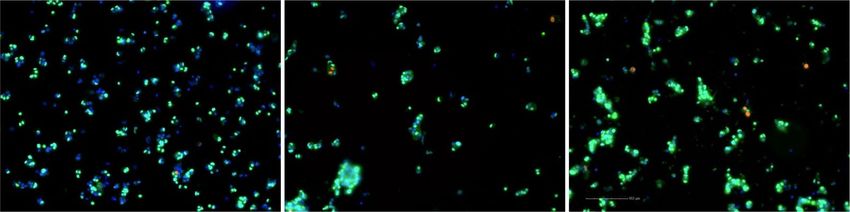
www.nature.com/scientificreports/

                                                                                                                                                               Vehicle
                                                                                                                                                               Harmine

                                                                                                                                                               2.8 Glu
                                                                                                                                                               16.7 Glu

                                                                                                                                                               Vehicle
                                                                                                                                                               HB-EGF

                                                                                                 8   P=0.0013                 18       P=0.0007

                                                                                                                              16

                                                                                                 6                            14
                                                                               % EdU+ of CPEP+

                                                                                                                % EdU+ of CPEP-

                                                                                                                              12
                                                                                                                              10
                                                                                                 4
                                                                                                                                  8
                                                                                                                                  6
                                                                                                 2
                                                                                                                                  4
                                                                                                                                  2
                                                                                  0                                               0
                                                                                 Low Glucose
                                                                                         Harmine
                                                                                             High Glucose
                                                                                                      HB-EGF                      Low Glucose
                                                                                                                                          Harmine
                                                                                                                                              High Glucose
                                                                                                                                                       HB-EGF
                                                                       Glucose, mM 2.8     2.8 16.7 2.8                               2.8   2.8   16.7   2.8
                                                                                      -    Har     -    HB                             -    Har    -     HB

          Scientific Reports |   (2021) 11:11283 |   https://doi.org/10.1038/s41598-021-90643-3                                                                       6

Vol:.(1234567890)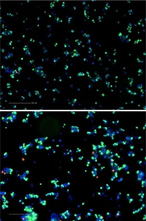
www.nature.com/scientificreports/
◂Figure 3. Analysis of β-cell proliferation in intact human islets by immunohistochemistry for C-peptide. (a–i)
Intact human islets were exposed to 2.8 mM glucose (Vehicle or 2.8 Glu; a–i), harmine (10 μM) (Har; a,b,e,f,i),
16.7 mM glucose (16.7 Glu; a,c,e,g,i) and HB-EGF (100 ng/ml) (HB; a,d,e,h,i) for 72 h. EdU (10 μM) was added
throughout. Proliferation was assessed by EdU staining and C-peptide (CPEP) to mark β cells. Representative
images of CPEP (green), EdU (red) and nuclei (blue) staining are shown (a). Arrows and arrowheads highlight
EdU+/CPEP+ and EdU+/CPEP− cells, respectively. Scale bar, 50 μm. Images were acquired using an Axio Imager
with ZEN 2012 software (https://www.zeiss.com/microscopy/int/products/microscope-software/zen.html).
A minimum of 1500 C PEP+ and CPEP− cells were counted for each sample. Proliferation is presented as the
percentage of EdU /CPEP+ over the total CPEP+ cells (b–e) or EdU+/CPEP− cells over the total CPEP− cells
+
(f–i). Graphs showing cell proliferation of individual (b–d,f–h) and combined (e,i) donors were generated
using GraphPad Prism 9 software (https://www.graphpad.com/scientific-software/prism/). Donor identifiers
(Supplementary Table S1 online) are indicated. Significance was tested using paired t tests (b–d,f–h) or one-way
ANOVA (e,i). P < 0.05 was considered significant. NS not significant.
Although the percentage of proliferating β cells detected by immunohistochemistry for Nkx6.1 or CPEP were
significantly correlated in all treatment conditions (Supplementary Fig. S5 online), the fraction of E dU+ cells
was markedly different. In general, the fraction of E dU+/Nkx6.1+ cells was lower than the corresponding E dU+/
+ 31
CPEP cells. This is reminiscent of a study by Aamodt et al. in dispersed human islets, who scored only β cells
that were both insulin+ and pancreatic and duodenal homeobox-1 (Pdx-1)+ and described a considerably lower
level of β-cell proliferation in response to harmine and glucose compared to studies where all i nsulin+ (or C PEP+)
cells were scored17–19,21,24–27,32. This phenomenon may be explained in part by the fact that actively proliferating
β-cells express lower levels of differentiation m arkers33, which would decrease the sensitivity of detection of
double labeling. Although CPEP is associated with functional β-cell differentiation, higher expression levels of
CPEP compared to Nkx6.1 may allow for the perdurance of CPEP protein in proliferating β cells.
By performing EdU and Ki67 staining of intact islets by immunohistochemistry we found that the percentage
of CPEP+ cells stained for each of these markers was comparable and significantly correlated in all treatment
conditions (Supplementary Fig. S5 online). This result was surprising given that EdU was added to the culture
medium throughout the 72-h exposure to mitogens. The expectation was that EdU should accumulate in more
cells than express Ki67 at the end of the assay. The similar levels may be due to both EdU and Ki67 capturing
all phases of the cell cycle. Taken together with the general absence of apoptosis in E dU+ cells, our data confirm
that EdU+ cells are proliferating.
The DYRK1A inhibitor harmine increased human β-cell proliferation irrespective of the assay used, and also
increased proliferation of GCG+, CPEP−/GCG−, and C K19+ cells in human islet batches. Whether the potency of
harmine differs between cell types, however, is not known as the mitogenic effects of harmine were only assessed
at 10 μM. Nevertheless, these data are consistent with previous studies demonstrating a potent mitogenic effect
of harmine on human β-cell replication both in vitro and in vivo21,26. Furthermore, the increase in proliferation
of non-β cells in response to harmine is expected given the ubiquitous nature of DYRK1A signaling, and is
consistent with several studies. Indeed, Wang et al.21 showed that combined inhibition of DYRK1A and TGFβ
signaling stimulates proliferation of C K19+ duct cells and endocrine α, δ and PP cells. Accordingly, the DYRK1A
inhibitor 5-iodotubercidin stimulates both β- and α-cell proliferation19. Finally, using single-cell mass cytometry
Wang et al.20 showed α cells to be considerably more responsive than β cells to the mitogenic effects of harmine.
Further studies will be required to identify drugs that selectively target the β cell for proliferation to advance
β-cell mass accretion as a viable therapeutic approach to treat diabetes.
We found that the capacity of glucose to increase human β-cell proliferation was dependent on the donor
and assay used. Glucose was inactive in the majority of donors in dispersed islets, whereas in intact islets β-cell
proliferation was increased in most donors when assessed by immunohistochemistry but not by flow cytometry.
In dispersed islets the loss of the cellular microenvironment and/or extended culture times (72 h) might con-
tribute to decreased β-cell survival and proliferation28. Our findings contrast with previous reports showing that
glucose (15–20 mM) can increase β-cell proliferation to up to 2% (approximately twofold over basal) in dispersed
human islets cultured from 16–96 h17,18,24,27,32, but are in line with our previous immunohistochemical data in
intact human islets34. Although the underlying reasons for the differences in the β-cell proliferative response
to glucose between our different assays ex vivo are unclear, it is noteworthy that variability in the response to
glucose has been observed in vivo. Transplantation of human islets into streptozotocin-induced diabetic non-
obese diabetic-severe combined immunodeficient mice revealed a correlation between glucose levels and β-cell
proliferation following glucose infusion16. Similarly, transplantation of human islets into spontaneously diabetic
immunodeficient non-obese diabetic-Rag1null IL2rγnull Ins2Akita mice led to β-cell proliferation that positively
correlated with glucose levels35. Yet, when adult human islets were transplanted into NSG-DT mice, a model of
hyperglycemia, no increase in β-cell proliferation was d etected36. In light of our ex vivo data, we speculate that
Scientific Reports | (2021) 11:11283 | https://doi.org/10.1038/s41598-021-90643-3 7
Vol.:(0123456789)www.nature.com/scientificreports/
Vehicle
Harmine
2.8 Glu
16.7 Glu
Vehicle
HB-EGF
Figure 4. Analysis of β-cell proliferation in intact human islets by immunohistochemistry for Ki67. (a–i)
Intact human islets were exposed to 2.8 mM glucose (Vehicle or 2.8 Glu; a–i), harmine (10 μM) (Har; a,b,e,f,i),
16.7 mM glucose (16.7 Glu; a,c,e,g,i) and HB-EGF (100 ng/ml) (HB; a,d,e,h,i) for 72 h. Proliferation was
assessed by staining for Ki67 and C-peptide (CPEP) to mark β cells. Representative images of CPEP (green),
Ki67 (red) and nuclei (blue) staining are shown (a). Arrows and arrowheads highlight Ki67+/CPEP+ and
Ki67+/CPEP− cells, respectively. Scale bar, 50 μm. Images were acquired using an Axio Imager with ZEN 2012
software (https://www.zeiss.com/microscopy/int/products/microscope-software/zen.html). A minimum of 1500
CPEP+ and CPEP− cells were counted for each sample. Proliferation is presented as the percentage of K i67+/
+ + + − −
CPEP over the total CPEP cells (b–e) or Ki67 /CPEP cells over the total C PEP cells (f–i). Graphs showing
cell proliferation of individual (b–d,f–h) and combined (e,i) donors were generated using GraphPad Prism 9
software (https://www.graphpad.com/scientific-software/prism/). Donor identifiers (Supplementary Table S1
online) are indicated. Significance was tested using paired t tests (b–d,f–h) or one-way ANOVA (e,i). P < 0.05
was considered significant. NS not significant.
Scientific Reports | (2021) 11:11283 | https://doi.org/10.1038/s41598-021-90643-3 8
Vol:.(1234567890)www.nature.com/scientificreports/
the effect of glucose on human β-cell proliferation in vivo may be mostly indirect, whereas the direct effects of
glucose, in striking contrast to the mouse β c ell9,10,18,37, may be relatively minor.
We recently demonstrated that the EGF receptor ligand HB-EGF exerts mitogenic activity on human β cells22.
In the present study we substantiated these findings by showing that β cells proliferate in response to HB-EGF
in the majority of human islet batches in both intact and dispersed islets. Although α cells were only weakly
responsive to HB-EGF, CPEP−/GCG− cells and CK19+ ductal cells were highly responsive to HB-EGF. However,
it is not known whether α cells respond to the mitogenic effects of HB-EGF at different concentrations since
HB-EGF was only tested at 100 ng/ml. In agreement with these findings, the EGF receptor is expressed in β, α
and ductal cells in the adult human pancreas38,39. Furthermore, ductal cells constitute a neogenic source of β
cells in rodents and h umans40 that are also responsive to EGF receptor ligands. In mice, adenoviral transduc-
tion of HB-EGF leads to ductal cell proliferation and differentiation into i nsulin+ cells41. In humans, ductal cells
proliferate in response to EGF and the related ligand betacellulin39, and the combination of EGF and gastrin
promotes ductal cell expansion and differentiation into Pdx-1+/insulin+ cells42. Hence, the proliferating β cells
detected in the presence of HB-EGF in this study could arise from replication of existing β cells and/or β-cell
neogenesis from ductal cells.
Discrepancies between the various approaches used in the present study and with previous reports point to
the sensitivity of human β cells to the culture conditions, for example using dispersed or intact islets, but also the
need for rigorous labeling and scoring of proliferating β cells. In particular, the abundance of non-β-cell types
in human islet batches could lead to the mis-identification of some proliferating cells as β cells. Importantly,
we note that non-β-cell types are rarely scored in human islet cell proliferation assays yet human islet batches
contain multiple cell types including other endocrine cells but also ductal and acinar as well as endothelial,
immune and stellate cells29.
By comparing immunohistochemistry and flow cytometry to assess proliferation we found that the percentage
of proliferating GCG+ cells was highly correlated in all conditions tested, whereas no significant correlation was
detected in the percentage of proliferating CPEP+ and C K19+ cells (Supplementary Fig. S5 online). We identified
a number of advantages and disadvantages of immunostaining and flow cytometry that may account for these
differences. In our experience accurately determining the cell type identity of a nucleus in images of dispersed
islet cells and intact islet sections is difficult, even when the operator is blinded to the experimental conditions,
which limits the reliability of immunostaining approach. In contrast, flow cytometry eliminates the subjectivity of
visual scoring and a higher number of cells can be easily and much more rapidly counted, increasing confidence
in the data. However, the need for more extensive manipulations in the processing of samples for flow cytometry
increases the level of cell death/loss compared to immunostaining. We conclude that multiple markers for β cells
(e.g. INS, CPEP, Nkx6.1, Pdx-1) and proliferating cells (e.g. EdU, Ki67, phospho-histone H3) as well as endo-
crine, exocrine and other cell types should be stained simultaneously or in parallel to enhance the robustness of
the conclusions and assess the relative efficacy of potential β-cell mitogens on several islet cell populations. We
hope that these findings will provide useful guidance to reliably assess human islet cell proliferation ex vivo in
the quest to identify novel and selective β-cell mitogens for potential therapeutic applications.
Methods
Human islets. Islets from non-diabetic human donors were provided by the Alberta Diabetes Institute
Islet Core, the Clinical Islet Laboratory at the University of Alberta, and the National Institute of Diabetes and
Digestive and Kidney Diseases-funded Integrated Islet Distribution Program (IIDP; Supplementary Table S1
online). The use of human islets was approved by the Institutional Ethics Committee of the Centre Hospitalier
de l’Université de Montréal (protocol # ND-05-035). All experiments were performed in accordance with the
Institutional Ethics Committee guidelines and regulations. Informed consent was obtained from all donors and/
or their legal guardians. Human islets were cultured in cGMP Prodo Islet Media (Standard) with 5% (vol./
vol.) human AB serum and 1% glutamine/glutathione mixture (Prodo Labs Inc., Irvine, CA) overnight prior to
experimentation.
Measurement of cell proliferation in dispersed islets by immunocytochemistry. Dispersed
escribed22. Briefly, hand-picked human islets were dis-
human islet proliferation was assessed essentially as d
persed in accutase for 10 min at 37 °C and plated in 96-well plates (Perkin Elmer Inc., Waltham, MA) treated
with Poly-d-Lysine Hydrobromide (Sigma-Aldrich, St. Louis, MO). The next day, cells were cultured in RPMI-
1640 (Thermo Fisher Scientific, Waltham, MA) with 1% (vol./vol.) human serum albumin (Celprogen, Torrance,
CA) for 3 days in the presence of EdU (10 μM) and glucose (2.8, 5.5 or 16.7 mM), harmine (10 μM; Millipore-
Sigma, Darmstadt, Germany) or HB-EGF (100 ng/ml; R&D Systems, Minneapolis, MN) as indicated in the
Scientific Reports | (2021) 11:11283 | https://doi.org/10.1038/s41598-021-90643-3 9
Vol.:(0123456789)www.nature.com/scientificreports/
Figure 5. Analysis of cytokeratin 19-positive cell proliferation in intact human islets by immunohistochemistry. ▸
(a–e) Intact human islets were exposed to 2.8 mM glucose (Vehicle or 2.8 Glu; a–e), harmine (10 μM) (Har;
a,b,e), 16.7 mM glucose (16.7 Glu; a,c,e) and HB-EGF (100 ng/ml) (HB; a,d,e) for 72 h. EdU (10 μM) was
added throughout. Proliferation was assessed by EdU staining and cytokeratin 19 (CK19) to mark ductal
cells. Representative images of CK19 (green), EdU (red) and nuclei (blue) staining are shown (a). Arrows and
arrowheads highlight EdU+/CK19+ and EdU+/CK19− cells, respectively. Scale bar, 50 μm. Images were acquired
using an Axio Imager with ZEN 2012 software (https://www.zeiss.com/microscopy/int/products/microscope-
software/zen.html). A minimum of 1000 CK19+ cells were counted for each sample. Proliferation is presented as
the percentage of E dU+/CK19+ over the total CK19+ cells (b–e). Graphs showing cell proliferation of individual
(b–d) combined (e) donors were generated using GraphPad Prism 9 software (https://www.graphpad.com/scien
tific-software/prism/). Donor identifiers (Supplementary Table S1 online) are indicated. Significance was tested
using paired t tests (b–d) or one-way ANOVA (e). P < 0.05 was considered significant. NS not significant.
Figure legends. At the end of the treatment, cells were fixed with 3.7% paraformaldehyde and immunostained
for Nkx6.1 (Supplementary Table S2 online). The proliferative marker EdU was detected using the Click-iT EdU
Imaging Kit with Alexa Fluor 594 (Thermo Fisher Scientific). Images were acquired with an Operetta CLS high-
content imaging system (Perkin Elmer Inc.) at 20X magnification using Harmony 4.5 software (https://www.
perkinelmer.com/en-ca/product/operetta-cls-system-hh16000000). Cell counts were determined using ImageJ
software (National Institutes of Health; https://imagej.net/Fiji). Proliferation was calculated as the percentage
of double-positive EdU+/Nkx6.1+ cells over the total Nkx6.1+ population. Between 1000 and 1500 Nkx6.1+ cells
from three different wells were manually counted per condition for each experimental replicate, as indicated in
the Figure legends.
Measurement of cell proliferation in intact islets by immunohistochemistry. Hand-picked
human islets (approximately 200/condition) were cultured in RPMI-1640 with 1% (vol./vol.) human serum
albumin in 6-well plates for 3 days in the presence of glucose (2.8 or 16.7 mM), HB-EGF (100 ng/ml) or harmine
(10 μM) as indicated in the Figure legends. EdU (10 μM) was added throughout. Media were replaced daily. At
the end of treatment, islets were embedded in optimal cutting temperature (OCT) compound, frozen, sectioned
at 8 μm and mounted on SuperFrost Plus slides (Thermo Fisher Scientific). Sections were fixed with 3.7% para-
formaldehyde and immunostained for Nkx6.1, CPEP, GCG or CK19 (Supplementary Table S2 online). Prolif-
eration was detected by immunostaining for Ki67 (Supplementary Table S2 online) or EdU using the Click-iT
EdU Imaging Kit with Alexa Fluor 594 or Pacific Blue. Secondary antibodies were from Jackson ImmunoRe-
search (West Grove, PA). Images were taken with an Axio Imager upright fluorescence microscope with ZEN
2012 imaging software (Zeiss, Oberkochen, Germany; https://www.zeiss.com/microscopy/int/products/micro
scope-software/zen.html). Between 1000 and 1500 Nkx6.1+, CPEP+, GCG+ or CK19+ cells from 7–21 islets on 2
separate slides were manually counted per condition for each experimental replicate, as indicated in the figure
legends.
Measurement of cell proliferation in intact islets by flow cytometry. At the end of the treatments
described above, islets were dispersed in Accutase (1 μl/islet; Innovative Cell Technologies, Inc., San Diego, CA)
for 10 min at 37 °C and dead cells were labeled using the LIVE/DEAD Fixable Aqua (405 nm) Dead Cell Stain
Kit (BD Biosciences, San Jose, CA). EdU detection, using the Click-iT Plus EdU Flow Cytometry Assay Kit with
Alexa Fluor 488, and immunostaining were performed according to the manufacturer’s instructions (Thermo
Fisher Scientific). Fluorophore-coupled primary antibodies and dilutions are listed in Supplementary Table S2
online. Flow cytometry analysis was carried out using a LSRIIB flow cytometer with BD FACSDiva software
(BD Biosciences). Data was analyzed using FlowJo v10.7 software (Ashland, OR; https://www.flowjo.com/solut
ions/flowjo). Dead-cell stain, EdU-, CPEP-, GCG-, and CK19-labeled cells were detected using the 405-, 488-,
640- and 561-nm lasers coupled with 525/50-, 530/30-, 670/14- and 586/15-nm BP filters, respectively. Prolif-
eration was calculated as the percentage of double-positive cells for EdU and each population marker over the
corresponding total cell population for that marker.
Measurement of apoptosis in intact islets. Apoptotic cells were visualized with the terminal deoxy-
nucleotidyl transferase (TdT)-FragEL DNA Fragmentation Detection Kit (Millipore, Etobicoke, ON, Canada)
according to the manufacturer’s instructions.
Scientific Reports | (2021) 11:11283 | https://doi.org/10.1038/s41598-021-90643-3 10
Vol:.(1234567890)www.nature.com/scientificreports/
Vehicle
Harmine
2.8 Glu
16.7 Glu
Vehicle
HB-EGF
4
P=0.005
3
% EdU+ of CK19+
2
1
0
Low Glucose
Harmine
High Glucose
HB-EGF
Glucose, mM 2.8 2.8 16.7 2.8
- Har - HB
Scientific Reports | (2021) 11:11283 | https://doi.org/10.1038/s41598-021-90643-3 11
Vol.:(0123456789)www.nature.com/scientificreports/
Vehicle 2.8 Glu Vehicle
Harmine 16.7 Glu HB-EGF
12 P=0.0015
10
% EdU+ of GCG+
8
6
4
2
0
Low Glucose
Harmine
High Glucose
HB-EGF
Glucose, mM 2.8 2.8 16.7 2.8
- Har - HB
P=0.039
30
28
12
% EdU+ of CPEP-, GCG-
10
8
6
4
2
0
Low Glucose
Harmine
High Glucose
HB-EGF
Glucose, mM 2.8 2.8 16.7 2.8
- Har - HB
9 P=0.046
8
7
% EdU+ of CK19+
6
5
4
3
2
1
0
Low Glucose
Harmine
High Glucose
HB-EGF
Glucose, mM 2.8 2.8 16.7 2.8
- Har - HB
Figure 6. Analysis of cell proliferation in intact human islets by flow cytometry. (a–p) Intact human islets
were exposed to 2.8 mM glucose (Vehicle or 2.8 Glu; a–p), harmine (10 μM) (Har; a–d,m–p), 16.7 mM glucose
(16.7 Glu; e–h,m–p) and HB-EGF (100 ng/ml) (HB; i–l,m–p) for 72 h. EdU (10 μM) was added throughout.
At the end of the treatment islets were dispersed into single cells and analyzed by flow cytometry for C-peptide
(CPEP), glucagon (GCG), cytokeratin 19 (CK19) and EdU. A minimum of 2000 C PEP+, GCG+, GCG−/
− +
CPEP and CK19 cells were counted for each sample. Proliferation is presented as the percentage of E dU+
cells over the total cells for each of CPEP+ (a,e,i,m), GCG+ (b,f,j,n), CPEP−, GCG− (c,g,k,o) and CK19+ (d,h,l,p)
cell types. Graphs showing cell proliferation of individual (a–l) and combined (m–p) donors were generated
using GraphPad Prism 9 software (https://www.graphpad.com/scientific-software/prism/). Donor identifiers
(Supplementary Table S1 online) are indicated. Significance was tested using paired t tests (a–l) and one-way
ANOVA (m–p). P < 0.05 was considered significant. NS, not significant.
Scientific Reports | (2021) 11:11283 | https://doi.org/10.1038/s41598-021-90643-3 12
Vol:.(1234567890)www.nature.com/scientificreports/
Vehicle 2.8 Glu Vehicle
Harmine 16.7 Glu HB-EGF
2.4 2.4 2.4
FC: NS FC: NS FC: P=0.043
IHC: P=0.0071 IHC: NS IHC: P=0.014
2.0 2.0 2.0
% EdU+ of CPEP+
% EdU+ of CPEP+
% EdU+ of CPEP+
1.6 1.6 1.6
1.2 1.2 1.2
0.8 0.8 0.8
0.4 0.4 0.4
0.0 0.0 0.0
IHC
C
IHC
C
IHC
C
IHC
C
IHC
C
IHC
C
IHC
C
IHC
IHC
IHC
C
IHC
C
C
C
IHC
C
IHC
C
IHC
C
IHC
C
F
F
F
F
F
F
F
F
F
F
F
F
F
F
F
29 30 31 32 33 29 30 31 32 33 29 30 31 32 33
14 1.4 1.4
FC: NS FC: NS FC: P=0.032
IHC: NS IHC: P=0.018 IHC: NS
12 1.2 1.2
10 1.0 1.0
% EdU+ of GCG+
% EdU+ of GCG+
% EdU+ of GCG+
8 0.8 0.8
6 0.6 0.6
4 0.4 0.4
2 0.2 0.2
0 0.0 0.0
IHC
C
IHC
C
IHC
C
IHC
C
IHC
C
IHC
C
IHC
C
IHC
C
IHC
C
IHC
C
IHC
C
IHC
C
F
F
F
F
F
F
F
F
F
F
F
F
30 31 32 33 30 31 32 33 30 31 32 33
9 0.8 2.0
FC: NS FC: P=0.022
FC: NS IHC: NS 1.8
8 IHC: P=0.01
IHC: NS 0.7
1.6
7 0.6
% EdU+ of CK19+
% EdU+ of CK19+
1.4
% EdU+ of CK19+
6
0.5
1.2
5
0.4 1.0
4
0.3 0.8
3
0.6
0.2
2 0.4
1 0.1
0.2
0 0.0 0.0
IHC
C
IHC
C
IHC
C
IHC
C
IHC
C
IHC
C
IHC
C
IHC
C
IHC
C
IHC
C
IHC
C
F
F
F
F
F
F
F
F
F
F
F
31 32 33 30 31 32 33 30 31 32 33
Figure 7. Comparison of immunohistochemistry and flow cytometry to assess human islet cell proliferation.
(a–l) Intact human islets were exposed to 2.8 mM glucose (Vehicle or 2.8 Glu; a–l), harmine (10 μM) (a,b,e,f,i,j),
16.7 mM glucose (16.7 Glu; a,c,e,g,i,k) and HB-EGF (100 ng/ml) (a,d,e,h,i,l) for 72 h. EdU (10 μM) was added
throughout. Proliferation was assessed in parallel for each donor by immunohistochemistry (IHC) and flow
cytometry (FC) following staining for C-peptide (CPEP), glucagon (GCG), cytokeratin 19 (CK19) and EdU.
(a,e,i) Representative images of CPEP (a, green), GCG (e, green), CK19 (i, green), EdU (red) and nuclei (blue)
staining are shown. Arrows and arrowheads highlight respectively, EdU+/CPEP+ and EdU+/CPEP− (a), EdU+/
GCG+ and EdU+/GCG− (e) and EdU+/CK19+ and EdU+/CK19− (i). Scale bar, 50 μm. Images were acquired
using an Axio Imager with ZEN 2012 software (https://www.zeiss.com/microscopy/int/products/microscope-
software/zen.html). A minimum of 1000 and 2000 CPEP+, GCG+ and CK19+ cells were counted for each sample
in immunohistochemistry and flow cytometry, respectively. Proliferation is presented as the percentage of E dU+
cells over the total cells for each of CPEP+ (a–d), GCG+ (e–h) and CK19+ (i–l) cell types. Graphs showing cell
proliferation of individual donors were generated using GraphPad Prism 9 software (https://www.graphpad.
com/scientific-software/prism/). Donor identifiers (Supplementary Table S1 online) are indicated. Significance
was tested using paired t tests (b-d,f-h,j-k). P < 0.05 was considered significant. NS, not significant.
Statistics. Significance was tested using two-tailed paired t tests or ordinary one-way ANOVA, using Graph-
Pad Prism 9 software (San Diego, CA; https://www.graphpad.com/scientific-software/prism/). P < 0.05 was con-
sidered significant. Fold-change data are expressed as mean ± SEM.
Data availability
All data generated or analyzed during this study are included in this published article (and its Supplementary
Information files).
Received: 29 October 2020; Accepted: 6 May 2021
Scientific Reports | (2021) 11:11283 | https://doi.org/10.1038/s41598-021-90643-3 13
Vol.:(0123456789)www.nature.com/scientificreports/
References
1. Kahn, S. E., Cooper, M. E. & Del Prato, S. Pathophysiology and treatment of type 2 diabetes: Perspectives on the past, present, and
future. Lancet 383, 1068–1083. https://doi.org/10.1016/s0140-6736(13)62154-6 (2014).
2. Okada, T. et al. Insulin receptors in beta-cells are critical for islet compensatory growth response to insulin resistance. Proc. Natl.
Acad. Sci. U.S.A. 104, 8977–8982. https://doi.org/10.1073/pnas.0608703104 (2007).
3. Park, S. et al. Exendin-4 uses Irs2 signaling to mediate pancreatic beta cell growth and function. J. Biol. Chem. 281, 1159–1168.
https://doi.org/10.1074/jbc.M508307200 (2006).
4. Kim, H. et al. Serotonin regulates pancreatic beta cell mass during pregnancy. Nat. Med. 16, 804–808. https://doi.org/10.1038/nm.
2173 (2010).
5. Hakonen, E. et al. Epidermal growth factor (EGF)-receptor signalling is needed for murine beta cell mass expansion in response
to high-fat diet and pregnancy but not after pancreatic duct ligation. Diabetologia 54, 1735–1743. https://doi.org/10.1007/s00125-
011-2153-1 (2011).
6. Kondegowda, N. G. et al. Osteoprotegerin and denosumab stimulate human beta cell proliferation through inhibition of the recep-
tor activator of NF-kappaB ligand pathway. Cell Metab. 22, 77–85. https://doi.org/10.1016/j.cmet.2015.05.021 (2015).
7. El Ouaamari, A. et al. SerpinB1 promotes pancreatic beta cell proliferation. Cell Metab. 23, 194–205. https://doi.org/10.1016/j.
cmet.2015.12.001 (2016).
8. Alonso, L. C. et al. Glucose infusion in mice: A new model to induce beta-cell replication. Diabetes 56, 1792–1801. https://doi.
org/10.2337/db06-1513 (2007).
9. Terauchi, Y. et al. Glucokinase and IRS-2 are required for compensatory beta cell hyperplasia in response to high-fat diet-induced
insulin resistance. J. Clin. Investig. 117, 246–257. https://doi.org/10.1172/jci17645 (2007).
10. Porat, S. et al. Control of pancreatic beta cell regeneration by glucose metabolism. Cell Metab. 13, 440–449. https://doi.org/10.
1016/j.cmet.2011.02.012 (2011).
11. Stamateris, R. E., Sharma, R. B., Hollern, D. A. & Alonso, L. C. Adaptive beta-cell proliferation increases early in high-fat feeding
in mice, concurrent with metabolic changes, with induction of islet cyclin D2 expression. Am. J. Physiol. Endocrinol. Metab. 305,
E149–E159. https://doi.org/10.1152/ajpendo.00040.2013 (2013).
12. Moulle, V. S. et al. Glucose and fatty acids synergistically and reversibly promote beta cell proliferation in rats. Diabetologia 60,
879–888. https://doi.org/10.1007/s00125-016-4197-8 (2017).
13. Kulkarni, R. N., Mizrachi, E. B., Ocana, A. G. & Stewart, A. F. Human beta-cell proliferation and intracellular signaling: Driving
in the dark without a road map. Diabetes 61, 2205–2213. https://doi.org/10.2337/db12-0018 (2012).
14. Bernal-Mizrachi, E. et al. Human beta-cell proliferation and intracellular signaling part 2: Still driving in the dark without a road
map. Diabetes 63, 819–831. https://doi.org/10.2337/db13-1146 (2014).
15. Stewart, A. F. et al. Human beta-cell proliferation and intracellular signaling: Part 3. Diabetes 64, 1872–1885. https://doi.org/10.
2337/db14-1843 (2015).
16. Levitt, H. E. et al. Glucose stimulates human beta cell replication in vivo in islets transplanted into NOD-severe combined immu-
nodeficiency (SCID) mice. Diabetologia 54, 572–582. https://doi.org/10.1007/s00125-010-1919-1 (2011).
17. Lakshmipathi, J. et al. PKCzeta Is essential for pancreatic beta-cell replication during insulin resistance by regulating mTOR and
Cyclin-D2. Diabetes 65, 1283–1296. https://doi.org/10.2337/db15-1398 (2016).
18. Stamateris, R. E. et al. Glucose induces mouse beta-cell proliferation via IRS2, MTOR, and Cyclin D2 but not the insulin receptor.
Diabetes 65, 981–995. https://doi.org/10.2337/db15-0529 (2016).
19. Dirice, E. et al. Inhibition of DYRK1A stimulates human beta-cell proliferation. Diabetes 65, 1660–1671. https://doi.org/10.2337/
db15-1127 (2016).
20. Wang, Y. J. et al. Single-cell mass cytometry analysis of the human endocrine pancreas. Cell Metab. 24, 616–626. https://doi.org/
10.1016/j.cmet.2016.09.007 (2016).
21. Wang, P. et al. Combined inhibition of DYRK1A, SMAD, and Trithorax pathways synergizes to induce robust replication in adult
human beta cells. Cell Metab. 29, 638-652.e635. https://doi.org/10.1016/j.cmet.2018.12.005 (2019).
22. Maachi, H. et al. HB-EGF signaling is required for glucose-induced pancreatic β-cell proliferation in rats. Diabetes 69, 369–380.
https://doi.org/10.2337/db19-0643 (2020).
23. Cox, A. R. et al. Resolving discrepant findings on ANGPTL8 in β-cell proliferation: A collaborative approach to resolving the
betatrophin controversy. PLoS One 11, e0159276. https://doi.org/10.1371/journal.pone.0159276 (2016).
24. Metukuri, M. R. et al. ChREBP mediates glucose-stimulated pancreatic beta-cell proliferation. Diabetes 61, 2004–2015. https://
doi.org/10.2337/db11-0802 (2012).
25. Shen, W. et al. Inhibition of DYRK1A and GSK3B induces human beta-cell proliferation. Nat. Commun. 6, 8372. https://doi.org/
10.1038/ncomms9372 (2015).
26. Wang, P. et al. A high-throughput chemical screen reveals that harmine-mediated inhibition of DYRK1A increases human pan-
creatic beta cell replication. Nat. Med. 21, 383–388. https://doi.org/10.1038/nm.3820 (2015).
27. Kumar, A. et al. Activation of Nrf2 is required for normal and ChREBPalpha-augmented glucose-stimulated beta-cell proliferation.
Diabetes 67, 1561–1575. https://doi.org/10.2337/db17-0943 (2018).
28. Aamodt, K. I. & Powers, A. C. Signals in the pancreatic islet microenvironment influence β-cell proliferation. Diabetes Obes. Metab.
19(Suppl 1), 124–136. https://doi.org/10.1111/dom.13031 (2017).
29. Segerstolpe, Å. et al. Single-cell transcriptome profiling of human pancreatic islets in health and type 2 diabetes. Cell Metab. 24,
593–607. https://doi.org/10.1016/j.cmet.2016.08.020 (2016).
30. Bonner-Weir, S. et al. In vitro cultivation of human islets from expanded ductal tissue. Proc. Natl. Acad. Sci. U.S.A. 97, 7999–8004.
https://doi.org/10.1073/pnas.97.14.7999 (2000).
31. Aamodt, K. I. et al. Development of a reliable automated screening system to identify small molecules and biologics that promote
human beta-cell regeneration. Am. J. Physiol. Endocrinol. Metab. 311, E859-e868. https://doi.org/10.1152/ajpendo.00515.2015
(2016).
32. Sharma, R. B. et al. Insulin demand regulates beta cell number via the unfolded protein response. J. Clin. Investig. 125, 3831–3846.
https://doi.org/10.1172/jci79264 (2015).
33. Russ, H. A., Bar, Y., Ravassard, P. & Efrat, S. In vitro proliferation of cells derived from adult human beta-cells revealed by cell-
lineage tracing. Diabetes 57, 1575–1583. https://doi.org/10.2337/db07-1283 (2008).
34. Vivot, K. et al. The regulator of G-protein signaling RGS16 promotes insulin secretion and beta-cell proliferation in rodent and
human islets. Mol. Metab. 5, 988–996. https://doi.org/10.1016/j.molmet.2016.08.010 (2016).
35. Diiorio, P. et al. Hyperglycemia-induced proliferation of adult human beta cells engrafted into spontaneously diabetic immunode-
ficient NOD-Rag1null IL2rgammanull Ins2Akita mice. Pancreas 40, 1147–1149. https://doi.org/10.1097/MPA.0b013e31821ffabe
(2011).
36. Dai, C. et al. Stress-impaired transcription factor expression and insulin secretion in transplanted human islets. J. Clin. Investig.
126, 1857–1870. https://doi.org/10.1172/jci83657 (2016).
37. Salpeter, S. J. et al. Glucose regulates cyclin D2 expression in quiescent and replicating pancreatic β-cells through glycolysis and
calcium channels. Endocrinology 152, 2589–2598. https://doi.org/10.1210/en.2010-1372 (2011).
Scientific Reports | (2021) 11:11283 | https://doi.org/10.1038/s41598-021-90643-3 14
Vol:.(1234567890)www.nature.com/scientificreports/
38. Miyagawa, J. et al. Immunohistochemical localization of betacellulin, a new member of the EGF family, in normal human pancreas
and islet tumor cells. Endocr. J. 46, 755–764 (1999).
39. Rescan, C. et al. EGF-induced proliferation of adult human pancreatic duct cells is mediated by the MEK/ERK cascade. Lab.
Investig. 85, 65–74. https://doi.org/10.1038/labinvest.3700204 (2005).
40. Dirice, E. et al. Human duct cells contribute to β cell compensation in insulin resistance. JCI Insight 4, e99576. https://doi.org/10.
1172/jci.insight.99576 (2019).
41. Kozawa, J. et al. Regenerative and therapeutic effects of heparin-binding epidermal growth factor-like growth factor on diabetes
by gene transduction through retrograde pancreatic duct injection of adenovirus vector. Pancreas 31, 32–42 (2005).
42. Suarez-Pinzon, W. L., Lakey, J. R., Brand, S. J. & Rabinovitch, A. Combination therapy with epidermal growth factor and gastrin
induces neogenesis of human islet {beta}-cells from pancreatic duct cells and an increase in functional {beta}-cell mass. J. Clin.
Endocrinol. Metab. 90, 3401–3409. https://doi.org/10.1210/jc.2004-0761 (2005).
Acknowledgements
This study was supported by the National Institutes of Health (Grant R01-DK-58096 to V.P.), the Canadian
Institutes of Health Research (Grant MOP 77686 to V.P.), and the Cardiometabolic Health, Diabetes and Obesity
research Network of Quebec. Human pancreatic islets were provided by the IIDP (RRID:SCR_014387) at City
of Hope, NIH Grant # 2UC4DK098085 and the JDRF-funded IIDP Islet Award Initiative. H.M. was supported
by a doctoral studentship from the Fonds de Recherche Québec—Santé. V.P. holds the Canada Research Chair
in Diabetes and Pancreatic B Cell Function.
Author contributions
J.G., H.M. and V.P. designed the experiments, analyzed the results, and wrote the manuscript. H.M., J.G., and
C.T. acquired the data. All authors revised the manuscript and approved the final version. V.P. is the guarantor
of this work and, as such, takes full responsibility for the work.
Competing interests
The authors declare no competing interests.
Additional information
Supplementary Information The online version contains supplementary material available at https://doi.org/
10.1038/s41598-021-90643-3.
Correspondence and requests for materials should be addressed to V.P.
Reprints and permissions information is available at www.nature.com/reprints.
Publisher’s note Springer Nature remains neutral with regard to jurisdictional claims in published maps and
institutional affiliations.
Open Access This article is licensed under a Creative Commons Attribution 4.0 International
License, which permits use, sharing, adaptation, distribution and reproduction in any medium or
format, as long as you give appropriate credit to the original author(s) and the source, provide a link to the
Creative Commons licence, and indicate if changes were made. The images or other third party material in this
article are included in the article’s Creative Commons licence, unless indicated otherwise in a credit line to the
material. If material is not included in the article’s Creative Commons licence and your intended use is not
permitted by statutory regulation or exceeds the permitted use, you will need to obtain permission directly from
the copyright holder. To view a copy of this licence, visit http://creativecommons.org/licenses/by/4.0/.
© The Author(s) 2021
Scientific Reports | (2021) 11:11283 | https://doi.org/10.1038/s41598-021-90643-3 15
Vol.:(0123456789)You can also read