Narcissistic personality traits and prefrontal brain structure
←
→
Page content transcription
If your browser does not render page correctly, please read the page content below
www.nature.com/scientificreports
OPEN Narcissistic personality traits
and prefrontal brain structure
Igor Nenadić1,2,3*, Carsten Lorenz3 & Christian Gaser3,4
Narcissistic traits have been linked to structural and functional brain networks, including the insular
cortex, however, with inconsistent findings. In this study, we tested the hypothesis that subclinical
narcissism is associated with variations in regional brain volumes in insular and prefrontal areas. We
studied 103 clinically healthy subjects, who were assessed for narcissistic traits using the Narcissistic
Personality Inventory (NPI, 40-item version) and received high-resolution structural magnetic
resonance imaging. Voxel-based morphometry was used to analyse MRI scans and multiple regression
models were used for statistical analysis, with threshold-free cluster enhancement (TFCE). We
found significant (p < 0.05, family-wise error FWE corrected) positive correlations of NPI scores with
grey matter in multiple prefrontal cortical areas (including the medial and ventromedial, anterior/
rostral dorsolateral prefrontal and orbitofrontal cortices, subgenual and mid-anterior cingulate
cortices, insula, and bilateral caudate nuclei). We did not observe reliable links to particular facets
of NPI-narcissism. Our findings provide novel evidence for an association of narcissistic traits with
variations in prefrontal and insular brain structure, which also overlap with previous functional studies
of narcissism-related phenotypes including self-enhancement and social dominance. However,
further studies are needed to clarify differential associations to entitlement vs. vulnerable facets of
narcissism.
Narcissism refers to a set of personality traits incorporating cognitive, emotional, and behavioural features, which
are commonly conceptualised around facets of grandiosity, entitlement, and v ulnerability1–3. Current concep-
tualisations of narcissism therefore consider a bipolarity of grandiose vs. vulnerable narcissism or multipolarity
of major facets, often evolving around deficits in maintaining functional levels of self-esteem, with such traits
being common in the general population and not uniformly linked to dysfunction or d istress2.
Narcissistic traits have been studied both in social or personality psychology as well as clinical contexts,
especially with reference to narcissistic personality disorder (NaPD)4–6. While the case has been made that
clinical research on narcissistic personality disorder might benefit from data obtained in non-clinical studies of
narcissistic traits7, the relation between the conceptualisations in these two different lines of research is by no
means clear and a matter of ongoing debates and research3,6,8. In the subclinical range, narcissistic traits can be
associated with positive effects in initial group formation and leadership, but often lead to adverse interactional
outcomes over time9,10.
Psychometric characterisation of the narcissistic phenotype in general population cohorts has relied on well-
established and validated questionnaires, in particular the Narcissistic Personality Inventory (NPI) by Raskin and
Hall11, which considers aspects of grandiosity, as well as leadership and entitlement12–16. Hence, while alternative
more recent scales have become available16–18, the NPI still remains a widely used instrument19,20 with a large
database of studies21,22.
Given the relevance of narcissism in both clinical and non-clinical research fields, there is an astonishing
paucity of neuroscience research relating narcissistic traits or behaviours to either brain function or structure.
A pioneering explorative functional magnetic resonance imaging (fMRI) study comparing 11 high-narcissistic
vs. 11 low-narcissistic subjects using an empathy paradigm implied decreased deactivation in the right anterior
insula in the high-narcissism g roup23, an area implicated in cognitive e mpathy24–26, which can be considered
a main factor in developing prosocial b ehaviours27,28. Further functional studies have found correlations of
narcissistic traits in clinically healthy subjects in anterior insula and dorsal anterior cingulate and subgenual
cingulate cortices during tasks involving social rejection29, as well as elevated dorsal anterior cingulate cortex
1
Department of Psychiatry and Psychotherapy, Philipps Universität Marburg,
Rudolf‑Bultmann‑Str. 8, 35039 Marburg, Germany. 2Department of Psychology, Goethe-Universität
Frankfurt, Frankfurt, Germany. 3Department of Psychiatry and Psychotherapy, Jena University Hospital,
Jena, Germany. 4Department of Neurology, Jena University Hospital, Jena, Germany. *email: nenadic@
staff.uni-marburg.de
Scientific Reports | (2021) 11:15707 | https://doi.org/10.1038/s41598-021-94920-z 1
Vol.:(0123456789)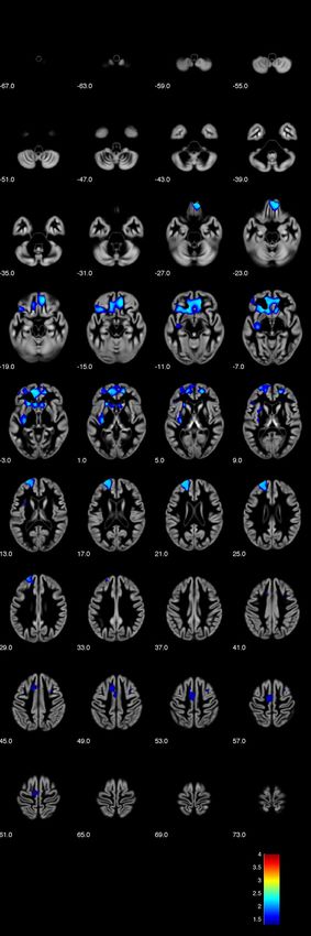
www.nature.com/scientificreports/
(dACC) response to social rejection s timuli30 and self-related visual stimulus p rocessing31. In an EEG study, feed-
back related negativity in midline frontal areas in an EEG study did not differ between low vs high narcissistic
subjects, but a difference in centro-parietal P3 e merged32. Together with studies implicating impaired structural
white matter connectivity in frontostriatal t racts33, this gives rise to (anterior) insula and prefrontal (esp. dACC)
involvement in narcissistic behaviours.
In contrast to these cues from functional imaging studies, there is no clear evidence on the brain structural
underpinnings, esp. for grey matter. One previous study using cortical thickness measurements reported a nega-
tive correlation of PNI (pathological narcissism inventory) scores with right dorsolateral and inferior prefrontal
thickness, and cortical volumes in the left medial prefrontal and right dorsolateral prefrontal cortices34, while
another showed an interaction of gender and NPI scores in the right superior parietal cortex using voxel-based
morphometry35.
The present study was conducted to test the association of brain structure and narcissistic traits in a non-
clinical cohort. In particular, we tested the hypothesis that subclinical narcissistic traits (assessed with the NPI)
would be correlated with prefrontal brain structures (as implicated in functional studies and one of the preceding
cortical mapping studies) as well as the (anterior) insula. We chose a whole-brain voxel-wise analysis for spatial
resolution to distinguish between different areas of the orbital, medial, and lateral prefrontal cortices.
Methods
Study cohort and phenotyping. For this study, we analysed data from a total of 103 psychiatrically
healthy subjects (53 female, 50 male) recruited from the local community. All participants gave written informed
consent to study participation as part of a study protocol approved by the local ethic committee of the Medical
School of Friedrich-Schiller-University of Jena, in accordance with the Declaration of Helsinki in its current
version. Inclusion criteria were age 18–65 years and ability to provide informed consent, while exclusion criteria
were any concurrent or previous psychiatric disorder (including current substance dependence) central nervous
neurological disorders (including traumatic brain injury/loss of consciousness), or learning disability/IQ lower
than 80, as well as intake of psychotropic medication.
Subjects were screened for absence of exclusion criteria, in particular any previous treatment for psychiatric
disorders. IQ was estimated using the MWT-B (Mehrfachwortschatztest B;36,37), and while IQ scores lower 70
would be considered suggestive of a learning disability, we defined an exclusion threshold of 80 to take into
account imprecisions and potential overestimations of this screening test (ultimately, however, none of our
recruited subjects was excluded as the minimum detected IQ in this sample was 88). Following screening and
formal inclusion, subjects underwent MRI scanning and phenotyping for narcissistic traits.
We used the narcissistic personality inventory N PI11, applying the full 40-item validated German v ersion38, to
characterise our sample for narcissistic traits. The NPI has been used in a large number of studies20,39, including
non-clinical and clinical samples, as well as several of the functional imaging studies cited above. While validity
studies of the NPI by Raskin and Terry suggested seven components defined as authority, exhibitionism, supe-
riority, vanity, exploitativeness, entitlement, and self-sufficiency40, there have been alternative accounts of four
factors labelled exploitativeness/entitlement, leadership/authority, superiority/arrogance, self-absorption/self-
admiration15, and more recently of two or three factors assigned ‘power’, ‘exhibitionism’, and ‘special person’41.
In particular, Ackerman and colleagues in a recent re-appraisal of the NPI including analyses of large college
student samples12, advocated a three-factor model (with facets: leadership/authority, grandiose exhibitionism,
and entitlement/exploitativeness). Based on findings of the validation study and factorial analysis of the German
NPI translation38, we calculated additional seven NPI subscales designated (sample items in brackets refer to the
original NPI text in English): authority (8 items, e.g.: “I am a born leader”), entitlement (6 items, e.g.; “I insist
upon the respect that is due me.”), exhibitionism (7 items, e.g.: “Modesty doesn’t become me.”), exploitativeness
(6 items, e.g.: “I can make anybody believe anything I want them to”), self-sufficiency (6 items, e.g.: “I rarely
depend on anyone else to get things done.”), superiority (5 items, e.g.: “I think I am a special person.”), vanity (3
items, e.g.: “I like to look at myself in the mirror”).
Demographic and psychometric data of the sample are summarised in Table 1.
Magnetic resonance image (MRI) acquisition. MRI scanning was done on a 3 Tesla Siemens Tim Trio
system (Siemens, Erlangen, Germany) using a T1-weighted high-resolution MPRAGE sequence (magnetisa-
tion-prepared rapid gradient echo) with a standard quadrature head coil (scanning parameters: TR 2300 ms,
TE 3.03 ms, flip angle α 9°, in-plane field-of-view 256 mm) acquiring 192 contiguous sagittal slices covering the
whole brain. Scanning duration was 5:21 min. All scans were visually inspected after scanning for gross artefacts
(e.g. movement, ghosting), and all scans passed this initial step of quality assurance.
Voxel‑based morphometry. We used a voxel-based morphometry (VBM) approach to analyse T1 scans,
using Statistical Parametric Mapping (SPM) software (Wellcome Institute of Imaging Neuroscience, Institute
of Neurology, London, UK) running on Matlab (Mathworks, Natik, MA, USA) and the VBM8 toolbox, r435
(C. Gaser, Jena University Hospital; http://www.dbm.neuro.uni-jena.de/vbm/vbm8), as in two previous studies
of personality traits and narcissistic personality disorder, respectively42,43. Our processing pipeline have been
described previously (e.g.43), including augmentation of segmentation through accounting for partial volume
effects44, adaptive maximum a posteriori estimation45, and hidden Markov Random Field models46. All scans
passed the automated quality assurance protocol in VBM8. After segmentation of grey matter maps, we applied
an internal grey matter threshold of 0.2, in order to eliminate potential artefacts at ambiguous grey matter bor-
ders; this threshold is more conservative than the often used 0.1 GM threshold. Anatomical labelling was avail-
able with the AAL atlas47.
Scientific Reports | (2021) 11:15707 | https://doi.org/10.1038/s41598-021-94920-z 2
Vol:.(1234567890)www.nature.com/scientificreports/
Mean (standard deviation) total sample Mean (standard deviation) female subjects Mean (standard deviation) male subjects
(n = 103) (n = 53) (n = 50)
Age 31.7 years (10.2) 33.4 yrs (10.8) 29.9 years (9.3)
Estimated IQ (MWT-B score) 108.5 (12.5) 108.68 (12.3) 108.34 (12.8)
NPI total score 12.23 (5.73) 11.28 (5.5) 13.24 (5.9)
NPI subscale authority 3.31 (1.88) 2.96 (1.7) 3.68 (2)
NPI subscale entitlement 1.17 (1.22) 0.85 (0.9) 1.52 (1.4)
NPI subscale exhibitionism 2.01 (1.54) 1.87 (1.5) 2.16 (1.6)
NPI subscale exploitativeness 1.60 (1.22) 1.49 (1.1) 1.72 (1.3)
NPI subscale self-sufficiency 1.55 (1.28) 1.58 (1.3) 1.52 (1.3)
NPI subscale superiority 1.42 (1.03) 1.3 (1) 1.54 (1.1)
NPI subscale vanity 1.17 (1.09) 1.23 (1.1) 1.1 (1.1)
Table 1. Demographic and psychometric data of the study cohort (n = 103), including data on female (n = 53)
versus male (n = 50) subjects.
Statistical analysis. For all VBM statistical analysis, we used threshold-free cluster enhancement (TFCE),
an approach introduced to increase sensitivity of voxel-based a nalyses48,49, applying 5000 permutations (Smith
method).
First, we tested our main hypothesis of brain structural associations with NPI scores using a general linear
model (GLM) in SPM with NPI total score as regressor and age and sex as nuisance variables (in order to remove
age and sex related effects). Based on TFCE, we then used a p = 0.05 family-wise error (FWE) correction to cor-
rect for multiple comparisons across whole-brain GM voxels, testing for both positive and negative correlations.
NPI skewness of 0.411 was in an acceptable range for this statistical approach.
Second, we followed up our main analysis by testing the hypothesis of sex interactions, i.e. that correlation
slopes might differ significantly between female and male study participants. For this purpose, we set up a new
GLM, again using age as a regressor, to reveal areas in which female subjects would show a higher/steeper increase
over males and vice versa. This analysis aimed at replicating the previous finding35 of sexually dimorphic associa-
tions for the parietal cortex in a VBM study (with unclear main effects of NPI total scores).
The exploratory nature of this analysis acknowledges limited statistical power in these (smaller) subgroups of
the study cohort, as well as interaction effects in VBM often being more difficult to detect given lack of sensitivity
even in decent sized samples.
Third, we performed exploratory analyses testing for potential associations of the seven NPI subscales with
brain structure, defining separate GLMs, each including the respective NPI subscale, as well as age and sex as
nuisance variables.
Results
Associations of NPI total score with brain structure. In our main analysis, we found significant
(p < 0.05, FWE-corrected, TFCE) positive correlations NPI total scores with regional brain grey matter volume
in four clusters including bilateral medial, orbital, and dorsolateral prefrontal as well as left insular cortices (see
Figs. 1 and 2).
Of the four significant clusters, the first cluster spanned a large confluence of regions mostly covering the
prefrontal areas (cluster size k = 15,419, maximum intensity voxel at MNI space co-ordinates 10; 39; − 14 with
pFWE-corr = 0.005, with additional local maxima at 10; 47; − 20 and − 21; 24; 6 – both at p FWE-corr = 0.006).
Additional clusters were k = 1377 voxels (maximum intensity voxel at − 12; 18; 48 with pFWE-corr = 0.035; addi-
tional local maxima − 9; − 15; 60 with p FWE-corr = 0.036 and − 3; − 4; 52 with p FWE-corr = 0.037), and smaller clusters
with k = 178 voxels (maximum intensity voxel at − 30; 9; 40 with p FWE-corr = 0.045; additional local maxima 32;8;49
with pFWE-corr = 0.047 and − 30; 11; 57 with p FWE-corr = 0.047) and k = 102 voxels (maximum intensity voxel at − 44;
27; 7 with p FWE-corr = 0.047), respectively, with clusters extending towards bilateral caudate nuclei.
Interaction effects with NPI total scores. We did not identify a significant interaction effect of sex and
NPI total scores on brain structure at corrected thresholds (p < 0.05, FWE-corrected, TFCE) apart from one
single voxel in the right lateral prefrontal cortex (k = 1; 58; 22; 3, p
FWE-corr = 0.05) with higher correlation slopes
in women. In particular, we did not identify any sex-by-NPI interaction in the superior parietal cortex, as impli-
cated in a previous study35.
In further exploratory analysis at uncorrected threshold levels (p < 0.001, uncorr.), women showed steeper
positive correlations with NPI total scores than men in two right dorsolateral prefrontal clusters (k = 930; maxi-
mum intensity voxel 58; 22; 3; and k = 233; 36;26;28) and one in the right posterior parietal/occipital cortex
(k = 116; 30; − 81; 40) and one single voxel at 8; − 25; 73. There were no inverse effects (i.e. steeper slopes in men
compared to women) even at p < 0.001 uncorrected thresholds.
Comparison of psychometric data between female and male participants did not show significant group-level
differences, apart from one single scale with male subjects scoring higher on the NPI subscale entitlement (T-test:
T = 2.898, p = 0.005; assuming unequal variances based on Levene-test F = 11.154, p = 0.001), and trend-level find-
ings for higher values of total NPI score in male subjects (T-test: T = 1.749, p = 0.083; assuming equal variance
Scientific Reports | (2021) 11:15707 | https://doi.org/10.1038/s41598-021-94920-z 3
Vol.:(0123456789)www.nature.com/scientificreports/
Figure 1. Voxel-based morphometry (VBM) analysis showing positive correlations of narcissistic personality
inventory (NPI) total score with grey matter (TFCE analysis, p < 0.05 FWE corrected, axial sections with z levels
given beneath each section) (Image created using the VBM8 toolbox, version r435; C. Gaser, Structural Brain
Mapping Group, Jena University Hospital, Jena, Germany; http://www.dbm.neuro.uni-jena.de/vbm/vbm8).
Scientific Reports | (2021) 11:15707 | https://doi.org/10.1038/s41598-021-94920-z 4
Vol:.(1234567890)www.nature.com/scientificreports/
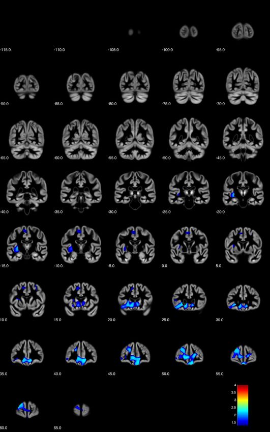
Figure 2. Voxel-based morphometry (VBM) analysis showing positive correlations of narcissistic personality inventory (NPI)
total score with grey matter (TFCE analysis, p < 0.05 FWE corrected, coronal sections with y levels given beneath each section)
(Image created using the VBM8 toolbox, version r435; C. Gaser, Structural Brain Mapping Group, Jena University Hospital,
Jena, Germany; http://www.dbm.neuro.uni-jena.de/vbm/vbm8).
Scientific Reports | (2021) 11:15707 | https://doi.org/10.1038/s41598-021-94920-z 5
Vol.:(0123456789)www.nature.com/scientificreports/
based on Levene-text F = 0.19, p = 0.664), and higher values for NPI subscale authority in male subjects (T-test:
T = 1.956, p = 0.053; assuming unequal variances based on Levene-test F = 4.216, p = 0.043).
Exploratory analysis of brain structure and NPI subscales. Exploratory analysis of the seven NPI
subscales (authority, entitlement, exhibitionism, exploitativeness, self-sufficiency, superiority, vanity) revealed
only small minor clusters in the following associations (only those with k > 15 reported): (a) for exhibition-
ism a positive correlation with two clusters in the left parietal lobe (k = 124; maximum at – 36; − 40; 52 with
pFWE-corr = 0.047) and right medial parietal/cingulate cortex (k = 17; maximum at 12; − 28; 33 with pFWE-corr = 0.048),
(b) for self-sufficiency a positive correlation with a cluster in the left medial prefrontal cortex (k = 84; maximum
at – 10; 12; − 11 with pFWE-corr = 0.048), (c) for superiority a positive correlation with a left anterior/rostral pre-
frontal cluster (k = 308; maximum at − 21; 56; 21 with p FWE-corr = 0.032). However, we did not identify any other
significant association on the brain structural level at p FWE-corr < 0.05 levels. While this exploratory analysis ini-
tially used uncorrected p < 0.001 thresholds, it is noteworthy that none of the above clusters would survive Bon-
ferroni adjustment for multiple comparisons (across multiple GLMs).
Discussion
The present study set out to test the hypothesis that subclinical narcissistic traits in a nonclinical population
would be associated with brain structural variation of grey matter, esp. in prefrontal systems. And indeed, our
findings provide evidence of a correlation of prefrontal cortical grey matter with NPI narcissism. Our interpreta-
tion of results is directed at the three main aspects of the study: first, the implication of insular and prefrontal cor-
tical regions (including orbitofrontal, ventromedial/medial prefrontal, and dorsolateral prefrontal areas) towards
a neurobiological model of narcissistic traits; second, the relation of our findings to the (limited) imaging studies
in clinical narcissistic personality disorder (NaPD); and thirdly, an overlap of our findings with studies of related
behavioural traits, such as social dominance or self-enhancement, which map to some of the identified regions.
Our findings extend the previous structural association studies of narcissism (measured with the PNI) and
reduced right dorsolateral prefrontal thickness34 by showing a (positive) correlation with a more widespread
network of prefrontal areas including the medial/ventromedial and orbitofrontal cortices, subgenual anterior
cingular as well as insular cortices. It is therefore the first to suggest multiple widespread prefrontal networks
to be involved in the narcissistic phenotype. This is of relevance, esp. given a previous VBM study failing to
demonstrate such an a ssociation35. This seems plausible, also given the multiple facets of narcissism on the phe-
notype level1,50, which do not make convergence on a single neuroanatomical region/network plausible. In fact,
the insular finding potentially links our finding to both studies of cognitive empathy27,51,52 as well as to studies
in patients with clinical narcissistic personality d isorder52. However, the latter study, similar to another pilot
study in N aPD42, only had small sample sizes, and rather hinted to a lateral prefrontal deficit. It is worthwhile
noting that, unlike the clinical studies, our findings showed a positive, rather than negative, correlation of the
narcissistic phenotype with brain volumes. It is interesting to note that comparable VBM studies of nonclinical
population assessing subclinical phenotypes, for example irritability/hostility53 or impulsivity54 have shown
such positive correlations and it has been suggested that this might be due to a non-linear association across a
broader continuum (from nonclinical to pathology), of which only a small proportion would be assessed in a
nonclinical study; hence, if narcissism, like irritability or hostility would show an inverted-U-shape relation across
the whole nonclinical-to-clinical spectrum, a study in the lower to mid nonclinical range might show positive
correlations (see, e.g.53). An additional interpretation might be that some aspects of narcissistic traits in a low
expression, might be beneficial or even desirable in a particular (e.g. competitive) social context, but our lack of
relevant social or other personality data in this sample does not allow for further testing in this particular cohort.
In comparing our findings to the literature, we also need to consider differences across narcissism inventories:
in contrast to the NPI, the PNI focuses more on pathological narcissism, with a more thorough focus on vulner-
able facets, which might be more closely associated with clinically relevant phenotypes (for discussion, see3,8,55).
The discrepancies to the two previous nonclinical association studies using the PNI34 and NPI35, respectively,
might additionally be explained by data analysis methodology as well as culturally different expressions (e.g.,
see56).
While our study only assessed brain structure, there are several links to functional imaging studies pertinent
to aspects of the narcissistic phenotype, which link our findings to prefrontal and insular networks to the expres-
sion of relevant behaviours. One of these is social rejection, which has been related to networks including the
anterior insula, dorsal ACC and subgenual ACC29—part of which also featured prominently in our findings.
Similarly, a recent study on cognitive emotion regulation training demonstrated that vmPFC activity exerts a
modulated emotional response in regulating emotions to aversive i mages57, which connects our study to previous
hypotheses of deficient emotion regulation in narcissism and prefrontal brain networks. The mPFC, also identi-
fied in our study, has previously been linked to self-enhancement in a series of brain stimulation s tudies58–60.
Given the relative paucity of imaging studies of narcissistic traits in the narrow sense, we should like to point
out that several previous studies have linked medial PFC structure and activity to social functions, especially
pertaining to social dominance and self-enhancement. The “dominance behavioral system”, which has been
linked to narcissistic and manic temperament phenotypes61,62 provides such a framework. In fact, at least two
recent fMRI studies of social dominance and hierarchies show brain activation foci in location similar to find-
ings of our study: one showed social hierarchy processing in an anterior dorsolateral prefrontal cluster, slightly
dorsal in localisation to our anterior prefrontal c lusters63, while another showed modulation of dominance and
subordination to a medial prefrontal/bilateral caudate n etwork64. While the latter in particular are consistent
with more general conceptualisations of biological dominance, it should be pointed out that this inference is
indirect at best, and that this interpretation should be considered with caution. It should, however, be noted that
Scientific Reports | (2021) 11:15707 | https://doi.org/10.1038/s41598-021-94920-z 6
Vol:.(1234567890)www.nature.com/scientificreports/
networks involving mPFC activity have consistently been linked to socially dominant behaviours even across a
more general biological conceptualisation of this phenotype across species27,65–68, which warrants further studies
of its overlap with the narcissistic phenotype studied in our sample.
Our study only found minor interactions of sex and narcissism in its relation to brain structure. While we need
to consider that our sample showed only minor differences in narcissism (sub)scales between females and males,
it might lack generalisability in that respect (as gender differences have been shown in large meta-analyses21).
The few findings of a sexually dimorphic effect were, however identified in the lateral prefrontal cortex and thus
no effects or trends were observed in medial prefrontal, orbitofrontal, or insular cortices.
Finally, we need to consider a few limitations of our study, including the moderate sample size, which is also
a potentially limiting factor in identifying sex interactions and correlations to those subscores, which are based
on a smaller number of NPI items, as well as the lack of functional MRI analyses. While our choice of the NPI
was guided based on its wide-spread application in the past, it might not cover some aspects of narcissism as
well as other inventories, and further studies are needed to differentiate the contribution of, for example, entitle-
ment vs. vulnerability to the different prefrontal network nodes. Despite our support for prefrontal involvement
in narcissism, the current evidence across the few available studies is not unequivocal, and additional studies
using more fine-grained phenotyping as well as possibly additional imaging modalities are needed to further
corroborate the available evidence, which is non unequivocal.
One major limitation is specificity: as our phenotyping only included the NPI, which defines a complex,
multi-faceted narcissism phenotype, we cannot exclude the possibility that other, less-specific factors or even
traits unrelated to narcissism (e.g. neuroticism) might similarly have explained variance in the identified brain
structure. Further studies with more in-depth phenotyping would be necessary to ascertain specificity and better
characterise which singular facets of narcissism or related traits might drive the associations to different brain
areas, esp. across the prefrontal cortex. Nevertheless, our study is a potentially important advance over previous
studies, as it shows for the first time, using a robust imaging and statistical approach, that multiple prefrontal
and insular cortical areas are correlated with the expression of narcissistic traits, even in the absence of manifest
pathology.
Received: 23 March 2021; Accepted: 16 July 2021
References
1. Ackerman, R. A., Donnellan, M. B. & Wright, A. G. C. Current conceptualizations of narcissism. Curr. Opin. Psychiatry 32, 32–37.
https://doi.org/10.1097/YCO.0000000000000463 (2019).
2. Miller, J. D., Lynam, D. R., Hyatt, C. S. & Campbell, W. K. Controversies in Narcissism. Annu. Rev. Clin. Psychol. 13, 291–315.
https://doi.org/10.1146/annurev-clinpsy-032816-045244 (2017).
3. Pincus, A. L. & Lukowitsky, M. R. Pathological narcissism and narcissistic personality disorder. Annu. Rev. Clin. Psychol. 6, 421–446.
https://doi.org/10.1146/annurev.clinpsy.121208.131215 (2010).
4. Alarcon, R. D. & Sarabia, S. Debates on the narcissism conundrum: trait, domain, dimension, type, or disorder?. J. Nerv. Ment.
Dis. 200, 16–25. https://doi.org/10.1097/NMD.0b013e31823e6795 (2012).
5. Cain, N. M., Pincus, A. L. & Ansell, E. B. Narcissism at the crossroads: phenotypic description of pathological narcissism across
clinical theory, social/personality psychology, and psychiatric diagnosis. Clin. Psychol. Rev. 28, 638–656. https://doi.org/10.1016/j.
cpr.2007.09.006 (2008).
6. Miller, J. D. & Campbell, W. K. Comparing clinical and social-personality conceptualizations of narcissism. J. Pers. 76, 449–476.
https://doi.org/10.1111/j.1467-6494.2008.00492.x (2008).
7. Miller, J. D. & Campbell, W. K. The case for using research on trait narcissism as a building block for understanding narcissistic
personality disorder. Pers. Disord. 1, 180–191. https://doi.org/10.1037/a0018229 (2010).
8. Krizan, Z. & Herlache, A. D. The narcissism spectrum model: A synthetic view of narcissistic personality. Pers. Soc. Psychol. Rev.
22, 3–31. https://doi.org/10.1177/1088868316685018 (2018).
9. Brunell, A. B. et al. Leader emergence: The case of the narcissistic leader. Pers. Soc. Psychol. Bull. 34, 1663–1676. https://doi.org/
10.1177/0146167208324101 (2008).
10. Ong, C. W., Roberts, R., Arthur, C. A., Woodman, T. & Akehurst, S. The leader ship is sinking: A temporal investigation of narcis-
sistic leadership. J. Pers. 84, 237–247. https://doi.org/10.1111/jopy.12155 (2016).
11. Raskin, R. N. & Hall, C. S. A narcissistic personality inventory. Psychol. Rep. 45, 590. https://doi.org/10.2466/pr0.1979.45.2.590
(1979).
12. Ackerman, R. A. et al. What does the narcissistic personality inventory really measure?. Assessment 18, 67–87. https://doi.org/10.
1177/1073191110382845 (2011).
13. Brown, R. P., Budzek, K. & Tamborski, M. On the meaning and measure of narcissism. Pers. Soc. Psychol. Bull. 35, 951–964. https://
doi.org/10.1177/0146167209335461 (2009).
14. Corry, N., Merritt, R. D., Mrug, S. & Pamp, B. The factor structure of the Narcissistic Personality Inventory. J. Pers. Assess 90,
593–600. https://doi.org/10.1080/00223890802388590 (2008).
15. Emmons, R. A. Factor analysis and construct validity of the Narcissistic Personality Inventory. J. Pers. Assess 48, 291–300. https://
doi.org/10.1207/s15327752jpa4803_11 (1984).
16. Miller, J. D., Price, J. & Campbell, W. K. Is the Narcissistic Personality Inventory still relevant? A test of independent grandiosity
and entitlement scales in the assessment of narcissism. Assessment 19, 8–13. https://doi.org/10.1177/1073191111429390 (2012).
17. Back, M. D. et al. Narcissistic admiration and rivalry: disentangling the bright and dark sides of narcissism. J. Pers. Soc. Psychol.
105, 1013–1037. https://doi.org/10.1037/a0034431 (2013).
18. Pincus, A. L. et al. Initial construction and validation of the Pathological Narcissism Inventory. Psychol. Assess 21, 365–379. https://
doi.org/10.1037/a0016530 (2009).
19. Briganti, G. & Linkowski, P. Exploring network structure and central items of the Narcissistic Personality Inventory. Int. J. Methods
Psychiatr. Res. 29, e1810. https://doi.org/10.1002/mpr.1810 (2020).
20. Miller, B. K., Nicols, K. M., Clark, S., Daniels, A. & Grant, W. Meta-analysis of coefficient alpha for scores on the Narcissistic
Personality Inventory. PLoS ONE 13, e0208331. https://doi.org/10.1371/journal.pone.0208331 (2018).
21. Grijalva, E. et al. Gender differences in narcissism: A meta-analytic review. Psychol. Bull. 141, 261–310. https://doi.org/10.1037/
a0038231 (2015).
Scientific Reports | (2021) 11:15707 | https://doi.org/10.1038/s41598-021-94920-z 7
Vol.:(0123456789)www.nature.com/scientificreports/
22. Wetzel, E. et al. The narcissism epidemic is dead; long live the narcissism epidemic. Psychol. Sci. 28, 1833–1847. https://doi.org/
10.1177/0956797617724208 (2017).
23. Fan, Y. et al. The narcissistic self and its psychological and neural correlates: An exploratory fMRI study. Psychol. Med. 41, 1641–
1650. https://doi.org/10.1017/S003329171000228X (2011).
24. Bernhardt, B. C. & Singer, T. The neural basis of empathy. Annu. Rev. Neurosci. 35, 1–23. https://doi.org/10.1146/annurev-neuro-
062111-150536 (2012).
25. Decety, J., Norman, G. J., Berntson, G. G. & Cacioppo, J. T. A neurobehavioral evolutionary perspective on the mechanisms
underlying empathy. Prog. Neurobiol. 98, 38–48. https://doi.org/10.1016/j.pneurobio.2012.05.001 (2012).
26. Engen, H. G. & Singer, T. Empathy circuits. Curr. Opin. Neurobiol. 23, 275–282. https://d oi.o
rg/1 0.1 016/j.c onb.2 012.1 1.0 03 (2013).
27. Decety, J., Bartal, I. B., Uzefovsky, F. & Knafo-Noam, A. Empathy as a driver of prosocial behaviour: Highly conserved neurobe-
havioural mechanisms across species. Philos. Trans. R. Soc. Lond. B Biol. Sci. 371, 20150077. https://doi.org/10.1098/rstb.2015.
0077 (2016).
28. Decety, J. & Svetlova, M. Putting together phylogenetic and ontogenetic perspectives on empathy. Dev. Cogn. Neurosci. 2, 1–24.
https://doi.org/10.1016/j.dcn.2011.05.003 (2012).
29. Cascio, C. N., Konrath, S. H. & Falk, E. B. Narcissists’ social pain seen only in the brain. Soc. Cogn. Affect. Neurosci. 10, 335–341.
https://doi.org/10.1093/scan/nsu072 (2015).
30. Chester, D. S. & DeWall, C. N. Sound the alarm: The effect of narcissism on retaliatory aggression is moderated by dACC reactivity
to rejection. J. Pers. 84, 361–368. https://doi.org/10.1111/jopy.12164 (2016).
31. Jauk, E., Benedek, M., Koschutnig, K., Kedia, G. & Neubauer, A. C. Self-viewing is associated with negative affect rather than
reward in highly narcissistic men: an fMRI study. Sci. Rep. 7, 5804. https://doi.org/10.1038/s41598-017-03935-y (2017).
32. Yang, Z. et al. Narcissism and risky decisions: A neurophysiological approach. Soc. Cogn. Affect. Neurosci. 13, 889–897. https://
doi.org/10.1093/scan/nsy053 (2018).
33. Chester, D. S., Lynam, D. R., Powell, D. K. & DeWall, C. N. Narcissism is associated with weakened frontostriatal connectivity: A
DTI study. Soc. Cogn. Affect. Neurosci. 11, 1036–1040. https://doi.org/10.1093/scan/nsv069 (2016).
34. Mao, Y. et al. Reduced frontal cortex thickness and cortical volume associated with pathological narcissism. Neuroscience 328,
50–57. https://doi.org/10.1016/j.neuroscience.2016.04.025 (2016).
35. Yang, W. et al. Gender differences in brain structure and resting-state functional connectivity related to narcissistic personality.
Sci. Rep. 5, 10924. https://doi.org/10.1038/srep10924 (2015).
36. Lehrl, S., Triebig, G. & Fischer, B. Multiple choice vocabulary test MWT as a valid and short test to estimate premorbid intelligence.
Acta Neurol. Scand. 91, 335–345. https://doi.org/10.1111/j.1600-0404.1995.tb07018.x (1995).
37. Lehrl, S. Mehrfachwahl-Wortschatz-Intelligenztest MWT-B. 5th ed. edn, (Spitta Verlag, 2005).
38. Schütz, A., Marcus, B. & Sellin, I. Die Messung von Narzissmus als Persönlichkeitskonstrukt: Psychometrische Eigenschaften einer
Lang- und einer Kurzform des Deutschen NPI (Narcissistic Personality Inventory). Diagnostica 50, 202–218 (2004).
39. Grosz, M. P. et al. A comparison of unidimensionality and measurement precision of the narcissistic personality inventory and the
narcissistic admiration and rivalry questionnaire. Assessment 26, 281–293. https://doi.org/10.1177/1073191116686686 (2019).
40. Raskin, R. & Terry, H. A principal-components analysis of the Narcissistic Personality Inventory and further evidence of its con-
struct validity. J. Pers. Soc. Psychol. 54, 890–902. https://doi.org/10.1037//0022-3514.54.5.890 (1988).
41. Kubarych, T. S., Deary, I. J. & Austin, E. J. The Narcissistic Personality Inventory: Factor structure in a non-clinical sample. Pers.
Individ. Differ. 36, 857–872. https://doi.org/10.1016/S0191-8869(03)00158-2 (2004).
42. Nenadic, I. et al. Brain structure in narcissistic personality disorder: A VBM and DTI pilot study. Psychiatry Res. 231, 184–186.
https://doi.org/10.1016/j.pscychresns.2014.11.001 (2015).
43. Nenadic, I. et al. Brain structural correlates of schizotypy and psychosis proneness in a non-clinical healthy volunteer sample.
Schizophr. Res. 168, 37–43. https://doi.org/10.1016/j.schres.2015.06.017 (2015).
44. Tohka, J., Zijdenbos, A. & Evans, A. Fast and robust parameter estimation for statistical partial volume models in brain MRI.
Neuroimage 23, 84–97. https://doi.org/10.1016/j.neuroimage.2004.05.007 (2004).
45. Rajapakse, J. C., Giedd, J. N. & Rapoport, J. L. Statistical approach to segmentation of single-channel cerebral MR images. IEEE
Trans. Med. Imag. 16, 176–186. https://doi.org/10.1109/42.563663 (1997).
46. Cuadra, M. B., Cammoun, L., Butz, T., Cuisenaire, O. & Thiran, J. P. Comparison and validation of tissue modelization and statisti-
cal classification methods in T1-weighted MR brain images. IEEE Trans. Med. Imag. 24, 1548–1565. https://doi.org/10.1109/TMI.
2005.857652 (2005).
47. Tzourio-Mazoyer, N. et al. Automated anatomical labeling of activations in SPM using a macroscopic anatomical parcellation of
the MNI MRI single-subject brain. Neuroimage 15, 273–289. https://doi.org/10.1006/nimg.2001.0978 (2002).
48. Smith, S. M. & Nichols, T. E. Threshold-free cluster enhancement: Addressing problems of smoothing, threshold dependence and
localisation in cluster inference. Neuroimage 44, 83–98. https://doi.org/10.1016/j.neuroimage.2008.03.061 (2009).
49. Salimi-Khorshidi, G., Smith, S. M. & Nichols, T. E. Adjusting the effect of nonstationarity in cluster-based and TFCE inference.
Neuroimage 54, 2006–2019. https://doi.org/10.1016/j.neuroimage.2010.09.088 (2011).
50. Ackerman, R. A., Hands, A. J., Donnellan, M. B., Hopwood, C. J. & Witt, E. A. Experts’ views regarding the conceptualization of
Narcissism. J. Pers. Disord. 31, 346–361. https://doi.org/10.1521/pedi_2016_30_254 (2017).
51. Singer, T., Critchley, H. D. & Preuschoff, K. A common role of insula in feelings, empathy and uncertainty. Trends Cogn. Sci. 13,
334–340. https://doi.org/10.1016/j.tics.2009.05.001 (2009).
52. Schulze, L. et al. Gray matter abnormalities in patients with narcissistic personality disorder. J. Psychiatr. Res. 47, 1363–1369.
https://doi.org/10.1016/j.jpsychires.2013.05.017 (2013).
53. Besteher, B. et al. Brain structural correlates of irritability: Findings in a large healthy cohort. Hum. Brain Mapp. 38, 6230–6238.
https://doi.org/10.1002/hbm.23824 (2017).
54. Besteher, B., Gaser, C. & Nenadic, I. Brain structure and trait impulsivity: A comparative VBM study contrasting neural correlates
of traditional and alternative concepts in healthy subjects. Neuropsychologia 131, 139–147. https://doi.org/10.1016/j.neuropsych
ologia.2019.04.021 (2019).
55. Pincus, A. L., Cain, N. M. & Wright, A. G. Narcissistic grandiosity and narcissistic vulnerability in psychotherapy. Pers. Disord. 5,
439–443. https://doi.org/10.1037/per0000031 (2014).
56. Zemojtel-Piotrowska, M. et al. Cross-cultural invariance of NPI-13: Entitlement as culturally specific, leadership and grandiosity
as culturally universal. Int. J. Psychol. 54, 439–447. https://doi.org/10.1002/ijop.12487 (2019).
57. Hermann, A. et al. Lasting effects of cognitive emotion regulation: Neural correlates of reinterpretation and distancing. Soc. Cogn.
Affect. Neurosci. https://doi.org/10.1093/scan/nsaa159 (2020).
58. Duran, K. A. et al. The medial prefrontal cortex: a potential link between self-deception and affect. Int. J. Neurosci. https://doi.org/
10.1080/00207454.2020.1753729 (2020).
59. Kwan, V. S. et al. Assessing the neural correlates of self-enhancement bias: A transcranial magnetic stimulation study. Exp. Brain
Res. 182, 379–385. https://doi.org/10.1007/s00221-007-0992-2 (2007).
60. Luber, B., Lou, H. C., Keenan, J. P. & Lisanby, S. H. Self-enhancement processing in the default network: A single-pulse TMS study.
Exp. Brain Res. 223, 177–187. https://doi.org/10.1007/s00221-012-3249-7 (2012).
61. Johnson, S. L. & Carver, C. S. The dominance behavioral system and manic temperament: motivation for dominance, self-percep-
tions of power, and socially dominant behaviors. J. Affect Disord. 142, 275–282. https://doi.org/10.1016/j.jad.2012.05.015 (2012).
Scientific Reports | (2021) 11:15707 | https://doi.org/10.1038/s41598-021-94920-z 8
Vol:.(1234567890)www.nature.com/scientificreports/
62. Johnson, S. L., Leedom, L. J. & Muhtadie, L. The dominance behavioral system and psychopathology: Evidence from self-report,
observational, and biological studies. Psychol. Bull. 138, 692–743. https://doi.org/10.1037/a0027503 (2012).
63. Ligneul, R., Girard, R. & Dreher, J. C. Social brains and divides: the interplay between social dominance orientation and the neural
sensitivity to hierarchical ranks. Sci. Rep. 7, 45920. https://doi.org/10.1038/srep45920 (2017).
64. Freeman, J. B., Rule, N. O., Adams, R. B. Jr. & Ambady, N. Culture shapes a mesolimbic response to signals of dominance and
subordination that associates with behavior. Neuroimage 47, 353–359. https://doi.org/10.1016/j.neuroimage.2009.04.038 (2009).
65. Chiao, J. Y. Neural basis of social status hierarchy across species. Curr. Opin. Neurobiol. 20, 803–809. https://doi.org/10.1016/j.
conb.2010.08.006 (2010).
66. Hiser, J. & Koenigs, M. The multifaceted role of the ventromedial prefrontal cortex in emotion, decision making, social cognition,
and psychopathology. Biol. Psychiatry 83, 638–647. https://doi.org/10.1016/j.biopsych.2017.10.030 (2018).
67. Wang, F., Kessels, H. W. & Hu, H. The mouse that roared: Neural mechanisms of social hierarchy. Trends Neurosci. 37, 674–682.
https://doi.org/10.1016/j.tins.2014.07.005 (2014).
68. Wang, F. et al. Bidirectional control of social hierarchy by synaptic efficacy in medial prefrontal cortex. Science 334, 693–697.
https://doi.org/10.1126/science.1209951 (2011).
Acknowledgements
Parts of this study were supported by a Junior Scientist Grant of the Friedrich-Schiller-University of Jena (to
I.N.). We would like to thank all colleagues at the Department of Psychiatry and Psychotherapy in Jena for their
help and assistance with subject recruitment and scanning, in particular Dr. Kerstin Langbein and Dipl.-Psych.
Maren Dietzek, who both contributed immensely to the lab’s MR studies, from which this cohort was drawn,
as well as the technicians of the Institute of Diagnostic and Interventional Radiology, Jena University Hospital,
for their help with scanning.
Author contributions
I.N. conceived of the study and its design, obtained funding, supervised recruitment, MRI scanning and data
analysis, interpreted data, and wrote the manuscript. C.L. analysed MRI data under supervision. C.G. supervised
MRI data analysis and consulted on methodology. All authors commented on the first draft and approved of the
final version of the manuscript.
Funding
Open Access funding enabled and organized by Projekt DEAL.
Competing interests
The authors declare no competing interests.
Additional information
Correspondence and requests for materials should be addressed to I.N.
Reprints and permissions information is available at www.nature.com/reprints.
Publisher’s note Springer Nature remains neutral with regard to jurisdictional claims in published maps and
institutional affiliations.
Open Access This article is licensed under a Creative Commons Attribution 4.0 International
License, which permits use, sharing, adaptation, distribution and reproduction in any medium or
format, as long as you give appropriate credit to the original author(s) and the source, provide a link to the
Creative Commons licence, and indicate if changes were made. The images or other third party material in this
article are included in the article’s Creative Commons licence, unless indicated otherwise in a credit line to the
material. If material is not included in the article’s Creative Commons licence and your intended use is not
permitted by statutory regulation or exceeds the permitted use, you will need to obtain permission directly from
the copyright holder. To view a copy of this licence, visit http://creativecommons.org/licenses/by/4.0/.
© The Author(s) 2021
Scientific Reports | (2021) 11:15707 | https://doi.org/10.1038/s41598-021-94920-z 9
Vol.:(0123456789)You can also read