A hypothesis on the capacity of plant odorant-binding proteins to bind volatile isoprenoids based on in silico evidences
←
→
Page content transcription
If your browser does not render page correctly, please read the page content below
RESEARCH ARTICLE

                                    A hypothesis on the capacity of plant
                                    odorant-binding proteins to bind volatile
                                    isoprenoids based on in silico evidences
                                    Deborah Giordano1, Angelo Facchiano1, Sabato D’Auria1,2*, Francesco Loreto3,4*
                                    1
                                     Institute of Food Science, CNR, Avellino, Italy; 2Department of Biology, Agriculture
                                    and Food Sciences, CNR, Rome, Italy; 3Department of Biology, University of Naples
                                    Federico II, Naples, Italy; 4Institute for Sustainable Plant Protection, CNR, Florence,
                                    Italy

                                    Abstract Volatile organic compounds (VOCs) from ‘emitting’ plants inform the ‘receiving’
                                    (listening) plants of impending stresses or simply of their presence. However, the receptors that
                                    allow receivers to detect the volatile cue are elusive. Most likely, plants (as animals) have odorant-
                                    binding proteins (OBPs), and in fact, a few OBPs are known to bind ‘stress-induced’ plant VOCs.
                                    We investigated whether these and other putative OBPs may bind volatile constitutive and stress-
                                    induced isoprenoids, the most emitted plant VOCs, with well-established roles in plant
                                    communication and defense. Molecular docking simulation experiments suggest that structural
                                    features of a few plant proteins screened in databases could allow VOC binding. In particular, our
                                    results show that monoterpenes may bind the same plant proteins that were described to bind
                                    other stress-induced VOCs, while the constitutive hemiterpene isoprene is unlikely to bind any
                                    investigated putative OBP and may not have an info-chemical role. We conclude that, as for animal,
                                    there may be plant OBPs that bind multiple VOCs. Plant OBPs may play an important role in
                                    allowing plants to eavesdrop messages by neighboring plants, triggering defensive responses and
                                    communication with other organisms.

*For correspondence:
sabato.dauria@cnr.it (SD’A);
francesco.loreto@unina.it (FL)
                                    Introduction
Competing interests: The
                                    Plants synthesize a variety of volatile organic compounds (VOCs) that are important for reproduction
authors declare that no
                                    and defense and in general to communicate with other organisms (Ninkovic et al., 2021). Insects
competing interests exist.
                                    and generalist herbivores, or carnivore insects that are also attracted by the volatile ‘cry for help’
Funding: See page 7                 released by plants upon herbivore attacks, are all able to sense plant volatiles (Dicke and Loreto,
Received: 21 January 2021           2010).
Accepted: 07 June 2021                 Whether volatiles are also important in plant–plant communication is a more fascinating, yet con-
Published: 23 June 2021             troversial, issue (Vickers et al., 2009). Growing reports show that VOCs influence plant–plant rela-
                                    tionships (Baldwin et al., 2002; Erb, 2019; Ninkovic et al., 2019). VOCs elicited in ‘emitting’ plants
Reviewing editor: Krishna
Persaud, The University of
                                    by abiotic or biotic stresses prime defensive responses in non-elicited ‘receiving’ plants (Zuo et al.,
Manchester, United Kingdom          2019; Frank et al., 2021). However, no study has so far looked for the primary events in such elusive
                                    plant–plant interaction, i.e., the receptors by which plants may sense VOCs emitted from neighbor-
   Copyright Giordano et al. This
                                    ing plants are largely unknown.
article is distributed under the
                                       Recently, it has been proposed that the passage of VOCs across the plasma membrane relies on
terms of the Creative Commons
Attribution License, which          their active transport. In particular, the presence of an ABC carrier protein involved in active trans-
permits unrestricted use and        port into plant cells has been hypothesized (Adebesin et al., 2017). Plant VOC receptors may
redistribution provided that the    belong to a similar category of transporters. An alternative explanation is that plants use odorant-
original author and source are      binding proteins (OBPs) as protein carriers, alike animals. Indeed, there have been at least three
credited.                           cases in which the presence of OBPs was postulated in plants. These are as follows: (1) the COI1

Giordano et al. eLife 2021;10:e66741. DOI: https://doi.org/10.7554/eLife.66741                                                       1 of 9
Research article Plant Biology
assembly with a jasmonate zinc finger inflorescence meristem (ZIM)-domain (JAZ) protein family
(COI1–JAZ), a high-affinity receptor protein for methyl-jasmonate (MeJa), and the volatile moiety of
jasmonic acid (JA) (Sheard et al., 2010). After JAZ degradation, transcription factors are released,
which activate downstream genes and the defensive metabolites in plants challenged by abiotic and
biotic stresses (Cheong and Choi, 2003). (2) The salicylic acid (SA)-binding protein 2 (SABP2), an
esterase of the a/b-fold hydrolase superfamily, that binds SA with high affinity and then converts the
biologically inactive methyl ester of SA (MeSA) to active SA-inducing systemic acquired resistance in
plants challenged by stresses (Park et al., 2007). (3) The TOPLESS-like protein (TPL) that specifically
binds b-caryophyllene, a stress-induced sesquiterpene and a volatile signal for herbivores and carni-
vores in multitrophic interactions. TPL and TPL-related proteins are transcriptional co-repressors
(also toward JA-mediated signaling). Interestingly, only the capacity to bind b-caryophyllene was
tested with emitting and receiving (eavesdropping) plants (Nagashima et al., 2019).
These three cases need confirmation, and all other plant VOCs (at least 1700 known so far;
Dicke and Loreto, 2010) wait for identification of receptors (if present). We report here an in silico
study based on current knowledge of plant protein structure, especially aiming at identifying best
candidates as plant OBPs for plant VOCs whose receptors are still unknown. We focused on the vol-
atile isoprenoids (isoprene and monoterpenes) produced by the methyl erythritol phosphate (MEP)
chloroplast pathway and representing the largest component of plant VOC emissions in the atmo-
sphere (Loreto and Schnitzler, 2010).
Results and discussion
We looked at plant OBPs following a two-level structural approach (identification of candidate plant
OBPs, and validation by in silico molecular simulation experiments of OBP capacity), as detailed in
the Materials and methods section. For the first level, we added to the three known plant OBPs
those potential OBPs resulting from plant protein sequence databases (approach a) and from com-
parison for sequence similarities with known animal OBPs (approach b, see Materials and methods
for details).
Approach (a) yielded five complete or partial protein sequences, three from Anthurium amnicola
(named OBP56d, OBP A10_1, and OBP A10_2), one from Nymphaea thermarum (named putative
OBP), and one from Pyrus x bretschneideri (named OBP-70 like) (Supplementary file 1). However,
when these sequences were further screened for similarities with annotated plant proteins in the
sequence databases, statistically relevant similarity and coverage was only found between the puta-
tive OBP from N. thermarum and the Flowering locus T (FT) and T1 proteins, and the heading date
3A and 3B. Comparison with animal OBPs, yielded similarities between (1) OBPs from insects and
plant OBP56d and OBP-70 like, (2) chemosensor proteins and plant OBP A10_1, and (3) phosphati-
dylethanolamine-binding proteins and the putative OBP from N. thermarum (Supplementary file 2).
Approach (b) was based on comparison between 432 OBP protein sequences from different ani-
mal sources with plant protein sequences. Only five animal protein families share sequence similarity
with plant proteins, as summarized in Table 1. Sequences were considered similar when showing
BLAST E-values close to 0, and sequence identity ranges (20–45%) with a confident query coverage
(highest values varying from 60% to 96%).
Interestingly, all plant protein sequences reported in Table 1 are related to inflorescence signal-
ing. HVA22 is induced by abscisic acid (ABA)/stress and has a role in the gibberellic acid (GA)-
induced cellular death and in the regulation of seed germination (Shen et al., 2001). FT is a florigen
that induces and promotes the transition from vegetative growth to flowering (Koornneef et al.,
1998). The protein MFT is involved in regulation of seeds germination by ABA/GA signaling
(Vaistij et al., 2018). Heading date 3A-like, as FT, is a probable florigen, which promotes the transi-
tion from vegetative growth to flowering downstream of HD1 and EHD1 under short day conditions
(Taoka et al., 2011). It is also remarkable that plant proteins previously reported in the literature
(COI1–JAZ, SABP2, and TPL, see Introduction) were not retrieved by our search based on plant–ani-
mal protein similarities. This suggests that plant proteins may be able to work as OBPs even if differ-
ent from animal OBPs, both at primary and tertiary structure levels.
The putative OBPs retrieved by approaches (a) and (b) were added to the three plant OBPs
already described in the literature (see Introduction), and all proteins were checked for availability of
experimental 3D structure data in the second step of our study. This search was successful for nine
Giordano et al. eLife 2021;10:e66741. DOI: https://doi.org/10.7554/eLife.66741 2 of 9Research article Plant Biology
Table 1. Similarities of animal and plant protein families with odorant receptor/transporter/channel functions.
Identity Sequence
percentage coverage BLAST
Animal protein family Similar plant proteins range range E-value
Bovine cyclic nucleotide gated olfactory Potassium voltage-gated channel 25–30% 60–75% 3e-27
channel of Bos Taurus
Chloride channel Anoctamin-2 of Mus anoctamin-like protein 22–27% 50–70% 3e-20
musculus
Human/mouse receptor expression HVA22 e HVA22-like plants proteins 27–44% 40–96% 7e-21
enhancing molecules
BPI fold containing family B member three putative BPI, lipid-binding protein, hypothetical protein, and 20–30% 25–60% 6e-07
from Mus musculus and Rattus norvegicus unnamed protein
putative OBP 5a from Drosophila FT, D3-like, protein ‘Mother of FT and TFL1-like (Terminal flower 28–41% 51–86% 5e-18
melanogaster 1-like)’ (MFT), ZCN9 (MFT-like), Heading Date 3A-like
putative plant OBPs that were then selected for molecular docking simulations of the interactions
between potential plant OBPs and selected VOCs, to finally identify candidate plant OBPs.
Table 2 reports the binding energy values obtained by docking simulations for each complex
between potential plant OBPs and ligands (plant VOCs), together with the binding energy values
obtained as a reference for experimental complexes after a redocking procedure, when available
(see Materials and methods).
The predicted binding constant (Ki) values are reported in Supplementary file 3 and should be
interpreted only as indicative values. Reported Ki values are very high compared to those found for
animal OBPs, but are consistent with other plant Ki studies, confirming that plants may sense VOCs
only when exposed to higher concentrations than animals (Nagashima et al., 2019).
Our results indicate that the three monoterpenes tested (a-pinene, b-myrcene, and limonene)
may bind some of the putative OBPs with energy values similar or lower than the values observed
for the reference complexes. For example, a-pinene binds the reference protein cytochrome P450
2B6 with an energy value of 5.42 kcal/mol and a predicted Ki of 103 mM. In our docking simula-
tions, a-pinene seems to dock better on SABP2 and on the complete JA receptor, with binding
energy value of 6.03 kcal/mol and 5.92 kcal/mol, and predicted Ki of 37 mM and 38.55 mM,
respectively. In both cases, this is a better interaction than with the reference complex. In the case of
GA receptor and heading date 3A, a-pinene binding energy values were similar to those of the ref-
erence complex. Similar to a-pinene, b-myrcene binds SABP2, GA receptor, and the JA receptor
1
Table 2. Binding energy values (Kcal mol ), obtained by docking simulations, between putative plant OBPs and isoprenoid VOCs.
OBPs a-Pinene Limonene b-Myrcene b-Caryophyllene Isoprene Linalool
TPL-like 5.06 4.76 3.95 6.16 3.39 3.94
ABA receptor 4.69 4.73 3.84 6.32 3.35 4.12
GA receptor 5.50 5.44 4.92 6.94 3.78 4.69
Heading Date 3A 5.46 4.99 4.29 6.83 3.44 4.79
FT 5.23 5.07 4.26 6.65 3.15 4.98
TFL1 5.30 5.53 4.38 7.15 3.70 4.98
Partial JA receptor 5.23 4.79 4.16 6.05 2.78 4.21
Complete JA receptor 5.92 5.11 4.41 6.61 2.82 4.49
SABP2 6.03 6.12 5.14 6.73 3.25 4.70
Reference protein* 5.42 6.29 4.15 Not available Not available Not available
*Reference protein is the protein for which it has been found an experimental complex with the ligand. For a-pinene, limonene, and b-myrcene, the refer-
ence proteins are cytochrome P450 2B6 complexed with a-pinene (PDB code: 4I91), lipid binding protein complexed with limonene 1,2 epoxide (PDB
code: 2A2G, and linalool dehydratase/isomerase complexed with b-myrcene [PDB code: 5HSS]), respectively.
Giordano et al. eLife 2021;10:e66741. DOI: https://doi.org/10.7554/eLife.66741 3 of 9Research article Plant Biology
better than the reference complex. Among the other candidate OBPs, protein heading date 3A, FT,
and tfl1 showed binding energy values similar to the reference complex for b-myrcene.
The reference complex found for limonene is a modified form of the plant volatile (limonene 1,2
epoxide), which may not interact with the protein-binding site exactly as the VOC does. Therefore,
its binding energy value represents a reference point less reliable than the values obtained by the
other two experimental protein–ligand complexes. However, as in the previous cases, SABP2
showed energy values lower than the other candidate OBPs, and similar to the reference complex.
Results obtained for b-caryophyllene, isoprene, and linalool cannot be compared to a reference
complex, as experimental complexes of these ligands with proteins are not available. In these cases,
protein-binding capacity can only be derived by comparing binding values of the different VOCs,
and with an even lower confidence. Remarkably, b-caryophyllene showed the lowest, and isoprene
the highest, energy binding values.
Our analysis overall confirms that OBPs might be present in plants, and also bind VOCs produced
by plants through the MEP pathway. MEP synthesizes isoprenoids that are emitted constitutively
(e.g. isoprene) or that are both constitutive and stress induced (e.g. monoterpenes) (Dicke and Lor-
eto, 2010). While monoterpenes are efficiently bound by OBPs, isoprene, the simplest and most
abundantly emitted volatile isoprenoid, does not seem to bind strongly enough any OBP. Ecological
observations report a role for monoterpenes in plant communication with other organisms
(Bouwmeester et al., 2019), which is arguably not observed for isoprene (e.g. Brilli et al., 2009),
However, isoprene influences many plant traits (Monson et al., 2021) and profoundly modifies prop-
erties of cellular and sub-cellular membranes (Velikova et al., 2015; Pollastri et al., 2019), which
may in turn activate signals reshaping plant genomes and phenomes (Harvey and Sharkey, 2016;
Miloradovic van Doorn et al., 2020). As isoprene is the main VOC emitted constitutively and not
induced by stresses, it may be tempting to generalize from our observations that, unlike induced
VOCs, constitutive VOCs are not bound by OBPs.
Interestingly, monoterpenes seem to bind more efficiently with OBPs that are also reported to
bind other plant volatiles. In particular, SABP2, the protein that strongly binds the stress-induced vol-
atile MeSA, also seems to be a candidate for three tested monoterpenes. Protein heading date 3A
and tfl1, GA receptor, and FT may also bind, perhaps more specifically, the three monoterpenes.
Our results suggest that, as reported for the OBPs from animals and insects (Ramoni et al., 2007),
the candidate plant OBPs have a broad ligand binding specificity and, consequently, they are likely
to bind several different VOCs. This should be tested experimentally by monitoring in vivo the dock-
ing patterns of constitutive and induced VOCs.
We noticed that in many cases binding of the ligands occurs at the same protein structure site, as
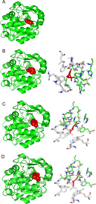
shown for SABP2 in the experimentally reported complex with SA (Figure 1A), and in the simulated
complexes with a-pinene, limonene, and b-myrcene (Figure 1B–D).
The SABP2 binding site (represented in the right panels of Figure 1B–D) is characterized by the
presence of aromatic side chains (two phenylalanines, one tyrosine, and one tryptophan), also
observed in other candidate plant OBPs (GA receptor: two phenylalanines and four tyrosines; JA
receptor: one phenylalanine, one tyrosine, and one tryptophan residue; FT receptor: two phenylala-
nines and one tryptophan). Other candidate plant OBPs have some aromatic side chains in the bind-
ing site, although in lower number (e.g., the ABA receptor has one phenylalanine and one tyrosine,
while TLF1 has two phenylalanines). This is also reminiscent of the binding site of OBPs from animal
organisms (Bianchet et al., 1996). Bovine OBPs include five phenylalanines and one tyrosine; Dro-
sophila OBPs have four phenylalanines and one tryptophan; and porcine OBPs have two phenylala-
nines. This conserved feature across biomes may reveal that a hydrophobic environment where
odorant molecules can be accommodated is needed. Analysis of the b-caryophyllene complexes
(see Figure 1—figure supplement 1) also suggests that larger ligands may interact with additional
aromatic or hydrophobic side chains in the binding pockets.
Overall, our study confirms that plant OBPs may exist and that they may be structurally and func-
tionally similar to OBPs described in animals. As in the case of animal OBPs, also plant OBPs seem
to be able to bind different VOCs in the same binding site, using the same amino acid sequences.
While our in silico results make the case that plants also have OBPs, with both common and different
features compared to animal OBPs, functional validation should follow. For example, chemical syn-
thesis of fluorescent VOCs could be used to confirm VOC binding by putative OBPs and to charac-
terize protein–ligand binding mechanisms and sites. Mutants or genetically modified plants that miss
Giordano et al. eLife 2021;10:e66741. DOI: https://doi.org/10.7554/eLife.66741 4 of 9Research article Plant Biology Figure 1. 3D models of protein-ligand interactions. (A) Experimental structure of SABP2 protein structure with salicylic acid in the binding site (PDB structure 1Y7I). The architecture of SABP2 is schematized by the backbone structure (green), with ribbons and arrows to evidence helices and beta strands, respectively. The salicylic acid molecule (colored in red) is in space-fill representation. (B) Left: Molecular docking simulation of SABP2 protein structure with a-pinene (red molecule) in the binding site. SABP2 is shown with the same spatial orientation as in panel (A) to emphasize that a-pinene Figure 1 continued on next page Giordano et al. eLife 2021;10:e66741. DOI: https://doi.org/10.7554/eLife.66741 5 of 9
Research article                                                                                                                  Plant Biology

Figure 1 continued
occupies the same binding site of salicylic acid. Right: Focus on the binding site of SABP2. a-Pinene (red molecule) interacts directly with amino acid
residues labeled with carbon atoms in green. Amino acid residues with carbon atom in grey are also part of the binding site, although not directly in
contact with a-pinene. Standard colors are used for the other amino acid atoms (red = oxygen, blue = nitrogen, yellow = sulphur, white = hydrogen).
(C) Left: SABP2 protein with limonene (red molecule) in the binding site. SABP2 is shown with the same spatial orientation of (A), to better show that
limonene occupies the same binding site of the salicylic acid. Right: Focus of the binding site of SABP2. Details as in panel (B). (D) Left: SABP2 protein
with b-myrcene (red molecule) in the binding site. SABP2 is shown with the same spatial orientation of (A), to better show that b-myrcene occupies the
same binding site of the salicylic acid. Right: Focus of the binding site of SABP2. Details as in (B).
The online version of this article includes the following figure supplement(s) for figure 1:
Figure supplement 1. The environment of the binding pocket of b-caryophyllene in the complexes with best binding energy values.
Figure supplement 2. Schematic workflow for the search of candidate odorant-binding proteins in plants.

                                      or have abundant OBP candidates could also be used. Retrieval and description of plant OBPs may
                                      be an important step to unveil how plants eavesdrop messages sent by other plants and how the
                                      information is then used to activate molecular and metabolic changes leading to defensive
                                      responses and patterns.

                                      Materials and methods
                                      The search for potential OBP proteins in plants was performed following the two-level investigation
                                      procedure schematized in Figure 1—figure supplement 2. The first level was about searching for
                                      plant proteins with potential OBP function. The second level included searching for experimental 3D
                                      structures of the candidate plant OBPs and validating by molecular simulations potential ability of
                                      putative OBPs to bind isoprenoid VOCs.
                                          In detail, for the first level, two steps were followed. Step (a) was a screening for proteins of inter-
                                      est performed on the UniProt (http://www.uniprot.org) and NCBI (http://www.ncbi.nih.nlm.gov) pro-
                                      tein databases. Initial screening was performed by using the protein name and entry annotations,
                                      with the query ‘odorant-binding protein’. Five plant proteins were found, annotated as ‘predicted
                                      proteins’, which means that they were obtained by nucleotide sequence translation, without evi-
                                      dence at protein or transcript levels, and the name was assigned to the proteins by similarity to
                                      other proteins. The protein sequence selected were further investigated by BLAST searches for simi-
                                      lar sequences, by using the BLAST interfaces at the database web sites. Standard BLAST search
                                      parameters were used. Step (b) was based on BLAST searches (using the same standard parameters
                                      of step a) for plant proteins and protein families similar to the 432 OBPs from animal sources avail-
                                      able in the protein databases (the list of the 432 OBPs is reported as Supplementary file 4).
                                          The second level of investigation was a molecular simulations of the interaction of the potential
                                      plant OBPs (i.e. those selected in steps 1a and 1b, and the three proteins for which OBP function
                                      has been reported [see Introduction]) with selected isoprenoid VOCs. First, we verified the availabil-
                                      ity of 3D structures of the candidate OBPs in plants. In particular, the Protein Data Bank (PDB)
                                      (http://www.rcsb.org), collecting the 3D structure of proteins, was interrogated for appropriate pro-
                                      tein structures of the candidate plant OBPs identified by the first level search. The screening allowed
                                      us to select the following plant proteins with potential primary or secondary function as OBP, and
                                      with available 3D structures: ABA Receptor from Arabidopsis thaliana (PDB code: 4dsb); GA recep-
                                      tor GID1 from Oryza sativa (3ebl); Flowering locus t (FT) from A. thaliana (1wkp); Terminal flower 1
                                      (tfl1) from A. thaliana (1wko); Protein Heading date DATE 3A from O. sativa (3axy); TPL-like protein
                                      from A. thaliana (5nqs); COI (partial JA receptor) from A. thaliana (3ogl); COI and JAZ (JA receptor
                                      complete) from A. thaliana (3ogl); and salicylic acid binding protein 2 (SA enzyme) from Nicotiana
                                      tabacum (1y7i).
                                          Molecular structures of VOCs were extracted from the PubChem database. The VOCs selected as
                                      ligands in our study were the isoprenoids a-pinene (PubChem code: 6654), limonene (22311), b-myr-
                                      cene (31253), b-caryophyllene (5281515), isoprene (6557), and linalool (6549).
                                          Molecular simulation experiments of protein–ligand interactions were carried out with Autodock
                                      4.2 and AutoDock Tools 1.5.6 (Morris et al., 2009), which allowed us to prepare the screening, per-
                                      form the docking simulation, and analyze the results. Molecular visualization of results was obtained
                                      with PyMOL Molecular Graphics System, Version 1.3 Schrödinger, LLC.

Giordano et al. eLife 2021;10:e66741. DOI: https://doi.org/10.7554/eLife.66741                                                                       6 of 9
Research article Plant Biology
The binding energy values obtained for the simulated protein–ligand complexes were compared
to the values for complexes used as reference. We found in the PDB database complexes of animal
proteins with a-pinene, limonene 1,2 epoxide, and b-myrcene. a-Pinene and b-myrcene are two of
the selected VOCs for our simulation, fully correspondent to the natural molecules synthesized and
emitted by plants. Limonene 1,2 epoxide is a modified form of the natural VOC. Although not identi-
cal to the corresponding plant VOC (limonene), it may be useful as additional reference value. For
available plant receptors (ABA receptor, GA receptor, JA receptor, and SA binding protein 2), the
reference structures are complexes with ABA, GA, JA-isoleucine, and SA, respectively. These com-
plexes may offer additional reference values of binding energy.
To validate the docking simulation experimental protocol, we applied a redocking procedure to
the reference complexes, following the procedure in use in our laboratory (Scafuri et al., 2016,
Scafuri et al., 2020). We depleted the ligand from the complex ligand–protein, and then the ligand-
depleted complex (the protein alone) was used to simulate the ligand docking. The redocking
experiments were carried out for the protein–ligand reference structures selected above. This
approach allowed us to check that the simulation procedure located correctly the ligand in the
expected binding site and to calculate the reference value of the binding energy expected in the
true protein–ligand complex. The redocking procedure also provided a computational estimation of
the binding energy in a true case of protein–ligand interaction. This estimation is used as reference
in comparison to the energy binding values computed for the putative protein–ligand interactions.
For each ligand a proper reference complex is needed, being the energy of interaction dependent
on the ligand chemical features. In the absence of a reference complex relative to an experimental
protein–ligand interaction (e.g. the cases of b-caryophyllene, isoprene, and linalool, see Table 1),
the computed binding energy values may be compared each other too, but only for a qualitative
ranking, without a reference threshold given by an effective binding.
Acknowledgements
FL acknowledges contribution from the Project PRIN – COFIN 2017 (Italian Ministry of University and
Research): ‘Plant multitROphic interactions for bioinspired Strategies of PEst ConTrol (PROSPECT)’.
We would like to thank Prof. Paolo Pelosi for the inspiring discussions about OBPs.
Additional information
Funding
Funder Grant reference number Author
Ministero dell’Istruzione, del- PRIN - COFIN 2017 Francesco Loreto
l’Università e della Ricerca
The funders had no role in study design, data collection and interpretation, or the
decision to submit the work for publication.
Author contributions
Deborah Giordano, Formal analysis, Validation, Investigation, Visualization, Methodology; Angelo
Facchiano, Formal analysis, Validation, Investigation, Methodology; Sabato D’Auria, Conceptualiza-
tion, Supervision, Writing - original draft, Writing - review and editing; Francesco Loreto, Conceptu-
alization, Supervision, Funding acquisition, Writing - original draft, Writing - review and editing
Author ORCIDs
Deborah Giordano https://orcid.org/0000-0002-5838-1913
Angelo Facchiano https://orcid.org/0000-0002-7077-4912
Francesco Loreto https://orcid.org/0000-0002-9171-2681
Decision letter and Author response
Decision letter https://doi.org/10.7554/eLife.66741.sa1
Author response https://doi.org/10.7554/eLife.66741.sa2
Giordano et al. eLife 2021;10:e66741. DOI: https://doi.org/10.7554/eLife.66741 7 of 9Research article Plant Biology
Additional files
Supplementary files
. Source data 1. Database accession numbers. Related to Table 1, Figure 1 and Figure 1—figure
supplement 1.
. Source data 2. Docking results. Related to Figure 1 and Table 2.
. Source data 3. PDB structures of SABP2 complexes. Related to Figure 1.
. Source data 4. PDB structures of protein - beta caryophyllene complexes. Related to Figure 1—
figure supplement 1.
.Supplementary file 1. Plant proteins from sequence databases annotated as ‘odorant-binding
protein’.
. Supplementary file 2. BLAST results for non-plant proteins with similarity to ‘general odorant-bind-
ing protein 56d’ from Anthurium amnicola. Only the best ten results are shown.
. Supplementary file 3. Complete results of docking simulation experiments. Docking simulations by
AutoDock suite generated results summarized in the table. For each receptor-ligand simulation, the
best 100 conformations are clusterized by AutoDock on the basis of ligand position, and three values
are reported for each cluster: the mean binding energy for the clusterized conformations, the best
binding energy, the number of conformations in the cluster. An additional parameter computed by
AutoDock is the predicted Ki value. We reported the results for the best cluster in terms of energy
and population, or in some cases, alternative clusters, with reference to different pockets on the
receptor surface.
. Supplementary file 4. List of 432 protein sequences selected as OBP from animal sources, used for
searching public database for similar plant proteins.
. Transparent reporting form
Data availability
All data generated during this study were obtained by analysing entries retrieved from public data-
bases, according to procedures described in the manuscript. Source data and supporting files report
complete list of accession numbers of entries and the models of 3D structures obtained by our study
and used for generating figures.
References
Adebesin F, Widhalm JR, Boachon B, Lefèvre F, Pierman B, Lynch JH, Alam I, Junqueira B, Benke R, Ray S,
Porter JA, Yanagisawa M, Wetzstein HY, Morgan JA, Boutry M, Schuurink RC, Dudareva N. 2017. Emission of
volatile organic compounds from Petunia flowers is facilitated by an ABC transporter. Science 356:1386–1388.
DOI: https://doi.org/10.1126/science.aan0826, PMID: 28663500
Baldwin IT, Kessler A, Halitschke R. 2002. Volatile signaling in plant-plant-herbivore interactions: what is real?
Current Opinion in Plant Biology 5:351–354. DOI: https://doi.org/10.1016/S1369-5266(02)00263-7, PMID: 1217
9970
Bianchet MA, Bains G, Pelosi P, Pevsner J, Snyder SH, Monaco HL, Amzel LM. 1996. The three-dimensional
structure of bovine odorant binding protein and its mechanism of odor recognition. Nature Structural Biology
3:934–939. DOI: https://doi.org/10.1038/nsb1196-934, PMID: 8901871
Bouwmeester H, Schuurink RC, Bleeker PM, Schiestl F. 2019. The role of volatiles in plant communication. The
Plant Journal 100:892–907. DOI: https://doi.org/10.1111/tpj.14496, PMID: 31410886
Brilli F, Ciccioli P, Frattoni M, Prestininzi M, Spanedda AF, Loreto F. 2009. Constitutive and herbivore-induced
monoterpenes emitted by Populus x euroamericana leaves are key volatiles that orient Chrysomela populi
beetles. Plant, Cell and Environment 32:542–552. DOI: https://doi.org/10.1111/j.1365-3040.2009.01948.x
Cheong JJ, Choi YD. 2003. Methyl jasmonate as a vital substance in plants. Trends in Genetics 19:409–413.
DOI: https://doi.org/10.1016/S0168-9525(03)00138-0, PMID: 12850447
Dicke M, Loreto F. 2010. Induced plant volatiles: from genes to climate change. Trends in Plant Science 15:115–
117. DOI: https://doi.org/10.1016/j.tplants.2010.01.007, PMID: 20137997
Erb M. 2019. Plant biology: evolution of Volatile-Mediated Plant-Plant interactions. Current Biology 29:R873–
R875. DOI: https://doi.org/10.1016/j.cub.2019.07.066, PMID: 31550472
Giordano et al. eLife 2021;10:e66741. DOI: https://doi.org/10.7554/eLife.66741 8 of 9Research article Plant Biology
Frank L, Wenig M, Ghirardo A, van der Krol A, Vlot AC, Schnitzler JP, Rosenkranz M. 2021. Isoprene and b-
caryophyllene confer plant resistance via different plant internal signalling pathways. Plant, Cell & Environment
44:1151–1164. DOI: https://doi.org/10.1111/pce.14010, PMID: 33522606
Harvey CM, Sharkey TD. 2016. Exogenous isoprene modulates gene expression in unstressed Arabidopsis
thaliana plants. Plant, Cell & Environment 39:1251–1263. DOI: https://doi.org/10.1111/pce.12660,
PMID: 26477606
Koornneef M, Alonso-Blanco C, Blankestijn-de Vries H, Hanhart CJ, Peeters AJ. 1998. Genetic interactions
among late-flowering mutants of Arabidopsis. Genetics 148:885–892. PMID: 9504934
Loreto F, Schnitzler JP. 2010. Abiotic stresses and induced BVOCs. Trends in Plant Science 15:154–166.
DOI: https://doi.org/10.1016/j.tplants.2009.12.006, PMID: 20133178
Miloradovic van Doorn M, Merl-Pham J, Ghirardo A, Fink S, Polle A, Schnitzler JP, Rosenkranz M. 2020. Root
isoprene formation alters lateral root development. Plant, Cell & Environment 43:2207–2223. DOI: https://doi.
org/10.1111/pce.13814, PMID: 32495947
Monson RK, Weraduwage SM, Rosenkranz M, Schnitzler JP, Sharkey TD. 2021. Leaf isoprene emission as a trait
that mediates the growth-defense tradeoff in the face of climate stress. Oecologia 1:04813-7. DOI: https://doi.
org/10.1007/s00442-020-04813-7
Morris GM, Huey R, Lindstrom W, Sanner MF, Belew RK, Goodsell DS, Olson AJ. 2009. AutoDock4 and
AutoDockTools4: automated docking with selective receptor flexibility. Journal of Computational Chemistry
30:2785–2791. DOI: https://doi.org/10.1002/jcc.21256, PMID: 19399780
Nagashima A, Higaki T, Koeduka T, Ishigami K, Hosokawa S, Watanabe H, Matsui K, Hasezawa S, Touhara K.
2019. Transcriptional regulators involved in responses to volatile organic compounds in plants. Journal of
Biological Chemistry 294:2256–2266. DOI: https://doi.org/10.1074/jbc.RA118.005843
Ninkovic V, Rensing M, Dahlin I, Markovic D. 2019. Who is my neighbor? volatile cues in plant interactions. Plant
Signaling & Behavior 14:1634993. DOI: https://doi.org/10.1080/15592324.2019.1634993
Ninkovic V, Markovic D, Rensing M. 2021. Plant volatiles as cues and signals in plant communication. Plant, Cell
& Environment 44:1030–1043. DOI: https://doi.org/10.1111/pce.13910, PMID: 33047347
Park SW, Kaimoyo E, Kumar D, Mosher S, Klessig DF. 2007. Methyl salicylate is a critical mobile signal for plant
systemic acquired resistance. Science 318:113–116. DOI: https://doi.org/10.1126/science.1147113, PMID: 17
916738
Pollastri S, Jorba I, Hawkins TJ, Llusià J, Michelozzi M, Navajas D, Peñuelas J, Hussey PJ, Knight MR, Loreto F.
2019. Leaves of isoprene-emitting tobacco plants maintain PSII stability at high temperatures. New Phytologist
223:1307–1318. DOI: https://doi.org/10.1111/nph.15847
Ramoni R, Bellucci S, Grycznyski I, Grycznyski Z, Grolli S, Staiano M, De Bellis G, Micciulla F, Pastore R, Tiberia A,
Conti V, Merli E, Varriale A, Rossi M, D’Auria S. 2007. The protein scaffold of the lipocalin odorant-binding
protein is suitable for the design of new biosensors for the detection of explosive components. Journal of
Physics: Condensed Matter 19:395012–395019. DOI: https://doi.org/10.1088/0953-8984/19/39/395012
Scafuri B, Marabotti A, Carbone V, Minasi P, Dotolo S, Facchiano A. 2016. A theoretical study on predicted
protein targets of apple polyphenols and possible mechanisms of chemoprevention in colorectal Cancer.
Scientific Reports 6:32516. DOI: https://doi.org/10.1038/srep32516, PMID: 27587238
Scafuri B, Bontempo P, Altucci L, De Masi L, Facchiano A. 2020. Molecular docking simulations on histone
deacetylases (HDAC)-1 and -2 to investigate the flavone binding. Biomedicines 8:568. DOI: https://doi.org/10.
3390/biomedicines8120568
Sheard LB, Tan X, Mao H, Withers J, Ben-Nissan G, Hinds TR, Kobayashi Y, Hsu FF, Sharon M, Browse J, He SY,
Rizo J, Howe GA, Zheng N. 2010. Jasmonate perception by inositol-phosphate-potentiated COI1-JAZ co-
receptor. Nature 468:400–405. DOI: https://doi.org/10.1038/nature09430, PMID: 20927106
Shen Q, Chen CN, Brands A, Pan SM, Ho TH. 2001. The stress- and abscisic acid-induced barley gene HVA22:
developmental regulation and homologues in diverse organisms. Plant Molecular Biology 45:327–340.
DOI: https://doi.org/10.1023/a:1006460231978, PMID: 11292078
Taoka K, Ohki I, Tsuji H, Furuita K, Hayashi K, Yanase T, Yamaguchi M, Nakashima C, Purwestri YA, Tamaki S,
Ogaki Y, Shimada C, Nakagawa A, Kojima C, Shimamoto K. 2011. 14-3-3 proteins act as intracellular receptors
for rice Hd3a florigen. Nature 476:332–335. DOI: https://doi.org/10.1038/nature10272, PMID: 21804566
Vaistij FE, Barros-Galvão T, Cole AF, Gilday AD, He Z, Li Y, Harvey D, Larson TR, Graham IA. 2018. MOTHER-
OF-FT-AND-TFL1 represses seed germination under far-red light by modulating phytohormone responses in
Arabidopsis thaliana. PNAS 115:8442–8447. DOI: https://doi.org/10.1073/pnas.1806460115, PMID: 30061395
Velikova V, Müller C, Ghirardo A, Rock TM, Aichler M, Walch A, Schmitt-Kopplin P, Schnitzler JP. 2015.
Knocking down of isoprene emission modifies the lipid matrix of thylakoid membranes and influences the
chloroplast ultrastructure in poplar. Plant Physiology 168:859–870. DOI: https://doi.org/10.1104/pp.15.00612,
PMID: 25975835
Vickers CE, Gershenzon J, Lerdau MT, Loreto F. 2009. A unified mechanism of action for volatile isoprenoids in
plant abiotic stress. Nature Chemical Biology 5:283–291. DOI: https://doi.org/10.1038/nchembio.158, PMID: 1
9377454
Zuo Z, Weraduwage SM, Lantz AT, Sanchez LM, Weise SE, Wang J, Childs KL, Sharkey TD. 2019. Isoprene acts
as a signaling molecule in gene networks important for stress responses and plant growth. Plant Physiology
180:124–152. DOI: https://doi.org/10.1104/pp.18.01391, PMID: 30760638
Giordano et al. eLife 2021;10:e66741. DOI: https://doi.org/10.7554/eLife.66741 9 of 9You can also read