Low pH alleviated salinity stress of ginger seedlings by enhancing photosynthesis, fluorescence, and mineral element contents - PeerJ
←
→
Page content transcription
If your browser does not render page correctly, please read the page content below
Low pH alleviated salinity stress of ginger
                                   seedlings by enhancing photosynthesis,
                                   fluorescence, and mineral element
                                   contents
                                   Fengman Yin1 ,2 ,3 ,4 ,* , Shanying Zhang5 ,* , Bili Cao1 ,2 ,3 ,4 and Kun Xu1 ,2 ,3 ,4
                               1
                                 College of Horticulture Science and Engineering, Shandong Agricultural University, Tai’an, Shandong, China
                               2
                                 Collaborative Innovation Center of Fruit & Vegetable Quality and Efficient Production in Shandong, Tai’an,
                                 China
                               3
                                 Key Laboratory of Biology and Genetic Improvement of Horticultural Crops in Huanghuai Region, Ministry
                                 of Agriculture and Rural Affairs, P.R. China, Tai’an, China
                               4
                                 State Key Laboratory of Crop Biology, Tai’an, China
                               5
                                 College of Food Science, Hainan University, Haikou, Hainan, China
                               *
                                 These authors contributed equally to this work.

                                       ABSTRACT
                                       We investigated the effects of low pH on the photosynthesis, chlorophyll fluorescence,
                                       and mineral contents of the leaves of ginger plants under salt stress. This experiment
                                       involved four treatments: T1 (pH 6, 0 salinity), T2 (pH 4, 0 salinity), T3 (pH 6, 100
                                       mmol L−1 salinity) and T4 (pH 4, 100 mmol L−1 salinity). This study showed that
                                       photosynthesis (Pn, Gs, WUE and Tr) and chlorophyll fluorescence (qP, 8 PSII, and
                                       Fv/Fm) significantly decreased under salt stress; however, all the parameters of the
                                       ginger plants under the low-pH treatment and salt stress recovered. Moreover, low
                                       pH reduced the content of Na and enhanced the contents of K, Mg, Fe and Zn in
                                       the leaves of ginger plants under salt stress. Taken together, these results suggest that
                                       low pH improves photosynthesis efficiency and nutrient acquisition and reduces the
                                       absorption of Na, which could enhance the salt tolerance of ginger.

Submitted 30 October 2020          Subjects Agricultural Science, Plant Science, Soil Science, Biogeochemistry, Environmental
Accepted 4 January 2021            Impacts
Published 11 February 2021
                                   Keywords Ginger, Salt stress, Low pH, Photosynthesis, Chlorophyll fluorescence, Mineral
Corresponding author               contents
Kun Xu, xukun@sdau.edu.cn
Academic editor
Mukhtar Ahmed                      INTRODUCTION
Additional Information and         Soil salinity has severe effects on plant growth and development and plant productivity in
Declarations can be found on
                                   arid and semiarid regions worldwide (Kochian et al., 2015; Krasensky & Jonak, 2012; Ryu &
page 13
                                   Cho, 2015). The increase in soil salinity and acidification is due to poor irrigation practices,
DOI 10.7717/peerj.10832
                                   improper fertilizer application, industrial pollution, and seawater intrusion caused by
    Copyright                      global warming (Gaudio et al., 2015; Ding et al., 2020). In particular, growers are advised
2021 Yin et al.
                                   to apply large amounts of chemical fertilizers for high yields during long-term vegetable
Distributed under                  production processes. This leads to a nutrient imbalance and excess salt accumulation in
Creative Commons CC-BY 4.0
                                   the soil. Salt causes several adverse effects on plant growth and development, including
OPEN ACCESS                        decreased leaf size, yellowing of the leaves, short internodes, short plant height, early

                                   How to cite this article Yin F, Zhang S, Cao B, Xu K. 2021. Low pH alleviated salinity stress of ginger seedlings by enhancing photosyn-
                                   thesis, fluorescence, and mineral element contents. PeerJ 9:e10832 http://doi.org/10.7717/peerj.10832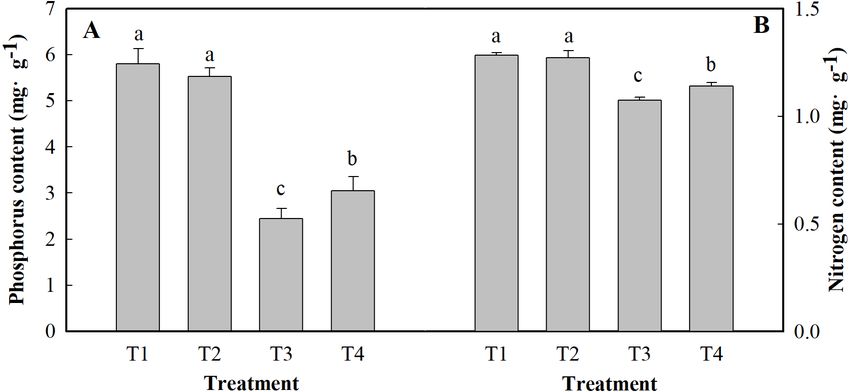
flowering and decreased yields (Acosta et al., 2011; Thouvenot, Haury & Thiébaut, 2012;
                                    Jan et al., 2017; Ahmad et al., 2018)
                                        Photosynthesis is an important biological process for maintaining plant life and plays a
                                    very important role in the evolution of ecosystems on Earth. Photosynthesis provides the
                                    energy and carbon required for the biosynthesis of organic compounds necessary for the
                                    growth and biomass production of plants. Increasing photosynthesis efficiency is critical to
                                    increasing crop yields to meet human demand for food (Long, Marshall-Colon & Zhu, 2015;
                                    Zhu, Long & Ort, 2010). Many researchers have studied the effects of salt stress on plant
                                    photosynthesis. There is considerable evidence for significant changes in the chlorophyll
                                    content (Chl) (Kalaji et al., 2016), net photosynthesis rate (Pn) (Jiang et al., 2017), stomatal
                                    conductance (Gs) (Janda et al., 2016), transpiration rate (Tr), maximum photochemical
                                    efficiency (Fv/Fm) (Singh, Singh & Prasad, 2016), amount of ribulose-1,5-bisphosphate
                                    carboxylase/oxygenase (RuBisco) and photochemical quenching (qP) (Moles et al., 2016).
                                    Chloroplast development and chlorophyll metabolism are important biological activities
                                    of photosynthesis in green plants. Studies have revealed chlorophyll synthesis-related
                                    enzymes and key regulators involved in chloroplast development (Bollivar, 2006; Cortleven
                                    & Schmulling, 2015). Tang et al. (2018) showed that NaCl stress significantly decreased the
                                    contents of Chl a, Chl b, and total chlorophyll in cucumber leaves. Ribose-1,5-bisphosphate
                                    ribulose carboxylation/oxygenase is a key enzyme that is involved in plant photosynthesis
                                    and controls both CO2 fixation and carbon. Rubisco is the key enzyme in the Calvin cycle,
                                    converting free CO2 in the atmosphere into energy-storage molecules, such as sucrose, and
                                    it plays a direct role in the photosynthesis rate.
                                        Ginger (Zingiber officinale Rose), a perennial plant species of the family Zingiberaceae,
                                    is native to tropical rainforest regions. Ginger has been cultivated and widely used for
                                    more than 2000 years in China as a spice and as an important ingredient in traditional
                                    Chinese medicine. Because the bioactive constituents in ginger are valuable and have
                                    been accepted gradually by people (Ali et al., 2008; Li et al., 2015a), ginger’s demand has
                                    increased annually worldwide. According to statistics from the FAO (Food and Agriculture
                                    Organization of the United Nations), global ginger production was 813 340 tons in 2010
                                    and 1 218 710 tons in 2016 but was 426 032 tons and 938 000 tons in mainland China,
                                    respectively.
                                        Many studies on the photosynthesis and chlorophyll content of ginger have focused
                                    mainly on antibiotics and drought (Li et al., 2013; Li et al., 2015b; Liu et al., 2018). Salt
                                    stress was shown to decrease the growth and biomass yield (leaf fresh weight and root
                                    fresh weight) of ginger seedlings in a preliminary study. Moreover, low pH significantly
                                    alleviated this inhibition under salt stress, perhaps by lowering the Na content, alleviating
                                    osmotic stress, and enhancing plant nutrient uptake (Yin, Cao & Xu, 2019; Yin et al., 2020).
                                    However, several studies have focused on the effects of low pH on the photosynthesis and
                                    chlorophyll content of ginger under salt stress. As such, the objectives of this study were to
                                    investigate the changes in photosynthesis and chlorophyll content in response to acidic salt
                                    stress, which is important for understanding the mechanism underlying plant tolerance to
                                    acidic salt stress.

Yin et al. (2021), PeerJ, DOI 10.7717/peerj.10832                                                                              2/19
MATERIALS & METHODS
Plant materials and experimental treatments
The plant materials and experiment treatment reference Yin et al. (2020). In Tai’an,
Shandong Province, China pot culture experiment was performed from April to October
2017. On May 14, the Zingiber officinale cultivar Shannong No. 1 were sown in pots
(diameter, 25 cm; height, 30 cm) filled with cleaned quartz sand (Yin et al., 2020). Neutral
salt stress was simulated with NaCl and Na2 SO4 (NaCl/Na2 SO4 =1/1), and pure water
with different pH values (HCl/H2 SO4 =1/1) was used to simulate hydrochloric acid stress
treatments. This experiment involved four treatments: T1 (pH 6, 0 salinity), T2 (pH 4, 0
salinity), T3 (pH 6, 100 mmol L−1 salinity) and T4 (pH 4, 100 mmol L−1 salinity). Each
treatment was replicated three times, with six individual plants in each replicate. Each pot
received 400 mL of treatment solution.
Analytical methods
Photosynthesis parameters
Functional leaves of ginger were selected, and the Pn, Gs, Tr and Ci were measured by a
portable photosynthesis system (Ciras-3, PP Systems, USA) using the method of Li et al.
(2013), with slight modifications. When the Pn reached a steady state at each light intensity
level, data were recorded 5 times per treatment, and the average value was calculated to
determine the final photosynthesis parameters. Natural light was used, and the CO2 gas
source was part of an open system.
Pigment concentrations
The chlorophyll content was measured according to the methods of Holm (1954).
Chlorophyll fluorescence
The qP, NPQ, 8PSII, and Fv/Fm were measured according to the methods of Hendrickson
et al. (2005) and Liu et al. (2018). At the time of measurements, 5 plants were averaged for
each treatment.
Photosynthesis enzyme (RuBPCase, FBPase, and FBA) activity assays
Fructose 1,6-diphosphatase (FBPase) activity was measured according to the methods of
Lazro et al. (1974).
RuBPCase activity was determined using an ELISA kit (Suzhou Keming), and fructose
1,6-bisphosphate aldolase (FBA) activity was determined using an ELISA kit (GenMed).
Sugar metabolism
Sucrose synthase (SS) and sucrose phosphate synthase (SPS) activity were estimated
following the methods of Batta & Singh (1986).
Reducing sugars and sucrose contents were measured according to the methods of
Handel (1968), using a standard graph of glucose.
The starch content was calculated according to the methods of Hannachi & Van Labeke
(2018).
Yin et al. (2021), PeerJ, DOI 10.7717/peerj.10832 3/19Mineral analysis
Ginger leaves were dried for 48 h at 75 ◦ C and ground separately in a Wiley mill to pass
through a 20-mesh screen. Afterward, 0.5 g of dried plant tissue was analyzed to determine
the following major and minor elements: N, P, K, Ca and Mg. The nitrogen concentration
in the plant tissues was determined by the Kjeldahl method after mineralization with
sulfuric acid (Bremner, 1965).
Phosphorus concentrations were determined by titration with molybdenum antimony
reagent in the presence of dinitrophenol (Shankar, Kumar & Agrawal, 2016).
K, Ca, Fe, Zn and Mg concentrations were determined by dry ashing at 400 ◦ C for 24 h,
dissolving the ash in 1/20 HNO3 , and assaying the solution obtained using an inductively
coupled plasma emission spectrometer (iCAP 7000 Series, Thermo Scientific).
Observations of ginger leaf chloroplast ultrastructure
Functional leaves were sampled (1 mm ×1 mm), quickly placed in a 2.5% glutaraldehyde
fixative solution, and then transferred to a 4 ◦ C refrigerator. The material was rinsed with
0.1 M PB (pH 7.4). Tissues avoid light post fixed with 1% OsO4 in 0.1 M PB (pH 7.4)
for 7 h at room temperature., after which it was rinsed 3 times with 0.1 M PB (pH 7.4)
again for 15 min each time. After that, the leaf tissue was then subjected to dehydration
and infiltration, after which it was embedded. Afterward, the material was sectioned (Leica
UC7), stained, the cuprum grids are observed under transmission electron microscope
(HT7700, Hitachi) and take images.
Statistical analysis
The data are presented as means ± one standard deviation (SD) for three independent
replicates. The data were processed with DPS software. All graphs were created using the
program SigmaPlot 10.0.
RESULTS
Photosynthesis parameters
Figure 1 shows the Pn, Gs, Ci, Tr, WUE and Ls of the leaves of ginger plants under salt
stress with or without H+ application. Compared with those under the control treatment
(T1), the Pn, Gs, Tr, and WUE decreased markedly (by 47.21%, 42.67%, 17.19%, 36.26%,
respectively) under the T3 treatment, and the Ci increased by 18.92%. Moreover, compared
with those under the control treatment (T1), the Pn, Gs, Tr, and WUE under the T4
treatment decreased by 25.42%, 31.90%, 12.50%, and 14.76%, respectively, and the Ci
increased by 8.62%. Compared with that under the control treatment (T1), the Ls under
the T3 and T4 treatments decreased by 41.75% and 20.26%, respectively. However, the pH
treatment had only a certain effect on the Pn and WUE of ginger seedling leaves, but they
did not significantly differ at the level of P < 0.05.
Pigment contents
As shown in Table 1, compared with the control treatment (T1), the T3 treatment reduced
the contents of Chl a, Chl b, Car, and Chl a+b by 9.33%, 28.28%, 7.59% and 13.01%,
respectively. Moreover, the contents of Chl a, Chl b, Car, and Chl a+b slightly increased
Yin et al. (2021), PeerJ, DOI 10.7717/peerj.10832 4/19Figure 1 Effects of low pH on photosynthesis parameters of the leaves of ginger plants under salt
stress. T1 (pH 6, 0 salinity), T2 (pH 4, 0 salinity), T3 (pH 6, 100 mmol L−1 salinity) and T4 (pH 4, 100
mmol L−1 salinity). The different small letters in a column of the same treatment days indicate significance
at the 5% level.
Full-size DOI: 10.7717/peerj.10832/fig-1
under the T4 treatment compared to the T3 treatment. There was no considerable difference
caused by low pH in the Chl a, Chl b, Car, and Chl a+b contents in the leaves. Salt stress
alone decreased the root activity by 26.94%. However, at low pH, salt stress decreased the
root activity by only 19.57% (Table 2).
Chlorophyll fluorescence
To analyze the changes in different ginger light systems in response to salt stress, chlorophyll
fluorescence parameters were measured. These chlorophyll fluorescence parameters display
significant negative effects under salt stress (Fig. 2). These effects were manifested by
decreased Fv/Fm, qP, and 8PSII values and an increased NPQ compared to the those of
controls (the T3 and T4 treatments). Salt stress alone (T3) reduced the Fv/Fm, qP, and
8PSII by 9.62%, 12.90%, and 28.96%, respectively, under the T3 treatment and by 6.85%,
7.87%, and 14.35%, respectively, under the T4 treatment compared to those under the
Yin et al. (2021), PeerJ, DOI 10.7717/peerj.10832 5/19Table 1 Effects of low pH on Chlorophyll contents in ginger leaves under salt stress. T1 (pH 6, 0 salinity), T2 (pH 4, 0 salinity), T3 (pH 6, 100
mmol L−1 salinity) and T4 (pH 4, 100 mmol L−1 salinity). Different small letters in a column of the same treatment days indicate significance at the
5% level.
Treatment Chl a Chl b Car Chl a+b Root activity
(mg g−1 FW) (mg g−1 FW) (mg g−1 FW) (mg g−1 FW) (µg h−1 g FW)
T1 1.93 ± 0.01a 0.99 ± 0.0211a 0.79 ± 0.0122a 2.92 ± 0.013a 64.84 ± 1.36a
T2 1.8 ± 0.01b 0.82 ± 0.0049b 0.76 ± 0.0166b 2.62 ± 0.016b 61.29 ± 1.21b
T3 1.75 ± 0.01c 0.71 ± 0.0142c 0.71 ± 0.0068c 2.45 ± 0.017c 47.37 ± 0.62d
T4 1.78 ± 0.01c 0.74 ± 0.0102c 0.73 ± 0.0115c 2.51 ± 0.019c 52.15 ± 1.46c
Table 2 Effects of low pH on the activities of Rubisco, FBA and FBPase in ginger leaves under salt
stress. T1 (pH 6, 0 salinity), T2 (pH 4, 0 salinity), T3 (pH 6, 100 mmol L−1 salinity) and T4 (pH 4, 100
mmol L−1 salinity) Different small letters in a column of the same treatment days indicate significance at
the 5% level.
Treatment Rubisco FBA FBPase
nmol min−1 g−1 nmol min−1 g−1 nmol min−1 g−1
T1 105.65 ± 3.43a 155.81 ± 13.38a 81.23 ± 3.46b
T2 94.5 ± 2.28b 162.17 ± 6.07a 91.22 ± 4.51a
T3 65.35 ± 4.66d 116.65 ± 4.33c 65.55 ± 1.35c
T4 74.65 ± 0.83c 134.55 ± 8.67b 76.4 ± 3.26b
normal conditions (T1). In contrast, the value of NPQ increased by 23.27% under the T3
treatment and by 14.35% under the T4 treatment compared to those under the control
(T1).
Photosynthesis enzyme activities
The results related to the activities of photosynthesis enzymes are depicted in Table 2. Low
pH increased FBA and FBP activities by 4.08 and 12.30% and decreased Rubisco activity by
10.55% in the absence of salt stress, respectively, compared to those of the control seedlings
(Table 2). Salt stress significantly decreased Rubisco, FBA, and FBP activities. Compared
with the T3 treatment, the T4 treatment increased the Rubisco, FBA, and FBP activities by
14.23%, 15.34%, and 16.55%, respectively.
Reducing sugar, sucrose, and starch contents
In the salt-affected ginger leaves, the reducing sugar, sucrose and starch contents decreased
significantly (Table 3). Salt stress alone (T3) reduced the reducing sugar, sucrose, and
starch contents by 51.75, 63.42, and 54.33%, respectively, compared to those of the control.
However, at low pH, salt stress decreased reducing sugar, sucrose, and starch contents by
only 35.06, 48.62, and 31.48%, respectively, compared to those under the T1 treatment.
Table 3 shows that low pH had a weak effect on reducing sugar, sucrose, and starch
contents, but they did not significantly differ at the level of P < 0.05.
Activity of SS and SPS
SS and SPS are key enzymes involved in carbon metabolism. The different treatments had
significant effects on the activity of SS and SPS in the ginger leaves (Fig. 3). Salt stress
Yin et al. (2021), PeerJ, DOI 10.7717/peerj.10832 6/19Figure 2 Effects of low pH on chlorophyll parameters of the leaves of ginger plants under salt stress.
T1 (pH 6, 0 salinity), T2 (pH 4, 0 salinity), T3 (pH 6, 100 mmol L−1 salinity) and T4 (pH 4, 100 mmol L−1
salinity). The different lowercase letters in a column of the same treatment days indicate significance at the
5% level.
Full-size DOI: 10.7717/peerj.10832/fig-2
Table 3 Effects of low pH on Reducing sugar, Sucrose and Starch content in ginger leaves under salt
stress. DW stands for dry weight. T1 (pH 6, 0 salinity), T2 (pH 4, 0 salinity), T3 (pH 6, 100 mmol L−1
salinity) and T4 (pH 4, 100 mmol L−1 salinity) Different small letters in a column of the same treatment
days indicate significance at the 5% level.
Treatment Reducing sugar Sucrose Starch
mg g−1 DW mg g−1 DW mg g−1 DW
T1 46.32 ± 0.73a 14.46 ± 0.59a 323.89 ± 2.68a
T2 40.51 ± 1.15b 12.31 ± 0.35b 319.44 ± 2.71a
T3 22.35 ± 1.05d 5.29 ± 0.08d 147.92 ± 5.12c
T4 30.08 ± 0.64c 7.43 ± 0.29c 221.94 ± 7.87b
alone reduced SS and SPS activity by 41.57 and 30.34%, respectively, compared to that of
the control (T1). The activity of SS and SPS was reduced by 30.3 and 6.15%, respectively,
under the salt +low pH treatment (T4) compared to the control treatment (T1).
Content of phosphorus and nitrogen
Figure 4 shows the results of the content of P and N in the leaves of ginger plants under
salt stress or under low pH. Compared with the control (T1), salt stress alone (T3) reduced
the content of P and N by 57.90 and 16.35%, respectively. Low pH with salt stress (T4)
decreased the contents of P and N by 47.48 and 11.08%, respectively, compared to those
Yin et al. (2021), PeerJ, DOI 10.7717/peerj.10832 7/19Figure 3 Effects of low pH on the activity of SS (A) and SPS (B) in the leaves of ginger plants under salt
stress. T1 (pH 6, 0 salinity), T2 (pH 4, 0 salinity), T3 (pH 6, 100 mmol L−1 salinity) and T4 (pH 4, 100
mmol L−1 salinity). The different small letters in a column of the same treatment days indicate significance
at the 5% level.
Full-size DOI: 10.7717/peerj.10832/fig-3
Figure 4 Effects of low pH on the contents of P (A) and N (B) in the leaves of ginger plants under salt
stress. T1 (pH 6, 0 salinity), T2 (pH 4, 0 salinity), T3 (pH 6, 100 mmol L−1 salinity) and T4 (pH 4, 100
mmol L−1 salinity).
Full-size DOI: 10.7717/peerj.10832/fig-4
of the control. Under low pH, the contents of P and N were slightly affected, but they did
not significantly differ at the level of P < 0.05.
Mineral composition content
As shown in Fig. 5, salt stress significantly increased the Na content in leaves of the plants
and significantly decreased the contents of K, Mg, Ca, Fe and Zn. The content of Na in
ginger seedling leaves increased by 101.55% under salt stress alone and increased by 40.62%
under low pH and salt stress. Salt stress alone significantly reduced the contents of K, Mg,
Ca, Fe and Zn by 27.27%, 32.52%, 28.08%, 47.01% and 41.73%, respectively, compared to
those of the control. The contents of K, Mg, Ca, Fe and Zn decreased by 17.36, 29.38, 13.78,
40.64 and 31.75%, respectively, under low pH with salt stress (T4), respectively, compared
to those under the control treatment (T1).
Yin et al. (2021), PeerJ, DOI 10.7717/peerj.10832 8/19Figure 5 Effects of low pH on the contents of Na (A), K (B), Mg (C), Ca (D), Fe (E) and Zn (F) in the
leaves of ginger plants under salt stress. T1 (pH 6, 0 salinity), T2 (pH 4, 0 salinity), T3 (pH 6, 100 mmol
L−1 salinity) and T4 (pH 4, 100 mmol L−1 salinity).
Full-size DOI: 10.7717/peerj.10832/fig-5
Ultrastructure morphological changes
Changes in whole mesophyll cells and chloroplasts are shown in Fig. 6. Seedlings grown
under normal conditions exhibited regular cell shape and typical chloroplasts; moreover,
there were several well-packed starch grains (Fig. 6). However, cell morphological
disturbance and plasmolysis occurred when the seedlings were treated with salt stress alone.
The shapes of chloroplasts were severely swollen. Furthermore, there was an abundance
of osmiophilic granules and fewer starch grains in the chloroplasts compared with those
of control (Fig. 6). For low-pH-treated seedlings under salt stress conditions, there was
a small improvement in cell morphology. The chloroplasts contained more starch grains
than did those of seedlings under salt stress alone (Fig. 6).
Yin et al. (2021), PeerJ, DOI 10.7717/peerj.10832 9/19Figure 6 Effects of low pH on the leaf ultrastructure in the leaves of ginger plants under salt stress: T1
(pH 6, 0 salinity), T2 (pH 4, 0 salinity), T3 (pH 6, 100 mmol L−1 salinity) and T4 (pH 4, 100 mmol L−1
salinity). GL: Granum lamellae; S: Starch grains; ch: Chloroplast; N: Cell nucleus.
Full-size DOI: 10.7717/peerj.10832/fig-6
DISCUSSION
Photosynthesis
The damage caused by salt to plants is primarily attributed to the inhibition and disruption
of photosynthesis, and the decrease of photosynthetic efficiency is one of the important
reasons for the decrease of plant biomass under salt stress. The Pn decreases because of
stomatal limitation or nonstomatal limitation. If Gs and Ci are positively correlated, then the
reason for the Pn decrease is related to stomatal limitation; if the two showed no correlation
or contrast, then it is related to nonstomatal limitation. In the present experiment, the Pn
and Gs decreased under the T3 treatment, and the Ci increased. This suggests that the main
factor of photosynthesis limitation is nonstomatal limitation. This is consistent with the
research results of Tang et al. (2018). However, damaged photosynthetic structures may be
another factor affecting the photosynthesis rate. The photosynthesis enzyme (Rubisco, FBA,
and FBP) activities were related to the degree of damage to the photosynthetic structure.
1,5-Ribulose diphosphate oxygenase/shuttle enzyme is an important enzyme involved in
CO2 fixation in plant leaves and plays an important role in maintaining photosynthesis
(Parry et al., 1997). In this study, the activity of Rubisco, FBA and FBP decreased under
the T3 treatment. These results suggest that the reason for photosynthesis under salt stress
Yin et al. (2021), PeerJ, DOI 10.7717/peerj.10832 10/19alone is nonstomatal limitation. Similar results were reported previously (Feng et al., 2014;
Ning et al., 2018) However, low pH with salt stress reduced the Ci and increased the Pn;
Gs; and the activities of Rubisco, FBA and FBP. Taken together, these results indicated that
low pH could protect photosynthetic structures and increase the photosynthesis enzyme
activities, thereby increasing the photosynthesis rate.
The water-use efficiency (WUE) of leaves is an important factor affecting whether
plants can adapt to extreme environmental conditions (Martin, Tauer & Lin, 1999). In our
experiment, the WUE value decreased under salt stress alone. This decrease was related
to the reduced leaf transpiration rate, which was caused by the decreased Gs (Bacha et al.,
2017). These conditions were unfavorable for substance transport in ginger. However, low
pH increased the WUE of leaves under salt stress and improved substance transport in
ginger.
Chlorophyll fluorescence
The absorption and transformation of light energy by plants are mainly divided into
three closely related parts: chlorophyll fluorescence, qP-related photosynthetic electron
transport, and qN-related heat consumption (Schreiber, Schliwa & Bilger, 1986). As an
important physiological index for evaluating plant growth and development characteristics,
the chlorophyll content can reflect plant health and adaptability of plants (Guo et al., 2015;
Xiao, Sang & Wang, 2008). Salt stress causes the decrease of chlorophyll and carotenoid
contents in mung bean leaves, which may be caused by the expansion of chloroplast
membrane and/or excess Na+ ions in the leaves (Alharby et al., 2019). In this study, the
levels of Chl a, Chl b, and carotenoids decreased under salt stress alone. This is consistent
with the research results of Do et al. (2018) and Patil et al. (2016). Chlorophyll content has
an obvious correlation with the photosynthesis ability of leaves (Dhanapal et al., 2016), and
a decrease in chlorophyll content can lead to an irreversibly decreased photosynthesis rate.
The maximal photochemical efficiency of PSII (Fv/Fm) was used to evaluate the primary
conversion efficiency of light energy in the PSII reaction center (Jagerbrand & Kudo, 2016).
8PSII is the actual light-harvesting efficiency of PSII when the reaction center is partially
closed, and 8PSII reflects the ratio of energy consumed by photosynthetic transmission
of electrons when the leaves absorb energy. The qP reflects the relative proportion of light
energy captured by light-harvesting pigments for photochemical electron transfer (Awlia et
al., 2017). The values of Fv/Fm, 8PSII, and qP were significantly reduced under salt stress,
suggesting that salinity induced the inhibition of PSII electron transport and dissipated
the excess excitation in the form of heat. This resulted in the reduction in the fluorescence
quantum yield (Mehta et al., 2010). The decrease in qP also indicated an increase in the
fraction of reduced QA in PSII (Bacha et al., 2017; Hu, Yan & Yu, 2016). The NPQ value,
which represents heat dissipation, increased by 23.27%, which indicated that a greater share
of excess energy was released as heat, whereas the ability to utilize light energy decreased.
The Fv/Fm, 8FPSII, and qP values increased significantly under low pH with salt stress,
indicating that the photochemical activity and electron transfer in ginger leaves were
positively affected and thereby enhanced the light energy conversion efficiency of PSII.
Yin et al. (2021), PeerJ, DOI 10.7717/peerj.10832 11/19Metal elements
Salt stress leads to specific ion toxicity and plant growth inhibition (Park, Kim & Yun, 2016).
Excessive accumulation of Na+ is harmful to plant cells, which can lead to significant
changes in metabolism and malnutrition (Liang et al., 2018). Nitrogen is an important
major element in plant growth and development. It is a component of many plant cell
components, including amino acids, proteins, and nucleic acids. The results show that salt
treatment induced a decrease in N concentration in ginger plants in our study. However,
the plants that were grown under low pH had consistently higher N concentration than the
normal plants under salt stress. The decrease in N concentration due to salt stress may be
caused by interference by salinity in N acquisition and utilization. Our study corroborates
the findings of Zhang et al. (2016), who reported that phosphorus concentration decreased
under salt stress. The effect is compounded by the deficiency of other elements (K, Mg,
etc.) due to the excessive Na content, which also severely reduces photosynthesis (Lu et al.,
2017). In this study, as expected, in the salt-stressed plants, Na accumulated excessively in
the leaves. On the other hand, the K content under salt stress alone was markedly lower than
that without salt stress treatment, which implied that there is a competitive relationship
between K+ and Na+ in ginger leaves. A similar result was reported by Wakeel et al. (2011).
However, low pH with salt stress decreased the Na content and increased the K content.
Moreover, the results showed that low pH with salt stress resulted in a stronger ability for
the absorption and transport of K+ to ensure an adequate concentration of the ions that
participate in key metabolic activities (e.g., photosynthetic metabolites) in leaves. As is well
known, Mg also plays an important role in photosynthetic metabolism. Mg accumulations
were shown to be significantly positively correlated with the relative photosynthesis rate
under salt stress (Ning et al., 2018). In the present study, the Mg content decreased under
salt stress alone. These results suggest that salt stress reduced chlorophyll concentrations
and photosynthesis by imparting a negative impact on Mg2+ uptake. Under salt stress,
the concentrations of micronutrients (Fe and Zn in) ginger leaves decreased. Similarly,
the concentration of Fe and Zn in chickpea plants decreased with NaCl stress, as reported
by Shankar, Kumar & Agrawal (2016). However, compared with salt stress alone, low pH
with salt stress resulted in a significantly higher concentration of micronutrients in plants.
This may be attributed to the low pH reducing the Na content and thus enhancing the
absorption of trace micronutrients.
CONCLUSIONS
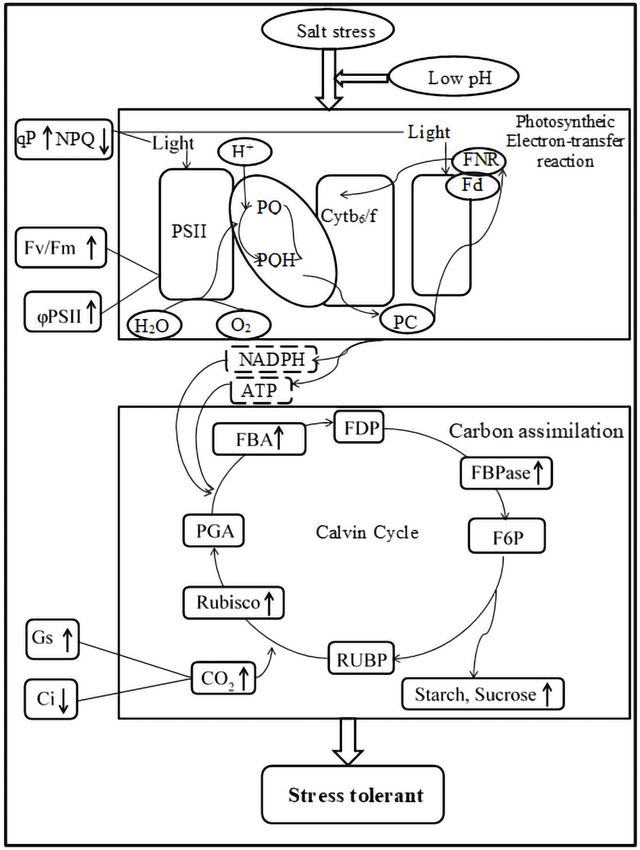
Salt stress is one of the major abiotic stresses that inhibit plant growth. As shown in Fig. 7,
salt stress significantly inhibited the growth and decreased the photosynthesis, pigment
contents and mineral contents of ginger leaves. Low pH with salt stress enhanced the
activities of RuBPCase, FBPase, and FBA and increased the pigment contents, increasing the
photosynthesis rate. Moreover, it is worth noting that low pH simultaneously increased the
accumulation of K, Mg, Ca, Fe, and Zn. In summary, the improvement of photosynthesis,
Yin et al. (2021), PeerJ, DOI 10.7717/peerj.10832 12/19Figure 7 Impacts of low pH on photosynthesis processes of ginger under salt stress. ‘‘ ↓ ’’ indicates a
decrease, ‘‘ ↑ ’’ indicates an increase.
Full-size DOI: 10.7717/peerj.10832/fig-7
pigment contents, and accumulation of minerals due to low pH ultimately increased the
biomass accumulation of ginger seedlings under salt stress.
ADDITIONAL INFORMATION AND DECLARATIONS
Funding
This work was supported by the TaiShan Industrial Experts Program, China (Grant No.
tscy20190105), the Project of agricultural excellent germplasm in Shandong province
(Grant No. 2020LZGC006), the Industrial technology system of national specialty
vegetables (Grant No. CARS-24-A-09), and the National Natural Science Foundation
Yin et al. (2021), PeerJ, DOI 10.7717/peerj.10832 13/19of China (Grant No. 31972399). The funders had no role in study design, data collection
and analysis, decision to publish, or preparation of the manuscript.
Grant Disclosures
The following grant information was disclosed by the authors:
TaiShan Industrial Experts Program, China: tscy20190105.
Project of agricultural excellent germplasm in Shandong province: 2020LZGC006.
Industrial technology system of national specialty vegetables: CARS-24-A-09.
National Natural Science Foundation of China: 31972399.
Competing Interests
The authors declare there are no competing interests.
Author Contributions
• Fengman Yin conceived and designed the experiments, performed the experiments,
analyzed the data, prepared figures and/or tables, authored or reviewed drafts of the
paper, and approved the final draft.
• Shanying Zhang performed the experiments, analyzed the data, prepared figures and/or
tables, authored or reviewed drafts of the paper, and approved the final draft.
• Bili Cao performed the experiments, analyzed the data, authored or reviewed drafts of
the paper, and approved the final draft.
• Kun Xu conceived and designed the experiments, performed the experiments, analyzed
the data, authored or reviewed drafts of the paper, and approved the final draft.
Data Availability
The following information was supplied regarding data availability:
Raw measurements are available as a Supplemental File.
Supplemental Information
Supplemental information for this article can be found online at http://dx.doi.org/10.7717/
peerj.10832#supplemental-information.
REFERENCES
Acosta JA, Faz A, Jansen B, Kalbitz K, Martínez-Martínez S. 2011. Assessment of salinity
status in intensively cultivated soils under semiarid climate, Murcia, SE Spain.
Journal of Arid Environments 75:1056–1066 DOI 10.1016/j.jaridenv.2011.05.006.
Ahmad P, Abd Allah EF, Alyemeni MN, Wijaya L, Alam P, Bhardwaj R, Siddique KHM.
2018. Exogenous application of calcium to 24-epibrassinosteroid pre-treated tomato
seedlings mitigates NaCl toxicity by modifying ascorbate-glutathione cycle and
secondary metabolites. Scientific Reports 8:13515 DOI 10.1038/s41598-018-31917-1.
Alharby HF, Al-Zahrani HS, Hakeem KR, Iqbal M. 2019. Identification of physiological
and biochemical markers for salt (NaCl) stress in the seedlings of mungbean [Vigna
radiata (L.) Wilczek] genotypes. Saudi Journal of Biological Sciences 26:1053–1060
DOI 10.1016/j.sjbs.2018.08.006.
Yin et al. (2021), PeerJ, DOI 10.7717/peerj.10832 14/19Ali BH, Blunden G, Tanira MO, Nemmar A. 2008. Some phytochemical, pharma-
cological and toxicological properties of ginger (Zingiber officinale Roscoe): a
review of recent research. Food and Chemical Toxicology: an International Journal
Published for the British Industrial Biological Research Association 46:409–420
DOI 10.1016/j.fct.2007.09.085.
Awlia M, Fajkus J, Oakey H, Panzarov aK, Trílek M, Negráo S, Roy SJ, Tester M,
M. Julkowska M. 2017. Mapping genetic components underlying early salt stress
responses in Arabidopis thaliana HapMap population. Journal of Horticultural
Science and Biotechnology 92:31e38.
Bacha H, Tekaya M, Drine S, Guasmi F, Touil L, Enneb H, Triki T, Cheour F, Ferchichi
A. 2017. Impact of salt stress on morpho-physiological and biochemical parameters
of Solanum lycopersicum cv. Microtom leaves. South African Journal of Botany
108:364–369 DOI 10.1016/j.sajb.2016.08.018.
Bollivar DW. 2006. Recent advances in chlorophyll biosynthesis. Photosynthesis Research
90:173–194 DOI 10.1007/s11120-006-9076-6.
Bremner JM. 1965. Total nitrogen. In: Black CA, Evans DD, White IL, Ensminger
LE, Clark FE, eds. Methods of soil analysis. Agronomy monograph (Part 2). vol. 9.
Madison: American Society of Agronomy, Inc., 1149–1178.
Cortleven A, Schmulling T. 2015. Regulation of chloroplast development and function
by cytokinin. Journal of Experimental Botany 66:4999–5013 DOI 10.1093/jxb/erv132.
Dhanapal AP, Ray JD, Singh SK, Hoyos-Villegas V, Smith JR, Purcell LC, Fritschi
FB. 2016. Genome-wide association mapping of soybean chlorophyll traits based
on canopy spectral reflectance and leaf extracts. BMC Plant Biology 16:174
DOI 10.1186/s12870-016-0861-x.
Ding Z, Koriem MA, Ibrahim SM, Antar AS, Ewis MA, He Z, Kheir AMS. 2020.
Seawater intrusion impacts on groundwater and soil quality in the northern part of
the Nile Delta, Egypt. Environmental Earth Sciences 79:313
DOI 10.1007/s12665-020-09069-1.
Do TD, Vuong TD, Dunn D, Smothers S, Patil G, Yungbluth DC, Chen P, Scaboo A, Xu
D, Carter TE. 2018. Mapping and confirmation of loci for salt tolerance in a novel
soybean germplasm, Fiskeby III. TAG Theoretical and Applied Genetics Theoretische
Und Angewandte Genetik 131:513–524 DOI 10.1007/s00122-017-3015-0.
Feng ZT, Deng YQ, Fan H, Sun QJ, Sui N, Wang BS. 2014. Effects of NaCl stress on
the growth and photosynthetic characteristics of Ulmus pumila L. seedlings in sand
culture. Photosynthetica 52:313–320 DOI 10.1007/s11099-014-0032-y.
Gaudio N, Belyazid S, Gendre X, Mansat A, Nicolas M, Rizzetto S, Sverdrup H, Probst
A. 2015. Combined effect of atmospheric nitrogen deposition and climate change
on temperate forest soil biogeochemistry: a modeling approach. Ecological Modelling
306:24–34 DOI 10.1016/j.ecolmodel.2014.10.002.
Guo R, Yang Z, Li F, Yan C, Zhong X, Liu Q, Xia X, Li H, Zhao L. 2015. Comparative
metabolic responses and adaptive strategies of wheat (Triticum aestivum) to salt and
alkali stress. BMC Plant Biology 15:170 DOI 10.1186/s12870-015-0546-x.
Yin et al. (2021), PeerJ, DOI 10.7717/peerj.10832 15/19Handel E. 1968. Direct microdetermination of sucrose. Analytical Biochemistry
22:280–283 DOI 10.1016/0003-2697(68)90317-5.
Hannachi S, Van Labeke M-C. 2018. Salt stress affects germination, seedling growth and
physiological responses differentially in eggplant cultivars (Solanum melongena L.).
Scientia Horticulturae 228:56–65 DOI 10.1016/j.scienta.2017.10.002.
Hendrickson L, For̈ster B, Pogson BJ, Chow WS. 2005. A simple chlorophyll fluores-
cence parameter that correlates with the rate coefficient of photoinactivation of
Photosystem II. Photosynthesis Research 84:43–49 DOI 10.1007/s11120-004-6430-4.
Holm G. 1954. Chlorophyll mutations in barley. Acta Agriculturae Scandinavica
4(1):457–471 DOI 10.1080/00015125409439955.
Hu WH, Yan XH, Yu JQ. 2016. Importance of the mitochondrial alternative ox-
idase (AOX) pathway in alleviating photoinhibition in cucumber leaves un-
der chilling injury and subsequent recovery when leaves are subjected to high
light intensity. The Journal of Horticultural Science and Biotechnology 92:31–38
DOI 10.1080/14620316.2016.1219239.
Jagerbrand AK, Kudo G. 2016. Short-term responses in maximum quantum yield
of PSII (Fv/Fm) to ex situ temperature treatment of populations of bryophytes
originating from different sites in Hokkaido, Northern Japan. Plants 5:22
DOI 10.3390/plants5020022.
Jan AU, Hadi F, Midrarullah , Nawaz MA, Rahman K. 2017. Potassium and zinc
increase tolerance to salt stress in wheat (Triticum aestivum L.). Plant Physiology and
Biochemistry 116:139–149 DOI 10.1016/j.plaphy.2017.05.008.
Janda T, Darko E, Shehata S, Kovacs V, Pal M, Szalai G. 2016. Salt acclimation processes
in wheat. Plant Physiology and Biochemistry 01:68–75
DOI 10.1016/j.plaphy.2016.01.025.
Jiang C, Zu C, Lu D, Zheng Q, Shen J, Wang H, Li D. 2017. Effect of exogenous selenium
supply on photosynthesis, Na+ accumulation and antioxidative capacity of maize
(Zea mays L.) under salinity stress. Scientific Reports 7:42039 DOI 10.1038/srep42039.
Kalaji HM, Jajoo A, Oukarroum A, Brestic M, Zivcak M, Samborska IA, Cetner MD,
Łukasik I, Goltsev V, Ladle RJ. 2016. Chlorophyll a fluorescence as a tool to monitor
physiological status of plants under abiotic stress conditions. Acta Physiologiae
Plantarum 38:102 DOI 10.1007/s11738-016-2113-y.
Kochian LV, Pineros MA, Liu J, Magalhaes JV. 2015. Plant adaptation to acid soils:
The molecular basis for crop aluminum resistance. Annual Review of Plant Biology
66:571–598 DOI 10.1146/annurev-arplant-043014-114822.
Krasensky J, Jonak C. 2012. Drought, salt, and temperature stress-induced metabolic
rearrangements and regulatory networks. Journal of Experimental Botany
63:1593–1608 DOI 10.1093/jxb/err460.
Lazro JJ, Chueca A, Gorge JL, Mayor F. 1974. Fructose 1, 6-diphosphatase from spinach
leaf chloroplasts: purification and heterogeneity. Phytochemistry 13:2455–2461
DOI 10.1016/S0031-9422(00)86920-4.
Li C, Chen J, Wang J, Ma Z, Han P, Luan Y, Lu A. 2015a. Occurrence of antibiotics in
soils and manures from greenhouse vegetable production bases of Beijing, China
Yin et al. (2021), PeerJ, DOI 10.7717/peerj.10832 16/19and an associated risk assessment. The Science of the Total Environment 521–
522:101–107 DOI 10.1016/j.scitotenv.2015.03.070.
Li H, Wang Y, Xiao J, Xu K. 2015b. Reduced photosynthetic dark reaction triggered
by ABA application increases intercellular CO2 concentration, generates H2 O2
and promotes closure of stomata in ginger leaves. Environmental and Experimental
Botany 113:11–17 DOI 10.1016/j.envexpbot.2015.01.002.
Li H-D, Zhang YZ, Xiao J, Xu K. 2013. Photosynthetic dark reaction is more sensitive to
ABA signaling caused by osmotic stress than Ca2+ signaling in ginger leaves. Scientia
Horticulturae 164:73–76 DOI 10.1016/j.scienta.2013.09.003.
Liang W, Ma X, Wan P, Liu L. 2018. Plant salt-tolerance mechanism: a review. Biochemi-
cal and Biophysical Research Communications 495:286–291
DOI 10.1016/j.bbrc.2017.11.043.
Liu X, Lv Y, Xu K, Xiao X, Xi B, Lu S. 2018. Response of ginger growth to a tetracycline-
contaminated environment and residues of antibiotic and antibiotic resistance genes.
Chemosphere 201:137–143 DOI 10.1016/j.chemosphere.2018.02.178.
Long SP, Marshall-Colon A, Zhu XG. 2015. Meeting the global food demand of the
future by engineering crop photosynthesis and yield potential. Cell 161:56–66
DOI 10.1016/j.cell.2015.03.019.
Lu Y, Lei JQ, Zeng FJ, Zhang B, Liu GJ, Liu B. 2017. Effect of NaCl-induced changes
in growth, photosynthetic characteristics, water status and enzymatic antioxidant
system of Calligonum caput-medusae seedlings. Photosynthetica 55:96–106
DOI 10.1007/s11099-016-0234-6.
Martin B, Tauer CG, Lin RK. 1999. Carbon isotope discrimination as a tool to improve
water-use efficiency in tomato. Crop Science 39:1775–1783
DOI 10.2135/cropsci1999.3961775x.
Mehta P, Jajoo A, Mathur S, Bharti S. 2010. Chlorophyll a fluorescence study revealing
effects of high salt stress on Photosystem II in wheat leaves. Plant Physiology and
Biochemistry 48:16–20 DOI 10.1016/j.plaphy.2009.10.006.
Moles TM, Pompeiano A, Reyes THuarancca, Scartazza A, Guglielminetti L.
2016. The efficient physiological strategy of a tomato landrace in response
to short-term salinity stress. Plant Physiology and Biochemistry 109:262–272
DOI 10.1016/j.plaphy.2016.10.008.
Ning L, Kan G, Shao H, Yu D. 2018. Physiological and transcriptional responses to salt
stress in salt-tolerant and salt-sensitive soybean (Glycine max [L.] Merr.) seedlings.
Land Degradation & Development 29:2707–2719 DOI 10.1002/ldr.3005.
Park HJ, Kim WY, Yun DJ. 2016. A new insight of salt stress signaling in plant. Molecules
and Cells 39:447–459 DOI 10.14348/molcells.2016.0083.
Parry MAJ, Andralojc PJ, Parmar S, Keys AJ, Habash D, Paul MJ, Alres R, Quick WP,
Servaites JC. 1997. Regulation of Rubisco by inhibitors in the light. Plant, Cell and
Environment 20(4):528–5340 DOI 10.1046/j.1365-3040.1997.d01-85.x.
Yin et al. (2021), PeerJ, DOI 10.7717/peerj.10832 17/19Patil G, Do T, Vuong TD, Valliyodan B, Lee JD, Chaudhary J, Shannon JG, Nguyen
HT. 2016. Genomic-assisted haplotype analysis and the development of high-
throughput SNP markers for salinity tolerance in soybean. Scientific Reports 6:19199
DOI 10.1038/srep19199.
Ryu H, Cho YG. 2015. Plant hormones in salt stress tolerance. Journal of Plant Biology
58:147–155 DOI 10.1007/s12374-015-0103-z.
Schreiber U, Schliwa U, Bilger W. 1986. Continuous recording of photochemical and
non-photochemical chlorophyll fluorescence quenching with a new type of modula-
tion fluorometer. Photosynthesis Research 10:51–62 DOI 10.1007/BF00024185.
Shankar V, Kumar D, Agrawal V. 2016. Assessment of antioxidant enzyme activity
and mineral nutrients in response to NaCl stress and its amelioration through
glutathione in Chickpea. Applied Biochemistry and Biotechnology 178:267–284
DOI 10.1007/s12010-015-1870-1.
Singh M, Singh VP, Prasad SM. 2016. Nitrogen modifies NaCl toxicity in eggplant
seedlings: Assessment of chlorophyll a fluorescence, antioxidative response
and proline metabolism. Biocatalysis and Agricultural Biotechnology 7:76–86
DOI 10.1016/j.bcab.2016.05.007.
Batta SK, Singh R. 1986. Sucrose metabolism in sugar cane grown under varying
climatic conditions synthesis and storage of sucrose in relation to the activities
of sucrose synthase, sucrose phosphate synthase and invertase. Phytochemistry
25(11):2431–2437 DOI 10.1016/S0031-9422(00)84484-2.
Tang YY, Yuan YH, Shu S, Guo SR. 2018. Regulatory mechanism of NaCl stress
on photosynthesis and antioxidant capacity mediated by transglutaminase in
cucumber (Cucumis sativus L.) seedlings. Scientia Horticulturae 235:294–306
DOI 10.1016/j.scienta.2018.02.045.
Thouvenot L, Haury J, Thiébaut G. 2012. Responses of two invasive macrophyte species
to salt. Hydrobiologia 686:213–223 DOI 10.1007/s10750-012-1013-4.
Wakeel A, Farooq M, Qadir M, Schubert S. 2011. Potassium substitution by sodium in
plants. Critical Reviews in Plant Sciences 30:401–413
DOI 10.1080/07352689.2011.587728.
Xiao CW, Sang WG, Wang RZ. 2008. Fine root dynamics and turnover rate in an
Asia white birch forest of Donglingshan Mountain, China. Forest Ecology and
Management 255:765–773 DOI 10.1016/j.foreco.2007.09.062.
Yin FM, Cao BL, Xu K. 2019. Effects of simulated soil acidification and salt interaction
on mineral elements and osmotic substance in ginger. Plant Physiology Journal
55(6):814–820 DOI 10.13592/j.cnki.ppj.2019.0116.
Yin FM, Liu XN, Cao BL, Xu K. 2020. Low pH altered salt stress in antioxidant
metabolism and nitrogen assimilation in ginger (Zingiber officinale) seedlings.
Physiologia Plantarum 168(3):648–659 DOI 10.1111/ppl.13011.
Zhang K, Liu H, Song J, Wu W, Li K, Zhang J. 2016. Physiological and comparative
proteome analyses reveal low-phosphate tolerance and enhanced photosynthesis in a
maize mutant owing to reinforced inorganic phosphate recycling. BMC Plant Biology
16:129 DOI 10.1186/s12870-016-0825-1.
Yin et al. (2021), PeerJ, DOI 10.7717/peerj.10832 18/19Zhu XG, Long SP, Ort DR. 2010. Improving photosynthetic efficiency for greater yield.
Annual Review of Plant Biology 61:235–261
DOI 10.1146/annurev-arplant-042809-112206.
Yin et al. (2021), PeerJ, DOI 10.7717/peerj.10832 19/19You can also read