DNA- and telomere-damage does not limit lifespan: evidence from rapamycin - Aging-US
←
→
Page content transcription
If your browser does not render page correctly, please read the page content below
www.aging-us.com                                                                 AGING 2021, Vol. 13, No. 3
                                                                                                 Research Perspective
DNA- and telomere-damage does not limit lifespan: evidence from
rapamycin
Mikhail V. Blagosklonny1
1
Roswell Park Cancer Institute, Buffalo, NY 14263, USA

Correspondence to: Mikhail V. Blagosklonny; email: Blagosklonny@oncotarget.com
Keywords: quasi-programmed aging, hyperfunction theory, antagonistic pleiotropy, natural selection, mTOR
Received: January 29, 2021       Accepted: February 10, 2021         Published: February 12, 2021

Copyright: © 2021 Blagosklonny. This is an open access article distributed under the terms of the Creative Commons
Attribution License (CC BY 3.0), which permits unrestricted use, distribution, and reproduction in any medium, provided the
original author and source are credited.

ABSTRACT

Failure of rapamycin to extend lifespan in DNA repair mutant and telomerase-knockout mice, while extending
lifespan in normal mice, indicates that neither DNA damage nor telomere shortening limits normal lifespan or
causes normal aging.

INTRODUCTION                                                       Quasi-programmed (hyperfunctional) aging

As a provocative title has recently announced,                     In proliferating cells, growth-promoting pathways such
“rapamycin fails to extend lifespan in DNA repair                  as mTOR (Target of Rapamycin) and MAPK drive
-deficient mice” [1]. The word “fails” implies bad news.           cellular growth, which is balanced by cell division. When
Rapamycin tried but failed. Yet, it is expected that the           the cell cycle is arrested, however, growth-promoting
anti-aging drug rapamycin should not restore lifespan of           pathways drive cellular senescence, which is a
short-lived mice that fail to grow and die young from              continuation of cellular growth in the absence of cell
causes other than normal aging [2]. In such growth-                division [5]. During geroconversion to senescence, cells
retarded mice, rapamycin, an inhibitor of cell growth,             become hypertrophic and hyperfunctional. One example
further retards weight gain.                                       of hyper-function is SASP or Senescence-Associated
                                                                   Secretory Phenotype [6]. Rapamycin can cause reversible
Similarly, rapamycin does not extend but even slightly             cycle arrest but suppresses geroconversion, thus ensuring
shortens lifespan in telomerase-deficient mice, which              quiescence instead of senescence. (Note: Rapamycin
die young from poor growth and intestinal atrophy                  does not prevent cell cycle arrest, it only prevents
caused by telomere shortening [3]. (As we will discuss,            geroconversion that makes this arrest permanent [7]. This
this is predictable by hyperfunction theory.) While                point is often miscited by others). Rapamycin slows
shortening lifespan by 18% in unnatural telomerase-                down both growth and geroconversion, figuratively
deficient mice, in the same study in natural mice,                 slowing down time [8]. Like cellular senescence is a
rapamycin increased lifespan by 39% and healthspan by              continuation of growth, organismal aging is a
58% (measured as tumor-free survival) [3]. In dozens of            continuation of growth too [9].
independent studies, rapamycin has not failed to extend
lifespan in normal mice [4]. However, while extending              According to hyperfunction theory, aging is quasi-
lifespan in normal mice, rapamycin may fail to save                programmed, a continuation of developmental
animals dying young from cellular growth retardation.              growth programs, driven in part by hyper-functional
But something important should not be overlooked. The              signaling pathways including the mTOR pathway
failure of rapamycin to extend lifespan in these short-            [9]. Hyperfunction is an excessive normal function
lived mice, dying from DNA damage, rules out the                   later in life. It’s not necessarily an increase of function;
damage theory of aging. To understand this point, we               it may even be insufficient decrease of function. For
must first discuss what limits animal lifespan.                    example, protein synthesis is decreased in C elegans but

www.aging-us.com                                         3167                                                         AGING
is still too high: its further inhibition extends lifespan Natural selection favors robust development and
[10, 11]. fitness early in life at the cost of aging. For example,
growth hormone receptor-deficient mice (GHR-KO
Hyperfunction leads to age-related diseases, secondary mice), with decreased mTORC1 activity, live longer
organ damage and loss of function. For example, but are small and weak early in life [27, 28]. In such
cellular hyperfunctions result in hypertension, mice mTORC1-driven aging is inhibited and mice live
culminating in stroke and damage of the brain. Aging is longer but would not survive in the wild and therefore
a sum of all age-related diseases [12, 13]. This theory do not exist in nature. As another example, knockout
was discussed in detail [9, 14–20] and has gained of PI3K, an activator of mTOR pathways, extends
experimental support [11, 16, 21–26]. I will not discuss lifespan 10-fold in C. elegans [29]. The mutant worm
it here, just to mention that accumulation of molecular undergoes prolonged developmental arrest, which
damage is not a driving force of development and would be lethal in the wild [29]. Therefore, natural
therefore of aging. It is hyperfunctional signaling selection favors hyperfunctional mTOR that is
pathways such as mTOR (one of many) that drive both optimal for development but drives age-related
growth and aging, causing age-related diseases that in diseases later in life.
turn damage organs, leading to secondary loss of
function. According to damage theories, aging is functional
decline caused by molecular damage. According to
Although molecular damage accumulates, this hyperfunction theory, quasi-programmed aging is not
accumulation is not life-limiting because quasi- functional decline but a hyperfunction: cellular and
programmed aging terminates life first (Figure 1A). systemic functions are higher than optimal for
Quasi-programmed (hyperfunctional) aging is life- longevity. They are optimal for early life fitness and in
limiting, because it is favored by natural selection. part (only in part) mTOR-dependent.
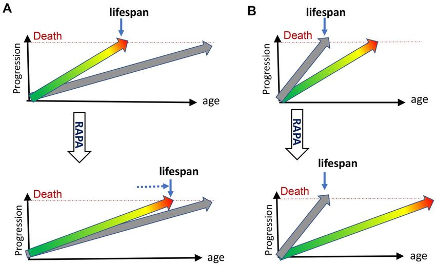
Figure 1. Rapamycin extends lifespan in natural but not progeroid mice. (A) Natural mice. Hyperfunctional aging (green/yellow/red
arrow) progresses from development (green) to diseases (red), reaching death threshold and limiting lifespan. Accumulation of molecular
damage (gray arrow) is slow and does not reach death threshold in animal lifetime. It would take longer to die from molecular damage.
Treatment with rapamycin (RAPA) extends lifespan by slowing down mTOR-driven aging (B) Progeroid, telomerase- or DNA-repair-deficient
mice. Accumulation of molecular damage (gray arrow) is artificially accelerated to become life-limiting. Treatment with rapamycin (RAPA)
cannot extend lifespan.
www.aging-us.com 3168 AGINGIn both molecular damage and hyperfunction theories, example, telomere shortening. Second-generation
aging exists because late-life is shadowed from natural telomerase-deficient mice (G2 Terc−/−) with critically
selection. But quasi-programmed aging is not simply short telomeres fail to grow and die young from
shadowed from, it is promoted by natural selection, unfamiliar diseases such as intestinal atrophy due to
because accelerated aging is hardwired with fitness failure of cell proliferation [3]. When telomeres reach
early in life. By selecting for fitness, nature indirectly critical length, it can cause DNA-damage response,
selects for accelerated aging. This makes quasi- leading to aplastic anemia, organ fibrosis, atrophy of the
programmed aging life-limiting. One of predictions of small intestine and the spleen, skin and hair lesions. In
hyperfunction theory is that rapamycin must extend humans, diseases of short telomeres cause death from
lifespan in animals [9]. This prediction has been bone marrow failure and pulmonary fibrosis [72]. This
confirmed. In dozens of studies, rapamycin prolongs does not resemble normal aging.
lifespan and healthspan in mice [3, 30–65]. Rapamycin
extends lifespan in C elegans [66] and Drosophila [67– In humans, mice and C. elegans, telomere shortening is
69]. Furthermore, rapamycin even extends life of the not life-limiting [73–75]. In mice lacking telomerase,
simplest animal, Hydra, which is thought to be even accelerated telomere shortening is still not life-
immortal. Depending on conditions, Hydra can be limiting in the first generation [76]. It took several
either immortal or undergo aging. Rapamycin slows generations to achieve critically short telomeres, leading
aging, stem cell exhaustion and extends life span in to syndromes strikingly different from normal aging. In
Hydra [70]. humans, telomere length does not reach telomere
threshold during life time [75, 77, 78]. Normal telomere
mTOR-driven aging is only one component of quasi- shortening would cause telomere-driven pathologies,
programmed (hyperfunction) aging. In addition, but normal animals do not live long enough to reach this
MEK/MAPK, NF-kB, p63, HIF-1 and many other threshold. Rapamycin prolongs life in normal mice,
signaling pathways are involved, interacting with the proving that telomere length does not constrain normal
mTOR pathway and forming networks. Rapamycin lifespan [3]. When artificially shortened, then telomeres
cannot affect all of them. In theory, mTOR-independent become life-limiting and rapamycin cannot extend
quasi-programmed aging can be life-limiting in some lifespan anymore [3].
conditions and diseases. I suggest that long-lived GHR-
KO mice with low mTORC1 activity undergo partially Ercc1∆/− mutant mice are defective in DNA repair, such
mTORC1-independent quasi-programmed senescence, as transcription-coupled repair, global-genome
because rapamycin cannot prolong lifespan in these nucleotide excision and crosslink repair [1, 2].
mice further, while prolonging lifespan in parental Therefore, multiple types of DNA damages accumulate.
normal mice [71]. Discussion of mTOR-independent This leads to decreased cell proliferation, arrested
components of quasi-programmed aging is beyond the development, poor growth, abnormal liver nuclei of
focus of this article. Let us return to stochastic liver and kidney, absence of subcutaneous fat, ferritin
accumulation of molecular damage. deposition, kidney malfunction and early death [2].
Unlike natural mice, short-lived Ercc1∆/− mice do not
How molecular damage can become life- develop tumors, probably because they do not live long
limiting enough to suffer typical age-related diseases [1, 2]. In
such mice, dying from molecular damage, rapamycin
Molecular damage can become life-limiting in two fails to extend lifespan [1].
ways. First, hyper-functional aging should be eliminated
or slowed down, so an organism lives long enough to CONCLUSIONS
die from accumulation of molecular damage. In this
scenario, accumulation of molecular damage causes Here I discussed new evidence that normal aging is not
post-aging. Such examples are unknown, but it is a very caused by accumulation of molecular damage or
intriguing possibility. Could a PI3K-null worm [29] telomere shortening: while extending normal lifespan in
with 10-fold longer lifespan die from molecular mice, rapamycin failed to do so in mice dying from
damage? molecular damage (Figure 1).
Second, accumulation of molecular damage can be Previously, several lines of evidence suggested that
greatly accelerated artificially by knockout of molecular damage does not cause normal aging. Their
repair/maintenance enzymes (Figure 1B). Such animals detailed discussion is beyond the focus of this article, so I
do not exist in nature. But artificially created, they may will just mention some of them, without referencing them
provide a glimpse of how post-aging may look. Their (I will reference these points in forthcoming review
pathology differs drastically from normal aging, for “When longevity drugs do not increase
www.aging-us.com 3169 AGINGlongevity: Unifying development-driven and damage- 1997; 7:427–39.
induced theories of aging”, In press). First, https://doi.org/10.1016/s0960-9822(06)00190-4
overexpression of enzymes that decrease damage does not PMID:9197240
extend lifespan in most studies. Similarly, antioxidants do
3. Ferrara-Romeo I, Martinez P, Saraswati S, Whittemore
not extend lifespan in animals and may increase mortality K, Graña-Castro O, Thelma Poluha L, Serrano R,
in humans. Furthermore, even data that support damage
Hernandez-Encinas E, Blanco-Aparicio C, Maria Flores J,
theory can be explained by other mechanisms. For
Blasco MA. The mTOR pathway is necessary for
example, N-Acetyl-L-Cysteine, a commonly used anti-
survival of mice with short telomeres. Nat Commun.
oxidant, can inhibit mTOR. Second, according to
2020; 11:1168.
calculations, molecular damage, especially mtDNA
https://doi.org/10.1038/s41467-020-14962-1
mutations and telomere shortening, cannot reach deadly
PMID:32127537
threshold during animal lifetime. Third, genetic knockout
of signaling pathways can extend lifespan without 4. Blagosklonny MV. The goal of geroscience is life
affecting molecular damage. Similarly, pharmacological extension. Oncotarget. 2021; 12:131–44
interventions can extend life without affecting damage https://doi.org/10.18632/oncotarget.27882
accumulation. Forth, dramatic intra- and inter-species 5. Demidenko ZN, Blagosklonny MV. Growth stimulation
differences in lifespan poorly correlate with the rate of leads to cellular senescence when the cell cycle is
molecular damage. Fifth, nuclear transfer and nuclear blocked. Cell Cycle. 2008; 7:3355–61.
reprogramming both rule out DNA damage as a cause of https://doi.org/10.4161/cc.7.21.6919 PMID:18948731
aging. Following adult somatic cell nuclear transfer,
cloned animals are healthy and have normal lifespan. 6. Laberge RM, Sun Y, Orjalo AV, Patil CK, Freund A, Zhou
Sixth, low levels of molecular damage may increase L, Curran SC, Davalos AR, Wilson-Edell KA, Liu S,
longevity. This phenomenon is known as hormesis. Limbad C, Demaria M, Li P, et al. MTOR regulates the
Regardless of mechanistic explanations, this indicates pro-tumorigenic senescence-associated secretory
that molecular damage is not-life-limiting even when phenotype by promoting IL1A translation. Nat Cell Biol.
moderately increased. Finally, rapamycin increases 2015; 17:1049–61.
lifespan in all normal animals tested, indicating that https://doi.org/10.1038/ncb3195 PMID:26147250
mTORC1-dependent quasi-program is life-limiting. The 7. Blagosklonny MV. Cell cycle arrest is not yet
list can go on and on. Once again, damage accumulates senescence, which is not just cell cycle arrest:
and must cause death eventually, but quasi-programmed terminology for TOR-driven aging. Aging (Albany NY).
(hyperfunctional) aging terminates life first. Molecular 2012; 4:159–65.
damage can become life-limiting, when artificially https://doi.org/10.18632/aging.100443
accelerated or, potentially, when quasi-programmed PMID:22394614
aging is decelerated. Then interventions to repair
molecular damage may increase life further. 8. Blagosklonny MV. Does rapamycin slow down time?
Oncotarget. 2018; 9:30210–12.
CONFLICTS OF INTEREST https://doi.org/10.18632/oncotarget.25788
PMID:30100983
The author declares that he has no conflicts of interest. 9. Blagosklonny MV. Aging and immortality: quasi-
programmed senescence and its pharmacologic
REFERENCES inhibition. Cell Cycle. 2006; 5:2087–102.
https://doi.org/10.4161/cc.5.18.3288
1. Birkisdóttir MB, Jaarsma D, Brandt RM, Barnhoorn S, PMID:17012837
van Vliet N, Imholz S, van Oostrom CT, Nagarajah B, 10. Pan KZ, Palter JE, Rogers AN, Olsen A, Chen D, Lithgow
Portilla Fernández E, Roks AJ, Elgersma Y, van Steeg H, GJ, Kapahi P. Inhibition of mRNA translation extends
Ferreira JA, et al. Unlike dietary restriction, rapamycin lifespan in caenorhabditis elegans. Aging Cell. 2007;
fails to extend lifespan and reduce transcription stress 6:111–19.
in progeroid DNA repair-deficient mice. Aging Cell. https://doi.org/10.1111/j.1474-9726.2006.00266.x
2021. [Epub ahead of print]. PMID:17266680
https://doi.org/10.1111/acel.13302 PMID:33484480
11. Dhondt I, Petyuk VA, Cai H, Vandemeulebroucke L,
2. Weeda G, Donker I, de Wit J, Morreau H, Janssens Vierstraete A, Smith RD, Depuydt G, Braeckman BP.
R, Vissers CJ, Nigg A, van Steeg H, Bootsma D, FOXO/DAF-16 activation slows down turnover of the
Hoeijmakers JH. Disruption of mouse ERCC1 results majority of proteins in C. Elegans. Cell Rep. 2016;
in a novel repair syndrome with growth failure, 16:3028–40.
nuclear abnormalities and senescence. Curr Biol. https://doi.org/10.1016/j.celrep.2016.07.088
www.aging-us.com 3170 AGINGPMID:27626670 PMID:31712450
12. Blagosklonny MV. Validation of anti-aging drugs by 21. Scialò F, Sriram A, Naudí A, Ayala V, Jové M, Pamplona
treating age-related diseases. Aging (Albany NY). 2009; R, Sanz A. Target of rapamycin activation predicts
1:281–88. lifespan in fruit flies. Cell Cycle. 2015; 14:2949–58.
https://doi.org/10.18632/aging.100034 https://doi.org/10.1080/15384101.2015.1071745
PMID:20157517 PMID:26259964
13. Blagosklonny MV. Prospective treatment of age- 22. de la Guardia Y, Gilliat AF, Hellberg J, Rennert P,
related diseases by slowing down aging. Am J Pathol. Cabreiro F, Gems D. Run-on of germline apoptosis
2012; 181:1142–46. promotes gonad senescence in C. Elegans. Oncotarget.
https://doi.org/10.1016/j.ajpath.2012.06.024 2016; 7:39082–96.
PMID:22841821 https://doi.org/10.18632/oncotarget.9681
14. Blagosklonny MV. Aging: ROS or TOR. Cell Cycle. 2008; PMID:27256978
7:3344–54. 23. Chen HY, Maklakov AA. The worm that lived: evolution
https://doi.org/10.4161/cc.7.21.6965 of rapid aging under high extrinsic mortality revisited.
PMID:18971624 Worm. 2013; 2:e23704.
15. Blagosklonny MV. Answering the ultimate question https://doi.org/10.4161/worm.23704 PMID:24778930
”what is the proximal cause of aging?”. Aging (Albany 24. Lind MI, Ravindran S, Sekajova Z, Carlsson H, Hinas A,
NY). 2012; 4:861–77. Maklakov AA. Experimentally reduced insulin/IGF-1
https://doi.org/10.18632/aging.100525 signaling in adulthood extends lifespan of parents and
PMID:23425777 improves darwinian fitness of their offspring. Evol Lett.
16. Gems D, de la Guardia Y. Alternative perspectives on 2019; 3:207–16.
aging in caenorhabditis elegans: reactive oxygen https://doi.org/10.1002/evl3.108 PMID:31007945
species or hyperfunction? Antioxid Redox Signal. 2013; 25. Cheng Z, Ristow M. Mitochondria and metabolic
19:321–29. homeostasis. Antioxid Redox Signal. 2013; 19:240–42.
https://doi.org/10.1089/ars.2012.4840 https://doi.org/10.1089/ars.2013.5255
PMID:22870907 PMID:23432475
17. Gems D, Partridge L. Genetics of longevity in model
26. de Verges J, Nehring V. A critical look at proximate
organisms: debates and paradigm shifts. Annu Rev
causes of social insect senescence: damage
Physiol. 2013; 75:621–44.
accumulation or hyperfunction? Curr Opin Insect Sci.
https://doi.org/10.1146/annurev-physiol-030212-
2016; 16:69–75.
183712 PMID:23190075
https://doi.org/10.1016/j.cois.2016.05.003
18. Wang H, Zhao Y, Ezcurra M, Benedetto A, Gilliat AF, PMID:27720053
Hellberg J, Ren Z, Galimov ER, Athigapanich T,
27. Dominick G, Berryman DE, List EO, Kopchick JJ, Li X,
Girstmair J, Telford MJ, Dolphin CT, Zhang Z, Gems D. A
Miller RA, Garcia GG. Regulation of mTOR activity in
parthenogenetic quasi-program causes teratoma-like
snell dwarf and GH receptor gene-disrupted mice.
tumors during aging in wild-type C. Elegans. NPJ Aging
Endocrinology. 2015; 156:565–75.
Mech Dis. 2018; 4:6.
https://doi.org/10.1210/en.2014-1690
https://doi.org/10.1038/s41514-018-0025-3
PMID:25456069
PMID:29928508
19. Ezcurra M, Benedetto A, Sornda T, Gilliat AF, Au C, 28. Bartke A, Sun LY, Longo V. Somatotropic signaling:
Zhang Q, van Schelt S, Petrache AL, Wang H, de la trade-offs between growth, reproductive
Guardia Y, Bar-Nun S, Tyler E, Wakelam MJ, Gems D. C. development, and longevity. Physiol Rev. 2013;
elegans Eats Its Own Intestine to Make Yolk Leading to 93:571–98.
Multiple Senescent Pathologies. Curr Biol. 2018; https://doi.org/10.1152/physrev.00006.2012
28:2544–56.e5. PMID:23589828
https://doi.org/10.1016/j.cub.2018.06.035 29. Ayyadevara S, Alla R, Thaden JJ, Shmookler Reis RJ.
PMID:30100339 Remarkable longevity and stress resistance of
20. Xi J, Cai J, Cheng Y, Fu Y, Wei W, Zhang Z, Zhuang Z, nematode PI3K-null mutants. Aging Cell. 2008;
Hao Y, Lilly MA, Wei Y. The TORC1 inhibitor Nprl2 7:13–22.
protects age-related digestive function in Drosophila. https://doi.org/10.1111/j.1474-9726.2007.00348.x
Aging (Albany NY). 2019; 11:9811–28. PMID:17996009
https://doi.org/10.18632/aging.102428 30. Chen C, Liu Y, Liu Y, Zheng P. mTOR regulation and
www.aging-us.com 3171 AGINGtherapeutic rejuvenation of aging hematopoietic stem 4:144ra103.
cells. Sci Signal. 2009; 2:ra75. https://doi.org/10.1126/scitranslmed.3003802
https://doi.org/10.1126/scisignal.2000559 PMID:22837538
PMID:19934433
38. Wilkinson JE, Burmeister L, Brooks SV, Chan CC,
31. Harrison DE, Strong R, Sharp ZD, Nelson JF, Astle CM, Friedline S, Harrison DE, Hejtmancik JF, Nadon N,
Flurkey K, Nadon NL, Wilkinson JE, Frenkel K, Carter CS, Strong R, Wood LK, Woodward MA, Miller RA.
Pahor M, Javors MA, Fernandez E, Miller RA. Rapamycin slows aging in mice. Aging Cell. 2012;
Rapamycin fed late in life extends lifespan in 11:675–82.
genetically heterogeneous mice. Nature. 2009; https://doi.org/10.1111/j.1474-9726.2012.00832.x
460:392–95. PMID:22587563
https://doi.org/10.1038/nature08221 PMID:19587680
39. Fang Y, Westbrook R, Hill C, Boparai RK, Arum O, Spong
32. Anisimov VN, Zabezhinski MA, Popovich IG, Piskunova A, Wang F, Javors MA, Chen J, Sun LY, Bartke A.
TS, Semenchenko AV, Tyndyk ML, Yurova MN, Antoch Duration of rapamycin treatment has differential
MP, Blagosklonny MV. Rapamycin extends maximal effects on metabolism in mice. Cell Metab. 2013;
lifespan in cancer-prone mice. Am J Pathol. 2010; 17:456–62.
176:2092–97. https://doi.org/10.1016/j.cmet.2013.02.008
https://doi.org/10.2353/ajpath.2010.091050 PMID:23473038
PMID:20363920
40. Flynn JM, O’Leary MN, Zambataro CA, Academia EC,
33. Anisimov VN, Zabezhinski MA, Popovich IG, Piskunova Presley MP, Garrett BJ, Zykovich A, Mooney SD, Strong
TS, Semenchenko AV, Tyndyk ML, Yurova MN, R, Rosen CJ, Kapahi P, Nelson MD, Kennedy BK, Melov
Rosenfeld SV, Blagosklonny MV. Rapamycin increases S. Late-life rapamycin treatment reverses age-related
lifespan and inhibits spontaneous tumorigenesis in heart dysfunction. Aging Cell. 2013; 12:851–62.
inbred female mice. Cell Cycle. 2011; 10:4230–36. https://doi.org/10.1111/acel.12109
https://doi.org/10.4161/cc.10.24.18486 PMID:23734717
PMID:22107964
41. Johnson SC, Yanos ME, Kayser EB, Quintana A,
34. Miller RA, Harrison DE, Astle CM, Baur JA, Boyd AR, de Sangesland M, Castanza A, Uhde L, Hui J, Wall VZ,
Cabo R, Fernandez E, Flurkey K, Javors MA, Nelson JF, Gagnidze A, Oh K, Wasko BM, Ramos FJ, et al. mTOR
Orihuela CJ, Pletcher S, Sharp ZD, et al. Rapamycin, but inhibition alleviates mitochondrial disease in a mouse
not resveratrol or simvastatin, extends life span of model of leigh syndrome. Science. 2013; 342:1524–28.
genetically heterogeneous mice. J Gerontol A Biol Sci https://doi.org/10.1126/science.1244360
Med Sci. 2011; 66:191–201. PMID:24231806
https://doi.org/10.1093/gerona/glq178
42. Livi CB, Hardman RL, Christy BA, Dodds SG, Jones D,
PMID:20974732
Williams C, Strong R, Bokov A, Javors MA, Ikeno Y,
35. Comas M, Toshkov I, Kuropatwinski KK, Chernova OB, Hubbard G, Hasty P, Sharp ZD. Rapamycin extends life
Polinsky A, Blagosklonny MV, Gudkov AV, Antoch MP. span of Rb1+/- mice by inhibiting neuroendocrine
New nanoformulation of rapamycin rapatar extends tumors. Aging (Albany NY). 2013; 5:100–10.
lifespan in homozygous p53-/- mice by delaying https://doi.org/10.18632/aging.100533
carcinogenesis. Aging (Albany NY). 2012; 4:715–22. PMID:23454836
https://doi.org/10.18632/aging.100496 43. Neff F, Flores-Dominguez D, Ryan DP, Horsch M,
PMID:23117593
Schröder S, Adler T, Afonso LC, Aguilar-Pimentel JA,
36. Komarova EA, Antoch MP, Novototskaya LR, Chernova Becker L, Garrett L, Hans W, Hettich MM, Holtmeier
OB, Paszkiewicz G, Leontieva OV, Blagosklonny MV, R, et al. Rapamycin extends murine lifespan but has
Gudkov AV. Rapamycin extends lifespan and delays limited effects on aging. J Clin Invest. 2013;
tumorigenesis in heterozygous p53+/- mice. Aging 123:3272–91.
(Albany NY). 2012; 4:709–14. https://doi.org/10.1172/JCI67674
https://doi.org/10.18632/aging.100498 PMID:23863708
PMID:23123616
44. Ye L, Widlund AL, Sims CA, Lamming DW, Guan Y, Davis
37. Ramos FJ, Chen SC, Garelick MG, Dai DF, Liao CY, JG, Sabatini DM, Harrison DE, Vang O, Baur JA.
Schreiber KH, MacKay VL, An EH, Strong R, Ladiges WC, Rapamycin doses sufficient to extend lifespan do not
Rabinovitch PS, Kaeberlein M, Kennedy BK. Rapamycin compromise muscle mitochondrial content or
reverses elevated mTORC1 signaling in lamin A/C- endurance. Aging (Albany NY). 2013; 5:539–50.
deficient mice, rescues cardiac and skeletal muscle https://doi.org/10.18632/aging.100576
function, and extends survival. Sci Transl Med. 2012; PMID:23929887
www.aging-us.com 3172 AGING45. Fok WC, Chen Y, Bokov A, Zhang Y, Salmon AB, Diaz V, 53. Johnson SC, Yanos ME, Bitto A, Castanza A, Gagnidze A,
Javors M, Wood WH 3rd, Zhang Y, Becker KG, Pérez VI, Gonzalez B, Gupta K, Hui J, Jarvie C, Johnson BM,
Richardson A. Mice fed rapamycin have an increase in Letexier N, McCanta L, Sangesland M, et al. Dose-
lifespan associated with major changes in the liver dependent effects of mTOR inhibition on weight and
transcriptome. PLoS One. 2014; 9:e83988. mitochondrial disease in mice. Front Genet. 2015;
https://doi.org/10.1371/journal.pone.0083988 6:247.
PMID:24409289 https://doi.org/10.3389/fgene.2015.00247
PMID:26257774
46. Hasty P, Livi CB, Dodds SG, Jones D, Strong R, Javors M,
Fischer KE, Sloane L, Murthy K, Hubbard G, Sun L, 54. Karunadharma PP, Basisty N, Dai DF, Chiao YA, Quarles
Hurez V, Curiel TJ, Sharp ZD. eRapa restores a normal EK, Hsieh EJ, Crispin D, Bielas JH, Ericson NG, Beyer RP,
life span in a FAP mouse model. Cancer Prev Res MacKay VL, MacCoss MJ, Rabinovitch PS. Subacute
(Phila). 2014; 7:169–78. calorie restriction and rapamycin discordantly alter
https://doi.org/10.1158/1940-6207.CAPR-13-0299 mouse liver proteome homeostasis and reverse aging
PMID:24282255 effects. Aging Cell. 2015; 14:547–57.
https://doi.org/10.1111/acel.12317 PMID:25807975
47. Khapre RV, Kondratova AA, Patel S, Dubrovsky Y,
Wrobel M, Antoch MP, Kondratov RV. BMAL1- 55. Arriola Apelo SI, Pumper CP, Baar EL, Cummings NE,
dependent regulation of the mTOR signaling pathway Lamming DW. Intermittent administration of
delays aging. Aging (Albany NY). 2014; 6:48–57. rapamycin extends the life span of female C57BL/6J
https://doi.org/10.18632/aging.100633 mice. J Gerontol A Biol Sci Med Sci. 2016; 71:876–81.
PMID:24481314 https://doi.org/10.1093/gerona/glw064
PMID:27091134
48. Leontieva OV, Paszkiewicz GM, Blagosklonny MV.
Weekly administration of rapamycin improves survival 56. Bitto A, Ito TK, Pineda VV, LeTexier NJ, Huang HZ,
and biomarkers in obese male mice on high-fat diet. Sutlief E, Tung H, Vizzini N, Chen B, Smith K, Meza D,
Aging Cell. 2014; 13:616–22. Yajima M, Beyer RP, et al. Transient rapamycin
https://doi.org/10.1111/acel.12211 PMID:24655348 treatment can increase lifespan and healthspan in
middle-aged mice. Elife. 2016; 5:e16351.
49. Miller RA, Harrison DE, Astle CM, Fernandez E, Flurkey
https://doi.org/10.7554/eLife.16351 PMID:27549339
K, Han M, Javors MA, Li X, Nadon NL, Nelson JF,
Pletcher S, Salmon AB, Sharp ZD, et al. Rapamycin- 57. Liao CY, Anderson SS, Chicoine NH, Mayfield JR,
mediated lifespan increase in mice is dose and sex Academia EC, Wilson JA, Pongkietisak C, Thompson
dependent and metabolically distinct from dietary MA, Lagmay EP, Miller DM, Hsu YM, McCormick MA,
restriction. Aging Cell. 2014; 13:468–77. O’Leary MN, Kennedy BK. Rapamycin reverses
https://doi.org/10.1111/acel.12194 PMID:24341993 metabolic deficits in lamin A/C-deficient mice. Cell Rep.
50. Popovich IG, Anisimov VN, Zabezhinski MA, 2016; 17:2542–52.
https://doi.org/10.1016/j.celrep.2016.10.040
Semenchenko AV, Tyndyk ML, Yurova MN,
PMID:27926859
Blagosklonny MV. Lifespan extension and cancer
prevention in HER-2/neu transgenic mice treated with 58. Felici R, Buonvicino D, Muzzi M, Cavone L, Guasti D,
low intermittent doses of rapamycin. Cancer Biol Ther. Lapucci A, Pratesi S, De Cesaris F, Luceri F, Chiarugi A.
2014; 15:586–92. Post onset, oral rapamycin treatment delays
https://doi.org/10.4161/cbt.28164 PMID:24556924 development of mitochondrial encephalopathy only at
supramaximal doses. Neuropharmacology. 2017;
51. Zhang Y, Bokov A, Gelfond J, Soto V, Ikeno Y, Hubbard
117:74–84.
G, Diaz V, Sloane L, Maslin K, Treaster S, Réndon S, van
Remmen H, Ward W, et al. Rapamycin extends life and https://doi.org/10.1016/j.neuropharm.2017.01.039
PMID:28161373
health in C57BL/6 mice. J Gerontol A Biol Sci Med Sci.
2014; 69:119–30. 59. Siegmund SE, Yang H, Sharma R, Javors M, Skinner O,
https://doi.org/10.1093/gerona/glt056 Mootha V, Hirano M, Schon EA. Low-dose rapamycin
PMID:23682161 extends lifespan in a mouse model of mtDNA depletion
syndrome. Hum Mol Genet. 2017; 26:4588–605.
52. Hurez V, Dao V, Liu A, Pandeswara S, Gelfond J, Sun L,
https://doi.org/10.1093/hmg/ddx341 PMID:28973153
Bergman M, Orihuela CJ, Galvan V, Padrón Á, Drerup J,
Liu Y, Hasty P, et al. Chronic mTOR inhibition in mice 60. Wang T, Tsui B, Kreisberg JF, Robertson NA, Gross AM,
with rapamycin alters T, B, myeloid, and innate Yu MK, Carter H, Brown-Borg HM, Adams PD, Ideker T.
lymphoid cells and gut flora and prolongs life of Epigenetic aging signatures in mice livers are slowed by
immune-deficient mice. Aging Cell. 2015; 14:945–56. dwarfism, calorie restriction and rapamycin treatment.
https://doi.org/10.1111/acel.12380 PMID:26315673 Genome Biol. 2017; 18:57.
www.aging-us.com 3173 AGINGhttps://doi.org/10.1186/s13059-017-1186-2 69. Castillo-Quan JI, Tain LS, Kinghorn KJ, Li L, Grönke S,
PMID:28351423 Hinze Y, Blackwell TK, Bjedov I, Partridge L. A triple
drug combination targeting components of the
61. Bielas J, Herbst A, Widjaja K, Hui J, Aiken JM, McKenzie
nutrient-sensing network maximizes longevity. Proc
D, Miller RA, Brooks SV, Wanagat J. Long term
rapamycin treatment improves mitochondrial DNA Natl Acad Sci USA. 2019; 116:20817–19.
https://doi.org/10.1073/pnas.1913212116
quality in aging mice. Exp Gerontol. 2018; 106:125–31.
PMID:31570569
https://doi.org/10.1016/j.exger.2018.02.021
PMID:29486228 70. Tomczyk S, Suknovic N, Schenkelaars Q, Wenger Y,
Ekundayo K, Buzgariu W, Bauer C, Fischer K, Austad S,
62. Weiss R, Fernandez E, Liu Y, Strong R, Salmon AB.
Galliot B. Deficient autophagy in epithelial stem cells
Metformin reduces glucose intolerance caused by
rapamycin treatment in genetically heterogeneous drives aging in the freshwater cnidarian Hydra.
Development. 2020; 147:dev177840.
female mice. Aging (Albany NY). 2018; 10:386–401.
https://doi.org/10.1242/dev.177840 PMID:31862842
https://doi.org/10.18632/aging.101401
PMID:29579736 71. Fang Y, Hill CM, Darcy J, Reyes-Ordoñez A, Arauz E,
McFadden S, Zhang C, Osland J, Gao J, Zhang T, Frank
63. Parihar M, Dodds SG, Hubbard G, Javors MA, Strong R,
SJ, Javors MA, Yuan R, et al. Effects of rapamycin on
Hasty P, Sharp ZD. Rapamycin extends life span in
ApcMin/+ colon cancer FAP model. Clin Colorectal growth hormone receptor knockout mice. Proc Natl
Acad Sci USA. 2018; 115:E1495–503.
Cancer. 2020; S1533-0028:30123–27.
https://doi.org/10.1073/pnas.1717065115
https://doi.org/10.1016/j.clcc.2020.08.006
PMID:29378959
PMID:33132009
72. Gramatges MM, Bertuch AA. Short telomeres: from
64. Strong R, Miller RA, Bogue M, Fernandez E, Javors MA,
dyskeratosis congenita to sporadic aplastic anemia and
Libert S, Marinez PA, Murphy MP, Musi N, Nelson JF,
Petrascheck M, Reifsnyder P, Richardson A, et al. Malignancy. Transl Res. 2013; 162:353–63.
https://doi.org/10.1016/j.trsl.2013.05.003
Rapamycin-mediated mouse lifespan extension: late-
PMID:23732052
life dosage regimes with sex-specific effects. Aging Cell.
2020; 19:e13269. 73. Raices M, Maruyama H, Dillin A, Karlseder J.
https://doi.org/10.1111/acel.13269 Uncoupling of longevity and telomere length in C.
PMID:33145977 Elegans. PLoS Genet. 2005; 1:e30.
65. Christy B, Demaria M, Campisi J, Huang J, Jones D, https://doi.org/10.1371/journal.pgen.0010030
PMID:16151516
Dodds SG, Williams C, Hubbard G, Livi CB, Gao X,
Weintraub S, Curiel T, Sharp ZD, Hasty P. P53 and 74. Simons MJ. Questioning causal involvement of
rapamycin are additive. Oncotarget. 2015; 6:15802–13. telomeres in aging. Ageing Res Rev. 2015; 24:191–96.
https://doi.org/10.18632/oncotarget.4602 https://doi.org/10.1016/j.arr.2015.08.002
PMID:26158292 PMID:26304838
66. Robida-Stubbs S, Glover-Cutter K, Lamming DW, 75. Steenstrup T, Kark JD, Verhulst S, Thinggaard M,
Mizunuma M, Narasimhan SD, Neumann-Haefelin E, Hjelmborg JV, Dalgård C, Kyvik KO, Christiansen L,
Sabatini DM, Blackwell TK. TOR signaling and Mangino M, Spector TD, Petersen I, Kimura M, Benetos
rapamycin influence longevity by regulating SKN-1/Nrf A, et al. Telomeres and the natural lifespan limit in
and DAF-16/FoxO. Cell Metab. 2012; 15:713–24. humans. Aging (Albany NY). 2017; 9:1130–42.
https://doi.org/10.1016/j.cmet.2012.04.007 https://doi.org/10.18632/aging.101216
PMID:22560223 PMID:28394764
67. Bjedov I, Toivonen JM, Kerr F, Slack C, Jacobson J, Foley 76. Herrera E, Samper E, Martín-Caballero J, Flores JM, Lee
A, Partridge L. Mechanisms of life span extension by HW, Blasco MA. Disease states associated with
rapamycin in the fruit fly drosophila melanogaster. Cell telomerase deficiency appear earlier in mice with short
Metab. 2010; 11:35–46. telomeres. EMBO J. 1999; 18:2950–60.
https://doi.org/10.1016/j.cmet.2009.11.010 https://doi.org/10.1093/emboj/18.11.2950
PMID:20074526 PMID:10357808
68. Wang A, Mouser J, Pitt J, Promislow D, Kaeberlein M. 77. Bischoff C, Petersen HC, Graakjaer J, Andersen-
Rapamycin enhances survival in a drosophila model of Ranberg K, Vaupel JW, Bohr VA, Kølvraa S, Christensen
mitochondrial disease. Oncotarget. 2016; 7:80131–39. K. No association between telomere length and
https://doi.org/10.18632/oncotarget.12560 survival among the elderly and oldest old.
PMID:27741510 Epidemiology. 2006; 17:190–94.
www.aging-us.com 3174 AGINGhttps://doi.org/10.1097/01.ede.0000199436.55248.10 mortality in the oldest old: a population-based study.
PMID:16477260 Aging Cell. 2005; 4:287–90.
https://doi.org/10.1111/j.1474-9726.2005.00171.x
78. Martin-Ruiz CM, Gussekloo J, van Heemst D, von
PMID:16300480
Zglinicki T, Westendorp RG. Telomere length in white
blood cells is not associated with morbidity or
www.aging-us.com 3175 AGINGYou can also read