Transposon silencing in the Drosophila female germline is essential for genome stability in progeny embryos
←
→
Page content transcription
If your browser does not render page correctly, please read the page content below
Published Online: 17 September, 2018 | Supp Info: http://doi.org/10.26508/lsa.201800179
Downloaded from life-science-alliance.org on 14 October, 2021

Research Article

Transposon silencing in the Drosophila female germline is
essential for genome stability in progeny embryos
Zeljko Durdevic1, Ramesh S Pillai2, Anne Ephrussi1

The Piwi-interacting RNA pathway functions in transposon con-                      at the transcriptional and posttranscriptional levels (reviewed in
trol in the germline of metazoans. The conserved RNA helicase                      Guzzardo et al [2013]). Besides PIWI proteins, other factors such as
Vasa is an essential Piwi-interacting RNA pathway component,                       Tudor domain proteins and RNA helicases are involved in piRNA
but has additional important developmental functions. Here, we                     biogenesis and transposon silencing. Mutations in most piRNA path-
address the importance of Vasa-dependent transposon control in                     way genes in Drosophila females cause transposon up-regulation that
the Drosophila female germline and early embryos. We find that                      leads to an arrest of oogenesis. This effect can be rescued by
transient loss of vasa expression during early oogenesis leads to                  suppression of the DNA damage checkpoint proteins of the ATR/
transposon up-regulation in supporting nurse cells of the fly egg-                  Chk2 pathway (Chen et al, 2007; Klattenhoff et al, 2007; Pane et al,
chamber. We show that elevated transposon levels have dramatic                     2007). By contrast, inhibition of DNA damage signaling cannot re-
consequences, as de-repressed transposons accumulate in the                        store embryonic development (Chen et al, 2007; Klattenhoff et al,
oocyte where they cause DNA damage. We find that suppression                        2007; Pane et al, 2007). Recent studies suggest that PIWI proteins
of Chk2-mediated DNA damage signaling in vasa mutant females                       might have additional roles during early embryogenesis inde-
restores oogenesis and egg production. Damaged DNA and up-                         pendent of DNA damage signaling (Khurana et al, 2010; Mani et al,
regulated transposons are transmitted from the mother to the                       2014). However, functions of the piRNA pathway during early em-
embryos, which sustain severe nuclear defects and arrest de-                       bryonic development remain poorly understood.
velopment. Our findings reveal that the Vasa-dependent pro-                            One of the essential piRNA pathway factors with an important
tection against selfish genetic elements in the nuage of nurse cell                 role in development is the highly conserved RNA helicase Vasa. First
is essential to prevent DNA damage–induced arrest of embryonic                     identified in Drosophila as a maternal-effect gene (Schüpbach &
development.                                                                       Wieschaus, 1986; Hay et al, 1988; Lasko & Ashburner, 1990), vasa
                                                                                   (vas) was subsequently shown to function in various cellular and
DOI 10.26508/lsa.201800179 | Received 27 August 2018 | Revised 4 September
                                                                                   developmental processes (reviewed in Lasko [2013]). In the Dro-
2018 | Accepted 5 September 2018 | Published online 17 September 2018
                                                                                   sophila female germline, Vasa accumulates in two different cyto-
                                                                                   plasmic electron-dense structures: the pole (or germ) plasm at the
                                                                                   posterior pole of the oocyte, and the nuage, the perinuclear region
Introduction                                                                       of nurse cells. In the pole plasm, Vasa interacts with the pole
                                                                                   plasm–inducer Oskar (Osk) (Markussen et al, 1995; Jeske et al, 2015)
Transposons and other selfish genetic elements are found in all                     and ensures accumulation of different proteins and mRNAs that
eukaryotes and comprise a large fraction of their genomes. Al-                     determine primordial germ cell (PGC) formation and embryonic
though transposons are thought to be beneficial in driving evo-                     patterning (Hay et al, 1988; Lasko & Ashburner, 1990). In the nuage,
lution (Levin & Moran, 2011), their mobilization in the germline can               Vasa is required for the assembly of the nuage itself (Liang et al,
compromise genome integrity with deleterious consequences:                         1994; Malone et al, 2009) and facilitates the transfer of transposon
insertional mutagenesis reduces the fitness of the progeny, and                     RNA intermediates from Aub to Ago3, driving the piRNA amplification
loss of germ cell integrity causes sterility. Therefore, it is of great            cycle and piRNA-mediated transposon silencing (Xiol et al, 2014;
importance for sexually reproducing organisms to firmly control                     Nishida et al, 2015). As Vasa’s involvement in many cellular processes
transposon activity in their germ cells. Metazoans have evolved                    renders it difficult to analyze its functions in each process in-
a germline-specific mechanism that, by relying on the activity of                   dividually, it remains unknown whether Vasa’s functions in devel-
Piwi family proteins and their associated Piwi-interacting RNAs                    opment and in the piRNA pathway are linked or independent.
(piRNAs), suppresses mobile elements.                                                 In this study, we address the role of Vasa in transposon control in
   Drosophila harbors three PIWI proteins: Piwi, Aubergine (Aub),                  Drosophila development. We find that failure to suppress trans-
and Argonaute 3 (Ago3), which, guided by piRNAs, silence transposons               posons in the nuage of nurse cells causes DNA double-strand

1                                                                                         2
Developmental Biology Unit, European Molecular Biology Laboratory, Heidelberg, Germany        Department of Molecular Biology, University of Geneva, Geneva, Switzerland

Correspondence: ephrussi@embl.de

© 2018 Ephrussi et al.                                                               https://doi.org/10.26508/lsa.201800179       vol 1 | no 5 | e201800179       1 of 9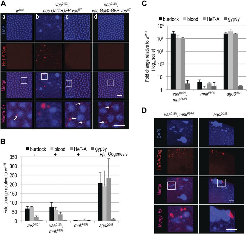
breaks (DSBs), severe nuclear defects, and lethality of progeny             Loss of Vasa during early oogenesis affects viability of progeny
embryos. Even transient interruption of Vasa expression in early            embryos
oogenesis de-represses transposons and impairs embryo via-
bility. Depletion of the Drosophila Chk2 ortholog maternal nuclear          Concentration of Vasa protein at the posterior pole of the embryo is
kinase (mnk) restores oogenesis in vas mutants, but does not                essential for PGC and abdomen formation during embryogenesis
suppress defects in transposon silencing or DSB-induced nuclear             (Schüpbach & Wieschaus, 1986; Hay et al, 1988; Lasko & Ashburner,
damage and embryonic lethality. We show that up-regulated                   1990). We analyzed the number of PGC-positive embryos and the
transposons invade the maternal genome, inducing DNA DSBs                   hatching rate of eggs produced by vasD1/D1 flies expressing GFP-VasWT
that, together with transposon RNAs and proteins, are maternally            either under control of the nos or the vas promoter (vasD1/D1; nos-
transmitted and consequently cause embryogenesis arrest. Our                Gal4>GFP-vasWT and vasD1/D1; vas-Gal4>GFP-vasWT embryos). Embryos
study thus demonstrates that Vasa function in the nuage of                  from vasD1/D1 mutant flies could not be included in these and all the
Drosophila nurse cells is essential to maintain genome integrity in         other experiments on embryos, as vasD1/D1 females arrest oogenesis
both the oocyte and progeny embryos, ensuring normal embry-                 early and do not lay eggs. PGC formation was restored in approximately
onic development.                                                           50% of vasD1/D1; nos-Gal4>GFP-vasWT and vasD1/D1; vas-Gal4>GFP-vasWT
                                                                            embryos (Fig 1C) (Table S1). However, DAPI staining revealed nuclear
                                                                            damage in some vasD1/D1; nos-Gal4>GFP-vasWT embryos (see below),
Results                                                                     which we excluded from the quantification.
                                                                               Expression of GFP-VasWT also partially rescued the hatching of eggs
Vasa-dependent transposon control is not essential for                      produced by vasD1/D1 flies (Fig 1D). However, expression of GFP-VasWT
oogenesis                                                                   led to a significantly lower hatching rate in vasD1/D1; nos-Gal4>GFP-vasWT
                                                                            than in vasD1/D1; vas-Gal4>GFP-vasWT flies (Fig 1D) (Table S2). Expression
Vasa is required for piRNA biogenesis and transposon silencing in           of GFP-VasWT in heterozygous loss-of-function vasD1/Q7 females led to
Drosophila, as in vas mutants piRNAs are absent and transposons             a low hatching rate similar to vasD1/D1 (Fig S2A and B) (Table S3),
are up-regulated (Vagin et al, 2004; Malone et al, 2009; Zhang et al,       excluding a possible secondary mutation as the cause of the low
2012; Czech et al, 2013; Handler et al, 2013). To investigate the im-       hatching rate. The fact that in spite of comparable GFP-VasWT levels
portance of transposon control in Drosophila development, we                (Fig S2C), the hatching rate of vasD1/D1; vas-Gal4>GFP-vasWT embryos
expressed WT GFP-Vasa fusion protein (GFP-VasWT; Fig S1A) in the            was higher than that of vasD1/D1; nos-Gal4>GFP-vasWT embryos,
female germline of loss-of-function (vasD1/D1) vas flies using two           suggests that transient loss of vas expression during early oogenesis
promoters with distinct expression patterns (Fig S1B and C): the vas        impairs viability of progeny embryos (Fig 1D).
promoter is active at all stages of oogenesis, whereas the nos
promoter is attenuated between stages 2 and 6 (Fig S1B and C).              Elevated transposon levels cause DNA and nuclear damage in
   We first assessed the ability of GFP-VasWT fusion protein to              progeny embryos
promote transposon silencing in the female germline, and exam-
ined the effect of GFP-VasWT on the level of expression of several          Elevated transposon activity leads to DNA damage and ultimately to
transposons in vas mutant ovaries. We chose the LTR retro-                  cell death. During our analysis of PGC formation, we observed nuclear
transposons burdock and blood, and the non-LTR retrotransposon              damage in a considerable fraction of vasD1/D1; nos-Gal4>GFP-vasWT
HeT-A, which were previously reported to be up-regulated upon               embryos. Quantification of embryos containing nuclei of aberrant
Vasa depletion (Vagin et al, 2004; Czech et al, 2013). The LTR ret-         nuclear morphology (Fig 2A, lower panel) compared with the nuclei
rotransposon gypsy, which belongs to the so-called somatic group            of WT embryos (Fig 2A, upper panel) revealed a high proportion of such
of transposons and is not affected by Vasa depletion, served as             nuclear defects among vasD1/D1; nos-Gal4>GFP-vasWT embryos (Fig
a negative control (Czech et al, 2013). Loss-of-function vasD1/D1           2A). Transposon mobilization causes DSBs in genomic DNA that are
ovaries contained elevated levels of burdock, blood, and HeT-A RNA          marked by the incorporation of a phosphorylated form of the H2A
(Fig 1A). Remarkably, silencing of transposons by GFP-VasWT in              variant (γH2Av), a histone H2A variant involved in DNA DSB repair.
vasD1/D1 flies depended on which Gal4 driver was used (Fig S1B and C):       Analysis of γH2Av occurrence showed that embryos displaying nu-
When driven by nos-Gal4, GFP-VasWT had no effect on transposon              clear damage were γH2Av-positive (Fig 2B), indicating that DNA DSBs
levels, whereas when driven by vas-Gal4, it led to the re-silencing of      cause nuclear defects. The levels of γH2Av were higher in vasD1/D1;
transposons (Fig 1A). This differential effect presumably reflects the       nos-Gal4>GFP-vasWT embryos compared with WT and vasD1/D1;
stages of oogenesis at which the nos and vas promoters are active, and      vas-Gal4>GFP-vasWT (Fig 2C) (Table S4).
suggests that lack of Vasa between stages 2 and 6 of oogenesis (Fig            The correlation between high levels of transposon expression
S1B) leads to transposon de-repression. Importantly, independent of         during oogenesis (Fig 1A, nos-Gal4-driven) and a high frequency of
Gal4 driver used, expression of GFP-VasWT restored oogenesis (Figs 1B       nuclear damage and DSBs in vasD1/D1; nos-Gal4>GFP-vasWT embryos
and S1D) and egg-laying (Fig S1E and F). The fact that in spite of          (Fig 2A–C) suggested that maternally transmitted transposons
transposon up-regulation oogenesis and egg-laying rates were largely        cause embryonic lethality. To test this, we compared transposon
restored in vasD1/D1 flies (Fig 1A, indicated by + and − and Fig S1D–F) is   RNA levels in embryos of vasD1/D1; nos-Gal4>GFP-vasWT and vasD1/D1;
consistent with the notion that transposon activation affects but does      vas-Gal4>GFP-vasWT flies, in which transposon RNAs are up- and down-
not completely block oogenesis unless the level of activation is so high    regulated, respectively (Fig 1A). Levels of maternally transmitted
as to cause its arrest.                                                     transposon RNA were significantly higher in vasD1/D1; nos-Gal4>GFP-vasWT

Transposons cause embryonic lethality   Ephrussi et al.                      https://doi.org/10.26508/lsa.201800179   vol 1 | no 5 | e201800179   2 of 9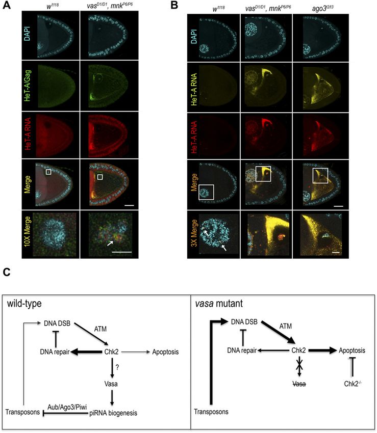
Figure 1. Silencing of transposon RNAs during
                                                                                                  oogenesis is essential for embryonic development.
                                                                                                  (A) qPCR analysis of LTR transposons burdock, blood,
                                                                                                  and gypsy and non-LTR transposon HeT-A RNAs in
                                                                                                  vasD1/D1, vasD1/D1; nos-Gal4>GFP-vasWT and vasD1/D1;
                                                                                                  vas-Gal4>GFP-vasWT, ovaries. Expression level of
                                                                                                  transposons in WT (w1118) was set to one and
                                                                                                  normalized to rp49 mRNA in individual experiments.
                                                                                                  Error bars represent the standard deviation among
                                                                                                  three biological replicates. P-values were determined
                                                                                                  by t test. P-values for burdock (0.006), blood (0.0002),
                                                                                                  and HeT-A (0.0007) were lower than 0.01 (indicated in
                                                                                                  the chart), whereas gypsy levels were not significantly
                                                                                                  different (P = 0.5). Oogenesis completion is indicated
                                                                                                  with + and −. (B) Immunohistochemical detection of
                                                                                                  Vasa in WT (w1118) and vasD1/D1 flies (upper panel), and
                                                                                                  GFP signal of GFP-VasWT fusion protein in vasD1/D1; nos-
                                                                                                  Gal4>GFP-vasWT and vaD1D/D1; vas-Gal4>GFP-vasWT flies
                                                                                                  (lower panel). Insets show enlarged images of nuage
                                                                                                  and oocyte posterior pole. Scale bars indicate 50 μm
                                                                                                  (egg-chambers) and 10 μm (nuage and pole plasm). (C)
                                                                                                  Quantification of PGC-positive embryos produced by
                                                                                                  WT (w1118), vasD1/D1; nos-Gal4>GFP-vasWT, and vasD1/D1;
                                                                                                  vas-Gal4>GFP-vasWT flies. Error bars represent the
                                                                                                  standard deviation among three biological replicates
                                                                                                  (Table S1). Panel (left) shows PGC-positive embryo (top)
                                                                                                  and PGC-negative embryo (bottom). Scale bars indicate
                                                                                                  100 μm (embryo) and 5 μm (PGCs). (D) Hatching rates of
                                                                                                  eggs laid by WT (w1118), vasD1/D1; nos-Gal4>GFP-vasWT,
                                                                                                  and vasD1/D1; vas-Gal4>GFP-vasWT flies. Error bars
                                                                                                  represent the standard deviation among three
                                                                                                  biological replicates (Table S2). P-value was
                                                                                                  determined by t test.

embryos (Fig 2D) suggesting that the increased lethality observed in      Chk2 mutation restores oogenesis but not transposon silencing
vasD1/D1; nos-Gal4>GFP-vasWT embryos is due to DNA damage (Fig            and embryogenesis in vas mutants
2A–C) caused by the high levels of maternally transmitted transposon
RNAs (Fig 2D).                                                            To test genetically whether DNA damage signaling contributes to
   One of the up-regulated transposons in vas mutants is HeT-A,           the oogenesis arrest of vas loss-of-function mutants (Schüpbach &
whose RNA and protein expression is strongly de-repressed in              Wieschaus, 1986;Hay et al, 1988; Lasko & Ashburner, 1990) (Fig 1A), we
piRNA pathway mutant ovaries (Aravin et al, 2001; Vagin et al, 2006;      introduced the mnkP6 loss-of-function allele into the vasD1 back-
Zhang et al, 2014; Lopez-Panades et al, 2015). Analysis of HeT-A/Gag      ground. Genetic removal of mnk (Fig S3B) suppressed the oogenesis
protein expression in 0–1 h old embryos showed that the levels of         arrest of vasD1/D1 mutants and partially rescued their egg laying (Figs
HeT-A/Gag were much higher in vasD1/D1; nos-Gal4>GFP-vasWT than           S3C and S4A). Importantly, mnkP6/P6 single mutants expressed Vasa at
in vasD1/D1; vas-Gal4>GFP-vasWT embryos (Fig 2E). In addition, we         WT levels and, as expected, the protein was not detected in vasD1/D1,
stained embryos with antibodies against HeT-A/Gag protein and             mnkP6/P6 double mutants (Fig S2C). Taken together, these findings
observed that in cellularized WT embryos, HeT-A localized in distinct     demonstrate that the oogenesis arrest of loss-of-function vas mutants
perinuclear foci (Figs 3A, panel a and S3A, panel a), as previously       results from activation of the Chk2-mediated DNA damage-signaling
described for HeT-A/Gag-HA-FLAG fusion protein (Olovnikov et al,          checkpoint.
2016). In vasD1/D1; nos-Gal4>GFP-vasWT embryos displaying nuclear            Although removal of mnk allowed oogenesis progression, it did
damage, HeT-A protein accumulated in large foci throughout the            not reduce transposon levels in vasD1/D1, mnkP6/P6 ovaries, and the
embryo (Figs 3A, panel b and S3A, panel b), whereas embryos of the        eggs laid failed to hatch (Figs 3B and S4B) (Table S5). Further analysis
same genotype lacking nuclear damage showed a WT distribution of          revealed that vasD1/D1, mnkP6/P6 early embryos contained elevated
the protein (Figs 3A, panel c and S3A, panel c). Finally, HeT-A/Gag       levels of maternally transmitted transposon RNAs (Fig 3C). This was
displayed WT localization in vasD1/D1; vas-Gal4>GFP-vasWT embryos         also the case of ago3 single mutant embryos, which displayed nuclear
(Figs 3A, panel d and S3A, panel d). Altogether, these results show       damage (Mani et al, 2014) (Fig S4D) similar to that of vasD1/D1; nos-
that up-regulation of transposon mRNAs and proteins during oogenesis      Gal4>GFP-vasWT embryos (Fig 2A and B). In addition to HeT-A RNA,
results in their maternal transmission to the progeny, where they cause   HeT-A/Gag protein was also up-regulated in vasD1/D1, mnkP6/P6, and
DSBs, nuclear damage, and arrest of embryogenesis.                        ago3 embryos during the syncytial blastoderm stage (Fig S4C). At

Transposons cause embryonic lethality   Ephrussi et al.                    https://doi.org/10.26508/lsa.201800179   vol 1 | no 5 | e201800179      3 of 9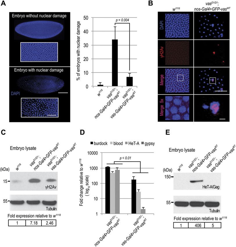
Figure 2. Maternally transmitted transposon RNAs
cause DNA double-strand breaks and nuclear damage
in progeny embryos.
(A) Quantification of nuclear damage determined by
NucBlue Fixed Cell Stain staining of WT (w1118), vasD1/D1;
nos-Gal4>GFP-vasWT, and vasD1/D1; vas-Gal4>GFP-vasWT
stage 5 embryos. Error bars represent the standard
deviation among three biological replicates (Table S4).
P-value was determined by t test. Panel shows an embryo
without (top) and an embryo with nuclear damage
(bottom). Scale bars indicate 100 μm (embryo) and 10 μm
(magnification). (B) Immunohistochemical detection of
DNA double-strand breaks using antibodies against H2Av
pS137 (γH2Av) in WT (w1118), and vasD1/D1; nos-Gal4>GFP-
vasWT stage 5 embryos. Whole embryos are presented in
(A). Scale bars indicate 5 and 2 μm (5× magnification). (C)
Western blot analysis using antibodies against H2Av
pS137 (γH2Av) showing protein levels in WT (w1118), vasD1/D1;
nos-Gal4>GFP-vasWT, and vasD1/D1; vas-Gal4>GFP-vasWT
1- to 3-h-old embryos. Tubulin was used as a loading
control. Table shows quantification of γH2Av levels
relative to WT. γH2Av signal was normalized to tubulin
signal in individual experiments and was set to one in WT.
(D) qPCR analysis of LTR transposons burdock, blood, and
gypsy and non-LTR transposon HeT-A RNAs in vasD1/D1;
nos-Gal4>GFP-vasWT and vasD1/D1; vas-Gal4>GFP-vasWT
early embryos. Expression level of transposons in WT
(w1118) was set to one and normalized to 18S rRNA in
individual experiments. Error bars represent the standard
deviation among three biological replicates. t Test
indicated P-values for burdock (0.004), blood (0.002), and
HeT-A (0.008) lower than 0.01 (indicated in the chart),
whereas gypsy levels were not significantly different
(P = 0.4). (E) Western blot analysis using antibodies against
HeT-A/Gag showing protein levels in early embryos
produced by WT (w1118), vasD1/D1; nos-Gal4>GFP-vasWT, and
vasD1/D1; vas-Gal4>GFP-vasWT flies. Tubulin was used as
a loading control. The table shows quantification of
HeT-A/Gag protein levels relative to WT. HeT-A/Gag
signal was normalized to tubulin signal in individual
experiments and was set to one in WT.
cellularization, the embryos displayed nuclear damage and HeT-A/ mnkP6/P6 double and in ago3 single mutant females up-regulated
Gag was present in large foci throughout the embryo (Figs 3D and transposons invade the maternal genome and are transmitted to
S4D), resembling the nuclear-damaged vasD1/D1; nos-Gal4>GFP-vasWT the progeny, causing severe nuclear defects and embryogenesis
embryos (Fig 3A, panel b). arrest. We conclude that tight regulation of transposons throughout
We next examined the distribution of HeT-A RNAs and occur- oogenesis is essential to maintain genome integrity in the oo-
rence of DNA DSBs by FISH and antibody staining of γH2Av, re- cyte and in early syncytial embryo, hence for normal embryonic
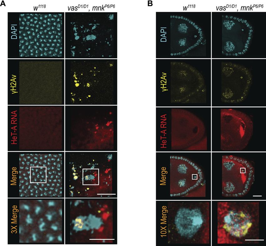
spectively. Damaged nuclei in vasD1/D1, mnkP6/P6 embryos were development.
γH2Av-positive (Figs 4A and S5A), indicating that DNA DSBs cause
nuclear defects. HeT-A RNAs localized in large foci in vasD1/D1,
mnkP6/P6 embryos, and was not detectable in WT embryos (Figs 4A Discussion
and S5A). Although, we did not detect HeT-A transcripts in the
damaged nuclei of vasD1/D1, mnkP6/P6 embryos, the oocyte nucleus Our study shows that a transient loss of vas expression during early
was positive both for HeT-A RNA and γH2Av (Fig 4B), indicating the oogenesis leads to up-regulation of transposon levels and com-
presence of DNA DSBs. Further analysis showed that HeT-A RNA promised viability of progeny embryos. The observed embryonic
and HeT-A/Gag protein co-localize in the oocyte cytoplasm and lethality is because of DNA DSBs and nuclear damage that arise as
nucleus (Fig 5A) indicating that transposon insertions into the a consequence of the elevated levels of transposon mRNAs and
maternal genome begin already during oogenesis. Additional FISH proteins, which are transmitted from the mother to the progeny. We
analyses showed that in WT egg-chambers, HeT-A and Burdock thus demonstrate that transposon silencing in the nurse cells is
transcripts were only detected at sites of transcription in the essential to prevent maternal transmission of transposons and
nurse cell nuclei, whereas in vasD1/D1, mnkP6/P6, and ago3 egg- DNA damage, protecting the progeny from harmful transposon-
chambers, transcripts of both transposons accumulated within mediated mutagenic effects.
the oocyte along the anterior margin, and around and within the Our finding that suppression of Chk2-mediated DNA damage
nucleus (Figs 5B and S5B). These results show that in vasD1/D1, signaling in loss-of-function vas mutant flies restores oogenesis,
Transposons cause embryonic lethality Ephrussi et al. https://doi.org/10.26508/lsa.201800179 vol 1 | no 5 | e201800179 4 of 9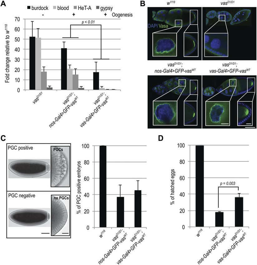
Figure 3. Loss of Chk2 DNA damage signaling does
                                                                                                not restore embryogenesis in vas mutant flies.
                                                                                                (A) Immunohistochemical detection of HeT-A/Gag
                                                                                                protein in WT (w1118; a), vasD1/D1; nos-Gal4>GFP-vasWT
                                                                                                (b and c), and vasD1/D1; vas-Gal4>GFP-vasWT (d) stage 5
                                                                                                embryos. Arrows indicate WT localization of HeT-A/
                                                                                                Gag. Staining of the whole embryos is presented in Fig
                                                                                                S3A. Scale bars indicate 10 and 5 μm (5× magnification).
                                                                                                (B) qPCR analysis of LTR transposons burdock, blood,
                                                                                                and gypsy and non-LTR transposon HeT-A RNAs in
                                                                                                ovaries from vasD1/D1 single and vasD1/D1, mnkP6/P6
                                                                                                double mutant flies, and mnkP6/P6 and agot2/t3 mutant
                                                                                                flies. The expression level of transposons in WT (w1118)
                                                                                                was set to one and normalized to rp49 mRNA in
                                                                                                individual experiments. Error bars represent the
                                                                                                standard deviation among three biological replicates.
                                                                                                Oogenesis completion is indicated with + and −. (C)
                                                                                                qPCR analysis of LTR transposons burdock, blood, and
                                                                                                gypsy and non-LTR transposon HeT-A RNAs in early
                                                                                                embryos produced by vasD1/D1, mnkP6/P6 double
                                                                                                mutant, and mnkP6/P6 and agot2/t3 single mutant flies.
                                                                                                The expression level of transposons in WT (w1118) was
                                                                                                set to one and normalized to 18S rRNA in individual
                                                                                                experiments. Error bars represent the SD among three
                                                                                                biological replicates. (D) Immunohistochemical
                                                                                                detection of HeT-A/Gag protein in stage 5 embryos
                                                                                                from vasD1/D1, mnkP6/P6 double mutant and agot2/t3
                                                                                                single mutant flies. Staining of the whole embryos is
                                                                                                presented in Fig S4D. Scale bars indicate 10 and 5 μm
                                                                                                (5× magnification).

and egg production demonstrates that Chk2 is epistatic to vas.          without deleterious effects on oogenesis or embryogenesis (Tiwari
However, hatching is severely impaired, because of the DNA              et al, 2017). It is possible that the absence of pole plasm in vas
damage sustained by the embryos. The defects displayed by vas,          mutants (Lehmann & Ephrussi, 1994) results in the release of the
mnk double mutant embryos resembled those of PIWI (piwi,                transposon products and their ectopic accumulation in the oocyte.
aub, and ago3) single and mnk; PIWI double mutant embryos               Localization of transposons to the germ plasm (Tiwari et al, 2017)
(Klattenhoff et al, 2007; Mani et al, 2014). Earlier observation that   may restrict their activity to the future germline and protect the
inactivation of DNA damage signaling does not rescue the devel-         embryo soma from transposon activity. Transposon-mediated
opment of PIWI mutant embryos led to the assumption that PIWI           mutagenesis in the germline would produce genetic variability,
proteins might have an essential role in early somatic development,     a phenomenon thought to play a role in the environmental ad-
independent of cell cycle checkpoint signaling (Mani et al, 2014). By   aptation and evolution of species. It would therefore be of interest
tracing transposon protein and RNA levels and localization from         to determine the role of pole plasm in transposon control in the
the mother to the early embryos, we suggest that, independent of        future.
Chk2 signaling, de-repressed transposons are responsible for               Transposon up-regulation in the Drosophila female germline
nuclear damage and embryonic lethality. Our study indicates that        triggers a DNA damage-signaling cascade (Chen et al, 2007;
transposon insertions occur in the maternal genome where they           Klattenhoff et al, 2007). In aub mutants, before their oogenesis
cause DNA DSBs that together with transposon RNAs and proteins          arrest occurs, Chk2-mediated signaling leads to phosphorylation
are passed on to the progeny embryos. Transposon activity and           of Vasa, leading to impaired grk mRNA translation and embryonic
consequent DNA damage in the early syncytial embryo cause               axis specification (Klattenhoff et al, 2007). Considering the genetic
aberrant chromosome segregation, resulting in unequal distri-           interaction of vas and mnk (Chk2) and the fact that Vasa is phos-
bution of the genetic material, nuclear damage and ultimately           phorylated in Chk2-dependent manner (Abdu et al, 2002; Klattenhoff
embryonic lethality. Our study shows that early Drosophila embryos      et al, 2007), it is tempting to speculate that phosphorylation of Vasa
are defenseless against transposons and will succumb to their           might stimulate piRNA biogenesis, reinforcing transposon silencing
mobilization if the first line of protection against selfish genetic      and thus minimizing transposon-induced DNA damage (Fig 5C). The
elements in the nuage of nurse cell fails.                              arrest of embryonic development as a first, and arrest of oogenesis as
   A recent study showed that in p53 mutants, transposon RNAs are       an ultimate response to DNA damage, thus, prevents the spreading of
up-regulated and accumulate at the posterior pole of the oocyte,        detrimental transposon-induced mutations to the next generation.

Transposons cause embryonic lethality   Ephrussi et al.                  https://doi.org/10.26508/lsa.201800179   vol 1 | no 5 | e201800179     5 of 9
Figure 4. Transposons invade maternal genome and
cause DNA DSBs in vas, mnk double mutant flies.
(A, B) In situ detection of HeT-A mRNA by FISH and
immunohistochemical detection of DNA DSBs using
antibodies against H2Av pS137 (γH2Av) in WT (w1118) and
vasD1/D1, mnkP6/P6 double mutant embryos (A) and
ovaries (B). Scale bars in (A) indicate 5 and 2 μm
(3× magnification); scale bars in (B) indicate 20 and
5 μm (10× magnification).
Experimental procedures Fecundity and hatching assays
Virgin females of all genetic backgrounds tested were mated with
Fly stocks and husbandry w1118 males for 24 h at 25°C. The crosses were then transferred to
The following Drosophila stocks were used: w1118; b1, vasD1/CyO apple-juice agar plates, and eggs collected in 24 h intervals over 3–4 d.
(vas3, Tearle and Nusslein-Volhard, 1987; Lasko & Ashburner, The number of eggs laid on each plate was counted; the plates
1990); b1, vasQ7, pr1/CyO (vas7, Tearle and Nusslein-Volhard, were kept at 25°C for 2 d, then the number of hatched larvae
1987; Lasko & Ashburner, 1990); vasD1/CyO; nos-Gal4-VP16/TM2 counted. Experiments were performed in three independent
(Xiol et al, 2014); vas-Gal4 (gift of Jean-René Huynh); GFP-vasWT/ replicates represented in Tables S2, S3, and S5. w1118 females were
TM2 (Xiol et al, 2014); mnkP6/CyO (Brodsky et al, 2004); bw1; used as a control.
st1, ago3t2/TM6B, Tb+ (FBst0028269), bw1; st1; ago3t3/TM6B, Tb1
(FBst0028270). All flies were kept at 25°C on standard Drosophila Ovarian morphology and Vasa localization analysis
medium. Ovaries of 3- to 7-d old flies were dissected in PBS. Ovarian mor-
phology was evaluated under an Olympus SZX16 stereo microscope.
Generation of mnk, vas double mutant flies Vasa localization was assessed in ovaries of 3- to 7-d old flies
To generate mnk, vas double mutants, +, +, mnkP6 [P{lacW}]/CyO and expressing the GFP-Vasa proteins after fixation in 2% PFA and 0.01%
b1, vasD1, +/CyO flies were crossed. F1 progeny +, +, mnkP6 [P{lacW}]/b1, Triton X-100 for 15 min at RT. Fixed ovaries were mounted on glass
vasD1, + females were then crossed to males of the balancer stock slides and GFP fluorescence examined under a Zeiss LSM 780
CyO/if. F2 progeny were screened for red eyes (mnkP6 marker P confocal microscope. Vasa localization in WT and vas mutant
{lacW}) and 200 individual red-eyed flies were mated to CyO/if ovaries and progeny embryos was analyzed by antibody staining
balancer flies. F3 generation stocks were established and screened (see below). Nuclei were visualized with NucBlue Fixed Cell Stain
for non-balanced flies of a dark body color (homozygous for b1, (Thermo Fisher Scientific).
a marker of the original vasD1 chromosome). Three lines were
obtained and tested for presence of the vasD1 mutation by Western Immunohistochemical staining of ovaries and embryos
blotting (Fig S2C) and for presence of the mnk mutation by RT–PCR Freshly hatched females were mated with WT males and kept for
(Fig S3B). A scheme of the crosses and recombination is shown in 2–3 d on yeast at 25°C before dissection. Ovaries were dissected in
Table S6 and sequences of primers used for RT–PCR reaction are PBS and immediately fixed by incubation at 92°C for 5 min in
shown in Table S7. preheated fixation buffer (0.4% NaCl, 0.3% Triton X-100 in PBS),
Transposons cause embryonic lethality Ephrussi et al. https://doi.org/10.26508/lsa.201800179 vol 1 | no 5 | e201800179 6 of 9Figure 5. Vasa couples the DNA damage response
machinery and the piRNA pathway in Drosophila
female germline.
(A) In situ detection of HeT-A mRNA by FISH and
immunohistochemical detection of HeT-A/Gag protein
in WT (w1118) and vasD1/D1, mnkP6/P6 double mutant
ovaries. Arrow indicates co-localization of HeT-A mRNA
and HeT-A/Gag protein signals. Scale bars indicate 20
and 5 μm (10× magnification). (B) In situ detection of
HeT-A mRNA by FISH in WT (w1118), vasD1/D1, mnkP6/P6
double mutant, and agot2/t3 single mutant ovaries.
Arrows indicate sites of HeT-A mRNA transcription.
Scale bars indicate 20 and 5 μm (3× magnification).
(C) In WT flies, the occurrence of DNA DSBs activates
Chk2 kinase that regulates several mechanisms that
together antagonize deleterious effects of DNA
damage. Chk2 might directly or indirectly target Vasa
that in turn affects piRNA biogenesis and transposon
control, reducing the transposon-induced DSBs.
Accordingly, DNA damage induced by high levels
of transposons in vas mutants triggers DNA
damage–induced apoptosis resulting in oogenesis
arrest. Oogenesis can be restored by depletion of Chk2;
however, transposon deregulation persists and causes
severe nuclear damage and embryogenesis arrest
preventing distribution of transposon-induced,
detrimental mutations within the population.
followed by extraction in 1% Triton X-100 for 1 h at RT. Fixed ovaries primary antibodies against H2Av pS137 (γH2Av; rabbit; 1:5,000; Rock-
were incubated with primary antibodies against Vasa (rat; 1:500; land) or stored in methanol at 20°C for later staining. The following
Tomancak et al, 1998) or HeT-A/Gag (rabbit 1:100; gift of Elena Casa- secondary antibodies were used: Alexa 488 conjugated goat anti-
cuberta). The following secondary antibodies were used: Alexa 488 rabbit (1:1,000; Invitrogen), Alexa 647 conjugated donkey anti-rat IgG
conjugated goat anti-rabbit (1:1,000; Invitrogen) and Alexa 647 conju- (1:1,000; Jackson ImmunoResearch), and Alexa 647 conjugated goat
gated donkey anti-rat IgG (1:1,000; Jackson ImmunoResearch). Nuclei anti-rabbit IgG (1:1,000; Invitrogen). Nuclei were stained with NucBlue
were stained with NucBlue Fixed Cell Stain (Thermo Fisher Scientific). Fixed Cell Stain (Thermo Fisher Scientific). The samples were observed
For embryo staining, freshly hatched females were mated with WT using a Zeiss LSM 780 or Leica SP8 confocal microscope.
males and fed with yeast for 2–3 d at 25°C before egg collection.
Embryos (0–1 h or 1–3 h) were collected and dechorionated in 50% Fluorescent in situ RNA hybridization
bleach, then fixed by incubation at 92°C for 30 s in preheated fixation All FISH experiments were performed as described in Gáspár et al
buffer (0.4% NaCl, 0.3% Triton X-100 in PBS), followed by devitellini- (2018). In brief, ovaries were dissected in PBS and immediately fixed
zation by rigorous shaking in a 1:1 mix of heptane and methanol. After in 2% PFA, 0.05% Triton X-100 in PBS for 20 min at RT. Embryos (1–3 h)
washing in 0.1% Tween-20, embryos were either immediately incubated were collected and dechorionated in 50% bleach, fixed for 25 min at
with primary antibodies against Vasa (rat; 1:500; Tomancak et al, 1998) RT in the heptane/2% PFA interface and devitellinized by vigorous
or HeT-A/Gag (rabbit 1:100; gift from Elena Casacuberta), or stored in shaking after adding 1 V of methanol. After washing in PBT (phosphate
methanol at −20°C for staining later on. For detection of DSBs, embryos buffered saline + 0.1% Triton X-100), samples were treated with 2 μg/ml
(1–3 h) were collected and dechorionated in 50% bleach, fixed for 25 min proteinase K in PBT for 5 min and then were subjected to 95°C in PBS +
at RT in the heptane/4% formaldehyde interface, and devitellinized 0.05% SDS for 5 min. Proteinase K treatment was omitted when
by rigorous shaking after adding 1 V of methanol. After washing in samples were subsequently to be immunohistochemically stained
0.1% Tween-20, the embryos were either immediately incubated with (see below). Samples were pre-hybridized in 200 μl hybridization
Transposons cause embryonic lethality Ephrussi et al. https://doi.org/10.26508/lsa.201800179 vol 1 | no 5 | e201800179 7 of 9buffer (300 mM NaCl, 30 mM sodium citrate pH 7.0, 15% ethylene deducting the measured value from the maximal pixel value. The net
carbonate, 1 mM EDTA, 50 μg/ml heparin, 100 μg/ml salmon sperm value of target proteins and the loading control was calculated by
DNA, and 1% Triton X-100) for 10 min at 42°C. Fluorescently labeled deducting the inverted background from the inverted protein value.
oligonucleotides (12.5–25 nM) were pre-warmed in hybridization The ratio of the net value of the target protein and the corresponding
buffer and added to the samples. Hybridization was allowed to loading control represents the relative expression level of the
proceed for 2 h at 42°C. Samples were washed 3 times for 10 min at 42°C target protein. Fold-change was calculated as the ratio of the relative
in pre-warmed buffers (1× hybridization buffer, then 1× hybridization expression level of the target protein in the WT control over that of
buffer:PBT 1:1 mixture, and then 1× PBT). The final washing step was a specific sample.
performed in pre-warmed PBT at RT for 10 min. The samples were
mounted in 80% 2,2-thiodiethanol in PBS and analyzed on a Leica RNA extraction and quantitative PCR analysis
SP8 confocal microscope. Total RNA was extracted from ovaries of 3- to 7-d-old flies or 0- to
For simultaneous FISH and immunohistochemical staining, 1-h-old embryos using Trizol reagent (Thermo Fisher Scientific). For
ovaries and embryos were fixed as described above. Samples were first-strand cDNA synthesis, RNA was reverse-transcribed using
simultaneously incubated with fluorescently labeled oligonucle- a QuantiTect Reverse Transcription Kit (QIAGEN). Quantitative PCR
otides (12.5–25 nM) complementary to HeT-A RNA and primary (qPCR) was performed on a StepOne real-time PCR system (Thermo
antibodies against γH2Av (rabbit; 1:5,000; Rockland) or HeT-A/Gag Fisher Scientific) using SYBR Green PCR Master Mix (Thermo Fisher
(rabbit 1:100; gift of Elena Casacuberta) overnight at 28°C in PBT. Scientific). Relative RNA levels were calculated by the 2−ΔΔCT method
Samples were washed 2 times for 20 min at 28°C in PBT and (Livak & Schmittgen, 2001) and normalized to rp49 mRNA levels for
subsequently incubated with secondary Alexa 488 conjugated goat ovaries, and 18S rRNA for embryos. Fold-enrichments were cal-
anti-rabbit antibodies (1:1,000; Invitrogen). The samples were culated by comparison with the respective RNA levels in w1118 flies.
mounted in 80% 2,2-thiodiethanol in PBS and analyzed on a Leica Sequences of primers used for qPCR reaction are shown in Table S7.
SP8 confocal microscope.
Labeling of DNA oligonucleotides for fluorescent in situ RNA Data Availability
hybridization
Labeling of the oligonucleotides was performed as described in The authors declare that all data supporting the findings of this study
Gáspár et al (2018). Briefly, non-overlapping arrays of 18–22 nt long are available within the manuscript and its supplementary files.
DNA oligonucleotides complementary to HeT-A or Burdock RNA (Table
S7) were selected using the smFISHprobe_finder.R script (Gáspár et al,
2018). An equimolar mixture of oligos for a given RNA was fluorescently
Supplementary Information
labeled with Alexa 565- or Alexa 633-labeled dideoxy-UTP using ter-
Supplementary Information is available at https://doi.org/10.26508/lsa.
minal deoxynucleotidyl transferase. After ethanol precipitation and 201800179.
washing with 80% ethanol, fluorescently labeled oligonucleotides
were reconstituted with nuclease-free water.
Acknowledgments
Protein extraction and Western blotting
To generate ovarian lysates, around 20 pairs of ovaries from 3- to We thank Elena Casacuberta for the gift of antibodies against HeT-A/Gag,
7-d-old flies were homogenized in protein extraction buffer (25 mM and Beat Suter and Jean-René Huynh for fly stocks. We are grateful to Anna
Tris pH 8.0, 27.5 mM NaCl, 20 mM KCl, 25 mM sucrose, 10 mM EDTA, Cyrklaff and Alessandra Reversi for their help with experiments. We thank
the EMBL Advanced Light Microscopy Core Facility for use of its microscopes.
10 mM EGTA, 1 mM DTT, 10% [vol/vol] glycerol, 0.5% NP40, 1% Triton This work was funded by the European Molecular Biology Laboratory (EMBL)
X-100, and 1× Protease inhibitor cocktail [Roche]). For embryo ly- and Z Durdevic by a postdoctoral fellowship from the EMBL Interdisciplinary
sates, 0- to 1-h-old or 1- to 3-h-old embryos were collected from Postdoc Program under Marie Curie COFUND actions.
apple-juice agar plates and homogenized in protein extraction
buffer. Samples were incubated on ice for 10 min, followed by two Author Contributions
centrifugations, each 15 min at 16,000 g. 50–100 μg of total protein
extracts were solubilized in SDS sample buffer by boiling at 95°C A Ephrussi: conceptualization, resources, formal analysis, super-
for 5 min, then analyzed by SDS polyacrylamide gel electrophoresis vision, funding acquisition, methodology, project administration,
(4–12% NuPAGE gel; Invitrogen). Western blotting was performed and writing—original draft, review, and editing.
using antibodies against Vasa (rat; 1:3,000; Tomancak et al [1998]), Z Durdevic: conceptualization, formal analysis, funding acquisition,
HeT-A/Gag (rabbit 1:750; gift from Elena Casacuberta), H2Av pS137 validation, investigation, visualization, methodology, project admin-
(γH2Av; rabbit; 1:1,000; Rockland), and Tub (mouse; 1:10,000; T5168; istration, and writing—original draft, review, and editing.
Sigma-Aldrich). Western blot analyses were performed in duplicates. RS Pillai: conceptualization, supervision, and writing—original draft,
Quantification of relative protein expression levels was performed review, and editing.
using ImageJ. A frame was placed around the most prominent band
on the image and used as a reference to measure the mean gray Conflict of Interest Statement
value of all other protein bands and the background. Next, the inverted
value of the pixel density was calculated for all measurements by The authors declare that they have no conflict of interest.
Transposons cause embryonic lethality Ephrussi et al. https://doi.org/10.26508/lsa.201800179 vol 1 | no 5 | e201800179 8 of 9Lopez-Panades E, Gavis ER, Casacuberta E (2015) Specific localization of the
References Drosophila telomere transposon proteins and RNAs, give insight in
their behavior, control and telomere biology in this organism. PLoS
Abdu U, Brodsky M, Schüpbach T (2002) Activation of a meiotic checkpoint during One 10: e0128573. doi:10.1371/journal.pone.0128573
Drosophila oogenesis regulates the translation of gurken through Chk2/
Malone CD, Brennecke J, Dus M, Stark A, McCombie WR, Sachidanandam R,
Mnk. Curr Biol 12: 1645–1651. doi:10.1016/s0960-9822(02)01165-x
Hannon GJ (2009) Specialized piRNA pathways act in germline and
Aravin AA, Naumova NM, Tulin AV, Vagin VV, Rozovsky YM, Gvozdev VA somatic tissues of the Drosophila ovary. Cell 137: 522–535. doi:10.1016/j.
(2001) Double-stranded RNA-mediated silencing of genomic cell.2009.03.040
tandem repeats and transposable elements in the D. melanogaster
Mani SR, Megosh H, Lin H (2014) PIWI proteins are essential for early Drosophila
germline. Curr Biol 11: 1017–1027. doi:10.1016/s0960-
embryogenesis. Dev Biol 385: 340–349. doi:10.1016/j.ydbio.2013.10.017
9822(01)00299-8
Markussen FH, Michon AM, Breitwieser W, Ephrussi A (1995) Translational
Brodsky MH, Weinert BT, Tsang G, Rong YS, McGinnis NM, Golic KG, Rio DC, Rubin
control of oskar generates short OSK, the isoform that induces pole
GM (2004) Drosophila melanogaster MNK/Chk2 and p53 regulate
plasma assembly. Development 121: 3723.
multiple DNA repair and apoptotic pathways following DNA damage.
Mol Cell Biol 24: 1219–1231. doi:10.1128/mcb.24.3.1219-1231.2004 Nishida KM, Iwasaki YW, Murota Y, Nagao A, Mannen T, Kato Y, Siomi H, Siomi
MC (2015) Respective functions of two distinct Siwi complexes
Chen Y, Pane A, Schüpbach T (2007) Cutoff and Aubergine mutations result in
assembled during PIWI-interacting RNA biogenesis in Bombyx germ
upregulation of retrotransposons and activation of a checkpoint in the
cells. Cell Rep 10: 193–203. doi:10.1016/j.celrep.2014.12.013
Drosophila germline. Curr Biol 17: 637–642. doi:10.1016/j.cub.2007.02.027
Olovnikov IA, Morgunova VV, Mironova AA, Kordyukova MY, Radion EI,
Czech B, Preall JB, McGinn J, Hannon GJ (2013) A transcriptome-wide
Olenkina OM, Akulenko NV, Kalmykova AI (2016) Interaction of
RNAi screen in the Drosophila ovary reveals factors of the
telomeric retroelement HeT-A transcripts and their protein product
germline piRNA pathway. Mol Cell 50: 749–761. doi:10.1016/j.
gag in early embryogenesis of Drosophila. Biochemistry (Mosc) 81:
molcel.2013.04.007
1023–1030. doi:10.1134/s000629791609011x
Gáspár I, Wippich F, Ephrussi A (2018) Terminal deoxynucleotidyl
Pane A, Wehr K, Schupbach T (2007) Zucchini and squash encode two
transferase mediated production of labeled probes for single-molecule
putative nucleases required for rasiRNA production in the Drosophila
FISH or RNA capture. Bio-Protocol 8: e2750. doi:10.21769/bioprotoc.2750
germline. Dev Cell 12: 851–862. doi:10.1016/j.devcel.2007.03.022
Guzzardo PM, Muerdter F, Hannon GJ (2013) The piRNA pathway in flies:
Schüpbach T, Wieschaus E (1986) Germline autonomy of maternal-effect
Highlights and future directions. Curr Opin Genet Dev 23: 44–52.
mutations altering the embryonic body pattern of Drosophila. Dev
doi:10.1016/j.gde.2012.12.003
Biol 113: 443–448. doi:10.1016/0012-1606(86)90179-x
Handler D, Meixner K, Pizka M, Lauss K, Schmied C, Gruber FS, Brennecke J
Tearle RG, Nusslein-Volhard C (1987) Tubingen mutants and stock list.
(2013) The genetic makeup of the Drosophila piRNA pathway. Mol Cell
Drosophila Info Serv 66: 209–269.
50: 762–777. doi:10.1016/j.molcel.2013.04.031
Tiwari B, Kurtz P, Jones AE, Wylie A, Amatruda JF, Boggupalli DP, Gonsalvez GB,
Hay B, Jan LY, Jan YN (1988) A protein component of Drosophila polar granules
Abrams JM (2017) Retrotransposons mimic germ plasm determinants
is encoded by vasa and has extensive sequence similarity to ATP-
to promote transgenerational inheritance. Curr Biol 27: 3010–3016.e3.
dependent helicases. Cell 55: 577–587. doi:10.1016/0092-8674(88)90216-4
doi:10.1016/j.cub.2017.08.036
Jeske M, Bordi M, Glatt S, Muller S, Rybin V, Muller CW, Ephrussi A (2015) The
Tomancak P, Guichet A, Zavorszky P, Ephrussi A (1998) Oocyte polarity
crystal structure of the Drosophila germline inducer oskar identifies
depends on regulation of gurken by Vasa. Development 125: 1723–1732.
two domains with distinct vasa helicase- and RNA-binding activities.
Cell Rep 12: 587–598. doi:10.1016/j.celrep.2015.06.055 Vagin VV, Klenov MS, Kalmykova AI, Stolyarenko AD, Kotelnikov RN,
Gvozdev VA (2004) The RNA interference proteins and vasa locus
Khurana JS, Xu J, Weng Z, Theurkauf WE (2010) Distinct functions for the
are involved in the silencing of retrotransposons in the female
Drosophila piRNA pathway in genome maintenance and telomere
germline of Drosophila melanogaster. RNA Biol 1: 54–58. doi:10.4161/
protection. PLoS Genet 6: e1001246. doi:10.1371/journal.pgen.1001246
rna.1.1.943
Klattenhoff C, Bratu DP, McGinnis-Schultz N, Koppetsch BS, Cook HA,
Vagin VV, Sigova A, Li C, Seitz H, Gvozdev V, Zamore PD (2006) A distinct small
Theurkauf WE (2007) Drosophila rasiRNA pathway mutations disrupt
RNA pathway silences selfish genetic elements in the germline.
embryonic axis specification through activation of an ATR/Chk2 DNA
Science 313: 320–324. doi:10.1126/science.1129333
damage response. Dev Cell 12: 45–55. doi:10.1016/j.devcel.2006.12.001
Xiol J, Spinelli P, Laussmann MA, Homolka D, Yang Z, Cora E, Coute
Lasko P (2013) The DEAD-box helicase Vasa: Evidence for a multiplicity of
Y, Conn S, Kadlec J, Sachidanandam R, et al (2014) RNA clamping
functions in RNA processes and developmental biology. Biochim
by Vasa assembles a piRNA amplifier complex on transposon
Biophys Acta 1829: 810–816. doi:10.1016/j.bbagrm.2013.04.005
transcripts. Cell 157: 1698–1711. doi:10.1016/j.cell.2014.05.018
Lasko PF, Ashburner M (1990) Posterior localization of vasa protein correlates
Zhang F, Wang J, Xu J, Zhang Z, Koppetsch BS, Schultz N, Vreven T, Meignin C,
with, but is not sufficient for, pole cell development. Genes Dev 4:
Davis I, Zamore PD, et al (2012) UAP56 couples piRNA clusters to the
905–921. doi:10.1101/gad.4.6.905
perinuclear transposon silencing machinery. Cell 151: 871–884.
Lehmann R, Ephrussi A (1994) Germ plasm formation and germ cell doi:10.1016/j.cell.2012.09.040
determination in Drosophila. Ciba Found Symp 182: 282–296;
Zhang L, Beaucher M, Cheng Y, Rong YS (2014) Coordination of transposon
discussion 296–300.
expression with DNA replication in the targeting of telomeric
Levin HL, Moran JV (2011) Dynamic interactions between transposable elements retrotransposons in Drosophila. EMBO J 33: 1148–1158. doi:10.1002/
and their hosts. Nat Rev Genet 12: 615–627. doi:10.1038/nrg3030 embj.201386940
Liang L, Diehl-Jones W, Lasko P (1994) Localization of vasa protein to the
Drosophila pole plasm is independent of its RNA-binding and
helicase activities. Development 120: 1201–1211.
License: This article is available under a Creative
Livak KJ, Schmittgen TD (2001) Analysis of relative gene expression data using Commons License (Attribution 4.0 International, as
real-time quantitative PCR and the 2(-Delta Delta C(T)) Method. described at https://creativecommons.org/
Methods 25: 402–408. doi:10.1006/meth.2001.1262 licenses/by/4.0/).
Transposons cause embryonic lethality Ephrussi et al. https://doi.org/10.26508/lsa.201800179 vol 1 | no 5 | e201800179 9 of 9You can also read