Directional Leads for Deep Brain Stimulation: Technical Notes and Experiences
←
→
Page content transcription
If your browser does not render page correctly, please read the page content below

Clinical Study

                                                            Stereotact Funct Neurosurg                                                           Received: November 18, 2019
                                                                                                                                                 Accepted: August 31, 2020
                                                            DOI: 10.1159/000512231                                                               Published online: January 5, 2021

Directional Leads for Deep Brain
Stimulation: Technical Notes and
Experiences
Patrick Fricke a Robert Nickl a Maria Breun a Jens Volkmann b Dalal Kirsch b
R.-I. Ernestus a Frank Steigerwald b Cordula Matthies a
aDepartmentof Neurosurgery, Würzburg University Hospital, Würzburg, Germany; bDepartment of Neurology,
Würzburg University Hospital, Würzburg, Germany

Keywords                                                                                        secutive series of 51 patients implanted with a DBS system,
Deep brain stimulation · Directional leads · Functional                                         31 patients (60.1%) were selected for implantation of D-
neurosurgery · Movement disorder surgery · Complications                                        leads and received 59 D-leads, 28 bilateral, and 3 unilateral
                                                                                                implantations. The control group consisted of a consecutive
                                                                                                series of a comparable time period, with 31 patients who re-
Abstract                                                                                        ceived conventional ring electrodes. Indication of D-lead im-
Objective: Deep brain stimulation (DBS) is an approved                                          plantation was based on the anatomic conditions of the tra-
treatment for movement disorders. Despite high precision in                                     jectory and target regions and the results of intraoperative
electrode placement, side effects do occur by current spread                                    test stimulations. In 1 patient, primary D-lead implantation
to adjacent fibers or nuclei. Directional leads (D-leads) are                                   on both sides was performed without any microelectrode
designed to adapt the volume of stimulation relative to the                                     implantation to minimize risk for hemorrhage. In the ab-
position within the target by horizontal and vertical current                                   sence of an externally visible marker, the control of implant
steering directions. The feasibility of implanting these new                                    depth and of the orientation of the D-lead needs to be con-
leads, possible difficulties, and complications were the focus                                  trolled by X-ray resulting in a longer fluoroscopy time and,
of this study. Material and Methods: This analysis is based                                     therefore, higher X-ray dose compared to conventional lead
on 31 patients who underwent a DBS procedure with D-                                            implantations (415.53 vs. 328.96 Gy cm2; p = 0.09). Mean pro-
leads and an implantable pulse generator (IPG) capable of                                       cedure duration for complete system implantation did not
multiple independent current control and 31 patients who                                        differ between either type of leads (ring electrodes vs. D-
received non-D-leads with a similar IPG. While trajectory                                       leads, 08:55 vs. 09:02 h:min). Surgical complications were un-
planning and most steps of the surgical procedure were                                          related to the type of electrode: surgical revision was neces-
identical to conventional DBS lead implantation, differences                                    sary and successfully performed in 1 subcutaneous hema-
in indication, electrode handling, lead control, and compli-
cations were documented and analyzed in comparison to a                                         Patrick Fricke, Robert Nickl, Frank Steigerwald, and Cordula Matthies
control group with ring electrodes. Results: During a con-                                      contributed equally.

karger@karger.com     © 2021 The Author(s)                                                      Robert Nickl
www.karger.com/sfn    Published by S. Karger AG, Basel                                          Department of Neurosurgery, Würzburg University Hospital
                      This is an Open Access article licensed under the Creative Commons
                                                                                                Josef-Schneider-St. 11
                      Attribution-NonCommercial-4.0 International License (CC BY-NC)            DE–97080 Würzburg (Germany)
                      (http://www.karger.com/Services/OpenAccessLicense), applicable to         Nickl_R @ ukw.de
                      the online version of the article only. Usage and distribution for com-
                      mercial purposes requires written permission.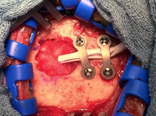
toma and 1 unilateral electrode dislocation. A rather rare                  an active tip as most distal and a conventional ring elec-
complication, symptomatic idiopathic delayed-onset ede-                     trode as the most proximal. Each of the 2 middle levels
ma, was observed in 4 patients with D-leads. They recovered                 consists of 3 “segmented” contacts spanning about 120°
completely within 1–3 weeks, spontaneously or after short-                  each, whereby the steering of current in the horizontal
term cortisone medication. In the control group, in a series                plane is enabled [10]. First clinical studies showed that ef-
of 31 patients (20 implanted with Medtronic 3389 lead and                   fect and adverse effect thresholds were altered in a highly
11 with Boston Scientific Vercise lead), not a single problem               individual manner by horizontal current steering, and the
of this kind was encountered at any time. Conclusion: Pre-                  therapeutic window compared to ring-mode DBS was ex-
cise positioning of D-leads is more challenging than that of                panded [11–13].
conventional DBS leads. By adding an external lead marker,                     After gathering experience and collecting data pro-
control of optimal lead position and orientation is enhanced.               spectively in a consecutive series of 59 D-lead implanta-
In case of supposed increased risk for hemorrhage because                   tions in 31 patients from September 2015 to September
of vessels crossing all possible trajectories in the pre-surgical           2016 at our institution, we highlight the special neurosur-
navigated simulation program, primary D-lead implantation                   gical aspects of directional DBS. The feasibility of apply-
instead of the sharper microelectrodes may be a feasible al-                ing these new leads in standard DBS procedures and pos-
ternative and it may offer more options than ring electrodes                sible early and long-term difficulties and complications
especially in these cases. Prospective studies comparing                    were the focus of this study.
ring-mode stimulation to directional stimulation to examine
the differences of the clinical effects have been started.
                                         © 2021 The Author(s)                  Patients, Material, and Methods
                                         Published by S. Karger AG, Basel
                                                                                During the period of 1 year, from September 2015 to September
                                                                            2016, technical and surgical data were prospectively collected and
    Introduction                                                            retrospectively analyzed for all patients implanted with Cartesia
                                                                            electrodes, as a control group served patients implanted with ring
    For over 2 decades, deep brain stimulation (DBS) has                    electrodes (3389, Medtronic, and Vercise, Boston Scientific) from
been shown to be a highly effective treatment for move-                     February 2015 and September 2016.
ment disorders, namely, Parkinson’s disease, dystonia,
                                                                                Target and Lead Planning
and essential tremor [1, 2]. Still, high precision in elec-                     We used image fusion of MRI to stereotactic CT (Leksell, Sur-
trode placement in the related target areas cannot always                   giPlan, Elekta, Stockholm, Sweden), MRI-based individual adjust-
prevent side effects from current spread to adjacent fibers                 ment of the atlas-based target coordinates, and multi-trajectory
or nuclei [3, 4]. The volume of tissue activated (VTA) has                  microelectrode mapping to guide the implantation identically to
                                                                            our procedure for conventional DBS leads. A decision for implant-
been identified to be of utmost importance for the effi-
                                                                            ing a D-lead instead of a conventional lead was made before sur-
cacy of the applied stimulation as well as for the avoidance                gery in 21 cases, based on clinical presentation and patients’ pref-
of side effects [5] and to be related to individual function-               erences and intraoperatively in another 10 cases, if only a single
al anatomy [6, 7].                                                          trajectory was feasible for implantation due to anatomic restric-
    Until 2015, commercially available leads for DBS con-                   tions and test stimulation via the microelectrodes indicated a nar-
                                                                            row therapeutic window (small amplitude range before reaching
tained ring electrodes in a linear arrangement, for exam-
                                                                            the adverse effect threshold) for the trajectory with the best thera-
ple, with either 4 (Medtronic Inc., Minneapolis, MN,                        peutic effect.
USA; Abbott (former St. Jude Medical); Little Canada,
MN, USA) or 8 electrodes (Boston Scientific, Valencia,                          Lead Handling
CA, USA), allowing limited control of a roughly spherical                       The Cartesia D-lead consists of 8 electrode contacts arranged
                                                                            in 4 levels. The 2 middle ones contain 3 segmented contacts each,
VTA by polarity, pulse duration, and amplitude settings                     which span 120° and can therefore direct the VTA in a specific di-
[5]. Intraoperative experimental studies with so-called D-                  rection. An electrode marker above the electrode contacts helps to
leads at various setups demonstrated the feasibility of                     adjust the orientation by fluoroscopy (Fig. 1). For efficient applica-
horizontal steering in humans [8, 9].                                       tion of directionality of DBS, it is important to carefully control for
    In September 2015, the first D-lead (Cartesia, Boston                   implantation depth and to position the 2 segmented electrodes at
                                                                            the best functional target level according to the intraoperative neu-
Scientific) in combination with a multiple independent                      rophysiological mapping and test stimulation.
current source control pulse generator was released in                          Besides the radio-opaque marker above the electrode contacts,
Europe and has been implanted for the first time in our                     the lead does not contain an external, extra-cerebral marker for
center. The lead is composed of 4 levels of contacts, with                  orientation and handling after insertion. Therefore, some techni-

2                     Stereotact Funct Neurosurg                                                    Fricke/Nickl/Breun/Volkmann/Kirsch/
                      DOI: 10.1159/000512231                                                        Ernestus/Steigerwald/Matthies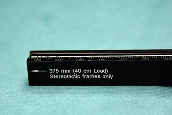
6
                                                                           3

Fig. 1. Scheme of D-lead with electrode
contacts 2 and 5 oriented in the same direc-                                                                      marker
tion as the closed part of the integrated
                                                                                              8
marker. Note the slits between the seg-                                        7     5
mented contacts, which become also visi-                             4     2
                                                                 1
ble by lateral fluoroscopy (© Boston Scien-
tific; reprinted with permission).

Fig. 2. The D-lead is marked for its length
and its orientation with the external white
stopper, by positioning the screw posteri-
orly towards the open part of the integrated
radiopaque marker.

cal support needed to be developed before implantation of the D-           stimulation in bipolar mode is performed via the upper 2 levels
lead. The lead is marked externally by the white “lead stopper” for        positive and lower 2 levels negative to reproduce effects of the for-
its length and the lead stopper screw is used to indicate the hori-        mer test stimulation via semi-macroelectrodes to confirm effect
zontal orientation by aligning it in posterior position, opposite to       and side effect thresholds.
the closed part of the integrated marker, to indicate the anterior-
posterior lead orientation and facilitate its handling (Fig. 2, 3). Fur-      Postoperative Surgical Control
thermore, we mark the lead itself with a permanent sterile marker             A postoperative cranial CT scan with image fusion to the pre-
pen.                                                                       surgically fused set of MRI and stereotactic CT were performed to
                                                                           assess possible surgical sequels as well as the electrode position.
    Lead Control
    As a standard, we use lateral fluoroscopy through the stereotac-           Clinical Programming
tic frame to control for implant depth. For D-leads, not only the              A monopolar review was performed on days 5–8 after surgery
depth and possible deviation from the planned trajectory are im-           as soon as testable off-period symptoms returned, at first by iden-
portant but also the rotation of the electrode which is controlled         tification of response and side effects level and related electrode
by the radiopaque marker above the electrode level. By lateral flu-        level, thereafter by evaluating the individual therapeutic window
oroscopy, a clear anterior-posterior orientation of the marker and         at contacts with best response.
the related electrode contact numbers 2 and 5 in anterior orienta-
tion is possible in contrast to a medial or lateral adjustment.                Follow-Up
    Among the 4 circular poles of the lead, the 2 middle contacts              At 2–3 months after implantation, patients were readmitted to
with the directional contacts should be placed at the estimated            the neurology department for an extensive monopolar review and
“sweet spot” chosen for chronic stimulation. After lead placement,         fine tuning of DBS settings. At the same time, a control CT or ro-
while its extra-cerebral part is still secured by the frame, the part      tational fluoroscopy was performed for final determination of lead
of the lead that will be within the bone fixation is coated with a         tip location and orientation in relation to preoperative planning.
silicone tube. The posterior side of this tube is marked with a per-
manent pen to facilitate handling the lead rotation during the next
steps. After releasing the electrode out of the frame, its depth and           Results
rotation are controlled by lateral fluoroscopy (Fig. 4). Correction
of the lead orientation should only be performed by clockwise ro-
tation in view of the rotation of the wires within the lead. Lead             From September 2015 to September 2016, out of a con-
fixation is carried out with 2 mini titanium plates and 4 screws at        secutive series of 51 patients undergoing a DBS proce-
the skull (Fig. 5). After securing and fixing the lead, a further test     dure, 31 patients (60.8%; 8 female, median age 60.5 years,

Experiences with Directional Leads for                                     Stereotact Funct Neurosurg                                         3
DBS                                                                        DOI: 10.1159/000512231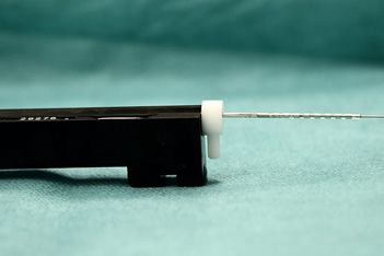
Fig. 4. Directional leads (Cartesia by Boston Scientific) by intraop-
                                                                       erative fluoroscopy by left lateral view. The lead marker is oriented
                                                                       anteriorly, the iron sights between anterior and posterolateral re-
                                                                       spectively between anterior and posteromedial (dotted arrow) di-
                                                                       rectional contacts are clearly visible.

Fig. 3. The screw of the white stopper indicates the horizontal ori-
entation of the directional segments and helps controlling the lead
orientation.

range 16–79 years) were implanted with 59 segmented
                                                                       Fig. 5. After coating the lead with a silicone tube, it is fixed in the
D-leads (Cartesia, Boston Scientific, Valencia, CA, USA),              bony groove by 2 mini titanium plates.
3 patients received unilateral and 28 bilateral implanta-
tions. DBS indications were Parkinson’s disease in 27 cas-
es with STN as target, 2 patients had essential tremor and
electrodes were implanted in the nucleus ventralis inter-
medius thalami and retro-subthalamic field, and 2 pa-                  ately beforehand. They received conventional ring elec-
tients with dystonia had implantations in the globus pal-              trodes, 20 with Medtronic 3389 and 11 with Boston Sci-
lidus internus (GPi).                                                  entific Vercise leads.
   Implantation of D-lead was offered to every DBS pa-                    We did not observe intracranial hematomas, cerebral
tient since September 2015 except patients with the need               infarctions, or infections in either patient group. Two
for any postoperative MRI, until MRI-approved pulse                    surgical complications required surgical revisions in the
generators were available. There were 4 cases of revision              D-lead group. A subcutaneous hematoma at the impulse
surgery of previously implanted conventional electrodes                generator site was evacuated on the implant day.
that were replaced by D-leads.                                            Suboptimal electrode position was identified in 1 pa-
   The control group consisted also of 31 patients (11 fe-             tient unilaterally (1 of 59) by postoperative CT imaging;
male, median age 58.1 years, range 8–80 years), 20 of the              as it occurred on the clinically less affected side, the pa-
same time period as the D-lead patients and 11 immedi-                 tient showed bilateral improvement with stimulation, but

4                      Stereotact Funct Neurosurg                                              Fricke/Nickl/Breun/Volkmann/Kirsch/
                       DOI: 10.1159/000512231                                                  Ernestus/Steigerwald/Matthies
had bothersome residual symptoms contralateral to the Change of Stereotactic Procedure with D-Lead
misplaced lead despite several programming attempts. In 1 patient, at stereotactic planning, multiple vessels
After discussing the findings and the possible long-term were crossing the trajectory and also all simulated alter-
importance, he agreed on revision which was carried out native trajectories on one side in this patient. By abdica-
successfully. tion of microelectrodes, direct primary D-lead implanta-
Idiopathic delayed-onset edema was encountered tion was performed on the specific side in order to mini-
solely in the D-lead group. Four patients became symp- mize the risk of hemorrhage and to compensate for the
tomatic after 1–2 uneventful weeks after surgery, by pro- lack of alternative trajectories.
longed drowsiness in 3 cases and by leg paresis and apathy
in 1 case. Cranial CT identified unilateral edema along the
electrode in 3 cases and bilateral edema in 1 patient. All 4 Discussion
patients recovered completely within a maximum of 3
weeks spontaneously and in 2 patients after IV/oral cor- Complications and Risks
tisone treatment. In this early series with D-leads, their application com-
pared to ring leads did not expose to greater risks such as
Surgical Methods and Technique cerebral hemorrhage, infection, or electrode deviation.
The specific technique of marking the D-lead for Electrode insertion was as smooth as with all the other
length and orientation with the external white stopper as available leads and did not cause any direct additional le-
well as the control by comparing the stopper to the fluo- sions.
roscopy findings was applied as reported above and could But 4 patients (12.9%) developed idiopathic delayed-
be fulfilled in each D-lead implantation in all the patients. onset edema around the course of 1 or both implanted
In each case, care was taken to achieve an anterior-poste- electrodes. The patients recovered completely after treat-
rior orientation with contacts 2 and 5 in an anterior posi- ment with cortisone or spontaneously. In the control
tion and with the slits between the anterior and the pos- group, none out of 31 patients (20 with Medtronic 3389
terolateral or posteromedial contacts appearing at the and 11 with Boston Scientific Vercise) revealed any brain
posterior third in the lateral view. edema. Furthermore, before the introduction of D-leads
in our institution, during the previous series of 142 con-
Intraoperative Fluoroscopy secutive DBS procedures implanted with ring leads over
A slightly longer fluoroscopy time (2.29 min vs. 1.63 5 years, there were only 2 unilateral cases of idiopathic
min) and therefore higher X-ray dose was needed for the delayed-onset edema (1.4% of the patients or 0.7% of the
placement of the D-leads compared to conventional leads implanted ring leads). This corresponds to the very low
(415.53 vs. 328.96 Gy cm2; p = 0.09), but the difference incidence reported about after conventional lead implan-
was not significant. tations [12, 14].
Hence, the increased edema incidence, in 5/59 (8.5%)
Duration of Surgery implanted D-leads (1 bilateral and 3 unilateral cases), is a
The mean duration of the whole procedure from place- matter of concern. At present, the underlying cause is
ment of the stereotactic frame until finishing the impulse completely unknown. Two aspects must be taken into
generator implantation did not differ between D-lead and consideration: Though D-leads appear very soft and
conventional lead implantation (09:02 vs. 08:55 h:min). smooth at the surface, some cause of irritation cannot be
All the patients showed good overall recovery and satisfy- excluded coming from the slit sites. In addition, these
ing response to DBS treatment. At a minimum follow-up electrodes were rotated while lying in the brain tissue,
of at least 2 months after the implantation, 47 (79.7%) what may cause additional irritation. In many cases, a
leads stimulated in steering current mode and 12 (20.3%) postoperative rotation of the leads was observed, so that
leads in ring mode. With the implementation of current the mechanical stress to the brain tissue might last for a
steering leads and the introduction of image-based pro- prolonged time. Furthermore, these leads are produced
gramming in our institution, the modeling of the approx- in a handmade fashion; whether this might lead to accu-
imated optimum VTA was more likely to be obtained by mulation of any artificial deposits or to significant elec-
a horizontal steering option. The clinical benefit of this is trostatic surface changes is a matter of speculation. As to
still under research. date, the phenomenon of idiopathic delayed-onset edema
is neither well understood nor explained; the higher inci-
Experiences with Directional Leads for Stereotact Funct Neurosurg 5
DBS DOI: 10.1159/000512231dence in D-leads compared to ring leads remains open to serves as a protection at the bony edge and towards the ti-
speculation [15]. Fortunately, no neurological long-term tanium plates. Furthermore, it may also serve as an external
sequelae were observed in any affected patient. marker. As is known from postsurgical investigations [19],
Electrode misplacement may occur in any stereotactic D-lead orientation may still change to some degree within
procedure, and at an increased risk especially in advanced the brain, but the primary positioning at surgery poses the
brain atrophy. Reported incidences in the literature vary important base for the individual long-term condition.
enormously from 5% to over 30–40% [16–18]. The en- If multiple vessel formation or other anatomical vari-
countered unilateral misplacement in 1 out of 59 im- ants put the implantation of multiple microelectrodes at
planted leads in this series was small (∼3.0 mm medial) a very high-risk level, primary D-lead implantation may
and most likely due to a combination of brain shift on the be a good compromise and alternative. Instead of implan-
second surgical side in brain atrophy and limited and tation of 1 central microelectrode and the possible shift
rather misleading cooperation by the patient. Therefore, after its removal, the direct application of D-lead allows
the revision in this patient was carried out in general an- good functional intraoperative testing at various direc-
esthesia. This lead dislocation and the indication for revi- tions. With only 1 inserted electrode, it is not possible to
sion were not attributable to the type of selected electrode. choose the best trajectory for final implantation out of the
In case of increased risk for hemorrhage because of the intraoperative microelectrode tests. In this case, D-leads
individual anatomy with multiple crossing vessels, pri- might provide a broader field for adjustment of the VTA
mary D-lead implantation instead of the more pointed than conventional leads.
microelectrodes may be a feasible alternative. In contrast
to conventional electrodes, if the position of the lead can- Optimal Lead Orientation
not be changed, D-leads provide more options to adapt Instead of the AP orientation of D-lead presented here,
the electric field. Still it is not yet known which deviation an implantation in medial-lateral orientation of the
of the electrode to the planned target point could be com- marker and related active segments is another option fa-
pensated for by D-leads. vored by some colleagues. This might make the fluoro-
scopic identification more difficult because the displayed
Surgical Handling marker could point medial or lateral. Knowing the orien-
For optimum outcome with D-leads, surgical handling tation of the D-lead makes it much easier to program an
needs special attention to adjust implantation depth, with effective VTA and saves time of testing. Therefore, based
the segmented levels aimed at the best stimulation depth on the experience in this case series, AP orientation of D-
and an adapted technique to control for horizontal lead lead appears as the technique of choice for easy and pre-
orientation. cise control of the electrode position.
Control of lead orientation, at present, is possible by
radiological control of the integrated marker above the New Lead Models
active contacts, to prove anterior-posterior orientation. When comparing the advantages of the D-lead to the
However, the product does not contain any markers that other recent lead model with 8 ring contacts, D-lead
will be visible after insertion of the marker and the active seems to bear more advantages for the majority of cases.
leads into the brain. In our experience, there are few indications where 8 elec-
The lead does not only tend to shift in a vertical direc- trode contacts over a length of 15.5 mm (in contrast to 7.5
tion, upward or downward dislocation, but also tend to mm in D-lead) will be necessary.
rotate horizontally. For minimizing fluoroscopy control From a surgeon’s point of view, the precise implanta-
shots, an external marker above the brain surface is help- tion of the D-leads is more challenging, but well feasible
ful during and after the lead insertion. This marker at first as demonstrated here. The increase in X-ray dose is small
may consist of the screw at the lead stopper and second and appears acceptable. The first impressions of the clin-
of a slit silicone layer or ink mark. ical effects indicate that the neurosurgical burden is paid
Correction of lead orientation, once inserted by 60–70 off by a larger therapeutic window [11] and reductions in
mm at depth from the surface, may be achieved by clock- the therapeutic current amplitude [12]. In this respect, D-
wise rotation (in the direction of the turn of the isolated lead leads might compensate small deviations of the lead from
wires within the lead carrier) to the optimal position, guid- an optimal functional target. Prospective studies to com-
ed by the external marker and the fluoroscopic control of pare ring-mode stimulation to directional stimulation are
the radiopaque marker. The externally applied silicone tube needed and are already under way.
6 Stereotact Funct Neurosurg Fricke/Nickl/Breun/Volkmann/Kirsch/
DOI: 10.1159/000512231 Ernestus/Steigerwald/MatthiesSummary and Conclusions Conflict of Interest Statement
In summary, this feasibility investigation for D-leads
Dr. Fricke reports no conflicts of interest. Dr. Nickl has received
proves their reliable and safe clinical applicability and, as grant support from Medtronic and Boston Scientific and honorar-
well, the necessity for specific technical care at surgical ia for speaking from Boston Scientific, outside the submitted work.
application. Optimal placement of the D-lead is of utmost Dr. Breun reports no conflict of interest. Dr. Volkmann reports
importance before a realistic comparison of the clinical grants and personal fees from Medtronic Inc., grants and personal
effects by D-leads versus ring leads can be accomplished. fees from Boston Scientific, and personal fees from St. Jude, outside
the submitted work. Dr. Kirsch reports no conflicts of interest. Dr.
The implantation of the segmented contacts needs to be Ernestus reports no conflicts of interest. Dr. Steigerwald has re-
exactly aligned in depth with the functional target and not ceived grant support from Medtronic and Boston Scientific and
every dislocation can be balanced by the adjusted electric honoraria for speaking from Boston Scientific, outside the submit-
field of the segmented contacts. The rotation of the lead ted work. Dr. Matthies reports congress fees and honoraria for pre-
is an additional surgical step, which needs exact align- sentations from Boston Scientific, outside the submitted work.
ment under fluoroscopy. In cases, in which by reason of
crossing vessels only 1 trajectory to the target point is pos-
sible, D-leads can compensate for the less favorable ana- Funding Sources
tomical circumstances and minimize the surgical risk. This study did not receive any funding.
If adequately placed at stereotactic implantation, D-
leads provide many options to shape the activated electric
field, if the programming neurologist invests sufficient Author Contributions
time for testing and adjusting the stimulation parameters.
Ongoing prospective studies evaluate the outcome of D- Each author has made substantial contributions to the concep-
leads with different time patterns of upcoming positive or tion or design of the work or to the acquisition, analysis, or inter-
negative effects compared to ring-mode stimulation. pretation of data for the work; participated in drafting the work or
revising it critically for important intellectual content; approved
the final version to be published; and agreed to be accountable for
all aspects of the work in ensuring that questions related to the ac-
Statement of Ethics
curacy or integrity of any part of the work are appropriately inves-
tigated and resolved literature.
All patients gave their written informed consent to the proce-
dure, and the study was approved by the local Ethics Committee.
References
1 Deuschl G, Schade-Brittinger C, Krack P, 5 Volkmann J, Moro E, Pahwa R. Basic algo- 10 Schüpbach WMM, Chabardes S, Matthies C,
Volkmann J, Schäfer H, Bötzel K, et al. A ran- rithms for the programming of deep brain Pollo C, Steigerwald F, Timmermann L, et al.
domized trial of deep-brain stimulation for stimulation in Parkinson’s disease. Mov Dis- Directional leads for deep brain stimulation:
Parkinson’s disease. N Engl J Med. 2006; ord. 2006 Jun;21(Suppl 14):S284–9. opportunities and challenges. Mov Disord.
355(9):896–908. 6 Astrom M, Diczfalusy E, Martens H, Wardell 2017 Oct;32(10):1371–5.
2 Benabid AL, Pollak P, Gervason C, Hoffmann K. Relationship between neural activation 11 Steigerwald F, Müller L, Johannes S, Matthies
D, Gao DM, Hommel M. Long-term suppres- and electric field distribution during deep C, Volkmann J. Directional deep brain stimu-
sion of tremor by chronic stimulation of the brain stimulation. IEEE Trans Biomed Eng. lation of the subthalamic nucleus: a pilot
ventral intermediate thalamic nucleus. Lan- 2015 Feb;62(2):664–72. study using a novel neurostimulation device.
cet. 1991 Feb 16;337(8738):403–6. 7 Reich MM, Brumberg J, Pozzi NG, Marotta G, Mov Disord. 2016;31(8):1240–3.
3 Maks CB, Butson CR, Walter BL, Vitek JL, Roothans J, Åström M, et al. Progressive gait 12 Rebelo P, Green AL, Aziz TZ, Kent A, Schafer
McIntyre CC. Deep brain stimulation activa- ataxia following deep brain stimulation for es- D, Venkatesan L, et al . Thalamic directional
tion volumes and their association with neu- sential tremor: adverse effect or lack of effi- deep brain stimulation for tremor: spend less,
rophysiological mapping and therapeutic cacy? Brain. 2016;139(11):2948–56. get more. Brain Stimul. 2018 May–Jun;11(3):
outcomes. J Neurol Neurosurg Psychiatry. 8 Contarino MF, Bour L, Verhagen R. Direc- 600–6.
2009;80(6):659–66. tional steering: a novel approach to deep brain 13 Dembek TA, Reker P, Visser-Vandewalle V,
4 Wodarg F, Herzog J, Reese R, Falk D, Pinsker stimulation. Neurology. 2014 Sep 23; 83(13): Wirths J, Treuer H, Klehr M, et al. Direction-
MO, Steigerwald F, et al. Stimulation site 1163–9. al DBS increases side-effect thresholds: a pro-
within the MRI-defined STN predicts postop- 9 Claudio Pollo C, Kaelin-Lang A, Schüpbach spective, double-blind trial. Mov Disord. 2017
erative motor outcome. Mov Disord. 2012 M. Directional deep brain stimulation: an in- Oct;32(10):1380–8.
Jun;27(7):874–9. traoperative double-blind pilot study. Brain. 14 Deogaonkar M, Nazzaro J, Machado JM,
2014;137(7):2015–26. Rezai A. Transient, symptomatic, post-oper-
ative, non-infectious hypodensity around the
deep brain stimulation (DBS) electrode. J Clin
Neurosci. 2011 Jul;18(7):910–5.
Experiences with Directional Leads for Stereotact Funct Neurosurg 7
DBS DOI: 10.1159/00051223115 de Cuba CM, Albanese A, Antonini A, Cossu failures: a retrospective analysis from 2 move- 18 Falowski SM, Bakay RA. Revision surgery of
G, Deuschl G, Eleopra R, et al. Idiopathic de- ment disorders centers. Arch Neurol. 2005; deep brain stimulation leads. Neuromodula-
layed-onset edema surrounding deep brain 62(8):1250–5. tion. 2016 Jul;19(5):443–50.
stimulation leads: insights from a case series 17 Rolston JD, Englot DJ, Starr PA, Larson PS. 19 Treuer H, Hellerbach A, Borggrefe J, Visser-
and systematic literature review. Parkinson- An unexpectedly high rate of revisions and re- Vandewalle V. Regarding “determining the
ism Relat Disord. 2016 Nov;32:108–15. movals in deep brain stimulation surgery: orientation of directional deep brain stimula-
16 Okun MS, Tagliati M, Pourfar M, Fernandez analysis of multiple databases. Parkinsonism tion electrodes using 3D rotational fluoros-
HH, Rodriguez RL, Alterman RL, et al. Man- & related disorders. Parkinsonism Relat Dis- copy”. AJNR Am J Neuroradiol. 2017 Dec;
agement of referred deep brain stimulation ord. 2016 Dec;33:72–7. 38(12):E105.
8 Stereotact Funct Neurosurg Fricke/Nickl/Breun/Volkmann/Kirsch/
DOI: 10.1159/000512231 Ernestus/Steigerwald/MatthiesYou can also read