Stereotactic Body Radiation Therapy for Primary Renal Cell Carcinoma in Inoperable Patients - Created By: Kevin Rogacki, MD Faculty Adviser: John ...
←
→
Page content transcription
If your browser does not render page correctly, please read the page content below
Stereotactic Body Radiation Therapy
 for Primary Renal Cell Carcinoma in
         Inoperable Patients
Created By: Kevin Rogacki, MD
Faculty Adviser: John P. Hayes, MD
Northwestern University Feinberg School of Medicine, Chicago IL

  May 21, 2021
ARROCase: Clinical presentation

• HPI: Woman in her early 70s who presents to
  urology with gross hematuria
• PMH/PSH: HTN, DM
• Social History: 30 pack-year former smoker
  (quit 2 years ago), no known occupational
  exposures
• Family History: No family history of cancer

May 21, 2021
ARROCase: Initial evaluation
• Cystoscopy: No lesions in the bladder, but atypical cells on cytology
• CT CAP w/ Contrast demonstrating a left upper pole mass suggestive of
  RCC, described as, “…mild exophytic solid left renal lesion. the bulk of
  the mass measures maximum diameter of 4 cm which is homogenously
  enhancing. A more medial component of this mass appears to insinuate
  into the renal pelvis based on its appearance.”
ARROCase: Initial evaluation
• CT Guided Biopsy demonstrating clear cell renal cell
  carcinoma (ccRCC)
• Renal function scan with mild impairment of tubular
  function in the kidneys bilaterally. The left kidney does not
  empty completely after furosemide administration.
• Outpatient urology consultation:
   – Surgical resection recommended, but patient declined
• Outpatient radiation oncology consultation:
    – Surgery emphasized as standard-of-care, but given patient refusal
      SBRT versus moderately hypofractionated radiation offered
    – Patient elected to proceed with SBRT

  May 21, 2021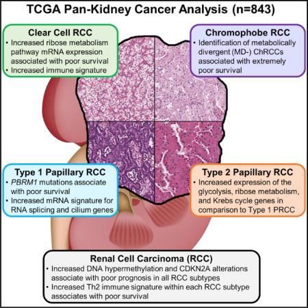
Renal Cell Carcinoma:                                    Epidemiology 1

• In the United States in 2020:
      – 73,750 estimated new cases (4.1% of new cancer cases)
      – 14,830 estimated deaths (2.4 % of all cancer deaths)
      – 8th most common cancer diagnosis
               • Incidence increased over past 50 years, stable since 2008 - 2017
• 65% diagnosed with localized disease, 16% regional disease,
  and 16% distant disease
• 5-year relative survival of 75.2% (from 2010 – 2016)
      – Localized: 92.5% 5-year survival
      – Regional: 70.4% 5-year survival
      – Distant: 13% 5-year survival

May 21, 2021
Risk Factors2
• Age (64 median age at diagnosis)
    – Most patients diagnosed between ages 65 – 74
• Sex (2 of 3 cases in males)
• Race
   – Highest incidence in American Indian/Alaska Native
   – Lowest incidence in Asian/Pacific Islander
• Smoking (1.5 – 1.6 RR of advanced disease)3
• Alcohol
• Obesity
• Poorly-controlled hypertension
• Kidney stones4
• Occupational exposures: benzene, vinyl chloride, coal tar, mineral oil,
  cadmium, herbicides, pesticides5
• Acetaminophen6
   May 21, 2021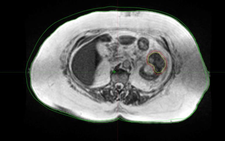
Genetic Syndromes
•   Von Hippel-Lindau syndrome
     – Mutation in VHL tumor suppressor gene, inherited in an autosomal
       dominant pattern
     – VHL regulates hypoxic inducible factor (HIF1α)
     – Predisposes to formation of cysts and tumors in central nervous
       system, retina, adrenal glands, pancreas, kidneys, epididymis (men),
       broad ligament (women)
     – Predominantly clear cell RCC (ccRCC), which develops in up to 2 of 3
       patients, may be bilateral7
•   Tuberous sclerosis8
     – Mutations in either TSC1 (hamartin protein) or TSC2 (tuberin protein)
       tumor suppressor genes, inherited in an autosomal dominant pattern
     – TSC1 and TSC2 regulate the mTOR signaling pathway
     – RCC occurs in 2-5% of patients with tuberous sclerosis
     – Most often ccRCC, but also associated with papillary renal cell
       carcinoma and hybrid oncocytic/chromophobe (HOCT) tumors
May 21, 2021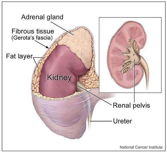
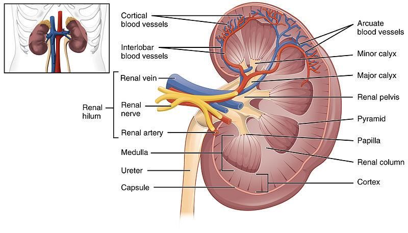
Anatomy

National Cancer Institute
By: Alan Hoofring, Public Domain

                                      By Henry Vandyke Carter - Henry Gray (1918) Anatomy of the Human Body,
                                      Bartleby.com: Gray’s Anatomy, Plate 1121, Public Domain

      May 21, 2021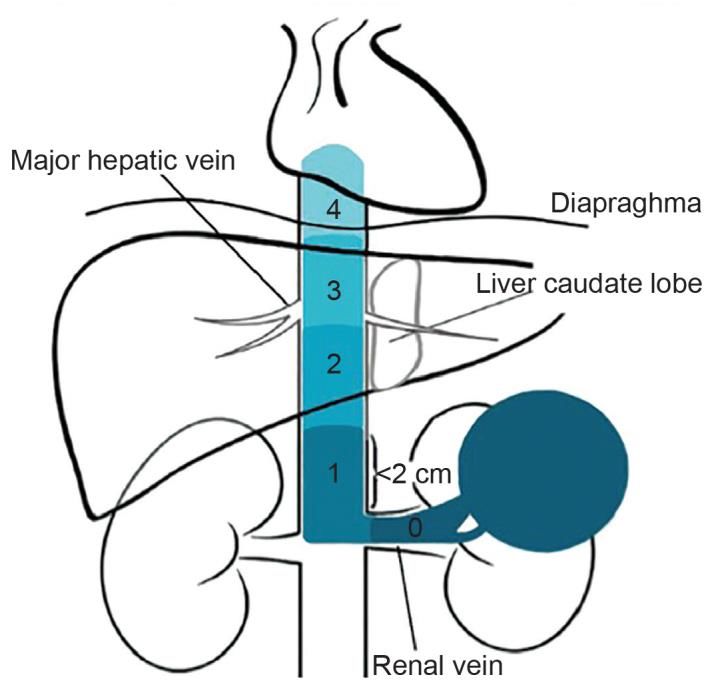
Anatomy

Nephron: functional unit of kidney

                                                     Proximal tubule epithelium10:
                                                     - Clear cell RCC (75 – 80%)
                                                     - Papillary RCC (10 – 15%)       Renal medullary carcinoma:
                                                                                      - Associated with sickle cell trait and
Distal nephron/collecting tubule10:                                                   hemoglobinopathies (rare)
- Chromophobe RCC (5%)                                                                Source: Anatomy & Physiology, Connexions Web
- Collecting duct RCC (< 1%)                                                          site. http://cnx.org/content/col11496/1.6/, Jun 19, 2013
                                                                                      https://creativecommons.org/licenses/by/3.0/deed.en
Created by ColnKurtz, Attribution-ShareAlike 4.0 International (CC BY-SA 4.0)

         May 21, 2021
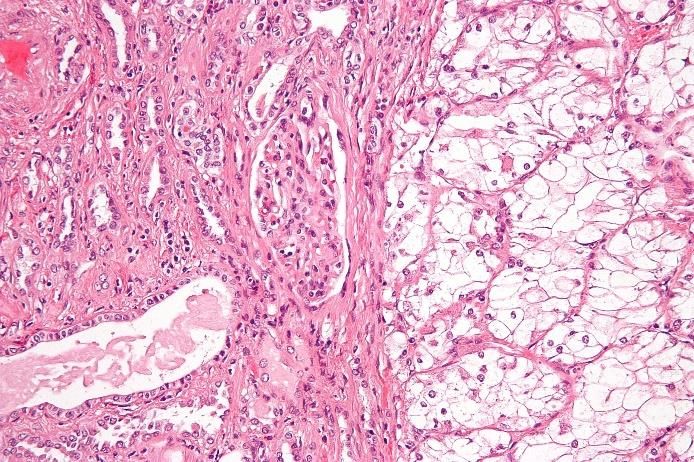
Histology
                                                                                Clear Cell Renal Cell Carcinoma11
                                                                                - Compact nests and sheets of cells with
                                                                                  clear cytoplasm and distinct membrane
                                                                                - Arborizing thin-walled vessels
                                                                                - Patterns: solid, alveolar (nested), acinar
                                                                                  (tubular), microcytic or macrocystic

                                                                                  Papillary Renal Cell Carcinoma12
                                                                                  - Often circumscribed with pseudocapsule
                                                                                  - Papillae or tubulopapillary architecture with
                                                                                    fibrovascular cores
                                                                                  - May contain foamy macrophages,
                                                                                    psammoma bodies, hemosiderin

Sources: https://commons.wikimedia.org/wiki/File:Clear_cell_renal_cell_carcinoma_high_mag.jpg (https://creativecommons.org/licenses/by-sa/3.0/deed.en),
https://commons.wikimedia.org/wiki/File:Histopathology_of_papillary_renal_cell_carcinoma_type_1.jpg (https://creativecommons.org/licenses/by/4.0/deed.en)

         May 21, 2021
The Cancer Genome Atlas: Molecular Profiling13 May 21, 2021
Clinical Presentation14
• Asymptomatic, discovered incidentally on
  imaging for other indications (better prognosis)
• Hematuria, flank pain, abdominal mass (classic
  triad suggests advanced disease)
• Scrotal varices (left-sided predominance)
• IVC involvement with lower extremity edema,
  ascites, pulmonary emboli, hepatic dysfunction,
  Budd-Chiari syndrome
• Symptoms related to metastases (lungs, lymph
  nodes, bone, liver and brain)
 May 21, 2021
Paraneoplastic Syndromes14
•   Anemia (iron-studies consistent with ACD)
•   Fever
•   Cachexia
•   Hepatic dysfunction (Stauffer syndrome if no liver metastases,
    possibly related to tumor cytokine production)
•   Hypercalcemia (lytic bone metastases, PTHrP production,
    prostaglandins → bone resorption)
•   Erythrocytosis (erythropoietic production)
•   Secondary amyloidosis (chronic inflammatory response)
•   Thrombocytosis (unclear mechanism)
•   Polymyalgia rheumatica (not steroid responsive, may respond
    to nephrectomy)
    May 21, 2021
Diagnostic Evaluation9,14
• H&P
• Labs: CBC with differential, CMP, UA
• Abdominal CT
• Abdominal MRI if CT inconclusive, or for further evaluation of
invasion of blood vessels and/or collecting system
• No biopsy if undergoing partial or radical nephrectomy
– NCCN states that biopsy of small lesions may confirm diagnosis of malignancy for surveillance or ablative
techniques
• Biopsy may also be appropriate when urothelial carcinoma or lymphoma is possible or suspected
• If inoperable, potential biopsy of primary to guide medical therapy
• If metastatic, biopsy of metastatic lesion preferred
• Chest CT or radiograph
• Bone scan (if pain or elevated alkaline phosphatase)
• Genetic evaluation
– ≤ 46 years old, close family history of kidney cancer, or multiple renal masses)
May 21, 2021Imaging Characteristics15
CT MRI
• Small lesions may • T1 heterogeneous
enhance homogenously – Blood, necrosis, solid
• Larger lesions may have components
irregular enhancement • T2 depends on histology
due to areas of necrosis – ccRCC: hyperintense
• 30% with calcification – Papillary RCC: hypointense
• 5-15% intraluminal • T1 contrast (Gd): arterial
growth into renal vein enhancement
• Prognosis worse for IVC • Distinguishes between
involvement bland and tumor
thrombus in vessels
May 21, 2021International Metastatic Renal Cell Carcinoma
Database Consortium (IMDC) Criteria
Prognostic Factors Prognostic Risk Groups
1. < 1 year from diagnosis to • Favorable = 0 risk factors
systemic therapy • Intermediate = 1 – 2 risk factors
2. PS < 80% (Karnofsky) • Poor = 3 – 6 risk factors
3. Hgb < lower limit normal
(12 g/dL)
4. Ca2+ > ULN (10.2 mg/dL)
5. Neutrophil > ULN (7x10⁹/L)
6. Platelets > ULN (400,000)
May 21, 2021AJCC 8th Edition TNM Staging
T1 N0
• T1a: ≤ 4 cm, limited to kidney • No regional lymph node metastases
• T1b: > 4 cm but ≤ 7 cm, limited to kidney N1
T2 • Involved regional lymph node(s)
• T2a: > 7 cm but ≤ 10 cm limited to kidney M0
• T2b: > 10 cm limited to kidney • No metastases
T3 M1
• T3a: extends to renal vein, pelvicalyceal • Distant metastases
system, perirenal or renal sinus fat, but
not beyond Gerota’s fascia
• T3b: Extends into vena cava below Prognostic Groups
diaphragm
Stage I = T1 N0M0
• T3c: Extends into the vena cava above the
diaphragm or invades wall of vena cava Stage II = T2 N0M0
T4 Stage III = T1-2 N1M0 & T3 NX,N0-N1M0
• T4: Invades beyond Gerota’s fascia Stage IV = T4 Any N, Any T/N M1
(including extension to ipsilateral adrenal
gland)
May 21, 2021Treatment Options 9
• Surgery
– Partial nephrectomy (preferred treatment for T1a & T1b)
– Radical nephrectomy
• Ablative Techniques (T1a tumors)
– Radiofrequency Ablation
– Cryotherapy
• Active surveillance (T1 patients with significant risk of morbidity or
death from intervention, small unbiopsied renal masses < 2 cm that may
be benign, or predominantly cystic T1a masses)
• SBRT (reserved for inoperable patients or those who decline surgery)
– Alternatively, moderately hypofractionated radiation (i.e. palliative intent)
• Systemic Treatment
– Immunotherapy
– Targeted therapy
May 21, 2021NCCN Treatment Guidelines9 • NCCN Treatment Kidney Cancer Guidelines v4.2021 does not include SBRT as a treatment strategy for primary RCC • No discussion of SBRT for inoperable or medically unfit patients • No discussion of SBRT for use in cytoreductive therapy • SBRT is a Category 2A recommendation for treatment of metastatic lesions May 21, 2021
Efficacy of SBRT for RCC
• RCC was considered radioresistant based on in
  vitro data and results of 1970s/1980s clinical trials
  that used conventionally fractionated radiation
  and failed to show a benefit with neoadjuvant or
  adjuvant regimens
• Early experience with SBRT to metastatic lesions
  demonstrated high rates of local control,
  suggesting RCC is responsive to high BED radiation
  (next slide)
Efficacy of SBRT for RCC
Extracranial stereotactic radiotherapy for primary and metastatic renal cell carcinoma16
Patient population and intervention:
• 58 patients with 162 lesions treated with SBRT (lung metastases most common)
• 50 patients with metastatic disease and 8 patients with primary or inoperable recurrent
disease
• Most common fractionation schedules: 8 Gy x4, 10 Gy x4, & 15 Gy x3
• Co-planar or non-coplanar conformal static fields with CT verification
Results:
• 30% of lesions with complete regression, 60% with partial regression or no change
• 3 local recurrences, local control of ~ 90% with median follow-up of 13 – 37 months
• Majority of patients developed new metastatic lesions and many were re-treated to new
sites of metastatic disease, with suggested survival benefit
Adverse events:
• 23 of 58 patients with adverse effects
– 50% Grade I-II, and most common cough, nausea, and pain
– 5x patients requiring steroid treatment for radiation pneumonitis
– 1x Grade 5 gastric hemorrhage after treatment for pancreatic metastasis
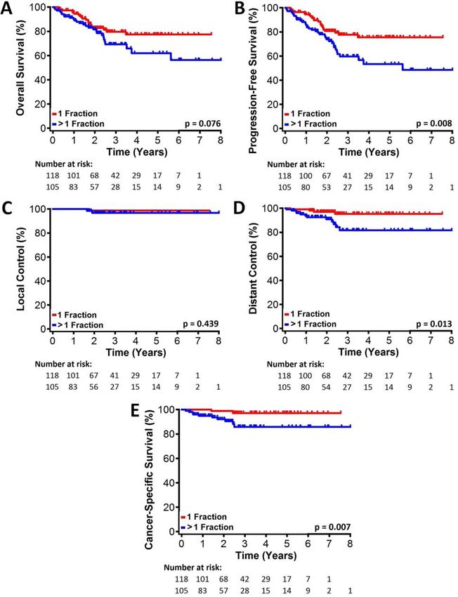
May 21, 2021Radiation for Primary RCC Patient population and intervention:
Pooled analysis of SABR for primary RCC: A report • 223 patients from 9 institutions
from the International Radiosurgery Oncology • 70% male, and mean age of 72
Consortium for Kidney (IROCK)17 • Mean maximal tumor dimension of 43.6 mm +/- 27.7 mm
• 118 patients received single-fraction SBRT (median BED 87.5 Gy)
and 105 patients received multi-fraction SBRT (median BED 80
Gy). Dose range of 14-26 Gy, median of 25 Gy.
– Patients receiving single-fraction SBRT were younger, had
better performance status, and had smaller tumors
Results:
• Local control at 2 & 4 years = 97.8%
• 2-year: CSS = 95.7%, OS = 82.1%, PFS = 77.4%
• 4-year: CSS = 91.9%, OS = 70.7%, PFS = 65.4%
• 3 patients with local recurrence, 16 with distant recurrence (1 of
with both local and distant)
• Mean change in eGFR - 5.5 +/- 13.3 mL
• Larger maximum tumor size and multi-fraction SBRT associated
with inferior CSS and PFS in both regimens
• Larger maximum tumor size associated with worse OS
Adverse events:
• 36% with Grade 1 or 2 toxicity only (nausea more common in
single-fraction 17% versus 6.8%)
• 1 patient with Grade 3 nausea and Grade 2 bowel toxicity
• 1 patient with Grade 4 bowel toxicity
• 1 patient with Grade 4 gastritis, followed by Grade 4 bowel
toxicityScenarios using SBRT for Primary RCC • Efficacious for tumors > 4 cm (T1b) when ablative therapies may be difficult to employ18 • Feasible and safe for treatment of solitary kidney tumors19 • Demonstrated success in treating tumors with IVC tumor thrombus (next slide) May 21, 2021
Inferior Vena Cava Tumor Thrombus
•    Inferior vena cava tumor thrombus
                                             •       SBRT used successfully to treat patients with IVC-TT20
     (IVC-TT)
                                                      – Of 2 patients reported in literature:
       – Level 3 and 4 tumor thrombus                      • One patient demonstrated ongoing response
         may involve more extensive                          at 24 months after treatment to level 4
         surgical resection with increased                   recurrent IVC-TT lesion
         morbidity                                         • One patient with metastatic disease and level
       – Patients with comorbidities may                     4 IVC-TT had better than expected clinical
         not be candidates for surgical                      course with 18-month survival after
         resection of advanced lesions                       treatment

                                                 Mayo Clinic RCC Tumor Thrombus Classification
                                             Level       Definition
                                                 0       Limited to renal vein or its tributaries
                                                 1       Extends into IVC < 2 cm above renal vein orifice

                                                 2       Extends into IVC > 2 cm above renal vein orifice,
                                                         but below hepatic veins
                                                 3       Extends above hepatic veins but below
                                                         diaphragm
    Source: Ramazan et al.21                     4       Extends above diaphragm
       May 21, 2021
Ongoing Phase II Clinical Trials
NCT02141919: SABR for Patients NCT01890590: A Phase II Study of
with Primary Renal Cancer Cyberknife Radiosurgery for RCC
• Estimated enrollment: 16 patients • Estimated enrollment: 46 patients
• Inclusion: Biopsy proven renal cancer • Inclusion: Biopsy proven T1N0M0
≤ 5 cm with growth ≥ 2 mm in a 1- RCC ≤ 8 cm, serum creatinine < 3
year period mg/dL, no coagulopathy, no
• Exclusion: No prior abdominal transaminitis
radiation, RFA, cryoablation or • Exclusion: Prior abdominal EBRT,
evidence of metastatic disease for ≥ 3 prior invasive malignancy within 2
ears prior to registration years, inability to target tumor or
• Technique: 12 Gy in 3 fractions, 10 Gy achieve dose constrains
in 4 fractions, 8 Gy in 5 fractions • Technique: ≥ 1 gold fiducials
• Primary outcome: 2-year tumor required, 3-4 fractions delivered with
growth and viability Cyberknife platform
• Secondary outcomes: Growth rate, • Primary outcome: Local control
renal function, disease progression, • Secondary outcome: Adverse events,
adverse events quality of life
May 21, 2021Ongoing Phase II Clinical Trials
NCT02613819: Focal Ablative
Stereotactic Radiosurgery for NCT03747133: SABR for Renal Tumors
Cancers of the Kidney (FASTRACK II)
• Estimated enrollment: 70 patients • Estimated enrollment: 30 patients
• Inclusion: Biopsy proven renal cancer • Inclusion: Solid kidney mass (primary
in high-risk, medically inoperable RCC or metastasis) ≤ 6 cm, inoperable,
patients or those who decline surgery high-risk or declined surgery
Exclusion: Tumors > 8 cm, high-dose • Exclusion: ≥ 5 active metastases, prior
radiation to overlapping region, < 30 abdominal XRT leading to excessive
mLs/min GFR, recent cytotoxic cumulative kidney dose, concurrent
chemotherapy, no concurrent chemo systemic therapy, ESRD, familial
or targeted agents syndrome with renal cancer
• Technique: ≤ 4 cm size 26 Gy in 1 predisposition
fraction, > 4 cm 42 Gy in 3 fractions • Technique: 27.5 – 40 Gy in 5 fractions
• Primary outcome: 1-year local • Primary outcome: Renal impairment
progression • Secondary outcome: Local control,
• Secondary outcomes: Tolerability, acute and late toxicity, CKD
survival, distant failure rate, renal progression, QOL
function changeOngoing Phase II Clinical Trials
NCT03108703: Assessment of QoL Outcomes with SBRT for RCC (AQuOS-RCC)
• Estimated enrollment: 30 patients
• Inclusion: Biopsy proven renal cancer, radiologic growth on surveillance in
medically inoperable patients or those who decline surgery, ≥ 2.5 cm or recurrence
after ablative therapy
• Exclusion: Prior abdominal radiation
• Technique: 35 – 40 Gy in 5 fractions
• Primary outcome: QoL up to 5-years
• Secondary outcomes: Oncologic outcomes, treatment-related toxicity, cost-
effectiveness
May 21, 2021ARROCase: CT/MRI Simulation
• SBRT delivered using MRI linac
– Note: Non-MRI based radiosurgery also appropriate
• Patient instructed to fast 3 hours prior to simulation to
limit stomach OAR volume
• Full-body immobilization
– Abdominal compression techniques may be desirable
• Positioned supine with arms above head
• Simulation scans:
- Free breathing CT without contrast, 2 mm slices
- Can use contrast if not MRI guided adaptive therapy and renal function WNL
- End exhale CT, 2 mm slices
- Can use bellows device with 4DCT for delineation of ITV if not MRI guided adaptive therapy
- T1 MRI simulation scan
May 21, 2021ARROCase: Treatment Planning
OAR Delineation NOTE: When using 4DCT,
• Bilateral kidneys evaluate OAR motion to
• Stomach ensure no movement into
tumor targets. Consider
• Bowel loops overlap planning structures
• Bowel substructures based on target:
• Duodenum
or PRV structures to better
• Jejunum/Ileum optimize OAR dose.
• Colon
• Pancreas
• Liver (more relevant for right
kidney targets)
• Left renal artery & vein
May 21, 2021ARROCase: Dose Constraints (5 fraction SBRT)
Volume Max Dose (Gy)
OAR Volume Max (Gy) [point max, unless noted]
Endpoint (≥ Grade 3)
Stomach < 5 cc 26.5 Gy < 32 Gy Ulceration/fistula
Duodenum* < 5 cc 18.3 Gy < 32 Gy Ulcer, bleeding, perforation
Jejunum/Ileum*
< 5 – 10 cc 25 Gy < 30 – 35 Gy [0.5 cc] Enteritis/obstruction
[UK protocols]22
Colon* < 20 cc 28.5 Gy < 38 Gy Colitis/fistula
Renal cortex **
200 cc (min. spared) < 17.5 Gy - Renal dysfunction
Solitary or 1 kidney 10 Gy22 < 45% mandatory
Renal hilum &
< 15 cc 23 Gy - Malignant hypertension
vascular trunk
Liver** 700 cc (min. spared) < 21 Gy - Liver dysfunction
< 28 Gy [to 0.035 cc]23
Spinal cord < 0.35 cc 22 Gy Myelitis
< 25.3 [point max]24
*Avoid circumferential radiation
**Or 1/3 of the native total organ volume (prior to resection or volume reducing disease), whichever is greater
See Also: Reference [25], Reference [26], Reference [27], and radoncreview.org -> Dose Constraints
May 21, 2021ARROCase: Target Delineation Approach: Dose paint PTV to 4000 cGy in 5 fractions with SIB to GTV of 5000 cGy in 5 fractions GTV_L_Kidney_5000 cGy PTV_L_Kidney_4000 cGy = GTV_L_Kidney + 3 mm uniform expansion Note: Delivered with a MRI linac using MRI guided adaptive replanning for each fraction, IMRT, and 12-fields May 21, 2021
ARROCase: OTVs and Follow-Up

• OTVs: Patient tolerated treatment with mild
  nausea and limited episode of diarrhea between
  2nd and 3rd fraction.

• Initial Follow-Up: At initial follow-up, patient felt
  well with no side-effects of treatment noted. 9-
  month follow-up with stable disease and no
  evidence of metastases on serial imaging.

  May 21, 2021
References
1.     Surveillance, Epidemiology, and End Results (SEER) Program Populations (1969-2018) (www.seer.cancer.gov/popdata),
       National Cancer Institute, DCCPS, Surveillance Research Program, released December 2019.
2.     Padala SA, Barsouk A, Thandra KC, Saginala K, Mohammed A, Vakiti A, Rawla P, Barsouk A. Epidemiology of Renal Cell
       Carcinoma. World J Oncol. 2020 Jun;11(3):79-87. doi: 10.14740/wjon1279. Epub 2020 May 14. PMID: 32494314; PMCID:
       PMC7239575.
3.     Tsivian M, Moreira DM, Caso JR, Mouraviev V, Polascik TJ. Cigarette smoking is associated with advanced renal cell
       carcinoma. J Clin Oncol. 2011 May 20;29(15):2027-31. doi: 10.1200/JCO.2010.30.9484. Epub 2011 Apr 18. PMID:
       21502558.
4.     van de Pol JAA, van den Brandt PA, Schouten LJ. Kidney stones and the risk of renal cell carcinoma and upper tract
       urothelial carcinoma: the Netherlands Cohort Study. Br J Cancer. 2019 Feb;120(3):368-374. doi: 10.1038/s41416-018-
       0356-7. Epub 2018 Dec 19. PMID: 30563989; PMCID: PMC6353869.
5.     Hu J, Mao Y, White K. Renal cell carcinoma and occupational exposure to chemicals in Canada. Occup Med (Lond). 2002
       May;52(3):157-64. doi: 10.1093/occmed/52.3.157. PMID: 12063361.
6.     Karami S, Daughtery SE, Schwartz K, Davis FG, Ruterbusch JJ, Wacholder S, Graubard BI, Berndt SI, Hofmann JN, Purdue
       MP, Moore LE, Colt JS. Analgesic use and risk of renal cell carcinoma: A case-control, cohort and meta-analytic
       assessment. Int J Cancer. 2016 Aug 1;139(3):584-92. doi: 10.1002/ijc.30108. Epub 2016 Apr 9. PMID: 27009534; PMCID:
       PMC6896985.
7.     Maher ER, Yates JR, Harries R, Benjamin C, Harris R, Moore AT, Ferguson-Smith MA. Clinical features and natural history of
       von Hippel-Lindau disease. Q J Med. 1990 Nov;77(283):1151-63. doi: 10.1093/qjmed/77.2.1151. PMID: 2274658.
8.     Yang P, Cornejo KM, Sadow PM, Cheng L, Wang M, Xiao Y, Jiang Z, Oliva E, Jozwiak S, Nussbaum RL, Feldman AS, Paul E,
       Thiele EA, Yu JJ, Henske EP, Kwiatkowski DJ, Young RH, Wu CL. Renal cell carcinoma in tuberous sclerosis complex. Am J
       Surg Pathol. 2014 Jul;38(7):895-909. doi: 10.1097/PAS.0000000000000237. PMID: 24832166; PMCID: PMC4139167.
9.     National Comprehensive Cancer Network. Kidney Cancer (Version 4.2021).
       https://www.nccn.org/professionals/physician_gls/pdf/kidney.pdf Accessed April 22, 2021.
10.    Cairns P. Renal cell carcinoma. Cancer Biomark. 2010;9(1-6):461-73. doi: 10.3233/CBM-2011-0176. PMID: 22112490;
       PMCID: PMC3308682.
11.    Nezami, B. G., MD; MacLennan, G., MD. (2021, April 20). Clear cell renal cell carcinoma. Retrieved April 23, 2021, from
       https://www.pathologyoutlines.com/topic/kidneytumormalignantrccclear.html

May 21, 2021
References
12. Andeen, N. K., MD; Tretkiatova, M., MD, PhD. (2018, March 1). Papillary Renal Cell Carcinoma. Retrieved April 23, 2021, from
https://www.pathologyoutlines.com/topic/kidneytumormalignantrccpap.html
13. Ricketts CJ, De Cubas AA, Fan H, Smith CC, Lang M, Reznik E, Bowlby R, Gibb EA, Akbani R, Beroukhim R, Bottaro DP, Choueiri TK, Gibbs RA,
Godwin AK, Haake S, Hakimi AA, Henske EP, Hsieh JJ, Ho TH, Kanchi RS, Krishnan B, Kwiatkowski DJ, Lui W, Merino MJ, Mills GB, Myers J,
Nickerson ML, Reuter VE, Schmidt LS, Shelley CS, Shen H, Shuch B, Signoretti S, Srinivasan R, Tamboli P, Thomas G, Vincent BG, Vocke CD, Wheeler
DA, Yang L, Kim WY, Robertson AG; Cancer Genome Atlas Research Network, Spellman PT, Rathmell WK, Linehan WM. The Cancer Genome Atlas
Comprehensive Molecular Characterization of Renal Cell Carcinoma. Cell Rep. 2018 Apr 3;23(1):313-326.e5. doi: 10.1016/j.celrep.2018.03.075.
Erratum in: Cell Rep. 2018 Jun 19;23(12):3698. PMID: 29617669; PMCID: PMC6075733.
14. Atkins, MB, Richie, JP, Shah, S. Clinical manifestations, evaluation, and staging of renal cell carcinoma. In. UpToDate, Post, TW (Ed.), UpToDate,
Waltham, MA, 2021
15. Asadov, D., & Morgan, M. A. (2021, September). Renal cell carcinoma: Radiology reference article. Retrieved April 23, 2021, from
https://radiopaedia.org/articles/renal-cell-carcinoma-1?lang=us#nav_radiographic-features
16. Wersäll PJ, Blomgren H, Lax I, Kälkner KM, Linder C, Lundell G, Nilsson B, Nilsson S, Näslund I, Pisa P, Svedman C. Extracranial stereotactic
radiotherapy for primary and metastatic renal cell carcinoma. Radiother Oncol. 2005 Oct;77(1):88-95. doi: 10.1016/j.radonc.2005.03.022. Epub
2005 Jun 20. PMID: 15972239.
17. Siva S, Louie AV, Warner A, Muacevic A, Gandhidasan S, Ponsky L, Ellis R, Kaplan I, Mahadevan A, Chu W, Swaminath A, Onishi H, Teh B, Correa RJ,
Lo SS, Staehler M. Pooled analysis of stereotactic ablative radiotherapy for primary renal cell carcinoma: A report from the International
Radiosurgery Oncology Consortium for Kidney (IROCK). Cancer. 2018 Mar 1;124(5):934-942. doi: 10.1002/cncr.31156. Epub 2017 Dec 20. PMID:
29266183.
18. Siva S, Correa RJM, Warner A, Staehler M, Ellis RJ, Ponsky L, Kaplan ID, Mahadevan A, Chu W, Gandhidasan S, Swaminath A, Onishi H, Teh BS, Lo
SS, Muacevic A, Louie AV. Stereotactic Ablative Radiotherapy for ≥T1b Primary Renal Cell Carcinoma: A Report From the International
Radiosurgery Oncology Consortium for Kidney (IROCK). Int J Radiat Oncol Biol Phys. 2020 Nov 15;108(4):941-949. doi:
10.1016/j.ijrobp.2020.06.014. Epub 2020 Jun 17. PMID: 32562838.
19. Correa RJM, Louie AV, Staehler M, Warner A, Gandhidasan S, Ponsky L, Ellis R, Kaplan I, Mahadevan A, Chu W, Swaminath A, Onishi H, Teh BS, Lo
SS, Muacevic A, Siva S. Stereotactic Radiotherapy as a Treatment Option for Renal Tumors in the Solitary Kidney: A Multicenter Analysis from the
IROCK. J Urol. 2019 Jun;201(6):1097-1104. doi: 10.1097/JU.0000000000000111. PMID: 30741849.
20. Hannan R, Margulis V, Chun SG, Cannon N, Kim DW, Abdulrahman RE, Sagalowsky A, Pedrosa I, Choy H, Brugarolas J, Timmerman RD. Stereotactic
radiation therapy of renal cancer inferior vena cava tumor thrombus. Cancer Biol Ther. 2015;16(5):657-61. doi:
10.1080/15384047.2015.1026506. PMID: 25800036; PMCID: PMC4622024.
21. Topaktaş R, Ürkmez A, Tokuç E, Kayar R, Kanberoğlu H, Öztürk Mİ. Surgical management of renal cell carcinoma with associated tumor thrombus
extending into the inferior vena cava: A 10-year single-center experience. Turk J Urol. 2019 Feb 4;45(5):345-350. doi: 10.5152/tud.2019.95826.
PMID: 30817278; PMCID: PMC6739079.
22. Hanna, G., Murray, L., Patel, R., Jain, S., Aitken, K., Franks, K., van As, N., Tree, A., Hatfield, P., Harrow, S., McDonald, F., Ahmed, M., Saran, F.,
Webster, G., Khoo, V., Landau, D., Eaton, D., & Hawkins, M. (2017). UK Consensus on Normal Tissue Dose Constraints for Stereotactic
Radiotherapy SABR Dose Constraints. Clinical Oncology. https://doi.org/10.1016/j.clon.2017.09.007
May 21, 2021References
23. NRG-BR002: A phase IIR/III trial of standard of care therapy with or without stereotactic body radiotherapy (SBRT) and/or surgical ablation for
newly oligometastatic breast cancer (NCT02364557). Steven J. Chmura, Kathryn A. Winter, Hania A Al-Hallaq, Virginia F. Borges, Nora T.
Jaskowiak, Martha Matuszak, Michael T. Milano, Joseph Kamel Salama, Wendy A. Woodward, and Julia R. White. Journal of Clinical Oncology 2019
37:15_suppl, TPS1117-TPS1117
24. Sahgal A, Chang JH, Ma L, Marks LB, Milano MT, Medin P, Niemierko A, Soltys SG, Tomé WA, Wong CS, Yorke E, Grimm J, Jackson A. Spinal Cord
Dose Tolerance to Stereotactic Body Radiation Therapy. Int J Radiat Oncol Biol Phys. 2021 May 1;110(1):124-136. doi:
10.1016/j.ijrobp.2019.09.038. Epub 2019 Oct 10. PMID: 31606528.
25. Pollom EL, Chin AL, Diehn M, Loo BW, Chang DT. Normal Tissue Constraints for Abdominal and Thoracic Stereotactic Body Radiotherapy. Semin
Radiat Oncol. 2017 Jul;27(3):197-208. doi: 10.1016/j.semradonc.2017.02.001. Epub 2017 Feb 20. PMID: 28577827.
26. Timmerman RD. An overview of hypofractionation and introduction to this issue of seminars in radiation oncology. Semin Radiat Oncol. 2008
Oct;18(4):215-22. doi: 10.1016/j.semradonc.2008.04.001. PMID: 18725106.
27. Benedict SH, Yenice KM, Followill D, Galvin JM, Hinson W, Kavanagh B, Keall P, Lovelock M, Meeks S, Papiez L, Purdie T, Sadagopan R, Schell MC,
Salter B, Schlesinger DJ, Shiu AS, Solberg T, Song DY, Stieber V, Timmerman R, Tomé WA, Verellen D, Wang L, Yin FF. Stereotactic body radiation
therapy: the report of AAPM Task Group 101. Med Phys. 2010 Aug;37(8):4078-101. doi: 10.1118/1.3438081. Erratum in: Med Phys. 2012
Jan;39(1):563. Dosage error in article text. PMID: 20879569.
May 21, 2021You can also read