Retinoic acid inhibits the pyroptosis of degenerated nucleus pulposus cells by activating Sirt1-SOD2 signaling
←
→
Page content transcription
If your browser does not render page correctly, please read the page content below
Retinoic acid inhibits the pyroptosis of degenerated
nucleus pulposus cells by activating Sirt1-SOD2
signaling
Peng-Fei Li
Zhejiang University School of Medicine
Fei Xiong
Zhejiang University School of Medicine
Ying Yin
Zhejiang University School of Medicine
Hong-Yuan Xing
Zhejiang University School of Medicine
Shao-Jun Hu
Zhejiang University School of Medicine
Ning Zhang ( zhangning@zju.edu.cn )
Zhejiang University School of Medicine
Research
Keywords: Intervertebral disc degeneration, pyroptosis, retinoic acid, Sirt1-SOD2 signaling,
ROS/TXNIP/NLRP3 signaling
Posted Date: August 17th, 2021
DOI: https://doi.org/10.21203/rs.3.rs-786450/v1
License: This work is licensed under a Creative Commons Attribution 4.0 International License.
Read Full License
Page 1/20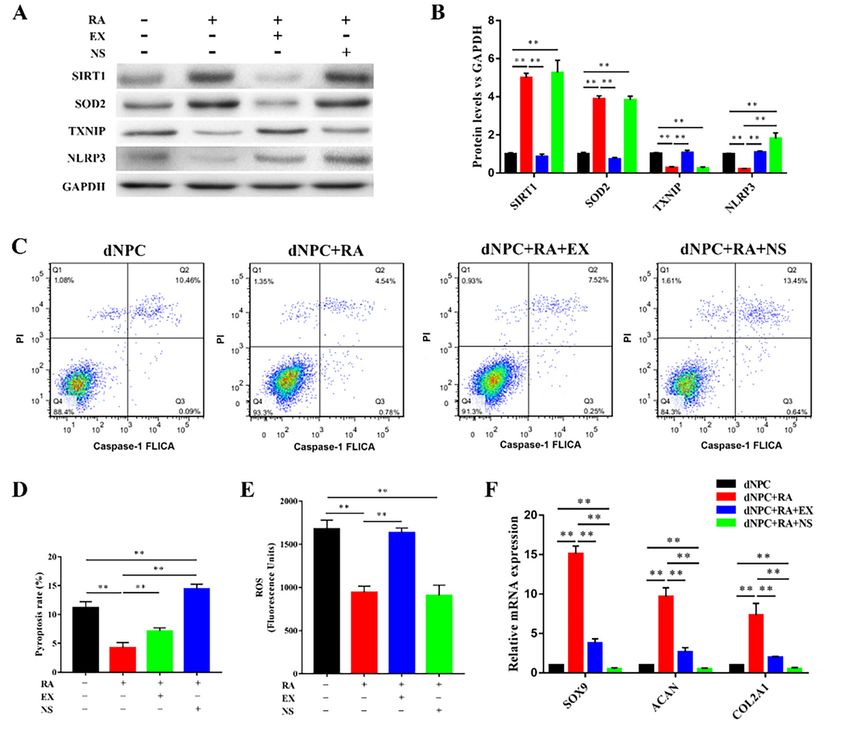
Abstract
Background:
Intervertebral disc (IVD) degeneration is a common disease and initiated by the degeneration of nucleus
pulposus (NP). The pyroptosis of degenerated NP cells (dNPCs) plays an important role in NP
degeneration and may be a potential target in the treatment of IVD degeneration. The purpose of this
study is to identify a feasible solution that can inhibit NP cell pyroptosis to therapy the degeneration of
the intervertebral disc.

Result:
In this study, we determined the effects of retinoic acid (RA) on dNPCs and investigated the underlying
mechanism of RA mediated pyroptosis in dNPCs. We also verified the effects of RA on IVD degeneration
in vivo. Our results demonstrated that RA significantly increased the proliferation and the protein
expression of sox9, aggrecan, and collagen II of dNPCs. Pyroptosis-related proteins such as cleaved
caspase-1, NT-GSDMD, IL-1β, IL-18, and the pyroptosis rate of dNPCs was significantly decreased by RA.
We also found that Sirt1-SOD2 signaling was activated, while ROS generation and TXNIP/NLRP3
signaling in dNPCs was inhibited after the addition of RA. Furthermore, RA also recovered the structure of
NP and increased the contents of sGAG and collagen in vivo.

Conclusion
Our study demonstrated that RA can inhibit the pyroptosis and increase the ECM synthesis function of
dNPCs and verified that RA has a protective effect in IVD degeneration.

Introduction
Intervertebral disc (IVD) degeneration is a common disease characterized by low back pain, and cause a
large economic burden (1, 2). The degeneration of nucleus pulposus (NP) which is surrounded by
annulus fibrosus and cartilaginous endplates is thought to be the origin of IVD degeneration (3). NP cells
(NPCs) located in NP and can synthesis glycosaminoglycan (GAG) and collagen II in normal condition.
Once the viability and function of NPCs are disturbed, the extracellular matrix (ECM) produced by NPCs
would alter and lead to NP degeneration (4).

Biological therapies targeted at NPCs can impact the future management of NP degeneration. Growth
factors can control the proliferation and function of NPCs, and maintain NP homeostasis (5). Many
growth factors such as growth differentiation factor 5 (6), bone morphogenetic protein-2 (BMP-2) (7), and
fibroblast growth factor (FGF) (8) have been used in enhancing the proliferation and ECM synthesis of
NPCs. Drugs are also used in promoting the viability and function of NPCs. Wogonin can ameliorate the

                                                  Page 2/20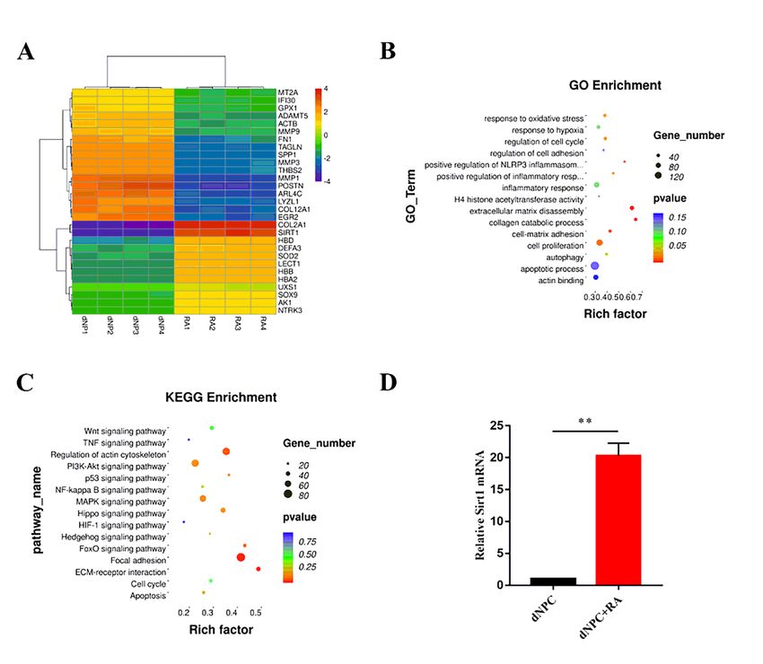
inflammation in NPCs and mitigates the progression of IVD (9). Liraglutide can protects NPCs against
high-glucose induced apoptosis (10). Small molecules are usually used to regulate the activity of
signaling pathways and can also regulate the function and activities of NPCs. Because of their high
efficiency and selectivity, they are now widely used in regulating cell activities. Retinoic acid (RA) is a
potent immunomodulator, and can inhibit inflammatory response and regulate cell activitjies in many
diseases, such as cardiac injury (11), macular degeneration (12) and even osteoarthritis (13). However,
the effects of RA in the treatment of NP degeneration have never been studied.

Many factors have been found to regulate the viability and function of NPCs, such as nutrient, aging, and
oxygen concentration (14). In addition, inflammation also plays an important role in inducing NP
degeneration (15). Apoptosis is the most studied form of inflammatory death in NPCs. Apoptosis of
NPCs leads to a down regulation of ECM synthesis and cell number, then furtherly aggravate NP
degeneration (16). However, other forms of inflammatory death can also regulate the viability and
function of NPCs and lead to NP degeneration. Pyroptosis is a novel form of inflammatory death of cell
and attract more and more attentions.

Pyroptosis is an inflammatory form of regulated cell death, and unlike apoptosis or necrosis, is always
driven by caspase-1 (17). Canonical pyroptosis is initiated after the liberation of N-terminal domain of
pore-forming protein gasdermin D (GSDMD). NT-GSDMD cleaved by active caspase-1 can also lead to the
release of IL-1β and IL-18, and then generated an inflammasome-associated inflammatory response (18).
Pyroptosis is related to different diseases, such as cardiac injury and diabetes mellitus (19, 20).
Pyroptosis can also influence the ECM synthesis function of chondrocytes (21). Resent study have
demonstrated that pyroptosis also exists in NP, and play an important role in decreasing cell viability and
ECM synthesis function of NPCs (22). Therefore, discovering new approach in inhibiting pyroptosis of
NPCs may be benefit for the treatment of IVD degeneration.

Our study aimed to clarify the effects of RA in inhibiting pyroptosis and promoting ECM synthesis
function of NPCs. The underlying mechanisms of RA in mediating pyroptosis of NPCs were also been
investigated. In addition, we verified the potential use of RA in preventing IVD degeneration in a rat model.
We hope our study will provide a new strategy for future IVD treatment.

Materials And Methods
Reagents

RA, EX 527, and Nigericin sodium salt (NS) were purchased from Selleck chem (Shanghai, China). Type II
collagenase and hyaluronidase was purchased from Gibco (Shanghai, China). The cell counting kit-8
(CCK8) was bought from Dojindo (Dalian, China). The lactate dehydrogenase (LDH) assay kit and the
hydroxyproline assay kit was bought from Jiancheng Bioengineering Institute (Nanjing, China). The in
situ cell death detection kit was purchased from Roche Life Science (Shanghai, China). DCFH-DA used for
the detection of cellular ROS was purchased from Sigma Aldrich (Shanghai, China). The FAM FLICA™
Caspase-1 kit was bought from Bio-rad (Shanghai, China). Primary antibodies against Sox9, aggrecan,
                                                   Page 3/20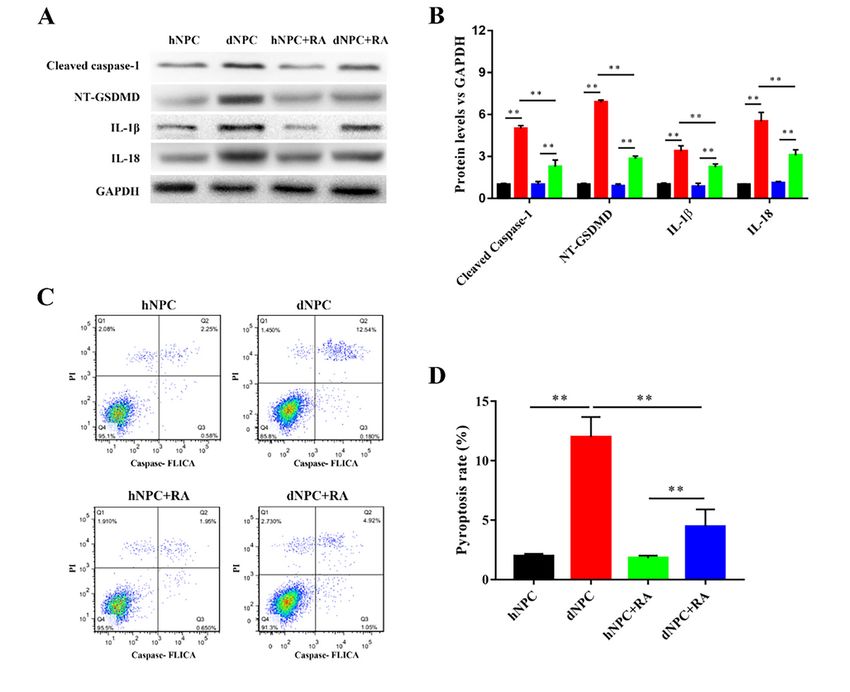
collagen II, cleaved caspase-1, NT-GSDMD, IL-18, SIRT1, TXNIP, NLRP3 and GAPDH were purchased from
Abcam (Shanghai, China). Primary antibodies against IL-1β, SOD2 were purchased from Cell Signaling
Technology (Shanghai, China). The RNAiso reagent, PrimeScript™ RT Master Mix and TB Green Premix Ex
Taq were purchased from TaKaRa (Shanghai, China). The Blyscan assay kit was bought from Biocolor
(Beijing, China).

Isolation and culture of NPCs

Fifteen 40- to 50-year-old patients suffer IVDD according to the results of magnetic resonance imaging
donated degenerative NP samples. The Pfirrmann classification of the degenerative NP samples were at
most grade III (23). The study was approved by the Ethics Committee of The Second Affiliated Hospital of
Zhejiang University School of Medicine, and informed consent was obtained from all the patients
involved in our study. The isolation of human NPCs was described previously (24). Firstly, the
degenerative NP samples were washed three times with phosphate-buffered saline (PBS) and cut into
small pieces. Then, the pieces were enzymatically dissociated by 0.2% collagenase II and 2 U/mL
hyaluronidase for 4 hours at 37 °C with gentle shaking. Next, the tissues were centrifuged at 1000 rpm for
5 min to isolated cells. Human healthy NPCs (hNPCs) were bought from Procell Life Science&Technology
(Hubei, China). Human degenerative NPCs (dNPCs) were cultured in Dulbecco's modified Eagle’s medium
(DMEM) supplemented with 10% fetal bovine serum (FBS), 1% penicillin-streptomycin, and 2 mM L-
glutamine in a humidified incubator at 37 °C with 5% CO2. The complete medium was changed every two
days. dNPCs of passage 2 were used for subsequent experiments.

Detection of cell viability and cytotoxicity

Cell viability and proliferation of each were measured by CCK8. Cells were firstly seeded into a 96-well
plate, and after treatment, were treated with 10% CCK8 in 100 μL DMEM- High Glucose according to the
manufacturer’s protocols. Absorbance values were taken at 450 nm using a microplate reader (Bio-Rad,
Hercules, CA, USA). The cytotoxicity of RA in each group was determined after 0, 7, and 14 days of
cultivation. The supernatant of each was collected and detected using a LDH assay kit following the
manufacturer’s protocols. Absorbance values at 450 nm were detected using a microplate reader (Bio-
Rad).

Fluorescence analysis

The terminal deoxynucleotidyl transferase dUTP nick end labeling (TUNEL) method was used for
measuring the death of NPCs. First, each group of NPCs were fixed with 4% paraformaldehyde for 10 min
at room temperature. Then, the cells were incubated with 3% H2O2 and 0.1% Triton X-100 for 10 min at
room temperature and washed three times with PBS. Next, an in situ cell death detection kit was used to
stain the death of cells according to the manufacturer’s instructions. 4′,6-diamidino-2-phenylindole (DAPI)
was used to stained the nuclei of cells. After staining, cells were observed under a fluorescence
microscope (Leica, Wetzlar, Germany).

                                                 Page 4/20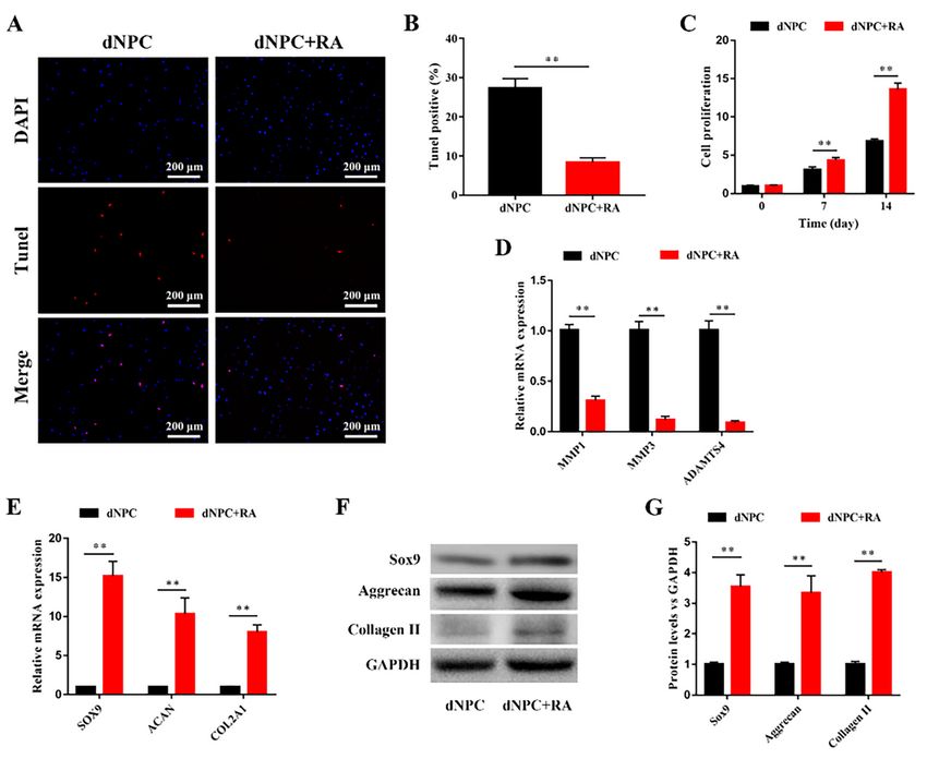
Total RNA isolation and real-time quantitative PCR (RT-qPCR)

Total RNA of each group was extracted using an RNAiso reagent, and quantified by measuring the
absorbance at 260 nm/280 nm. PrimeScriptTM RT Master Mix was used in reverse‐transcription and TB
Green Premix Ex Taq was used in RT-qPCR. The procedures of RT-qPCR were performed on an ABI
StepOnePlus System (Applied Biosystems, Warrington, UK). 18s was used as a housekeeping
gene, and the data was analyzed using the 2(−ΔΔCT) method. All primer sequences were synthesized by
Sangon Biotech (Table 1).

Western blotting analyses

Total protein was extracted from each group of cells by RIPA buffer supplemented with a proteasome
inhibitor (Beyotime, Shanghai, China). Protein concentrations were tested by a BCA protein assay kit
(Beyotime) and equivalent amounts of protein were separated by sodium dodecyl sulfate polyacrylamide
gel electrophoresis and transferred to a polyvinylidene fluoride membrane (Millipore, Shanghai, China) by
electroblotting. All membrane was blocked for 2 hours at room temperature in 5% non‐fat milk and
incubated with primary antibodies overnight at 4 °C. Then, the blots were incubated with horseradish
peroxidase-conjugated secondary antibody (Beyotime) for 1 hour at room temperature. The blots were
visualized using an enhanced chemiluminescence substrate (Millipore) and imaged using the Bio-Rad
XRS chemiluminescence detection system (Bio-Rad).

Flow Cytometry analyses

The pyroptosis of cells was assessed by flow cytometry analyses. The FAM FLICA™ Caspase-1 kit was
used according to the manufacturer’s protocols and pyroptotic death of cells was detected by flow
cytometry. PI (+) and caspase-1 FLICA (+) was defined as pyroptosis.

Microarray analysis

After the extraction and quantification of total RNA in NPCs, the total RNA was purified with a QIAGEN
RNeasy Kit (QIAGEN, Shanghai, China), amplified and labeled with Cy-3. After RNA was hybridized at 65
°C for 17 h, array images were acquired using Agilent Scanner G5761A (Agilent Technologies) and
analyzed using Agilent Feature Extraction software (version 12.0.1.1). Quantile normalization and
subsequent data processing were performed by using the GeneSpring v14.8 software package (Agilent
Technologies). After that, miRNAs that at least four out of the eight samples have flags in Detected were
chosen for further data analysis. Differentially expressed miRNAs were screened by a statistical
significance of p < 0.01. The biological function of the differentially expressed miRNAs were indicated by
gene ontology (GO) and Kyoto Encyclopedia of Genes and Genomes (KEGG) enrichment analysis.

Measurement of cellular ROS

Before the measurement of cellular ROS, each group of cells was cultured in 24-well plates. During the
measurement, cells was rinsed and incubated with 5 μM DCFH-DA in the dark at 37℃ for 30 minutes. A
                                                 Page 5/20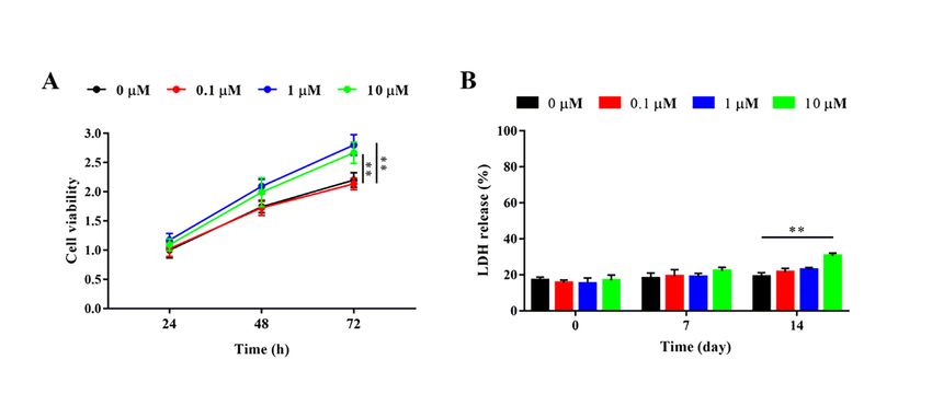
fluorescence microscope (DM5500; Leica) was used to detect cellular fluorescence.

Animal experiments

Male Sprague-Dawley (SD) rats (200 g) were obtained from the Animal Center of the Academy of Medical
Science of Zhejiang Province. All surgical interventions, treatments and postoperative animal care
procedures were performed in strict accordance with the Institutional Animal Care and Use Committee of
Zhejiang University. All SD rats were anesthetized with 1% sodium pentobarbital (Sigma Aldrich), and rat
tail disc degeneration model were fabricated by needle puncture of a 20-G sterile needle in the disc of
coccygeal vertebrae (Co)7/Co8 and Co8/Co9 (25). After that, 3 μL of PBS or RA were injected using a
microsyringe with a 31-G needle. The rats without needle puncture and injection were set as the normal
group, the rats with needle puncture and PBS injection were set as the negative control (NC) group, and
the rats with needle puncture and RA injection were set as the RA group. The follow-up experiments were
conducted 12 weeks after needle puncture and injection.

Histological and biochemical analysis

The IVD tissues of SD rats were collected after sacrifice. Firstly, all tissues were fixed with 4%
paraformaldehyde for 48 hours. Next, all tissues were immersed in a decalcifying solution and embedded
in paraffin. The tissues were sectioned at a thickness of 5 μm using a microtome. For histological
analysis, consecutive tissue sections were stained with hematoxylin and eosin (H&E) and Safranine O-
fast green. Cellularity and morphology of the IVDs were then assessed and represented by histological
score according to a previously described grading scale (26). For biochemical analysis, the Blyscan assay
kit was used to detect the contents of sulfate glycosaminoglycans (sGAG), and the hydroxyproline assay
kit was used to detect the contents of collagen. The results were normalized to the dry weight of NP.

Statistical analysis

All experiments were repeated three times and data were presented as the mean ± standard deviation.
Statistical analyses were performed using SPSS 19.0 (IBM, Armonk, NY, USA) and Statistical significance
was determined using a two-tailed Student’s t-test and one-way ANOVA following Tukey’s post hoc test. A
value of p < 0.05 was considered to indicate statistical significance.

Results
RA increased the viability of dNPCs

Different concentrations of RA were added to dNPCs. After 72 hours treatment, 1 and 10 μM RA
significantly increased the viability of dNPCs, and 0.1 μM RA showed no significant influence on the
viability of dNPCs (Figure 1A). The cytotoxicity of RA was also detected by measuring LDH release. 0.1, 1,
and 10 μM RA had no cytotoxicity on dNPCs on day 0 and 7. However, 10 μM RA significantly increased
the LDH release of dNPCs after 14 days of treatment (Figure 1B). Therefore, we chosen 1 μM of RA to
treat dNPCs in subsequent experiments.
                                                 Page 6/20
Appropriate concentration of RA decreased the death and increased the ECM synthesis function of dNPCs Fluorescence results showed that RA significantly decreased the death of dNPCs (Figure 2A and B). In addition, the proliferation of dNPCs were significantly increased by RA on days 7 and 14 (Figure 2C). ECM synthesis related gene markers were measured after 7 days of treatment. MMP1 (0.31-fold), MMP3 (0.12- fold), and ADAMTS4 (0.09-fold) were significantly decreased by RA, while SOX9 (15.21-fold), ACAN (10.34-fold), and COL2a1 (8.01-fold) were significantly increased by RA (Figure 2D and E). The protein expression levels of sox9, aggrecan, and collagen II in dNPCs were also significantly increased by RA (Figure 2F and G). Pyroptosis of dNPCs was inhibited by RA The protein expression levels of cleaved Caspase-1, NT-GSDMD, IL-1β, and IL-18 reflected the pyroptosis of NPCs. Our results showed that the protein expression levels of cleaved Caspase-1, NT-GSDMD, IL-1β, and IL-18 were significantly increased in dNPCs compared to those of the hNPCs. After the addition of RA, the expression of cleaved Caspase-1, NT-GSDMD, IL-1β, and IL-18 in hNPCs were not significantly changed. However, the expression of cleaved Caspase-1, NT-GSDMD, IL-1β, and IL-18 in dNPCs were significantly decreased after the addition of RA (Figure 3A and B). We also detected changes in caspase-1 in NPCs by flow cytometry to investigate the role of pyroptosis. The pyroptosis rate in the dNPC group was markedly higher and caspase-1 activity was markedly increased compared with the hNPC group. RA had no significant influence on pyroptosis rate of hNPCs, while RA significantly decreased the pyroptosis rate and caspase-1 activity of dNPCs (Figure 3C and D). Sirt1 was involved in RA mediated pyroptosis miRNA microarray analysis was performed to search differentially expressed miRNA and signaling pathways between dNPCs and RA treated dNPCs. Only miRNAs with a mean fold change >5 or
We further investigated the underlying mechanism of RA in inhibiting pyroptosis. We measured the
protein expression levels of SIRT1, SOD2, TXNIP, and NLRP3. Our results showed that after the addition of
RA, the expression levels of SIRT1 and SOD2 were increased while the expression levels of TXNIP and
NLRP3 were decreased. EX 527 is a specific inhibitor of SIRT1. After the addition of EX527, the expression
levels of SIRT1 and SOD2 were decreased, while the expression levels of TXNIP and NLRP3 were
increased compared with the RA+EX-NS- group. NS is a specific inhibitor of NLRP3. The expression levels
of SIRT1, SOD2, and TXNIP between the RA+EX-NS- and RA+EX-NS+ groups showed no significant
difference, while the expression of NLRP3 was significantly higher in the RA+EX-NS+ group than in the
RA+EX-NS- group (Figure 5A and B). The pyroptosis rate in the dNPC group was significant higher
compared with that in the dNPC+RA group. The pyroptosis rate of the dNPC+RA+EX group was higher
than the dNPC+RA group and lower than the dNPC group. The dNPC+RA+NS group showed the highest
pyroptosis rate and caspase-1 activity in the four groups (Figure 5C and D). The ROS generation of the
RA-EX-NS- and RA+EX+NS- groups showed no significant difference, but both of these two groups had
higher ROS generation than the RA+EX-NS- and RA+EX-NS+ groups (Figure 5E). The dNPC+RA group
showed the highest mRNA expression levels of SOX9, ACAN, and COL2a1 in the four groups, while the
dNPC+RA+EX group was the second highest. The dNPC+RA+NS group expressed the lowest mRNA levels
of SOX9, ACAN, and COL2a1 in the four groups (Figure 5F).
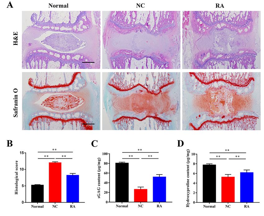
RA prevented the degeneration of IVDs.
We performed H&E and Safranine O-fast green staining to show the morphology, cellularity, and ECM of
the IVD. The normal group showed well-organized collagen lamellae in the AF and round NP with normal
cellularity and rich ECM. The morphology of AF in the NC group was inward bulging and the NP was
irregularly shaped. The cells and ECM of the NP in the NC group was severe decreased. The morphology
and cellularity of AF and NP in the RA group was better compared with those in the NC group, but worse
than the normal group (Figure 6A). The histological score in the normal group was the lowest (5.2 ± 0.45).
The RA group (8.2 ± 1.3) had lower histological score than the NC group (12.0± 0.71) (Figure 6B). The
contents of sGAG and hydroxyproline in the normal group were significantly higher than those in the other
groups. The RA group showed higher contents of sGAG and hydroxyproline than those of the NC group
(Figure 6C and D).
Discussion
The dysfunction and death of NPCs lead to NP degeneration, and subsequently IVD degeneration (27).
Pyroptosis is a new inflammatory form of programmed cell death, and played an important role in
regulating the ECM synthesis function and viability of NPCs (28). RA can inhibit inflammatory response
and regulate cell viability, so it had been used in treating many diseases (29). However, the effectiveness
of RA in treating IVD degeneration has not been studied, and the effects of RA in the pyroptosis of NPCs
was also unknown. In this study, we aimed to clarify the effects of RA in treating NP degeneration and
inhibiting pyroptosis of NPCs. We also investigated the mechanisms of RA regulated pyroptosis in NPCs.
Page 8/20We first evaluated the effects of different concentrations of RA on the viability and cytotoxicity of dNPCs.
1 and 10 µM of RA could promote the viability of dNPCs, but 10 µM of RA also showed cytotocixity on
dNPCs after 14 days of cultivation. Therefore, we think 1 µM of RA is appropriate in treating dNPCs, and
this concentration was used in the subsequent experiments. The effects of RA on ECM synthesis are
controversial. Some studies reported that RA induce matrix loss in chondrocytes by the activation of Wnt/
β-catenin pathways (30). However, other studies also reported the activation of RA receptors enhanced
aggrecan production in chondrocytes (31). We think the effects of RA on ECM synthesis are complicated,
for RA both regulate the anabolism and catabolism of ECM (32, 33). Which role of RA play depends on
the cells and environment. In our study, we found that RA can increase ECM synthesis in dNPCs. We think
it is mainly because that RA inhibited the catabolism of dNPCs by downregulating the expression of
MMP1, MMP3, and ADAMTS4, and increased cell number by promoting cell proliferation and protecting
dNPCs form death.
Both pyroptosis and apoptosis are existed in NPCs, and regulate the function and viability of NPCs (34,
35). Pyroptosis was involved in the degeneration process of NP (36). So inhibiting pyroptosis is important
in the treatment of IVD degeneration. In this study, we found that RA significantly inhibited the expression
of cleaved caspase-1, NT-GSDMD, IL -1β and IL-18, and decreased the pyroptosis of dNPCs. It means that
RA has the potential in treating IVD degeneration. We further discovered the mechanisms of RA in
regulating pyroptosis of dNPCs. The results of microarray analysis showed that Sirt1 was differentially
expressed between dNPCs and RA treated dNPCs. Sirt1 was important in regulating inflammatory
reactions of cell and was positive for the activity of NPCs (37). Sirt1 can inhibit apoptosis in NPCs via
ERK signaling pathway (38). Sirt1-overexpressed chondrocytes can also delay the degeneration of NPCs
during co-culture (39). In our study, we found that RA could increase the expression of Sirt1. After the
addition of Sirt1 specific inhibitor, pyroptosis of dNPCs increased and the ECM synthesis function of
NPCs was weakened. These results further demonstrated that Sirt1 induced by RA positively regulates the
function of dNPCs. In addition, Sirt1 can also inhibits the pyroptosis of dNPCs.
Sirt1 is an important mediator in FoxO signaling and regulates the mitochondrial oxidative stress of cells
(40). Our results of KEGG enrichment analysis also demonstrated the FoxO signaling pathway involved in
RA regulated pyroptosis in dNPCs. SOD2 is also involved in FoxO signaling, and is the downstream
regulator of Sirt1. Sirt1-SOD2 signaling mediates oxidative stress and ROS generation in different cell
types (41). We measured the expression of SOD2 and cellular generation of ROS. The results
demonstrated that Sirt1 activates SOD2 and reduced ROS generation in dNPCs. TXNIP/NLRP3
inflammasome signaling is the key activator in pyroptosis (42). Several studies have reported that ROS
can activate TXNIP/NLRP3 signaling and induce pyroptosis (22, 43). We also found that RA inhibited the
expression of TXNIP and NLRP3. In addition, NLRP3 inhibitor reversed the inhibition effects of RA in
pyroptosis. Therefore, we consider that RA inhibits pyroptosis in dNPCs by activating Sirt1-SOD2
signaling and thereby inhibiting ROS/TXNIP/NLRP3 signaling.
We also explored the in vivo effects of RA in a rat tail disc degeneration model. The structure of IVD was
abnormal, and the contents of sGAG and collagen were decreased in the degenerative NP. RA increased
Page 9/20the contents of sGAG and collagen, and recovered the structure of NP, indicating that RA has protective
effects in IVD degeneration. However, there were still differences between the normal and RA treated IVD,
so it is difficult to consider that RA has regenerative effects in degenerative NP. RA inhibits the pyroptosis
and enhances the ECM synthesis function of dNPCs in degenerative NP. However, the degeneration of IVD
was induced by nutrition, mechanical loading, inflammation and so on (44). We think that it is the
complicated factors in IVD degeneration leads to the limited effects of RA in degenerated IVD. There is
also a limitation in our study, for we did not measure the pyroptosis in the disc degeneration model. We
demonstrated the effects of RA in inhibiting pyroptosis of dNPCs, and discovered the underlying
mechanisms. Our in vivo experiments also demonstrated the protective effects of RA in degenerated NP.
However, further more studies should be performed before the clinical treatment of IVD degeneration by
RA.
Conclusions
We demonstrated that RA has the ability to inhibit the pyroptosis and increase the ECM synthesis
function of dNPCs. The inhibition of pyroptosis in dNPCs induced by RA was activated by Sirt1-SOD2
signaling. The activation of Sirt1-SOD2 signaling decreased the ROS generation and then inhibited the
TXNIP/NLRP3 signaling, which led to the decrease in pyroptosis. We furtherly verified the protective
effects of RA on degenerated IVD in an animal model. Our findings identified pyroptosis in dNPCs as a
novel target for NP degeneration, and provided a candidate therapy for the treatment of IVD degeneration.
Declarations
Ethics approval and consent to participate
The study was approved by the Ethics Committee of The Second Affiliated Hospital of Zhejiang
University School of Medicine, and informed consent was obtained from all the patients involved in our
study.
Consent for publication
Not applicable.
Competing interests
The authors declare that they have no competing interests
Funding
This work is supported by the National Natural Science Foundation of China (no. 81603126, no.
81972514).
Authors' contributions
Page 10/20Peng-Fei Li, Fei Xiong, Ying Yin contributed equally to this work by conducting the experiment and
collected and analyzed the data. Shaojun Hu and Hongyuan Xing arranged the article format. Zhang
Ning, the corresponding author of the article, was responsible for the review of the article and the
guidance of the experimental ideas
Acknowledgements
Not applicable
Availability of data and material
The datasets used and/or analyzed during the current study are available from the corresponding author
on reasonable request.
References
1. Bjornsdottir G, Benonisdottir S, Sveinbjornsson G, Styrkarsdottir U, Thorleifsson G, Walters GB, et al.
Sequence variant at 8q24.21 associates with sciatica caused by lumbar disc herniation. Nat
Commun. 2017;8:14265.
2. Freemont AJ. The cellular pathobiology of the degenerate intervertebral disc and discogenic back
pain. Rheumatology. 2009;48(1):5–10.
3. Neidlinger-Wilke C, Wurtz K, Urban JP, Borm W, Arand M, Ignatius A, et al. Regulation of gene
expression in intervertebral disc cells by low and high hydrostatic pressure. Eur Spine J.
2006;15(Suppl 3):372-8.
4. Gopal D, Ho AL, Shah A, Chi JH. Molecular basis of intervertebral disc degeneration. Adv Exp Med
Biol. 2012;760:114–33.
5. Longo UG, Petrillo S, Franceschetti E, Maffulli N, Denaro V. Growth factors and anticatabolic
substances for prevention and management of intervertebral disc degeneration. Stem Cells Int.
2012;2012:897183.
6. Peng BG. Pathophysiology, diagnosis, and treatment of discogenic low back pain. World J Orthop.
2013;4(2):42–52.
7. Kim DJ, Moon SH, Kim H, Kwon UH, Park MS, Han KJ, et al. Bone morphogenetic protein-2 facilitates
expression of chondrogenic, not osteogenic, phenotype of human intervertebral disc cells. Spine
(Phila Pa 1976). 2003;28(24):2679–84.
8. Thompson JP, Oegema TR Jr, Bradford DS. Stimulation of mature canine intervertebral disc by
growth factors. Spine (Phila Pa 1976). 1991;16(3):253–60.
9. Fang W, Zhou X, Wang J, Xu L, Zhou L, Yu W, et al. Wogonin mitigates intervertebral disc
degeneration through the Nrf2/ARE and MAPK signaling pathways. Int Immunopharmacol.
2018;65:539–49.
Page 11/2010. Yao M, Zhang J, Li Z, Bai X, Ma J, Li Y. Liraglutide Protects Nucleus Pulposus Cells Against High-
Glucose Induced Apoptosis by Activating PI3K/Akt/ mTOR/Caspase-3 and
PI3K/Akt/GSK3beta/Caspase-3 Signaling Pathways. Front Med (Lausanne). 2021;8:630962.
11. Liu Y, Zhao J, Lu M, Wang H, Tang F. Retinoic acid attenuates cardiac injury induced by
hyperglycemia in pre- and post-delivery mice. Can J Physiol Pharmacol. 2020;98(1):6–14.
12. Wu J, Cui D, Li H, Zeng J. Protective effects of NAC and salubrinal on apoptosis of retinal pigment
epithelial cells induced by all-trans retinoic acid. Eur J Ophthalmol. 2021:11206721211000674.
13. Takahata Y, Nakamura E, Hata K, Wakabayashi M, Murakami T, Wakamori K, et al. Sox4 is involved in
osteoarthritic cartilage deterioration through induction of ADAMTS4 and ADAMTS5. FASEB J.
2019;33(1):619–30.
14. Barakat AH, Elwell VA, Lam KS. Stem cell therapy in discogenic back pain. J Spine Surg.
2019;5(4):561–83.
15. Molinos M, Almeida CR, Caldeira J, Cunha C, Goncalves RM, Barbosa MA. Inflammation in
intervertebral disc degeneration and regeneration. J R Soc Interface. 2015;12(104):20141191.
16. Song Y, Li S, Geng W, Luo R, Liu W, Tu J, et al. Sirtuin 3-dependent mitochondrial redox homeostasis
protects against AGEs-induced intervertebral disc degeneration. Redox Biol. 2018;19:339–53.
17. Liu X, Zhang Z, Ruan J, Pan Y, Magupalli VG, Wu H, et al. Inflammasome-activated gasdermin D
causes pyroptosis by forming membrane pores. Nature. 2016;535(7610):153–8.
18. Xue Y, Enosi Tuipulotu D, Tan WH, Kay C, Man SM. Emerging Activators and Regulators of
Inflammasomes and Pyroptosis. Trends Immunol. 2019;40(11):1035–52.
19. Zheng X, Zhong T, Ma Y, Wan X, Qin A, Yao B, et al. Bnip3 mediates doxorubicin-induced
cardiomyocyte pyroptosis via caspase-3/GSDME. Life Sci. 2020;242:117186.
20. Yu ZW, Zhang J, Li X, Wang Y, Fu YH, Gao XY. A new research hot spot: The role of NLRP3
inflammasome activation, a key step in pyroptosis, in diabetes and diabetic complications. Life Sci.
2020;240:117138.
21. Hu J, Zhou J, Wu J, Chen Q, Du W, Fu F, et al. Loganin ameliorates cartilage degeneration and
osteoarthritis development in an osteoarthritis mouse model through inhibition of NF-kappaB activity
and pyroptosis in chondrocytes. J Ethnopharmacol. 2020;247:112261.
22. Xu Q, Xing H, Wu J, Chen W, Zhang N. miRNA-141 Induced Pyroptosis in Intervertebral Disk
Degeneration by Targeting ROS Generation and Activating TXNIP/NLRP3 Signaling in Nucleus
Pulpous Cells. Front Cell Dev Biol. 2020;8:871.
23. Pfirrmann CW, Metzdorf A, Zanetti M, Hodler J, Boos N. Magnetic resonance classification of lumbar
intervertebral disc degeneration. Spine (Phila Pa 1976). 2001;26(17):1873–8.
24. Cheng X, Zhang G, Zhang L, Hu Y, Zhang K, Sun X, et al. Mesenchymal stem cells deliver exogenous
miR-21 via exosomes to inhibit nucleus pulposus cell apoptosis and reduce intervertebral disc
degeneration. J Cell Mol Med. 2018;22(1):261–76.
Page 12/2025. Liu Y, Zhou W, Chen FF, Xiao F, Zhu HY, Zhou Y, et al. Overexpression of LMP-1 Decreases Apoptosis
in Human Nucleus Pulposus Cells via Suppressing the NF-kappaB Signaling Pathway. Oxid Med Cell
Longev. 2020;2020:8189706.
26. Han B, Zhu K, Li FC, Xiao YX, Feng J, Shi ZL, et al. A simple disc degeneration model induced by
percutaneous needle puncture in the rat tail. Spine (Phila Pa 1976). 2008;33(18):1925–34.
27. Croft AS, Illien-Junger S, Grad S, Guerrero J, Wangler S, Gantenbein B. The Application of
Mesenchymal Stromal Cells and Their Homing Capabilities to Regenerate the Intervertebral Disc. Int
J Mol Sci. 2021;22(7).
28. Zhao K, An R, Xiang Q, Li G, Wang K, Song Y, et al. Acid-sensing ion channels regulate nucleus
pulposus cell inflammation and pyroptosis via the NLRP3 inflammasome in intervertebral disc
degeneration. Cell Prolif. 2021;54(1):e12941.
29. Larange A, Cheroutre H. Retinoic Acid and Retinoic Acid Receptors as Pleiotropic Modulators of the
Immune System. Annu Rev Immunol. 2016;34:369–94.
30. Yasuhara R, Yuasa T, Williams JA, Byers SW, Shah S, Pacifici M, et al. Wnt/beta-catenin and retinoic
acid receptor signaling pathways interact to regulate chondrocyte function and matrix turnover. J
Biol Chem. 2010;285(1):317–27.
31. Williams JA, Kondo N, Okabe T, Takeshita N, Pilchak DM, Koyama E, et al. Retinoic acid receptors are
required for skeletal growth, matrix homeostasis and growth plate function in postnatal mouse. Dev
Biol. 2009;328(2):315–27.
32. Shingleton WD, Jones D, Xu X, Cawston TE, Rowan AD. Retinoic acid and oncostatin M combine to
promote cartilage degradation via matrix metalloproteinase-13 expression in bovine but not human
chondrocytes. Rheumatology. 2006;45(8):958–65.
33. Abidin FZ, Gouveia RM, Connon CJ. Application of retinoic acid improves form and function of tissue
engineered corneal construct. Organogenesis. 2015;11(3):122–36.
34. Bai Z, Liu W, He D, Wang Y, Yi W, Luo C, et al. Protective effects of autophagy and NFE2L2 on reactive
oxygen species-induced pyroptosis of human nucleus pulposus cells. Aging. 2020;12(8):7534–48.
35. Chen D, Xia D, Pan Z, Xu D, Zhou Y, Wu Y, et al. Metformin protects against apoptosis and senescence
in nucleus pulposus cells and ameliorates disc degeneration in vivo. Cell Death Dis.
2016;7(10):e2441.
36. He D, Zhou M, Bai Z, Wen Y, Shen J, Hu Z. Propionibacterium acnes induces intervertebral disc
degeneration by promoting nucleus pulposus cell pyroptosis via NLRP3-dependent pathway.
Biochem Biophys Res Commun. 2020;526(3):772–9.
37. Guo J, Shao M, Lu F, Jiang J, Xia X. Role of Sirt1 Plays in Nucleus Pulposus Cells and Intervertebral
Disc Degeneration. Spine (Phila Pa 1976). 2017;42(13):E757-E66.
38. He F, Li Q, Sheng B, Yang H, Jiang W. SIRT1 Inhibits Apoptosis by Promoting Autophagic Flux in
Human Nucleus Pulposus Cells in the Key Stage of Degeneration via ERK Signal Pathway. Biomed
Res Int. 2021;2021:8818713.
Page 13/2039. Lei B, Wang K, Yang D, Liao L, Dong X, Huang Z. Co-culture with Sirt1-overexpressed chondrocytes
delays the nucleus pulposus cells degeneration. Cell Tissue Bank. 2021.
40. Mouchiroud L, Houtkooper RH, Moullan N, Katsyuba E, Ryu D, Canto C, et al. The NAD(+)/Sirtuin
Pathway Modulates Longevity through Activation of Mitochondrial UPR and FOXO Signaling. Cell.
2013;154(2):430–41.
41. Li K, Zhai M, Jiang L, Song F, Zhang B, Li J, et al. Tetrahydrocurcumin Ameliorates Diabetic
Cardiomyopathy by Attenuating High Glucose-Induced Oxidative Stress and Fibrosis via Activating
the SIRT1 Pathway. Oxid Med Cell Longev. 2019;2019:6746907.
42. Bharti V, Tan H, Zhou H, Wang JF. Txnip mediates glucocorticoid-activated NLRP3 inflammatory
signaling in mouse microglia. Neurochem Int. 2019;131:104564.
43. Gu C, Draga D, Zhou C, Su T, Zou C, Gu Q, et al. miR-590-3p Inhibits Pyroptosis in Diabetic
Retinopathy by Targeting NLRP1 and Inactivating the NOX4 Signaling Pathway. Invest Ophthalmol
Vis Sci. 2019;60(13):4215–23.
44. Urban JP, Roberts S. Degeneration of the intervertebral disc. Arthritis Res Ther. 2003;5(3):120–30.
Tables
Table 1.
Gene Forward primer (5’ to 3’) Reverse primer (5’ to 3’)
18s GAATTCCCAGTAAGTGCGGGTCATA CGAGGGCCTCACTAAACCATC
MMP1 AAAATTACACGCCAGATTTGCC GGTGTGACATTACTCCAGAGTTG
MMP3 TGATGGGCCTGGAATGGTC TTCATGAGCAGCAACCAGGAATAG
ADAMTS4 CTCCTGCCTTTAGCCTGGTC CCCAAAGGCTGGTAATCGGT
ACAN CTAGCTGCTTAGCAGGGATAACG GATGACCCGCAGAGTCACAAAG
SOX9 AGGAAGCTGGCAGACCAGTACC GGGTCTCTTCTCGCTCTCGTTCA
COL2A1 CTGGTGGAGCAGCAAGAGC GTGGACAGTAGACGGAGGAAAG
SIRT1 TAGCCTTGTCAGATAAGGAAGGA ACAGCTTCACAGTCAACTTTGT
Figures
Page 14/20Figure 1
RA increased the viability of dNPCs. (A) Cell viability was detected at 24, 48, and 72 hours after treated
with different concentrations of RA. (B) Cytotoxicity of different concentrations RA was measured on
days 0, 7, and 14 by LDH release. Data represent mean ± SD; **p < 0.01.
Page 15/20Figure 2
Appropriate concentration of RA decreased the death and increased the ECM synthesis function of
dNPCs. (A) TUNEL method was performed to measure death of dNPCs (red) before and after the
treatment of RA, and the results was observed by fluorescence. Nuclei (blue) were stained by DAPI. (B)
Cell death was quantified according to the results of TUNEL. (C) Cell proliferation of dNPCs was
measured on days 0, 7, and 14 by CCK8. (D) The gene expression levels of MMP-1, MMP-3, and
ADAMTS4 in each group on day 7 were determined by RT-qPCR and normalized to 18s. (E) The gene
expression levels of SOX9, ACAN, and COL2a1 in each group on day 7 were determined by RT-qPCR and
normalized to 18s. (F) The protein expression levels of Sox9, aggrecan, and collagen II of dNPCs in each
group was measured and (G) quantified on day 14. Data represent mean ± SEM; **p < 0.01. Scale bar =
200 µm.
Page 16/20Figure 3
Pyroptosis of dNPCs was inhibited by RA. (A) Protein expression levels of Cleaved caspase-1, NT-GSDMD,
IL-1β, and IL-18 in hNPCs and dNPCs before and after the treatment of RA were measured on day 3. (B)
Quantification of the protein expression levels in each group. (C) Pyroptosis in hNPCs and dNPCs before
and after the treatment of RA was assessed using FLICA/PI staining and flow cytometric analysis. Data
represent mean ± SEM; **p < 0.01.
Page 17/20Figure 4
Sirt1 was involved in RA mediated pyroptosis. (A) The heatmap showed differentially expressed mRNAs
in dNPCs before and after the treatment of RA. (B) GO terms with significant p values for biological
processes, molecular function, cellular component. (C) KEGG terms with significant p values were
analyzed. (D) The expression level of Sirt1 in dNPCs before and after the treatment of RA was verified by
RT-PCR and normalized to 18s. Data represent mean ± SEM; **p < 0.01.
Page 18/20Figure 5
RA inhibited pyroptosis of dNPCs by activating Sirt1-SOD2 pathway. (A) Protein expression levels of
SIRT1, SOD2, TXNIP and NLRP3 in dNPCs after the addition of RA, EX 527, and NS were measured on day
3 and (B) quantified. (C) Pyroptosis in dNPCs after the addition of RA, EX 527, and NS was assessed
using FLICA/PI staining and flow cytometric analysis. (D) Quantification of pyroptosis rate in each group.
(E) Intracellular ROS generation in dNPCs after using RA, EX 527, and NS was measured. (F) The gene
expression levels of SOX9, ACAN, and COL2a1 in each group on day 7 were determined by RT-qPCR and
normalized to 18s. Data represent mean ± SEM; **p < 0.01.
Page 19/20Figure 6
RA prevented the degeneration of IVDs. (A) Representative H&E and Safranin O staining of discs at 12
weeks after injection were observed. (B) Histological grade of each group was evaluated. (C) The content
of sGAG in each group at 12 weeks after injection were quantified by Blyscan assay. (D) The contents of
collagen in each group at 12 weeks after injection were quantified by hydroxyproline assay. Data
represent mean ± SEM; **p < 0.01. Scale bar = 500 µm.
Page 20/20You can also read