Effect of Aspartame on the Frontal Cortex of Adult Male Albino Rats. A Light and Electron Microscopic Study
←
→
Page content transcription
If your browser does not render page correctly, please read the page content below
Egypt. J. Histol. Vol. 32, No. 2, Dec. 2009: 346 - 357                                                  (ISSN: 1110 - 0559)

Original Article

 Effect of Aspartame on the Frontal Cortex of Adult Male Albino Rats.
 A Light and Electron Microscopic Study
 Sahar M. M. Omar
 Histology Department, Faculty of Medicine, Ain Shams University

                                                         ABSTRACT
 Introduction: Aspartame is an artificial sweetener added to 9,000 food & drink products. Studies and investigations are
 thus important to prove or disapprove the existing fears concerning aspartame.
 Aim of the Work: Was to evaluate, the toxic effects of long term aspartame administration on the frontal cortex. And to
 investigate whether immunostaining for neuron-specific enolase (NSE) and glial fibrillary acidic protein (GFAP), could
 help as valuable markers for evaluating neuronal and glial response to aspartame-induced injury.
 Materials and Methods: Fifteen adult male albino rats were used; the rats were divided into three equal groups. Group
 I served as a control group. Group II received aspartame orally in a dose of (250mg\ kg\day) for 8 weeks. Group III
 received aspartame as in group II, but rats were then left for 4 weeks to recover.
 Results: Pyramidal cells of animals receiving aspartame showed significant morphological necrotic changes and
 appeared darkly stained or vacuolated, irregular in shape with pyknotic or faint nuclei. Ultra structurally, the cytoplasm
 of pyramidal cells showed prominent vacuolization, mitochondria with indistinct cristae. Neurons in aspartame group
 were statistically significantly less stained by anti-NSE antibody than control group. A significant increase in the number
 of GFAP immunoreactive astrocytes was also detected. Whereas, sections obtained from rats of group III showed
 significant improvement of the aspartame produced changes but never returned to control ones.
 The results of the study demonstrated that the content of NSE of neurons and the number of GFAP (+) astrocytes could
 serve as molecular markers for neuronal injury, regeneration and astrocytic proliferation, respectively.
 Conclusion: Chronic aspartame ingestion could result in marked affection of the frontal cortex. Four weeks of cessation
 was not sufficient to obtain a normal histological appearance.
Key Words: Aspartame, frontal cortex, neuron-specific             Corresponding Author: Sahar M. M. Omar
enolase, glial fibrillary acidic protein.                         Tel.: 0101428289 E-mail: saharhistology@yahoo.com

INTRODUCTION

   Aspartame is a widely used artificial, non-                        So aspartate (one of the aspartame metabolite)
carbohydrate sweetener, L-α-aspartyl-L-phenylalanine-             was one of the more commonly known excitotoxins.
l-methyl ester1.                                                  This amino acid had been known to be a normal
                                                                  neurotransmitter in the brain. However, aspartate as a
    Aspartame was found in more than 6,000 products,              neurotransmitter existed in the extracellular fluid only in
including carbonated soft drinks, hot chocolate, chewing          very, very small concentrations. When the concentration
gum, candy, deserts, tabletop sweeteners and some                 of this transmitter rose above certain level, the neurons
pharmaceutical products, such as vitamins and sugar-free          began to fire abnormally. At higher concentrations, the
cough drops1.                                                     cells underwent a specialized process of delayed cell
                                                                  death known as excitotoxicity, that is, they were excited
    It was estimated by the Aspartame Information                 to death4.
Center (2005) to be consumed by more 200 million
people worldwide, including children between 2-5 years               Aspartame consumption was suggested to be
of age and pregnant females2.                                     implicated with various symptoms from the central
                                                                  nervous system such as seizures5, memory loss6,
    Aspartame could be metabolized to l-phenylalanine,            Cholinergic symptoms, headaches7 and oncogenesis1.
l-aspartate and methanol. Each metabolite could produce
toxic or excitatory effects in animal model systems,                 Aspartame had been the subject of controversy
hence termed excitotoxins3.                                       regarding its safety and the circumstances of its approval
33 (1158-2009)

                                                            346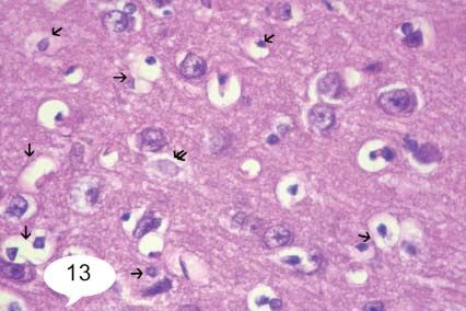
Effect of Aspartame on the Frontal Cortex of Adult Male Albino Rats

by American food and drug administration FDA8.                           Then the chosen dose for aspartame could be easily
                                                                     within the acceptable limits of human consumption after
    Since, the everyday consumption of aspartame by                  species factor correction. Dose comparisons between
people is increasing. Further investigations and studies             humans and rats had usually been corrected by a factor of
are recommended to prove or disapprove the existing                  5, since rats metabolize aspartame faster than humans14.
fears concerning aspartame.
                                                                        Aspartame was purchased from Al-Ameryia Pharma
    Neuron-specific enolase (NSE) is a glycolytic                    Company, Egypt. It was available in the form of tablets.
isoenzyme.     Immunocytochemical      surveys     had               Each tablet contained 20mg of aspartame.
demonstrated that NSE is confined solely to neurons,
not only central ones but those of dorsal root ganglia,                  At the end of the experiment the animals were
autonomic ganglia and some neuroendocrine cells as                   dissected under ether anesthesia, 2% glutaraldehde
well9.                                                               mixed with heparin was injected into the beating hearts
                                                                     of the animals for fixation of the brains by perfusion.
    It had been demonstrated that NSE served not only                After 5 minutes brains were dissected out.
as an identifying marker for neurons but, as an index of
neuronal injury, regeneration and reinnervation as well10.               For light microscopic study, samples from frontal
                                                                     parts of the cerebral hemispheres were taken; after
    On the other hand, it had been reported that, the                fixation in 10% neutral buffered formalin, they were
astrocyte-specific protein, the glial fibrillary acidic              dehydrated through alcohols, cleared in xylene and
protein (GFAP), this intermediate filaments found in                 embedded in paraffin wax. Later on, 5um thick sections
astrocyte, could also serve as a marker for glial cells11.           were stained with haematoxylin and eosin15. Other fresh
                                                                     specimens of frontal cortex were fixed in Golgi Cox
    So the specific aim of the current study was to                  solution for 2 months. Then they were processed as the
evaluate, the possible toxic effect of long term aspartame           usual paraffin technique. Paraffin sections of 10um thick
administration (8 weeks) on the rat frontal cortex. And              were cut and stained by Golgi Cox technique16.
to investigate whether the immunostaining by neurotypic
protein (NSE) and the gliotypic protein (GFAP), could                    For electron microscopy, other parts of the
help as valuable markers for evaluating neuronal and                 specimens were post fixed in 1% osmium tetra oxide at
glial response to aspartame-induced injury.                          4c, dehydrated and embedded in epoxy resin. Semi thin
                                                                     sections were cut and stained with toludine blue. Ultra
MATERIALS AND METHODS                                                thin sections were stained with uranyl acetate and lead
                                                                     citrate17 and examined and photographed by transmission
    The present study was carried out on fifteen adult               electron microscope (Jeol-Ex1010 TEM) in Al Azhar
male albino rats, weighing 200-250grams. The animals                 University.
were kept in adequate ventilation and temperature in
plastic cages and were fed standard laboratory food and                  For immunohistochemical study, staining was
water adlibitum.                                                     performed for glial fibrillary acidic protein as an indicator
                                                                     for glial reactivity.
The rats were divided into three equal groups:
                                                                         Primary antibodies monoclonal mouse anti-human
Group I: Served as a control group and received ordinary             GFAP was purchased from Dako Carpenteria, Ca, USA.
diet and water.                                                      Sections were treated with 0.01 m citrate buffer (ph
                                                                     6.0) for 10 minutes to unmask antigen. Sections were
GroupII: Received aspartame (250mg\kg\day)12                         incubated in o.3 % hydrogen peroxide for 30 min to
dissolved in 5ml distilled water and administered by                 abolish endogenous peroxidase activity, before blocking
gastric tube for 8 weeks.                                            with 5% horse serum for 1-2h. Slides were incubated
                                                                     with the primary antibody (1:500 monoclonal mouse
Group III: Received aspartame as in groupII, but rats                anti-GFAP) at 4c for 18-20h and after washing, they were
were then left for 4 weeks to recover before they were               incubated with biotinylated secondary antibodies (ABC
sacrificed.                                                          kit, 1:200) and then with avidin-biotin complex. Finally,
                                                                     sections were developed with 0.05% diaminobenzidine.
   The dose of aspartame had been consistent with                    Slides were counterstained with Mayer’s haematoxylin,
similar values in the previous literature and thus was               dehydration, clearing and mounting were done15.
chosen12.
                                                                         Immunostaining with anti-human NSE (as an
Since the acceptable daily dose for aspartame is 50mg\kg             indicator for neuron reactivity) mouse monoclonal
body weight in the United States and in Europe 40mg\kg               IgG antibody (Dako Japan, Kyoto, Japan) was also
body weight13.                                                       performed. Formalin-fixed, paraffin-embedded sections
                                                              347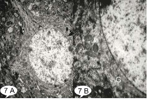
Sahar M. M. Omar

were treated as mentioned above, to unmask antigen and                For statistical analysis: All data were collected,
abolish endogenous peroxidase activity. Slides were then          revised and then subjected to statistical analysis using
incubated with the primary antibody (1:250 monoclonal             student`s “t”test& paired“t”test. The significance of the
mouse anti-NSE) at room temperature for 1h and then               data was determined by P value P>0.05 non-significant
washed and treated as previously mentioned15.                     (NS), P
Effect of Aspartame on the Frontal Cortex of Adult Male Albino Rats

    Toludine blue stained semi- thin sections showed                           The ultra structural examination of the frontal
pyramidal cells with vesicular nuclei but with fewer                       cortex showed slight vacuolation in cytoplasm of some
Nissl’s granules in the cytoplasm, others with faintly                     pyramidal cells, with numerous ribosomes, rough
stained cytoplasm and nuclei, while some others appeared                   endoplasmic reticulum, Golgi complex, but mitochondria
deeply stained and irregular in shape (Fig. 9-A). Golgi                    appeared ranging from electron dense to electron lucent
Cox- stained sections showed most of pyramidal cells                       with inapparent cristae (Fig. 17).
with deformed (moth-eaten) outline and absent processes
(Fig. 9-B).                                                                Statistical results:

    Immunohistochemical staining showed an apparent                        A- number of astrocytes/H.P.F.:
increase in the number of GFAP immunoreactive
astrocytes as compared to control group (Fig. 10).                              The aspartame group (Group II) showed a highly
Sections stained with anti-NSE-antibodies revealed                         significant increase in number of astrocytes/H.P.F. as
that some neurons had lost their normal stain ability for                  compared to control group P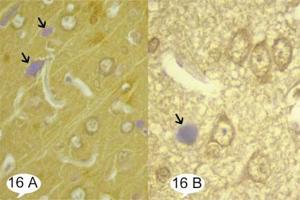
Sahar M. M. Omar

Table 2: Comparison between number of astrocytes in aspartame (GII) and after recovery (GIII):

                                                 aspartame (GII)             recovery (GIII)     Paired difference
                                                                                                                        t           P        Sig.
                                         Mean           ±SD            Mean            ±SD       Mean          ±SD

 No. of astrocytes                       11.05          ±2.16              8.10        ±1.89      2.95         ±2.93   4.50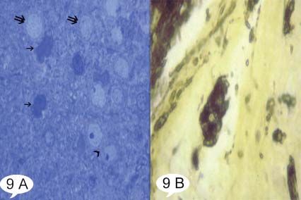
Effect of Aspartame on the Frontal Cortex of Adult Male Albino Rats

                             60                                 GII
                             50
                                                                       GIII
                             40

                             30
                                                            GI
                             20

                             10

                               0
                                                                HNI
 Histogram 3: Showing comparison between different groups as regard HNI ⁄ H.P.F.

Table 4: Comparison between HNI in Aspartame (GII) and after recovery (GIII) regarding HNI and percentages of positive cells:
                                        Aspartame                  Recovery              Paired difference
                                                                                                                     t            P          Sig.
                                    Mean          SD±          Mean         SD±         Mean           SD±

 HNI                                53.28        ±10.84        37.13        ±8.41       16.15        ±13.61        5.93       0.0001>         HS

 % of NSE +ve cells                  0.47        ±0.19          0.70        ±0.15       -0.23         ±0.26        4.34       0.0001>         HS

Paired t test:
Mean HNI showed decrease by significant amount (16.15) between Aspartame (GII) and after 4 weeks recovery (GIII) and percentage of NSE
positive cells showed statistically significant increase (by 23%) P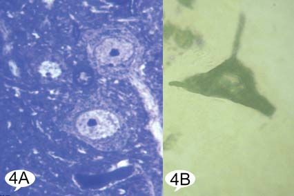
Sahar M. M. Omar
Fig. 5: Showing few GFAP positive neuroglia cells with brown
cytoplasmic granules (arrow ↑).
Fig. 2: Showing the frontal cortex covered by pia matter (arrow ↑) Control group avidin biotin peroxidase for GFAP X 640.
containing a blood vessel (arrow head ^). The upper five layers of the
frontal cortex can be identified; the outer molecular layer I, external
granular layer II, external pyramidal layer III, inner granular layerIV,
inner pyramidal layer. Control group H&E X100.
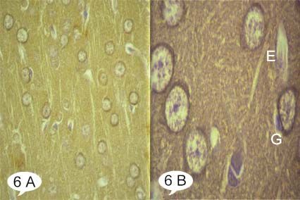
Fig. 6: Showing A: many NSE positive pyramidal cells. B: the cytoplasm
of neurons appears filled with reaction product. Glial cells (G) and
endothelial cells (E) are devoid of staining.
Fig. 3: Showing the pyramidal cells (arrow ↑) with their open face nuclei
Control group avidin biotin peroxidase for NSE A X 640, B X 1600.
and basophilic cytoplasm. The smaller neuroglia cells (double arrow
↑↑) cells are scattered, while blood capillaries appear lined by simple
squamous epithelium (arrow head ^). Control group H&E X 640.
Fig. 7: Showing A: A pyramidal cell with large euchromatic nucleus and
Fig. 4: Showing A: Pyramidal cells with Toludine blue positive cytoplasm surrounded by well defined regular nuclear envelop. B: The cytoplasm
containing Nissl’s granules and large nucleus with prominent nucleolus. is studded with scattered free ribosomes, rough endoplasmic reticulum,
B: The well defined cell body of a pyramidal cell and its processes. mitochondria (M) and Golgi complex (G).
Control group A: T.B. X 1600, B: Golgi Cox- X 1600. Control group T.E.M. A X 600, B X 20000.
352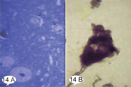
Effect of Aspartame on the Frontal Cortex of Adult Male Albino Rats
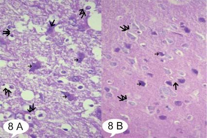
Fig. 8: Showing A: Some irregular darkly stained pyramidal cells with
pyknotic nuclei (arrow ↑), others show marked cytoplasmic vacuolization
(double arrow ↑↑). B: Almost all pyramidal cells appearing with darkly Fig. 11: Showing that some neurons are deprived from their normal
stained nuclei and surrounded by halos (arrow ↑), or with faintly stained staining ability for NSE (double arrow ↑↑); while fewer remained with
nuclei and cytoplasm (double arrow ↑↑). Group II H&E A&B X 640. darkly stained brown cytoplasmic granules (arrow↑).
Group II avidin biotin peroxidase for NSE X 640.
Fig. 9: Showing A: Pyramidal cells with vesicular nuclei but with fewer
Nissl’s granules (arrowhead ^), some are deeply stained and irregular in
shape (arrow ↑), while others appearing with faintly stained nuclei and Fig. 12: Showing the cytoplasm of a pyramidal cell with prominent
cytoplasm (double arrow ↑↑). B: Pyramidal cell with deformed (moth- vacuolization, electron lucent mitochondria with inapparent cristae (M)
eaten) outline and absent processes. and deformed Golgi complex (G). Group II T.E.M. X 25000.
Group II A: T.B. X 1600, B: Golgi Cox- X 1600.
Fig. 10: Showing numerous GFAP immunoreactive astrocytes. Notice! Fig. 13: Showing many pyramidal cells still vacuolated with acidophilic
An apparent increase in GFAP positive neuroglia cells. cytoplasm (arrow ↑). Notice! One pyramidal cell appears faintly stained
Group II avidin biotin peroxidase for GFAP X 640. (double arrow ↑↑). Group III H&E X 640.
353Sahar M. M. Omar
Fig. 14: Showing A: Pyramidal cells with vesicular nuclei and vacuolated
cytoplasm (arrow ↑). B: A pyramidal cell with festooned outline.
Group III A: T.B. X 1600, B: Golgi Cox- X 1600.
Fig. 17: Showing slight cytoplasmic vacuolation, with numerous
ribosomes, rough endoplasmic reticulum, Golgi complex. Notice!
Mitochondria appear ranged from electron dense to electron lucent with
indistinct cristae (M). Group III T.E.M. X 25000.
DISCUSSION
Numerous reports and various issues, concerning the
toxic effects of aspartame have continued to be raised
(more than 20 years after its approval by FDA)12. So the
specific aim of this study was to determine if long term
aspartame administration (for 8 weeks) would lead to
cytopathological effect on frontal cortex.
Immunohistochemistry performed in the present
study using anti-human NSE had enabled to assess the
Fig. 15: Showing many immunoreactive astrocytes. However, apparent neuronal damage besides HNI of H&E sections. Anti-
decrease in number of GFAP positive neuroglia cells as compared to NSE immunostaining was statistically significantly less
group II. in frontal cortex following aspartame administration.
Group III avidin biotin peroxidase for GFAP X 640. Moreover, histological necrosis index was statistically
much elevated when compared to control group. The
results of the current study indicated that there was
a significant negative correlation between anti-NSE
immunostaining and HNI of H&E sections. Therefore,
NSE could reflect vital reaction and could be useful in
evaluating neuronal damage. Similar findings had been
recognized in a study assessing NSE-immunostaining
and neuronal necrosis assessed by HNI in various forensic
autopsy cases20. It was also reported that, the number of
NSE (+) cells was well correlated with Nissl’s staining.
It was recently hypothesized that, NSE immunoreactivity
might be a valuable marker for determination of the
number of metabolically active neurons in different brain
regions after single and repeated experimental seizures21.
Moreover, it has been demonstrated that, neurons
undergo certain sequence of morphological and
Fig. 16: Showing A: Almost most neurons appear with their normal metabolic changes following axonal injury. Hence,
brown cytoplasmic stain ability; while few appear blue and devoid of any
studying the changes in NSE neurons content could serve
positive granules (arrow ↑). B: Immunoreactive pyramidal cells, except
one which appear deprived from its brown reactivity (arrow ↑). as a molecular marker of axon injury, regeneration and
Group III avidin biotin peroxidase for NSE A X 640, B X 1600. target reinnervation10.
354Effect of Aspartame on the Frontal Cortex of Adult Male Albino Rats
Additionally, the current study reported a statistically point of cell death, hence the name excitotoxins4,28.
significant increase in number of astrocytes after
aspartame intake as compared to control group. Evidence Interestingly, free radicals had been shown to prevent
potentially had been implicated revealing high incidence uptake of excitotoxins by astrocytes as well, which
of brain tumors in aspartame-fed rats compared to no would significantly increase extra cellular aspartame
brain tumors in concurrent control22. metabolites levels. This created a vicious cycle that
would multiply any resulting damage and malfunctioning
Meanwhile, several experimental studies suggested of neurophysiologic system29.
that, astrocytomas were the exact kind of brain tumor
found in aspartame dosed rats1. Other authors believed It was also added that, aspartame metabolites induced
that, the increase in number of astrocytes might be a amino acids imbalance within neuron microenviroment,
reaction to the degeneration of neighboring neurons23. thus producing ultimate damage30,31.
It was well recognized that, upon ingestion, aspartame Recently, others reported significant increase in
broke down into triple toxins (excitotoxins) including NA+, K+-ATPase activity of rat frontal cortex induced
aspartic acid, phenylalanine, methanol and its further by aspartame metabolites. This might be related to
breakdown products including formaldehyde and formic memory-retention problems that chronic consumption of
acid24. aspartame might be partially responsible12,32.
An interesting observation was that chronic The present study demonstrated that, four weeks after
elevations of blood aspartate could seep through the dosing of aspartame, the histological necrosis index,
normal blood-brain barrier25. anti- NSE immunostaining and the amount of GFAP in
frontal cortex showed significant improvement but never
An additional critical factor was that high levels of returned to near control values.
dietary aspartame might disrupt the normal blood-brain
barrier, thus allowing more aspartame metabolites to This agreed with other authors12 who stated that,
enter the brain, creating a vicious cycle. On the other improvement from aspartame- induced toxicity was
hand, when the blood-brain barrier became dysfunction gradual and incomplete.
like in various pathological conditions, including
hypertension, diabetes and minor strokes, brain levels of Furthermore, free radical scavengers including
aspartate reflect blood levels26. the functional reserve of antioxidant, vitamins and
minerals could be necessary for neural protection and
In the current study, examination of H&E stained regeneration27.
sections of aspartame-treated rats revealed the presence
of many degenerated pyramidal cells which appeared Over 200 million people now consume aspartame
either darkly stained with pyknotic nuclei or with faintly products. There is sufficient medical literature
stained cytoplasm and nuclei. Moreover, there was documenting serious injury by aspartame in the
significant elevation in HNI as compared to control group. concentrations below the acceptable daily intake. Hence,
Golgi-cox staining showed deformed pyramidal cell warning the public (especially pregnant women and
with irregular outline. Ultra-structurally, the pyramidal children) of these dangers is justified. Although, the case
cells showed mitochondria with inapparent cristae and against aspartame is strong, further in vitro and in vivo
vacuolated cytoplasm. Previous investigators reported investigations (not industry funded) are needed aiming
that, excessive aspartame stimulation could trigger the to explain the various toxic effects related to aspartame
generation of large numbers of free radical species, both consumption.
as nitrogen and oxygen species. These free radicals had
been shown to damage cellular proteins and DNA. The REFRENCES
most immediate DNA damage was to the mitochondrial
DNA27. 1. Soffritti M, Belpoggi F, Degli Esposti D, Lambertini L, Tibaldi
E and Rigano A. (2006): First experimental demonstration of the
More recent molecular studies had disclosed the multipotential carcinogenic effects of aspartame administered
mechanism of neuronal destruction in some details. It in the feed to Sprague-Dawley rats. Environ. Health Perspect.
had shown that aspartate and other excitatory amino Mar; 114(3):379-385.
acids could activate calcium channels via a specialized 2. Aspartame Information Center. Aspartame Information
family of receptors, which in turn allowed calcium to Center Homepage. 2005; Available at: http://www.aspartame.
flood into the cell. This calcium would further trigger a org/. Accessed Oct 27, 2005.
cascade of reactions, including free radical generation 3. Fisher RS. (1989): Aspartame, neurotoxicity and seizures: A
and lipid per oxidation, which would destroy the cell. review. J. Epilepsy;2(2):55-64.
With this calcium triggered stimulation, the neuron 4. Blaylock RL. (1999): Excitotoxins, neurodegeneration and
became excited, firing its impulses repetitively until the neurodevelopment. Med. Sentinel J. ;4(6):212-215.
355Sahar M. M. Omar
5. Camfield PR, Camfield CS, Dooley JM, Gordon K, Jollymore gamma subunit specific anti-peptide monoclonal antibodies. J.
S and Weaver DF. (1992): Aspartame exacerbates EEG spike- Clin. Pathol. Nov;46(11):993-996.
wave discharge in children with generalized absence epilepsy: A 19. Van Nassauw L, Harrisson F, Cras P and Callebaut M.
double-blind controlled study. Neurology May; 42(5): 1000-1003. (1987): Immunohistochemical localization of S-100 protein,
6. Moser RH. (1994): Aspartame and memory loss. JAMA glial fibrillary acidic protein and neuron-specific enolase
Nov, 16;272(19):1543. in the pars distalis of quail, rat and human hypophyses.
7. Van den Eeden SK, Koepsell TD, Longstreth JR, Van Belle Histochemistry ;86(4):353-358.
G, Daling JR and McKnight B. (1994): Aspartame ingestion 20. Nogami M, Takatsu A and Ishiyama I. (1998):
and headaches: A randomized crossover trial. Neurology Immunohistochemical study of neuron-specific enolase in
Oct; 44(10):1787-1793. human brains from forensic autopsies. Forensic Sci. Int.
8. Roberts HJ. (1991): Does aspartame cause human brain cancer. Jun, 8;94(1-2):97-109.
J. Advanc. Med. ;4(4):231-241. 21. Yardimoǧlu M, Ilbay G, Dalçik C, Dalçik H, Sahin D and
9. Pickel VM, Reis DJ, Marangos PJ and Zomzely Neurath Ateş N. (2008): Immunocytochemistry of neuron specific enolase
C. (1976): Immunocytochemical localization of nervous (NSE) in the rat brain after single and repeated epileptic seizures.
system specific protein (NSP-R) in rat brain. Brain Res. Int. J. Neurosci. ;118(7):981-993.
Mar, 19;105(1):184-187. 22. Olney JW, Farber NB, Spitznagel E and Robins LN. (1996):
10. Kirino T, Brightman MW, Oertel WH, Schmechel DE Increasing brain tumor rates: Is there a link to aspartame? J.
and Marangos PJ. (1983): Neuron-specific enolase as an Neuropathol. Exp. Neurol. Nov;55(11):1115-1123.
index of neuronal regeneration and reinnervation. J. Neurosci. 23. Goss JR, Finch CE and Morgan DG. (1991): Age-related
May; 3(5):915-923. changes in glial fibrillary acidic protein mRNA in the mouse
11. Brock TO and O’Callaghan JP. (1987): Quantitative changes in brain. Neurobiol. Aging Mar-Apr;12(2):165-170.
the synaptic vesicle proteins synapsin I and p38 and the astrocyte- 24. Trocho C, Pardo R, Rafecas I, Virgili J, Remesar X, Fernandez
specific protein glial fibrillary acidic protein are associated with Lopez JA and Alemany M. (1998): Formaldehyde derived
chemical-induced injury to the rat central nervous system. J. from dietary aspartame binds to tissue components in vivo. Life
Neurosci. Apr; 7(4):931-942. Sci. ;63(5):337-349.
12. Christian B, McConnaughey K, Bethea E, Brantley S, 25. Toth E and Lajtha A. (1981): Elevation of cerebral levels of
Coffey A, Hammond L, Harrell S, Metcalf K, Muehlenbein nonessential amino acids in vivo by administration of large doses.
D, Spruill W, Brinson L and McConnaughey M. (2004): Neurochem. Res. Dec;6(12):1309-1317.
Chronic aspartame affects T-maze performance, brain cholinergic 26. Novelli A, Reilly JA, Lysko PG and Henneberry RC. (1988):
receptors and Na+,K+-ATPase in rats. Pharmacol. Biochem. Glutamate becomes neurotoxic via the N-methyl-D-aspartate
Behav. May;78(1):121-127. receptor when intracellular energy levels are reduced. Brain Res.
13. Butchko HH, Stargel WW, Comer CP, Mayhew DA, Jun, 7;451(1-2):205-212.
Benninger C, Blackburn GL, de Sonneville LM, Geha RS, 27. Beal MF, Hyman BT and Koroshetz W. (1993): Do
Hertelendy Z, Koestner A, Leon AS, Liepa GU, McMartin defects in mitochondrial energy metabolism underlie the
KE, Mendenhall CL, Munro IC, Novotny EJ, Renwick pathology of neurodegenerative diseases? Trends Neurosci.
AG, Schiffman SS, Schomer DL, Shaywitz BA, Spiers Apr; 16(4):125-131.
PA, Tephly TR, Thomas JA and Trefz FK. (2002): Intake 28. Lipton SA and Nicotera P. (1998): Calcium, free radicals
of aspartame vs. the acceptable daily intake. Regul. Toxicol. and excitotoxins in neuronal apoptosis. Cell Calcium
Pharm. ;35:S13-S16. Feb-Mar; 23(2-3):165-171.
14. Fernstrom JD. (1989): Oral aspartame and plasma phenylalanine: 29. Sorg O and Horn TF. (1997): Inhibition of astrocytes excitotoxins
Pharmacokinetic difference between rodents and man and uptake by reactive oxygen species: Role of antioxidant enzymes.
relevance to CNS effects of phenylalanine. Short note. J. Neural Neurochem. Res. ;6:1309-1317.
Transm. ;75(2):159-164. 30. Beck B, Burlet A, Max JP and Stricker Krongrad A. (2002):
15. Bancroft JD and Stevens A. (1996): Theory and practice of Effects of long-term ingestion of aspartame on hypothalamic
histological techniques. 4th ed. Churchill Livingstone: Edinburgh. neuropeptide Y, plasma leptin and body weight gain and
16. Kiernan JA. (2000): Histological and histochemical methods: composition. Physiol. Behav. Feb 1-15;75(1-2):41-47.
Theory and practice. 3rded. Butterworth Heinemann: Oxford. 31. Goerss AL, Wagner GC and Hill WL. (2000): Acute effects
Laemmli, U.K. of aspartame on aggression and neurochemistry of rats. Life Sci.
17. Glauret AM and Lewis PR. (1998): Biophysical, specimen Aug 4;67(11):1325-1329.
preparation for transmission electron microscopy. Parhand Press: 32. Simintzi I, Schulpis KH, Angelogianni P, Liapi C and Tsakiris
London. S. (2008): L-Cysteine and glutathione restore the modulation of
18. Murray GI, Duncan ME, Melvin WT and Fothergill JE. rat frontal cortex Na+, K+-ATPase activity induced by aspartame
(1993): Immunohistochemistry of neurone specific enolase with metabolites. Food Chem. Toxicol. ;46(6):2074-2079.
356ﺗﺄﺛﻴﺮ ﻣﺎدة اﻷﺳﺒﺮﺗﺎم ﻋﻠﻰ اﻟﻘﺸﺮة اﻟﻤﺨﻴﺔ اﻷﻣﺎﻣﻴﺔ ﻟﺬآﻮر اﻟﺠﺮذان اﻟﺒﻴﻀﺎء .دراﺳﺔ ﺑﺎﺳﺘﺨﺪام اﻟﻤﻴﻜﺮوﺳﻜﻮب اﻟﻀﻮﺋﻲ
و اﻹﻟﻜﺘﺮوﻧﻲ.
ﺳﺤﺮ ﻣﺤﻤﺪ ﻣﻬﺪي ﻋﻤﺮ
ﻗﺴﻢ اﻟﻬﺴﺘﻮﻟﻮﺟﻴﺎ ،آﻠﻴﺔ اﻟﻄﺐ ،ﺟﺎﻣﻌﺔ ﻋﻴﻦ ﺷﻤﺲ
ﻣﻠﺨﺺ اﻟﺒﺤﺚ
اﻟﺨﻠﻔﻴﺔ :ﻳﻌﺘﺒﺮ اﻷﺳﺒﺮﺗﺎم ﻣﺤﻠﻰ ﺻﻨﺎﻋﻲ ﻳﺘﻢ إﺿﺎﻓﺘﻪ ﻟﺤﻮاﻟﻲ 9000ﻣﻨﺘﺞ ﻣﻦ اﻟﻤﺄآﻮﻻت و اﻟﻤﺸﺮوﺑﺎت .ﻟﺬﻟﻚ ﻓﻤﻦ اﻟﻤﻬﻢ إﺟﺮاء اﻟﻌﺪﻳﺪ ﻣﻦ
اﻷﺑﺤﺎث و اﻟﺘﺤﻘﻴﻘﺎت ﻹﺛﺒﺎت أو ﻧﻔﻰ اﻟﻤﺨﺎوف اﻟﻤﺜﺎرة ﺑﺨﺼﻮص هﺬا اﻟﻤﻨﺘﺞ.
هﺪف اﻟﺪراﺳﺔ :هﻮ ﺗﻘﻴﻢ اﻟﺘﺄﺛﻴﺮ اﻟﻀﺎر ﻟﻤﺎدة اﻷﺳﺒﺮﺗﺎم ﻋﻠﻲ اﻟﻘﺸﺮة اﻟﻤﺨﻴﺔ اﻷﻣﺎﻣﻴﺔ .آﻤﺎ ﺗﻬﺪف اﻟﺪراﺳﺔ إﻟﻰ ﻣﺤﺎوﻟﺔ اﻟﺘﺤﻘﻖ ﻣﻦ ﻣﺪى ﻗﺪرة
اﻟﺼﺒﻐﺘﺎن اﻟﻤﻨﺎﻋﻴﺘﺎن اﻹﻧﻴﻮﻟﻴﺰ اﻟﺨﺎص ﺑﺎﻟﺨﻼﻳﺎ اﻟﻌﺼﺒﻴﺔ و اﻟﺒﺮوﺗﻴﻦ اﻟﻠﻴﻔﻰ اﻟﺤﺎﻣﻀﻰ اﻟﺨﺎص ﺑﺎﻟﺨﻼﻳﺎ اﻟﻨﺠﻤﻴﺔ ﻋﻠﻲ ﻗﻴﺎس ﻣﺪى ﺗﺠﺎوب اﻟﺨﻠﻴﺘﻴﻦ
ﻟﻤﺎدة اﻻﺳﺒﺮﺗﺎم.
اﻟﻤﻮاد و اﻟﻄﺮق :ﺧﻤﺴﺔ ﻋﺸﺮ ﺟﺮذًا أﺑﻴﻀًﺎ ﺗﻢ ﺗﻘﺴﻴﻤﻬﻢ اﻟﻲ ﺛﻼث ﻣﺠﻤﻮﻋﺎت ﻣﺘﺴﺎوﻳﺔ .اﻟﻤﺠﻤﻮﻋﺔ اﻻوﻟﻲ:هﻰ اﻟﻤﺠﻤﻮﻋﺔ اﻟﻀﺎﺑﻄﺔ .اﻣﺎ اﻟﺜﺎﻧﻴﺔ
ﻓﺘﻜﻮﻧﺖ ﻣﻦ اﻟﺤﻴﻮاﻧﺎت اﻟﺘﻲ أﺧﺬت اﻻﺳﺒﺮﺗﺎم ﺑﺎﻟﻔﻢ و ﺑﺠﺮﻋﺔ 250ﻣﻠﻠﻴﺠﺮام ﻟﻜﻞ آﻴﻠﻮﺟﺮام ﻳﻮﻣﻴًﺎ ﻟﻤﺪة 8أﺳﺎﺑﻴﻊ .أﻣﺎ اﻟﻤﺠﻤﻮﻋﺔ اﻟﺜﺎﻟﺜﺔ ﻓﺘﻜﻮﻧﺖ
ﻣﻦ اﻟﺤﻴﻮاﻧﺎت اﻟﺘﻲ أﺧﺬت اﻻﺳﺒﺮﺗﺎم آﻤﺎ ﻓﻲ اﻟﻤﺠﻤﻮﻋﺔ اﻟﺜﺎﻧﻴﺔ ﺛﻢ ﺗﺮآﺖ ﺑﻌﺪ ذﻟﻚ ﻟﻤﺪة 4أﺳﺎﺑﻴﻊ ﻟﺘﺸﻔﻰ.
اﻟﻨﺘﺎﺋﺞ :ﻗﺪ اﺗﺨﺬت اﻟﺨﻼﻳﺎ اﻟﻬﺮﻣﻴﺔ ﻟﻠﻔﺌﺮان اﻟﺘﻲ أﻋﻄﻴﺖ اﻻﺳﺒﺮﺗﺎم ﺗﻨﺨﺮ ﺷﻜﻠﻲ ﻣﻠﺤﻮظ و ذو دﻻﻟﻪ إﺣﺼﺎﺋﻴﺔ ﻋﺎﻟﻴﺔ .ﻓﻘﺪ ﻇﻬﺮت اﻟﺨﻼﻳﺎ أﻣﺎ داآﻨﺔ
اﻟﺼﺒﻐﺔ او ﻋﻠﻰ اﻟﻌﻜﺲ ﻣﻔﺮﻏﺔ .آﻤﺎ آﺎﻧﺖ ﻣﺘﻌﺮﺟﺔ اﻟﺸﻜﻞ و ﻏﻴﺮ ﻣﻨﺘﻈﻤﺔ .أﻣﺎ اﻻﻧﻮﻳﺔ ﻓﻘﺪ ﻇﻬﺮت أﻣﺎ ﻣﺘﻐﻠﻈﺔ أو ﺑﺎهﺘﺔ اﻟﻠﻮن .أﻣﺎ ﻋﻦ
اﻟﻤﻴﻜﺮوﺳﻜﻮب اﻹﻟﻜﺘﺮوﻧﻲ ﻓﻘﺪ أوﺿﺢ ﺗﺠﺎوﻳﻒ ﻓﻲ اﻟﺴﻴﺘﻮﺑﻼزم ،آﻤﺎ آﺎﻧﺖ أرﻓﻒ اﻟﻤﻴﺘﻮآﻮﻧﺪرﻳﺎ ﻏﻴﺮ ﻇﺎهﺮة .آﻤﺎ اﺳﺘﻮﻋﺒﺖ اﻟﺨﻼﻳﺎ ﻓﻲ
ﻣﺠﻤﻮﻋﺔ اﻻﺳﺒﺮﺗﺎم اﻟﺼﺒﻐﺔ ﺑﺎﻷﺟﺴﺎم اﻟﻤﻀﺎدة ﺿﺪ اﻹﻧﻴﻮﻟﻴﺰ اﻟﺨﺎص ﺑﺎﻟﺨﻼﻳﺎ اﻟﻌﺼﺒﻴﺔ ﺑﺸﻜﻞ اﺣﺼﺎﺋﻰ أﻗﻞ ﻣﻦ اﻟﺨﻼﻳﺎ ﻓﻲ اﻟﻤﺠﻤﻮﻋﺔ اﻟﻀﺎﺑﻄﺔ.
آﻤﺎ زاد ﻋﺪد اﻟﺨﻼﻳﺎ اﻟﻨﺠﻤﻴﻪ ﺑﺸﻜﻞ إﺣﺼﺎﺋﻲ ﻣﻠﺤﻮظ ﻣﻦ ﺧﻼل ﻓﺤﺺ اﻟﺒﺮوﺗﻴﻦ اﻟﻠﻴﻔﻰ اﻟﺤﺎﻣﻀﻰ .ﻋﻠﻲ اﻟﻌﻜﺲ ،أﻇﻬﺮت اﻟﻤﻘﺎﻃﻊ اﻟﻤﺄﺧﻮذة ﻣﻦ
اﻟﻤﺠﻤﻮﻋﺔ اﻟﺜﺎﻟﺜﺔ وﺟﻮد ﺗﺤﺴﻦ ﻣﻠﺤﻮظ ﻟﻠﺘﻐﻴﺮات اﻟﺘﻲ ﻧﺘﺠﺖ ﻋﻦ اﻻﺳﺒﺮﺗﺎم و ﻟﻜﻦ ﻟﻢ ﺗﻌﺪ أﺑﺪًا إﻟﻰ ﻧﺘﺎﺋﺞ اﻟﻤﺠﻤﻮﻋﺔ اﻟﻀﺎﺑﻄﺔ .ﻗﺪ أﺑﺎﻧﺖ ﻧﺘﺎﺋﺞ هﺬﻩ
اﻟﺪراﺳﺔ إن ﻣﺤﺘﻮى اﻹﻧﻴﻮﻟﻴﺰ اﻟﺨﺎص ﺑﺎﻟﺨﻼﻳﺎ اﻟﻌﺼﺒﻴﺔ وﻋﺪد اﻟﺨﻼﻳﺎ اﻟﻨﺠﻤﻴﺔ اﻟﻘﺎﺑﻠﺔ ﻟﻠﺒﺮوﺗﻴﻦ اﻟﻠﻴﻔﻰ اﻟﺤﺎﻣﻀﻰ ﻳﻤﻜﻦ اﺳﺘﺨﺪاﻣﻬﻢ آﺪﻻﺋﻞ ﺟﺰﺋﻴﺔ
ﻋﻠﻰ إﺻﺎﺑﺔ أو ﺗﺠﺪد اﻟﺨﻼﻳﺎ اﻟﻌﺼﺒﻴﺔ و ﺗﻜﺎﺛﺮ اﻟﺨﻼﻳﺎ اﻟﻨﺠﻤﻴﺔ ﻋﻠﻲ اﻟﺘﻮاﻟﻲ.
اﻟﺨﻼﺻﺔ :ﺗﻨﺎول اﻻﺳﺒﺮﺗﺎم ﻟﻔﺘﺮات ﻃﻮﻳﻠﺔ ﻟﻪ ﺗﺄﺛﻴﺮات ﺟﻠﻴﺔ ﻋﻠﻰ اﻟﻘﺸﺮة اﻟﻤﺨﻴﺔ اﻷﻣﺎﻣﻴﺔ .آﻤﺎ إن اﻟﺘﻮﻗﻒ ﻋﻦ ﺗﻨﺎوﻟﻪ ﻟﻤﺪة 4أﺳﺎﺑﻴﻊ ﻟﻢ ﻳﻜﻦ آﺎﻓﻴًﺎ
ﻻﺳﺘﻌﺎدة اﻟﺸﻜﻞ اﻟﻬﺴﺘﻮﻟﻮﺟﻰ اﻟﻌﺎدي.
357You can also read


