Intravenous Administration of Toll-Like Receptor Inhibitory Peptide 1 is Effective for the Treatment of Systemic Lupus Erythematosus in a Mus ...
←
→
Page content transcription
If your browser does not render page correctly, please read the page content below
pISSN: 2093-940X, eISSN: 2233-4718
                               Journal of Rheumatic Diseases Vol. 28, No. 3, July, 2021
                               https://doi.org/10.4078/jrd.2021.28.3.133                                                      Original Article

Intravenous Administration of Toll-Like Receptor Inhibitory
Peptide 1 is Effective for the Treatment of Systemic Lupus
Erythematosus in a Mus musculus Model
                                     1,2                                1                                  1                         1                                 2
Wook-Young Baek, Ph.D. *, Sung-Min Lee, MSc *, Sang-Won Lee, MSc , In-Ok Son, MSc , Sangdun Choi, Ph.D. ,
                           1,2
Chang-Hee Suh, M.D., Ph.D.
1
 Department of Rheumatology, Ajou University School of Medicine, 2Department of Molecular Science and Technology, Ajou University,
Suwon, Korea

Objective. Systemic lupus erythematosus (SLE) is a common chronic autoimmune inflammatory disease. According to recent
studies, signaling through Toll-like receptor (TLR) protein, which promotes the production of inflammatory cytokines, leads to
the development of SLE. TLR-inhibitory peptide 1 (TIP1) has been newly identified for the treatment of autoimmune diseases.
Methods. The effect of TIP1 was analyzed in an SLE mouse model (MRL/lpr). The mice in the control treatment group (n=5)
were administered an intravenous injection of phosphate-buffered saline twice weekly, whereas the mice in the TIP1 treatment
group (n=6) were administered an intravenous injection of TIP1 (1 nmol/g) twice weekly. MRL/mpj mice (n=5) were selected
as normal controls. The mice were injected for 4 weeks between 14 and 18 weeks of age, followed by assays of their spleen,
kidneys, lymph nodes, serum, and urine. Results. The antinuclear antibody and inflammatory cytokine (interferon-α) in the se-
rum as well as levels of albumin in the urine of the mice in the TIP1 treatment group had decreased when compared to those
of mice in the control treatment group. Kidney inflammation in mice in the TIP1 treatment group was alleviated. The mRNA
expression levels of TLR7- or TLR9-related downstream signaling molecules also decreased in all organs of the mice in the TIP1
treatment group. Conclusion. Intravenous treatment with TIP1 reduces symptoms and markers of inflammation in MRL/lpr
mice. Hence, TIP1 is a promising medication for the treatment of SLE. (J Rheum Dis 2021;28:133-142)

Key Words. Systemic lupus erythematosus, Inflammation, MRL lpr mice, Toll-like receptor

                       INTRODUCTION                                                      antibodies (ANAs) and anti-dsDNA antibodies are pro-
                                                                                         duced, along with the development of lymphadenitis that
  Being an autoimmune disease, systemic lupus eryth-                                     results in hyperplasia of the lymph nodes [4].
ematosus (SLE), commonly called lupus, causes damage                                      Causes of SLE have not yet been clearly identified, but
to body parts, including skin, joints, blood, lymph nodes,                               abnormal immunological reactions are commonly as-
kidneys, and spleen, owing to excessive production of in-                                sumed to cause SLE. It has been suggested that immuno-
flammatory cytokines [1]. SLE also induces skin in-                                      logical reactions are associated with genetic, environ-
flammation, which leads to skin rashes. Further, invasion                                mental, and hormonal factors [5]. Considering SLE is
of cytokines in the kidneys causes lupus nephritis, fol-                                 most common in young women of reproductive age (in
lowed by the development of nephrotic syndrome, and                                      their 20s or 30s) and its incidence decreases with age, SLE
deteriorates kidney function, leading to renal failure                                   may also be related to female hormones [6,7]. It has also
[2,3]. Furthermore, autoantibodies including antinuclear                                 been reported that several types of immune cells, such as

Received:January 11, 2021, Revised:February 23, 2021, Accepted:March 1, 2021
Corresponding to:Chang-Hee Suh http://orcid.org/0000-0001-6156-393X
                  Department of Rheumatology, Ajou University School of Medicine, 164 Worldcup-ro, Yeongtong-gu, Suwon 16499, Korea.
                  E-mail:chsuh@ajou.ac.kr
*These authors contributed equally to this work.

Copyright ⓒ 2021 by The Korean College of Rheumatology. All rights reserved.
This is an Open Access article, which permits unrestricted non-commerical use, distribution, and reproduction in any medium, provided the original work is properly cited.

                                                                                  133
Wook-Young Baek et al.

macrophages and dendritic cells, are associated with an       lected for the control treatment group (n=5) and TIP1
overactive phenotype and function [8-10]. Recent studies      treatment group (n=6). MRL/mpj mice had the same ge-
have demonstrated that abnormal control of the Toll-like      netic background as MRL/lpr mice. All mice were pur-
receptor (TLR) pathway involved in the innate immune          chased from The Jackson Laboratory (Bar Harbor, ME,
system, the TLR7 or TLR9 pathway, may play a crucial          USA). The mice were injected from 14 to 18 weeks of age,
role in the activation of macrophages and dendritic cells     for a total of 4 weeks. Further, 1X phosphate-buffered sal-
[11-13]. In addition, abnormal control of the TLR path-       ine was injected twice weekly in the mice in the normal
way may also be involved in the development and pro-          control and MRL/lpr control treatment group. TIP1 (1
gression of SLE as well as the activation of immune cells,    nmol/g) was injected twice weekly in the mice in the TIP1
considering that the increased production of TLR7 leads       treatment group. All mice were weighed weekly for 4
to higher level of disease activity [14-16].                  weeks. After the injection period, 18-week-old mice were
  Kwon et al. [17] traced a Toll/interleukin-1 receptor       anesthetized with respiratory anesthesia using isoflurane.
(TIR) domain functioning as a TLR signaling adaptor and       Blood and urine samples were collected immediately be-
discovered a peptide controlling TLR activation signals. It   fore euthanasia, and spleen, lymph node, and kidney sam-
was reported that the TLR-inhibiting peptide (TIP)1, de-      ples were obtained immediately after euthanasia. Blood
rived from the TIR domain, specifically penetrates the cell   was immediately collected in a serum separation tube
membrane and blocks the downstream signaling cascade          (BD Microtainer SST; BD Biosciences, San Jose, CA, USA)
through the myeloid differentiation primary response          and centrifuged at 3,393 g for 30 minutes at 4°C to sepa-
gene 88 (MyD88). In addition, it has been found that TIP1     rate the serum, which was then stored at −70°C. Each or-
effectively inhibits cytokine secretion primarily induced     gan was cut into 5 mm pieces, stored in an RNA
by the signaling pathways of TLR4, TLR7, TLR8, and            Stabilization Reagent (RNAlater, Qiagen, Hilden, Germany),
TLR9 [17]. In our study, TIP1 was intravenously injected      and maintained at −20°C.
in mouse models of SLE (MRL/lpr), and their skin le-
sions, lymph nodes, spleen, and kidneys were analyzed to      Histologic assessment of kidney
determine whether their SLE symptoms or signs could be          Fragments of the kidney from MRL/lpr mice were pre-
alleviated. In addition, we examined TLR7- or TLR9-related    pared immediately after euthanasia. After fixation in 4%
downstream signaling molecules, including MyD88, in-          paraformaldehyde for 24 hours, the samples were dehy-
terleukin-1 receptor-associated kinase 4 (IRAK4), tumor       drated in ethanol, embedded in Paraplast (Sigma-
necrosis factor receptor-associated factor 6 (TRAF6), in-     Aldrich, St. Louis, MO, USA). And then, 2∼3 μm sec-
terferon regulatory factor 7 (IRF7), and inflammatory cy-     tions were then stained with hematoxylin and eosin
tokines such as interferon-α (IFN-α).                         (H&E) and Periodic Acid Schiff (PAS). In addition,
                                                              stained slides were read and interpreted in a blinded man-
         MATERIALS AND METHODS                                ner and evaluated as histopathological nephritis index.
                                                              The nephritis index indicated the score of the kidney sec-
Peptides                                                      tion on a scale of 0∼4 for the intensity of glomerular in-
 TIP1 peptides were constructed by solid-phase syn-           flammatory cell infiltration and the presence of glomer-
thesis (PEPTRON, Daejeon, Korea) and the purity above         ular necrosis. (Where glomerular inflammation is 0, nor-
95% of each peptides was confirmed by high-perform-           mal; 1, 2, 3, and 4 each represent few inflammatory cells,
ance liquid chromatography.                                   moderate inflammation, severe inflammation, and severe
                                                              abnormalities, respectively) [18].
Animals
 All animal procedures were reviewed and approved by          Real-time polymerase chain reaction assay
the animal ethics committee of our institute (approval no.     The mRNAs of the spleen, kidney, and lymph nodes
2018-0029). Mice were maintained in accordance with           were extracted using the FavorPrep Tissue Total RNA
the guidelines of the animal facility at our institution in   Purification Mini Kit (Favorgen Biotech Corporation,
pathogen-free conditions to acclimate them for 1 week af-     Pingtung, Taiwan) according to the manufacturer’s
ter purchase. Female MRL/mpj mice (n=5) were selected         protocol. The mRNA isolated from each tissue was syn-
as normal controls, and female MRL/lpr mice were se-          thesized using the AMPIGENE cDNA Synthesis Kit

134                                                                                 J Rheum Dis Vol. 28, No. 3, July, 2021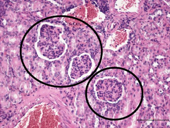
TIP1 is Effective Against SLE in a Mouse Model
(Enzo Life Sciences, Inc., Farmingdale, NY, USA) accord- lymph node tissue proteins were separated by electro-
ing to the manufacturer's instructions. The synthesized phoresis and transferred to a polyvinylidene fluoride
cDNA was subjected to a real-time polymerase reaction membrane. Then, the membrane was blocked with 5%
using AMPIGENE qPCR Green Mix (Enzo Life Sciences, bovine serum albumin (MP Biomedicals, Irvine, CA,
Inc.). Real-time PCR was performed 35 times and in- USA). Further, the membrane was stained with β-actin
cluded initialization at 95°C for 5 minutes, denaturation antibody (A300-491A; Bethyl Laboratories, Montgomery,
at 95°C for 30 seconds, annealing at 60°C for 1 minute, TX, USA), TLR7 antibody (#2633; Cell Signaling
and extension at 72°C for 30 seconds. mRNA expression Technology, Inc., Danvers, MA, USA), TLR9 antibody
levels of TLR7, TLR9, and downstream signaling proteins (LS-B688; LS Bio, Seattle, WA, USA), MyD88 antibody
were measured. (LS-C295471, LS Bio), IRAK4 antibody (LS-C334563, LS
Bio), TRAF6 antibody (LS-C331172, LS Bio), and IRF7
Western blotting antibody (LS-B577, LS Bio). Western blot bands were an-
For Western blotting, the total protein was isolated us- alyzed using Image J (Bethesda, MD, USA) software bun-
ing RIPA Buffer (Intron Biotechnology, Seoul, Korea), dled with 64-bit Java 1.8.0_112. The expression level of a
and each tissue was quantified according to the manu- specific protein was compared with the β-actin value and
facturer's protocol using the Pierce BCA Protein Assay Kit then statistically analyzed.
(Thermo Fisher Scientific, Rockford, IL, USA). After eu-
thanizing the mice, the extracted spleen, kidney, and
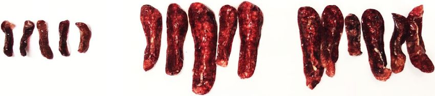
Figure 1. Effect of treatment with Toll-like receptor inhibitory peptide 1 (TIP1) in an SLE mouse model. (A) Whole body images of
MRL/lpr mice and age- and sex-matched controls (MRL/mpj) are shown. These images show the (B) change in the weight of the mice
during the 4-week injection period and (C) effect of TIP1 treatment on spleen hypertrophy in MRL/lpr mice. SLE: systemic lupus
erythematosus. *p<0.05.
www.jrd.or.kr 135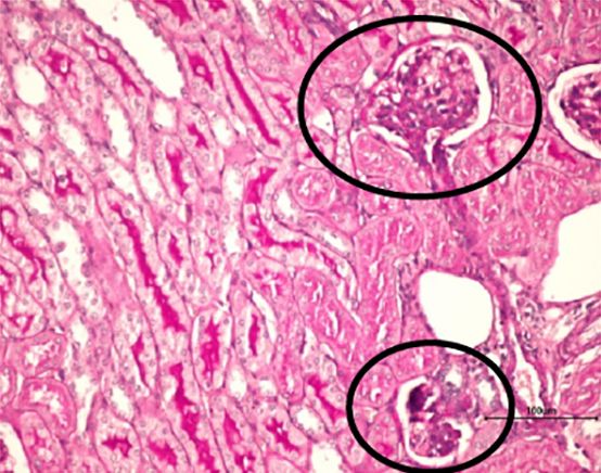
Wook-Young Baek et al.
Enzyme-linked immunosorbent assay (ELISA) enlargement (Figure 1A). Because it is known that body
For ELISA, Mouse Albumin ELISA Kit (Alpco weight may increase owing to organomegaly when symp-
Diagnostics, Salem, NH, USA), ANA ELISA Kit toms of SLE appear [4], we investigated the changes in
(MyBioSource, San Diego, CA, USA), anti-dsDNA ELISA the body weight of mice following treatment with TIP1.
Kit (Mybiosource), and Verikine-HS Mouse IFN-α All mice were weighed once a week, starting from imme-
ELISA Kit (PBL Assay Science, Piscataway, NJ, USA) were diately before injection to just before euthanasia. The
used according to the manufacturer’s instructions. average body weight of mice in the control treatment
Albumin was identified using mouse urine, and mouse group showed a tendency to increase continuously, but
serum was used for tests with ANA, anti-dsDNA anti- the average body weight of mice in the TIP1 treatment
body, and IFN-α. group tended to stabilize (Figure 1B). The difference in
the average weight of the spleen between mice in the two
Statistical analysis groups was not statistically significant, but the spleen
To confirm the significance of the difference in protein weight decreased slightly in the mice in the TIP1 treat-
expression levels between the control treatment and TIP1 ment group. However, the length of the spleen did not
treatment groups, the results of the tests were verified by change in the mice in either group (Figure 1C). The aver-
the Mann–Whitney U-test using the IBM Statistical age kidney weight of the mice in the TIP1 treatment
Package for the Social Sciences (SPSS) Statistics 25.0 group reduced to a small extent when compared to that of
(IBM Corporation, Armonk, NY, USA) program. When the mice in the control treatment group, but the differ-
the p-value was less than 0.05, the difference was consid- ence was not statistically significant (data not shown).
ered statistically significant. Further, the lymph nodes of mice in the control treatment
group were enlarged, but the enlargement was reduced
RESULTS marginally in mice in the TIP1 treatment group.
Body weight change and organomegaly due to Kidney tissue and inflammation after TIP1 treatment
TIP1 treatment in mice SLE usually invades the kidney tissue and causes in-
In this study, we analyzed the phenotype of the entire flammation, resulting in lupus nephritis. Lupus nephritis
mouse body for the first time. The mice in the control induces inflammation of blood vessels that filter waste
treatment group showed lymph node enlargement, which products from the kidneys, causing mesangial cells to
is the main symptom of SLE. However, TIP1 treatment of proliferate. Further, the basement membrane cells out-
MRL/lpr mice resulted in reduction of the lymph node side the glomerular blood vessels invade, and plasma pro-
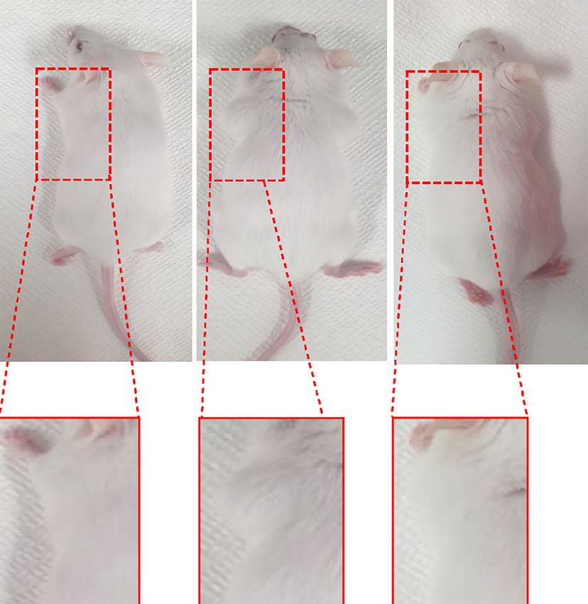
Figure 2. Toll-like receptor inhibitory peptide 1 (TIP1) treatment improved inflammation of the kidneys in lupus nephritis in
MRL/lpr mice. (A) Urine albumin analysis. The results of the TIP1 treatment group and control treatment group were compared us-
ing the Mann–Whitney U-test. (B) H&E staining (×200) and (C) PAS staining (×200) in the kidney tissues of the MRL/mpj normal
control group, MRL/lpr control treatment group, and MRL/lpr TIP1 treatment group. Representative micrographs obtained from
H&E and PAS staining of kidneys show differences in the glomeruli and surrounding inflammatory cells in mice of each group. The
shape of the glomerulus in the control treatment group was destroyed, but that was maintained in the TIP1 treatment group (circle).
136 J Rheum Dis Vol. 28, No. 3, July, 2021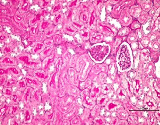
TIP1 is Effective Against SLE in a Mouse Model

teins are excreted in urine [19]. The urinary concen-            anti-dsDNA antibody levels (Figure 3B) between the
tration of albumin, known as an indicator of renal in-           mice in the two groups. However, serum IFN-α levels
flammation, was decreased in the mice in the TIP1 treat-         were significantly (p<0.05) lower in the mice in the TIP1
ment group when compared to the mice in the control              treatment group than in the mice in the control treatment
treatment group (Figure 2A). In this study, the kidneys of       group (Figure 3C).
mice were analyzed by H&E staining and PAS staining,
and the proliferation of vascular mesentery cells, infiltra-     TLR7 and TLR9 downstream signaling pathways in
tion of surrounding inflammatory cells, and the size and         major organs
shape of glomeruli were observed. The shape of the glo-           It is known that the expression of TLR7 and TLR9 in-
merulus of the mice in the control treatment group was           creases inflammatory cytokine levels and is thus related
destroyed, and the glomerulus was infiltrated by in-             to the onset and progression of SLE [14-16]. Therefore, in
flammatory cells, resulting in an irregular shape. However,      this study, the protein expression levels of MyD88,
in the mice in the TIP1 treatment group, the overall struc-      IRAK4, TRAF6, and IRF7, which are downstream mole-
ture and shape of the glomerulus was constantly main-            cules in the signaling pathways of TLR7 and TLR9, were
tained (black circles, Figure 2B and 2C). In addition, the       analyzed. The expression of TLR7, MyD88, and IRAK4 in
histopathological nephritis index also decreased in the          the spleen and MyD88, IRAK4, and IRF7 in the kidneys
mice of the TIP1 treatment group (Supplementary Figure           was significantly decreased in the mice in the TIP1 treat-
1). Therefore, intravenous treatment with TIP1 can im-           ment group, compared with those in the mice in the con-
prove inflammation in the kidney of lupus-prone mice.            trol treatment group (Figure 4A and 4B). Analysis of the
                                                                 mRNA of TLR7 and TLR9 in major tissues showed no sig-
Autoantibodies and inflammatory cytokines after                  nificant difference between the TIP1 treatment and con-
TIP1 treatment                                                   trol treatment in the spleen and kidney (Figure 5A and
  ANAs, anti-dsDNA antibodies, and serum IFN-α, which            5B), but the expression of MyD88, IRAK4, TRAF6, IRF7,
are indicators of the presence of lupus in mouse serum,          and IFN-α was significantly reduced in the lymph nodes
were measured by ELISA. ANAs tended to decrease in the           of the mice in the TIP1 treatment group compared with
mice in the TIP1 treatment group when compared with              that in the mice in the control treatment group (Figure
the mice in the control treatment group, but the differ-         5C). From these results, it was concluded that the ex-
ence was not statistically significant (Figure 3A). In addi-     pression of TLR7 and several downstream signaling mol-
tion, there was no statistically significant difference in the   ecules was significantly decreased in the spleen, kidney,

                                                                  Figure 3. Serological and inflammatory markers after treat-
                                                                  ment of MRL/lpr mice with Toll-like receptor inhibitory pep-
                                                                  tide 1 (TIP1). (A) Antinuclear antibody (ANA), (B) anti-ds
                                                                  DNA antibody, and (C) interferon-alpha (IFN-α). The results
                                                                  of the TIP1 treatment group and control treatment group
                                                                  were compared using the Mann–Whitney U-test. *p<0.05.

www.jrd.or.kr                                                                                                            137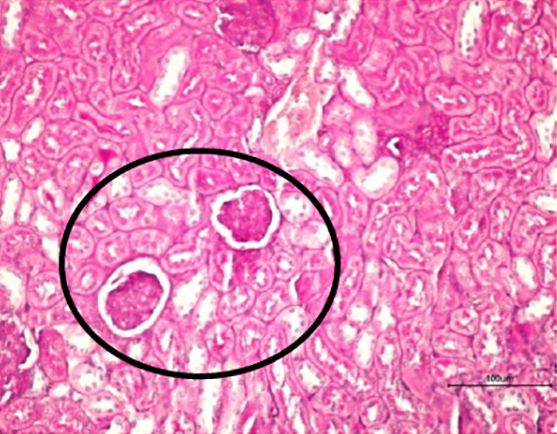
Wook-Young Baek et al.

Figure 4. Toll-like receptor (TLR) inhibitory peptide (TIP) 1 blocks the TLR 7/9 downstream signaling pathway in major organs in
mice. Western blot analysis of the expression levels the TLR 7/9 signaling pathway molecules in (A) spleen and (B) kidney of
MRL/lpr mice. The bar graph shows the band intensity presented as the average value of the target protein to β-actin ratio (*p<
0.05, **p<0.01).

and lymph nodes of mice owing to TIP1 treatment.                   well-known autoimmune diseases, is known for its ad-
                                                                   verse effects on many different organs and tissues [20]. In
                    DISCUSSION                                     addition, SLE is characterized by the production of auto-
                                                                   antibodies against nuclear antigens, which are deposited
  The incidence of autoimmune diseases and thus the de-            in tissues to form immune complexes, causing in-
mand for understanding immune tolerance and activa-                flammation [21,22].
tion is increasing globally. SLE, one of the most                    Several studies have consistently been conducted to es-

138                                                                                      J Rheum Dis Vol. 28, No. 3, July, 2021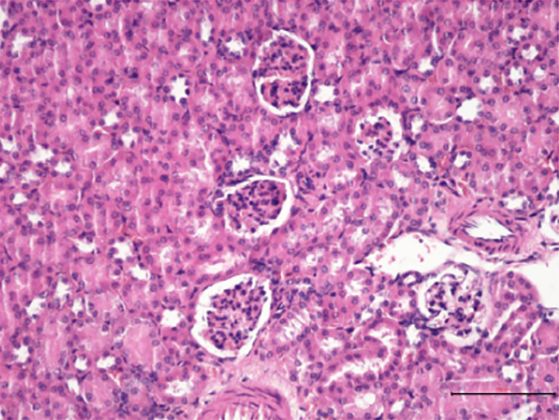
TIP1 is Effective Against SLE in a Mouse Model

Figure 5. The mRNA expression level of proteins of the Toll-like receptor (TLR) 7/9 signaling pathway were analyzed using re-
al-time polymerase chain reaction analysis in mouse (A) spleen, (B) kidney, and (C) lymph node. Using the Mann–Whitney U-test
in the analysis, p-values less than 0.05 or 0.01 were considered statistically significant (*p<0.05, **p<0.01).

www.jrd.or.kr                                                                                                           139
Wook-Young Baek et al.

tablish a suitable treatment for SLE; however, there are     duced symptoms. As a result of TIP1 treatment, an-
considerable limitations to subjecting any of these treat-   ti-dsDNA levels did not decrease, whereas urinary levels
ments to clinical trials, although their effects were suc-   of albumin decreased, although not significantly, in com-
cessfully demonstrated in animal testing. Nevertheless,      parison with the mice in the control treatment group.
several researchers are attempting to develop a more ef-       Several studies on murine lupus models have suggested
fective and safer treatment for SLE.                         the role of TLR signaling in the pathogenesis of lupus
  To date, many in vitro studies have provided evidence      nephritis [34]. Excessive production of TLR7 and TLR9 is
supporting the role of TLRs in the pathogenesis of SLE.      commonly observed with the progression of lupus neph-
TLRs are known to play an important role in the early de-    ritis [35]. Recently reported findings have shown a corre-
tection of pathogen-associated molecular patterns and        lation between TLR7 and TLR9 overexpression and clin-
the subsequent activation of adaptive immune responses.      ical pathological indicators, supporting the role of media-
Particularly, the recognition of nucleic acid motifs by      tors in the pathogenesis of lupus nephritis [29]. Similarly,
TLRs is reported to induce disease progression after acti-   in our study, the levels of TLR7, TLR9, and downstream
vation of antinuclear B cells and formation of immune        signaling proteins MyD88, IRAK4, TRAF6, and IRF7
complexes [23]. Many studies have reported the associa-      were reduced in TIP1-treated mice. These results indicate
tion of SLE with TLR7 and TLR9 [24-26]. Further, it was      that when TIP1 binds to the BB loop region of the TIR do-
identified that the activation of TLRs leads to the pro-     main, it blocks the TLR downstream signaling cascade
duction of inflammatory cytokines [14-16]. Therefore,        through the MyD88 or TRIF adapter. Additionally, H&E
TLRs can play an important role the pathogenesis of SLE      and PAS staining showed that the glomeruli of the mice in
[27]. Based on several prior studies, we decided that        the TIP1 treatment group retained their shape, and the
TLRs would be the most suitable target for the treatment     proliferation of cells was limited, which explains the im-
of SLE. SLE results in inflammation of several organs and    provement in kidney inflammation.
tissues and eventually causes damage. Thus, we inves-
tigated whether inflammatory cytokines decreased and                            CONCLUSION
SLE symptoms could be relieved when TIP1 is applied to
inhibit TLR signaling in a lupus-prone animal model           In this study, a representative SLE animal model
(MRL/lpr).                                                   (MRL/lpr) was used to determine whether SLE symp-
  MRL/lpr mice are representative animal models that         toms and signs reduced with TIP1 treatment. Treatment
present many organ pathologies and features of SLE           with TIP1 ameliorated lupus nephritis by reducing the
[28,29]. According to existing studies using MRL/lpr         concentration of urine albumin, retaining glomerular
models, the first SLE symptoms occurred at 14 weeks of       shape, and controlling the proliferation of mesenteric
age, and skin lesions and enlargement of the spleen and      cells. In addition, TLR7, TLR9, and downstream signaling
lymph nodes were observed [29,30]. In our study, how-        proteins decreased significantly in the lymph node,
ever, skin lesions were observed only on the faces of the    spleen, and kidney of the mice treated with TIP1. Further,
mice in each group, with no significant difference be-       IFN-α, an inflammatory cytokine, was also significantly
tween the lesions of the mice in the TIP1 treatment and      decreased. Therefore, TIP1 reduced the signs and symp-
control treatment groups. In addition, the weight of the     toms of SLE by inhibiting TLR7, TLR9, and downstream
spleen tended to decrease to a small extent with TIP1        signaling proteins. TIP1 could be a potential therapeutic
treatment, but the length of the spleen or kidney weight     agent for treating SLE.
did not decrease significantly.
  In lupus nephritis, glomeruli are damaged by kidney in-                 ACKNOWLEDGMENTS
flammation, and anti-dsDNA associated with the for-
mation of immune complexes is detected [31-33]. In ad-        This research was supported by a grant from the Korea
dition, exacerbating lupus nephritis results in higher       Health Technology R&D Project through the Korea
urine albumin levels. Therefore, anti-dsDNA antibodies       Health Industry Development Institute (KHIDI), funded
and urinary albumin can be indicators of lupus nephritis.    by the Ministry of Health & Welfare, Republic of Korea
In our study, experiments were conducted using various       (grant number: HI16C0992).
indicators of SLE to ascertain whether TIP1 treatment re-

140                                                                                J Rheum Dis Vol. 28, No. 3, July, 2021

TIP1 is Effective Against SLE in a Mouse Model

                                                                         plexes activate B cells by dual engagement of IgM and
                                                                         Toll-like receptors. Nature 2002;416:603-7.
             CONFLICT OF INTEREST                                  11.   Klonowska-Szymczyk A, Wolska A, Robak T, Cebula-Obrzut
                                                                         B, Smolewski P, Robak E. Expression of toll-like receptors
 No potential conflict of interest relevant to this article              3, 7, and 9 in peripheral blood mononuclear cells from pa-
was reported.                                                            tients with systemic lupus erythematosus. Mediators Inflamm
                                                                         2014;2014:381418.
                                                                   12.   Celhar T, Magalhães R, Fairhurst AM. TLR7 and TLR9 in
          AUTHOR CONTRIBUTIONS                                           SLE: when sensing self goes wrong. Immunol Res 2012;53:
                                                                         58-77.
 Conception and design of study: C.-H.S., S.C., W.-Y.B.            13.   Christensen SR, Kashgarian M, Alexopoulou L, Flavell RA,
                                                                         Akira S, Shlomchik MJ. Toll-like receptor 9 controls an-
Acquisition of data: W.-Y.B., S.-M.L., S.-W.L., I.-O.S.                  ti-DNA autoantibody production in murine lupus. J Exp
Analysis and/or interpretation of data: C.-H.S., W.-Y.B.,                Med 2005;202:321-31.
S.-M.L. Drafting the manuscript: W.-Y.B., S.-M.L.                  14.   Chauhan SK, Singh VV, Rai R, Rai M, Rai G. Distinct auto-
                                                                         antibody profiles in systemic lupus erythematosus patients
Revising the manuscript critically for important in-
                                                                         are selectively associated with TLR7 and TLR9 upregulation.
tellectual content: C.-H.S. All authors have read and ap-                J Clin Immunol 2013;33:954-64.
proved the final version of the manuscript.                        15.   Akira S, Uematsu S, Takeuchi O. Pathogen recognition and
                                                                         innate immunity. Cell 2006;124:783-801.
                                                                   16.   Guiducci C, Gong M, Xu Z, Gill M, Chaussabel D, Meeker T,
             SUPPLEMENTARY DATA                                          et al. TLR recognition of self nucleic acids hampers gluco-
                                                                         corticoid activity in lupus. Nature 2010;465:937-41.
 Supplementary data can be found with this article online          17.   Kwon HK, Patra MC, Shin HJ, Gui X, Achek A, Panneerselvam
                                                                         S, et al. A cell-penetrating peptide blocks Toll-like re-
at https://doi.org/10.4078/jrd.2021.28.3.133.
                                                                         ceptor-mediated downstream signaling and ameliorates au-
                                                                         toimmune and inflammatory diseases in mice. Exp Mol Med
                                                                         2019;51:1-19.
                      REFERENCES                                   18.   Watson ML, Rao JK, Gilkeson GS, Ruiz P, Eicher EM,
                                                                         Pisetsky DS, et al. Genetic analysis of MRL-lpr mice: rela-
 1. Tsokos GC. Systemic lupus erythematosus. N Engl J Med                tionship of the Fas apoptosis gene to disease manifestations
    2011;365:2110-21.                                                    and renal disease-modifying loci. J Exp Med 1992;176:
 2. Klippel JH. Systemic lupus erythematosus: demographics,              1645-56.
    prognosis, and outcome. J Rheumatol Suppl 1997;48:67-71.       19.   Chen D, Hu W. Lupus podocytopathy: a distinct entity of lu-
 3. Petri M. Long-term outcomes in lupus. Am J Manag Care                pus nephritis. J Nephrol 2018;31:629-34.
    2001;7(16 Suppl):S480-5.                                       20.   Celhar T, Lu HK, Benso L, Rakhilina L, Lee HY, Tripathi S,
 4. Toblli JE, Trigo M, Mendoza G, Fellner JP, Genaro O,                 et al. TLR7 protein expression in mild and severe lu-
    Houssay R, et al. [Hematologic manifestations in systemic            pus-prone models is regulated in a leukocyte, genetic, and
    lupus erythematosus. Experience collected from 150 cases].           IRAK4 dependent manner. Front Immunol 2019;10:1546.
    Rev Clin Esp 1983;169:257-61. Spanish.                         21.   Suurmond J, Diamond B. Autoantibodies in systemic auto-
 5. Kamen DL. Environmental influences on systemic lupus er-             immune diseases: specificity and pathogenicity. J Clin
    ythematosus expression. Rheum Dis Clin North Am 2014;                Invest 2015;125:2194-202.
    40:401-12, vii.                                                22.   Moulton VR, Suarez-Fueyo A, Meidan E, Li H, Mizui M,
 6. Kalok A, Abdul Cader R, Indirayani I, Abdul Karim AK,                Tsokos GC. Pathogenesis of human systemic lupus eryth-
    Shah SA, Mohamed Ismail NA, et al. Pregnancy outcomes in             ematosus: a cellular perspective. Trends Mol Med 2017;23:
    systemic lupus erythematosus (SLE) women. Horm Mol                   615-35.
    Biol Clin Investig 2019 Sep 25 [Epub]. DOI:10.1515/            23.   Marshak-Rothstein A. Toll-like receptors in systemic auto-
    hmbci-2019-0007.                                                     immune disease. Nat Rev Immunol 2006;6:823-35.
 7. Wincup C, McDonnell TCR, Rahman A. Menorrhagia: an             24.   Nickerson KM, Christensen SR, Shupe J, Kashgarian M,
    underappreciated problem in pre-menopausal women with                Kim D, Elkon K, et al. TLR9 regulates TLR7- and MyD88-
    systemic lupus erythematosus. Lupus 2019;28:916-7.                   dependent autoantibody production and disease in a murine
 8. Tsokos GC, Lo MS, Costa Reis P, Sullivan KE. New insights            model of lupus. J Immunol 2010;184:1840-8.
    into the immunopathogenesis of systemic lupus erythematosus.   25.   Celhar T, Fairhurst AM. Toll-like receptors in systemic lu-
    Nat Rev Rheumatol 2016;12:716-30.                                    pus erythematosus: potential for personalized treatment.
 9. Leonard D, Eloranta ML, Hagberg N, Berggren O, Tandre K,             Front Pharmacol 2014;5:265.
    Alm G, et al. Activated T cells enhance interferon-α pro-      26.   Christensen SR, Shupe J, Nickerson K, Kashgarian M,
    duction by plasmacytoid dendritic cells stimulated with              Flavell RA, Shlomchik MJ. Toll-like receptor 7 and TLR9
    RNA-containing immune complexes. Ann Rheum Dis                       dictate autoantibody specificity and have opposing in-
    2016;75:1728-34.                                                     flammatory and regulatory roles in a murine model of lupus.
10. Leadbetter EA, Rifkin IR, Hohlbaum AM, Beaudette BC,                 Immunity 2006;25:417-28.
    Shlomchik MJ, Marshak-Rothstein A. Chromatin-IgG com-          27.   Wu YW, Tang W, Zuo JP. Toll-like receptors: potential tar-

www.jrd.or.kr                                                                                                                  141
Wook-Young Baek et al.
gets for lupus treatment. Acta Pharmacol Sin 2015;36:1395-407. unknown. Clin Dev Immunol 2012;2012:139365.
28. Tilstra JS, Avery L, Menk AV, Gordon RA, Smita S, Kane LP, 32. Rekvig OP. The dsDNA, anti-dsDNA antibody, and lupus
et al. Kidney-infiltrating T cells in murine lupus nephritis nephritis: what we agree on, what must be done, and what
are metabolically and functionally exhausted. J Clin Invest the best strategy forward could be. Front Immunol 2019;10:
2018;128:4884-97. 1104.
29. Liu J, Karypis G, Hippen KL, Vegoe AL, Ruiz P, Gilkeson GS, 33. Goilav B, Putterman C. The role of anti-DNA antibodies in
et al. Genomic view of systemic autoimmunity in MRLlpr the development of lupus nephritis: a complementary, or al-
mice. Genes Immun 2006;7:156-68. ternative, viewpoint? Semin Nephrol 2015;35:439-43.
30. Kim YY, Park KT, Jang SY, Lee KH, Byun JY, Suh KH, et al. 34. Devarapu SK, Anders HJ. Toll-like receptors in lupus
HM71224, a selective Bruton's tyrosine kinase inhibitor, at- nephritis. J Biomed Sci 2018;25:35.
tenuates the development of murine lupus. Arthritis Res 35. Conti F, Spinelli FR, Truglia S, Miranda F, Alessandri C,
Ther 2017;19:211. Ceccarelli F, et al. Kidney expression of Toll like receptors in
31. Yung S, Chan TM. Autoantibodies and resident renal cells in lupus nephritis: quantification and clinicopathological
the pathogenesis of lupus nephritis: getting to know the correlations. Mediators Inflamm 2016;2016:7697592.
142 J Rheum Dis Vol. 28, No. 3, July, 2021You can also read