Transient forebrain ischemia under hyperthermic condition in the gerbil hippocampus by increasing NMDAR1 expression accelerates memory impairment ...
←
→
Page content transcription
If your browser does not render page correctly, please read the page content below
MOLECULAR MEDICINE REPORTS 23: 256, 2021

        Transient forebrain ischemia under hyperthermic condition
            accelerates memory impairment and neuronal death
       in the gerbil hippocampus by increasing NMDAR1 expression
                       BORA KIM1*, JI HYEON AHN1,2*, DAE WON KIM3, TAE‑KYEONG LEE4,
                 YOON SUNG KIM5,6, MYOUNG CHEOL SHIN5, JUN HWI CHO5, YOUNG‑MYEONG KIM7,
                       JOON HA PARK8, IL JUN KANG9, JAE‑CHUL LEE1 and MOO‑HO WON1

          1
         Department of Neurobiology, School of Medicine, Kangwon National University, Chuncheon, Gangwon 24341;
          2
         Department of Physical Therapy, College of Health Science, Youngsan University, Yangsan, Gyeongnam 50510;
     3
      Department of Biochemistry and Molecular Biology, and Research Institute of Oral Sciences, College of Dentistry,
Gangnung‑Wonju National University, Gangneung, Gangwon 25457; 4Department of Biomedical Science, Research Institute of
   Bioscience and Biotechnology, Hallym University, Chuncheon, Gangwon 24252; 5Department of Emergency Medicine,
  Kangwon National University Hospital, School of Medicine, Kangwon National University, Chuncheon, Gangwon 24289;
         6
           Department of Emergency Medicine, Samcheok Medical Center, Samcheok, Kangwon 25920; 7Department of
   Molecular and Cellular Biochemistry, School of Medicine, Kangwon National University, Chuncheon, Gangwon 24341;
           8
             Department of Anatomy, College of Korean Medicine, Dongguk University, Gyeongju, Gyeongbuk 38066;
       9
         Department of Food Science and Nutrition, Hallym University, Chuncheon, Gangwon 24252, Republic of Korea

                                     Received August 11, 2020; Accepted November 30, 2020

                                                    DOI: 10.3892/mmr.2021.11895

Abstract. Altered expression levels of N‑methyl‑D‑aspartate          under hyperthermia compared with that under normothermia.
receptor (NMDAR), a ligand‑gated ion channel, have a                 NMDAR1 immunoreactivity was not observed in the somata
harmful effect on cellular survival. Hyperthermia is a proven        of pyramidal neurons of sham gerbils with normothermia.
risk factor of transient forebrain ischemia (tFI) and can cause      However, its immunoreactivity was strong in the somata and
extensive and severe brain damage associated with mortality.         processes at 12 h post‑tFI. Thereafter, NMDAR1 immuno‑
The objective of the present study was to investigate whether        reactivity decreased with time after tFI. On the other hand,
hyperthermic preconditioning affected NMDAR1 immunore‑               NMDAR1 immunoreactivity under hyperthermia was signifi‑
activity associated with deterioration of neuronal function in       cantly increased in the somata and processes at 6 h post‑tFI.
the gerbil hippocampal CA1 region following tFI via histolog‑        The change pattern of NMDAR1 immunoreactivity under
ical and western blot analyses. Hyperthermic preconditioning         hyperthermia was different from that under normothermia.
was performed for 1 h before tFI, which was developed by             Overall, accelerated tFI‑induced neuronal death under hyper‑
ligating common carotid arteries for 5 min. tFI‑induced cogni‑       thermia may be closely associated with altered NMDAR1
tive impairment under hyperthermia was worse compared                expression compared with that under normothermia.
with that under normothermia. Loss (death) of pyramidal
neurons in the CA1 region occurred fast and was more severe          Introduction

                                                                     Transient forebrain ischemia (tFI) can be induced by tempo‑
                                                                     rary deprivation of arterial blood flow to the forebrain through
                                                                     occlusion of bilateral common carotid arteries for five minutes.
Correspondence to: Professor Moo‑Ho Won or Dr Jae‑Chul Lee,          A tFI can cause death (loss) of pyramidal cells (neurons) only
Department of Neurobiology, School of Medicine, Kangwon National     in the Conus Ammonis 1 region (CA1) of the hippocampus
University, 1 Kangwondaehak‑gil, Chuncheon, Gangwon 24341,
                                                                     of the gerbil (1). Pyramidal cells of the CA1 are called CA1
Republic of Korea
                                                                     pyramidal cells. They will die at 4‑5 days after tFI for 5 min.
E‑mail: mhwon@kangwon.ac.kr
E‑mail: anajclee@kangwon.ac.kr                                       This phenomenon of CA1 pyramidal cell death is called
                                                                     delayed neuronal death (DND) (1). Afterward, DND has been
*
    Contributed equally                                              reported in the hippocampal CA1 of rodents including rats and
                                                                     mice (2,3).
Key words: cognition, hyperthermic preconditioning, ligand‑gated         Possible mechanisms of DND after tFI have been reported.
ion channel, neuronal death, pyramidal neurons, transient ischemia   Among them, transient increase in excitatory amino acid
                                                                     transmitter efflux is probably associated with the degrees of
                                                                     neuronal damage or death in ischemic brains (4). In detail,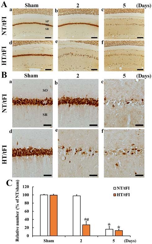
2                      KIM et al: HYPERTHERMIA-MEDIATED NMDAR1 CHANGE IN ISCHEMIC HIPPOCAMPUS

during ischemia, high extracellular glutamate level can acti‑       a heating pad until rectal temperature reached 39.5±0.2˚C for
vate ionotropic glutamate receptors (i.e., N‑methyl‑D‑aspartate     1 h before tFI induction. For normothermia, rectal temperature
receptor (NMDAR) that are ligand‑gated ion channels leading         was regulated at 37±0.2˚C. tFI was induced as follows. Both
to channel opening and overflow of calcium ions into neurons.       common carotid arteries were isolated from the carotid sheath
Finally, neuronal damage/death occurs (5,6). Many researchers       and occluded using aneurysm clips for 5 min. Thereafter, the
have reported that brain diseases and dysfunctions are related      gerbils were kept in thermal incubator (temperature, 23˚C;
to abnormal NMDAR expressions (7,8). For example, it has            humidity, 60%) to recover to normothermic level. The gerbils
been reported that pathological activation of NMDAR1 is a           of NT/sham and HT/sham groups received the same surgical
major cause of neuronal death following brain ischemia (9‑12).      process of tFI without bilateral carotid artery occlusion.
    Brain or body temperature has been thought to be one of
major factors affecting neuronal survival or death after brain      Passive avoidance test (PAT). Short term memory was
ischemic injury (13‑15). Preclinical studies have provided          evaluated by PAT used by previous researchers (24,25) with
evidence that hyperthermia has harmful effects on animal            modifications. In short, we used a Gemini Avoidance System
models of transient global cerebral ischemia (16,17) and tran‑      from GEM 392 (San Diego Instruments) consisting of two
sient focal cerebral ischemia models (18,19). Clinical reports      (light and dark) compartments which communicates through
have confirmed that hyperthermia can accelerate infarction          a sliding door was used. The gerbils received training of
and worsen outcomes of ischemic stroke patients (20,21).            PAT for three days before tFI. Namely, the gerbils were
Recently, we have reported that hyperthermia can accelerate         allowed to explore light and dark compartments for 1 min
and worsen neuronal damage or death in the gerbil hippo‑            while the sliding door was opened. And, when the gerbils
campus after tFI (13,22,23).                                        enter the dark compartment, the door was closed, and they
    As described above, transient ischemic insults under            received an electric foot‑shock (0.5 mA for 5 sec) from a
hyperthermic condition can lead to extensive brain damage.          steel grid in the floor. Two and five days after tFI, PAT was
However, molecular mechanisms involved in deleterious               performed. The PAT was elevated as latency time. Namely,
effects of hyperthermic condition on results from tFI have          the gerbils were put in the light compartment, while the door
not yet been fully clarified. Thus, the objective of this study     was opened. The latency time was that the gerbils in the
was to investigate changes of NMDAR1 expressions in the             light compartment went into the dark compartment after
hippocampus and relationships between NMDAR1 changes                receiving the electric foot‑shock. Maximum latency time
and neuronal damage or death after tFI induction under              was 180 sec, which was to stay in the light compartment
hypothermic condition in gerbils.                                   with the an electric foot‑shock.

Materials and methods                                               Western blot analysis. The gerbils (n=7 at 6 h, 1 day, 2 days,
                                                                    and 5 days after tFI) in each group were deeply anesthe‑
Experimental animals, protocol and groups. Male                     tized by intraperitoneal injection of 60 mg/kg pentobarbital
Mongolian gerbils (age, 6 months; body weight, 64‑74 g)             sodium and sacrificed to analyze the level of NMDAR1
were used. They were obtained from the Experimental                 protein in the hippocampal CA1 region after tFI. As previ‑
Animal Center located in Kangwon National University                ously described (26), the hippocampi were removed, and
(Chuncheon, Kangwon, Korea). The protocol of this                   the CA1 regions were obtained. The tissues of the CA1
research was approved (approval number, KW‑200113‑1)                regions were homogenized in PBS (5 mM, pH 7.4), which
on January 13, 2020 by the Institutional Animal Care and            contained 0.2% Nonidet P‑40, 0.1 mM ethylene glycol bis
Use Committee (IACUC) located in Kangwon National                   (2‑aminoethyl Ether)‑N,N,N',N' tetraacetic acid (EGTA)
University. The gerbils used in this study were cared under         (pH 8.0), 10 mM ethylendiamine tetraacetic acid (EDTA)
constant temperature (~23˚C) and humidity (~55%) with               (pH 8.0), 15 mM sodium pyrophosphate, 150 mM NaCl,
a 12‑h light/dark cycle. The handling and caring animals            50 mM NaF, 2 mM sodium orthovanadate, 100 mM
conformed to the guidelines in the ‘Current international           β ‑glycerophosphate, 1 mM dithiothreitol (DTT) and 1 mM
laws and policies’ of the ‘NIH Guide for the Care and Use           phenylmethylsulfonyl fluoride (PMSF). The homogenates
of Laboratory Animals’ (The National Academies Press,               were separated by centrifugation, and the protein level was
8th edition, 2011). Number of the gerbils used for this study       determined in the supernatants with Micro BCA protein
were minimized, and the suffering caused by our procedures          assay kit (Pierce Chemical Co.). Aliquots containing total
used in this experiment was minimized.                              protein (20 µg) were boiled in loading buffer (150 mM Tris,
    Experimental animals (total number=120) were assigned           pH 6.8) containing 6% SDS, 0.3% bromophenol blue, 3 mM
to four groups: i) sham‑operated animals with normothermia          DTT, and 30% glycerol and loaded onto 12.5% polyacryl‑
(NT) (NT/sham group, n=12); ii) tFI‑operated animals with           amide gel. Subsequently, they were received electrophoresis,
NT (NT/tFI group, n=12); iii) sham‑operated animals with            and the gels were transferred to nitrocellulose transfer
hyperthermia (HT) (HT/sham group, n=48); and iv) tFI‑oper‑          membranes. To block non‑specific staining, the membranes
ated animals with HT (HT/tFI group, n=48).                          were incubated with 5% defatted milk at room temperature
                                                                    for 60 min. The membranes, thereafter, were immunore‑
Induction of tFI. As previously described (23), the gerbils to be   acted with rabbit anti‑NMDAR1 (diluted 1:1,000, Abcam),
used for tFI were anesthetized with mixture of 2.5% isoflurane      peroxidase‑conjugated goat anti‑rabbit IgG (diluted 1:250,
in 33% oxygen and 67% nitrous oxide via inhalation. Under           Sigma‑Aldrich Co.; Merck KGaA) and ECL kit (Pierce
anesthesia, hyperthermia was induced by heating them with           Chemical Co.).
MOLECULAR MEDICINE REPORTS 23: 256, 2021                                                                3

    The analysis of western blotting was done according to
a published method (26). In brief, the western bands were
scanned with computer scanner, and the quantification of
the analysis was done using a Scion Image software from
Scion Corp. The expression rate of NMDAR1 protein was
normalized through the corresponding expression rate of
β ‑actin.

Tissue processing for histology. Sections containing the
hippocampus were prepared as previously described (13). In
short, the gerbils in each group were deeply anesthetized by
                                                                     Figure 1. Passive avoidance test in the NT/sham, HT/sham, NT/tFI and HT/tFI
intraperitoneal injection of 60 mg/kg pentobarbital sodium           groups at 2 and 5 days after tFI. At 2 and 5 days after tFI, the latency time in
at 6 h, 1 day, 2 days, and 5 days after tFI. Under anesthesia, the   the HT/tFI groups was significantly shorter than that in the respective NT/tFI
brains of the gerbils were perfused with 4% paraformaldehyde         group. The data are presented as the mean ± SEM (n=7). *P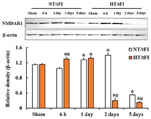
4                          KIM et al: HYPERTHERMIA-MEDIATED NMDAR1 CHANGE IN ISCHEMIC HIPPOCAMPUS

                                                                               Figure 3. (A) Low (x10; scale bar, 200 µm) and (B) high (x40; scale bar,
Figure 2. (A) Low (x10; scale bar, 200 µm) and (B) high (x40; scale bar,       50 µm) magnification of histofluorescence with F‑JB in the CA1 region of
50 µm) magnification of immunohistochemical staining for NeuN in the           the (a) NT/sham, (b and c) NT/tFI at 2 and 5 days after tFI, (d) HT/sham
CA1 region of the (a) NT/sham, (b and c) NT/tFI at 2 and 5 days after tFI,     and (e and f) HT/tFI groups at 2 and 5 days post‑tFI. In the NT/tFI group,
(d) HT/sham and (e and f) HT/tFI groups at 2 and 5 days after tFI. Five days   numerous F‑JB+ cells were detected in the SP 5 days after tFI. However, in
after tFI, NeuN+ neurons were rare in the SP. However, in the HT/tFI group,    the HT/tFI group, numerous F‑JB+ cells were observed from 2 days post‑tFI.
NeuN+ neurons in the SP were dramatically decreased from 2 days after tFI.     (C) Numbers of F‑JB+ cells in the CA1 region. The data are presented as the
(C) Numbers of NeuN+ neurons in the CA1 region. The data are presented         mean ± SEM (n=7). *P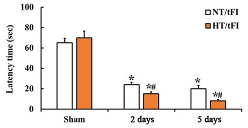
MOLECULAR MEDICINE REPORTS 23: 256, 2021 5
reduce short‑term memory function evaluated by PAT
compared to the control (NT/tFI group). This finding agrees
with previous studies showing that cognitive impairment is
accelerated after brain injury under hyperthermia (35,36).
It has been reported that death of pyramidal cells in
the gerbil hippocampal CA1 region following tFI under
normothermic condition occurs at 4‑5 days after tFI, and
this phenomenon is known as ‘delayed neuronal death’ (1).
However, in the HT/tFI group with memory deterioration,
death (loss) of pyramidal neurons in the CA1 region was
accelerated and deteriorated than that in the NT/tFI group.
In particular, neuronal loss in the HT/tFI group began
at 2 days post‑tFI, while neuronal loss in the NT/tFI group
was found at 5 days post‑tFI. Although the present study
did not confirm neuronal death at 6 h and 1day after tFI,
this finding is similar to our previously published results
Figure 4. Western blot analyses of NMDAR1 in the CA1 region of the showing that, under hyperthermic conditions before and
NT/sham, NT/tFI, HT/sham and HT/tFI groups at 6 h, 1 day, 2 days and during ischemia‑reperfusion (IR), neuronal death began
5 days after tFI. The change in NMDAR1 expression in the NT/tFI group at 1 day post‑tFI and was accelerated at 2‑3 days post‑tFI,
was different from that in the HT/tFI group. The graph indicates the rela‑ and that hyperthermia increased the extent and severity
tive optical density of the immunoblot bands. The data are presented as the
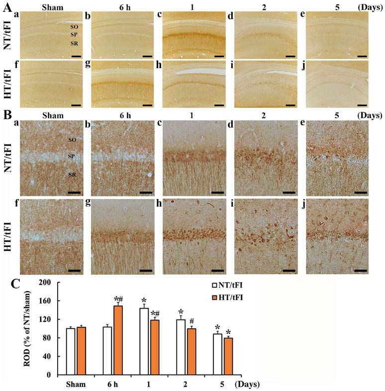
mean ± SEM (n=7). *P6 KIM et al: HYPERTHERMIA-MEDIATED NMDAR1 CHANGE IN ISCHEMIC HIPPOCAMPUS Figure 5. (A) Low (x10; scale bar, 200 µm) and (B) high (x40; scale bar, 50 µm) magnification of immunohistochemical staining for NMDAR1 in the CA1 region of the (a) NT/sham, (b‑e) NT/tFI at 6 h, 1 day, 2 days and 5 days after tFI, (f) HT/sham and (g‑j) HT/tFI groups at 6 h, 1 day, 2 days and 5 days after tFI. NMDAR1 immunoreactivity was hardly observed in the SP of the sham groups. In the NT/tFI group, strong NMDAR1 immunoreactivity was observed in the somata and processes at 1 and 2 days post‑tFI. In the HT/tFI group, NMDAR1 immunoreactivity was increased from 6 h post‑tFI, indicating that the change pattern of NMDAR1 immunoreactivity was different from that in the NT/tFI group. (C) ROD of NMDAR1 immunoreactivity in the CA1 region. The data are presented as the mean ± SEM (n=7). *P
MOLECULAR MEDICINE REPORTS 23: 256, 2021                                                        7

Institutional Animal Care and Use Committee of Kangwon                  18. Morikawa E, Ginsberg MD, Dietrich WD, Duncan RC,
                                                                            Kraydieh S, Globus MY and Busto R: The significance of brain
National University (Chuncheon, Republic of Korea).                         temperature in focal cerebral ischemia: Histopathological conse‑
                                                                            quences of middle cerebral artery occlusion in the rat. J Cereb
Patient consent for publication                                             Blood Flow Metab 12: 380‑389, 1992.
                                                                        19. de Jonge JC, Wallet J and van der Worp HB: Fever worsens
                                                                            outcomes in animal models of ischaemic stroke: A systematic
Not applicable.                                                             review and meta‑analysis. Eur Stroke J 4: 29‑38, 2019.
                                                                        20. Seo WK, Yu SW, Kim JH, Park KW and Koh SB: The impact
                                                                            of hyperthermia and infection on acute ischemic stroke
Competing interests                                                         patients in the intensive care unit. Neurocrit Care 9: 183‑188,
                                                                            2008.
The authors declare that they have no competing interests.              21. Wang Y, Lim LL, Levi C, Heller RF and Fisher J: Influence
                                                                            of admission body temperature on stroke mortality. Stroke 31:
                                                                            404‑409, 2000.
References                                                              22. Kim DW, Cho JH, Cho GS, Kim IH, Park JH, Ahn JH, Chen BH,
                                                                            Shin BN, Tae HJ, Hong S, et al: Hyperthermic preconditioning
 1. Kirino T: Delayed neuronal death in the gerbil hippocampus              severely accelerates neuronal damage in the gerbil ischemic
    following ischemia. Brain Res 239: 57‑69, 1982.                         hippocampal dentate gyrus via decreasing SODs expressions.
 2. Kirino T, Tamura A and Sano K: Delayed neuronal death in the            J Neurol Sci 358: 266‑275, 2015.
    rat hippocampus following transient forebrain ischemia. Acta        23. Lee JC, Cho JH, Lee TK, Kim IH, Won MH, Cho GS, Shin BN,
    Neuropathol 64: 139‑147, 1984.                                          Hwang IK, Park JH, Ahn JH, et al: Effect of hyperthermia on
 3. Chen J, Nagayama T, Jin K, Stetler RA, Zhu RL, Graham SH                calbindin‑D 28k immunoreactivity in the hippocampal forma‑
    and Simon RP: Induction of caspase‑3‑like protease may mediate          tion following transient global cerebral ischemia in gerbils.
    delayed neuronal death in the hippocampus after transient cere‑         Neural Regen Res 12: 1458‑1464, 2017.
    bral ischemia. J Neurosci 18: 4914‑4928, 1998.                      24. Chen BH, Park JH, Lee YL, Kang IJ, Kim DW, Hwang IK,
 4. Benveniste H, Drejer J, Schousboe A and Diemer NH: Elevation            Lee CH, Yan BC, Kim YM, Lee TK, et al: Melatonin improves
    of the extracellular concentrations of glutamate and aspartate in       vascular cognitive impairment induced by ischemic stroke by
    rat hippocampus during transient cerebral ischemia monitored by         remyelination via activation of ERK1/2 signaling and restoration
    intracerebral microdialysis. J Neurochem 43: 1369‑1374, 1984.           of glutamatergic synapses in the gerbil hippocampus. Biomed
 5. Arundine M and Tymianski M: Molecular mechanisms of                     Pharmacother 108: 687‑697, 2018.
    calcium‑dependent neurodegeneration in excitotoxicity. Cell         25. Lee JC, Park JH, Ahn JH, Kim IH, Cho JH, Choi JH, Yoo KY,
    Calcium 34: 325‑337, 2003.                                              Lee CH, Hwang IK, Cho JH, et al: New GABAergic neurogenesis
 6. Kristian T and Siesjo BK: Calcium in ischemic cell death.               in the hippocampal CA1 region of a gerbil model of long‑term
    Stroke 29: 705‑718, 1998.                                               survival after transient cerebral ischemic injury. Brain Pathol 26:
 7. Young AB, Greenamyre JT, Hollingsworth Z, Albin R,                      581‑592, 2016.
    D'Amato C, Shoulson I and Penney JB: NMDA receptor losses           26. Lee JC, Kim IH, Park JH, Ahn JH, Cho JH, Cho GS, Tae HJ,
    in putamen from patients with Huntington's disease. Science 241:        Chen BH, Yan BC, Yoo KY, et al: Ischemic preconditioning
    981‑983, 1988.                                                          protects hippocampal pyramidal neurons from transient isch‑
 8. Stanika RI, Winters CA, Pivovarova NB and Andrews SB:                   emic injury via the attenuation of oxidative damage through
    Differential NMDA receptor‑dependent calcium loading and                upregulating heme oxygenase‑1. Free Radic Biol Med 79: 78‑90,
    mitochondrial dysfunction in CA1 vs. CA3 hippocampal                    2015.
    neurons. Neurobiol Dis 37: 403‑411, 2010.                           27. Radtke‑Schuller S, Schuller G, Angenstein F, Grosser OS,
 9. Gao S, Yu Y, Ma ZY, Sun H, Zhang YL, Wang XT, Wang C,                   Goldschmidt J and Budinger E: Brain atlas of the Mongolian
    Fan WM, Zheng QY and Ma CL: NMDAR‑mediated hippo‑                       gerbil (Meriones unguiculatus) in CT/MRI‑aided stereotaxic
    campal neuronal death is exacerbated by activities of ASIC1a.           coordinates. Brain Struct Funct 221 (Suppl 1): S1‑S272, 2016.
    Neurotox Res 28: 122‑137, 2015.                                     28. Schmued LC and Hopkins KJ: Fluoro‑Jade B: A high affinity
10. Seo JY, Yan BC, Park JH, Ahn JH, Kim IH, Lee JC, Kwon YG,               fluorescent marker for the localization of neuronal degeneration.
    Kim YM, Cho JH and Won MH: Comparison of the immuno‑                    Brain Res 874: 123‑130, 2000.
    reactivities of NMDA receptors between the young and adult          29. Sugawara T, Lewen A, Noshita N, Gasche Y and Chan PH:
    hippocampal CA1 region induced by experimentally transient              Effects of global ischemia duration on neuronal, astroglial,
    cerebral ischemia. J Neurol Sci 325: 108‑114, 2013.                     oligodendroglial, and microglial reactions in the vulnerable
11. Benquet P, Gee CE and Gerber U: Transient brain ischemia:               hippocampal CA1 subregion in rats. J Neurotrauma 19: 85‑98,
    NMDA receptor modulation and delayed neuronal death. Med                2002.
    Sci (Paris) 24: 185‑190, 2008 (In French).                          30. Corbett D and Thornhill J: Temperature modulation (hypo‑
12. Yanli L, Xizhou Z, Yan W, Bo Z, Yunhong Z, Zicheng L,                   thermic and hyperthermic conditions) and its influence on
    Lingling Y, Lingling Y, Zhangao C, Min Z and Zhi H: Clonidine           histological and behavioral outcomes following cerebral isch‑
    preconditioning alleviated focal cerebral ischemic insult               emia. Brain Pathol 10: 145‑152, 2000.
    in rats via up‑regulating p‑NMDAR1 and down‑regulating              31. Dietrich WD, Halley M, Valdes I and Busto R: Interrelationships
    NMDAR2A/p‑NMDAR2B. Eur J Pharmacol 793: 89‑94, 2016.                    between increased vascular permeability and acute neuronal
13. Kim MJ, Cho JH, Cho JH, Park JH, Ahn JH, Tae HJ, Cho GS,                damage following temperature‑controlled brain ischemia in rats.
    Yan BC, Hwang IK, Lee CH, et al: Impact of hyperthermia                 Acta Neuropathol 81: 615‑625, 1991.
    before and during ischemia‑reperfusion on neuronal damage and       32. Barber PA, Hoyte L, Colbourne F and Buchan AM:
    gliosis in the gerbil hippocampus induced by transient cerebral         Temperature‑regulated model of focal ischemia in the mouse: A
    ischemia. J Neurol Sci 348: 101‑110, 2015.                              study with histopathological and behavioral outcomes. Stroke 35:
14. Busto R, Dietrich WD, Globus MY and Ginsberg MD: The                    1720‑1725, 2004.
    importance of brain temperature in cerebral ischemic injury.        33. Campos F, Blanco M, Barral D, Agulla J, Ramos‑Cabrer P
    Stroke 20: 1113‑1114, 1989.                                             and Castillo J: Influence of temperature on ischemic brain:
15. Hsu SF, Niu KC, Lin CL and Lin MT: Brain cooling causes                 Basic and clinical principles. Neurochem Int 60: 495‑505,
    attenuation of cerebral oxidative stress, systemic inflammation,        2012.
    activated coagulation, and tissue ischemia/injury during heat‑      34. Reith J, Jørgensen HS, Pedersen PM, Nakayama H, Raaschou HO,
    stroke. Shock 26: 210‑220, 2006.                                        Jeppesen LL and Olsen TS: Body temperature in acute stroke:
16. Dietrich WD, Busto R, Valdes I and Loor Y: Effects of normo‑            Relation to stroke severity, infarct size, mortality, and outcome.
    thermic versus mild hyperthermic forebrain ischemia in rats.            Lancet 347: 422‑425, 1996.
    Stroke 21: 1318‑1325, 1990.                                         35. Titus DJ, Furones C, Atkins CM and Dietrich WD: Emergence
17. Baena RC, Busto R, Dietrich WD, Globus MY and Ginsberg MD:              of cognitive deficits after mild traumatic brain injury due to
    Hyperthermia delayed by 24 hours aggravates neuronal damage             hyperthermia. Exp Neurol 263: 254‑262, 2015.
    in rat hippocampus following global ischemia. Neurology 48:         36. Walter EJ and Carraretto M: The neurological and cognitive
    768‑773, 1997.                                                          consequences of hyperthermia. Crit Care 20: 199, 2016.
8 KIM et al: HYPERTHERMIA-MEDIATED NMDAR1 CHANGE IN ISCHEMIC HIPPOCAMPUS
37. Wass CT, Lanier WL, Hofer RE, Scheithauer BW and 43. Dhawan J, Benveniste H, Luo Z, Nawrocky M, Smith SD and
Andrews AG: Temperature changes of > or=1 degree C alter Biegon A: A new look at glutamate and ischemia: NMDA agonist
functional neurologic outcome and histopathology in a canine improves long‑term functional outcome in a rat model of stroke.
model of complete cerebral ischemia. Anesthesiology 83: Future Neurol 6: 823‑834, 2011.
325‑335, 1995. 44. Castillo J, Davalos A, Marrugat J and Noya M: Timing for
38. Moriyoshi K, Masu M, Ishii T, Shigemoto R, Mizuno N and fever‑related brain damage in acute ischemic stroke. Stroke 29:
Nakanishi S: Molecular cloning and characterization of the rat 2455‑2460, 1998.
NMDA receptor. Nature 354: 31‑37, 1991. 45. Campos F, Pérez‑Mato M, Agulla J, Blanco M, Barral D,
39. Choi DW: Excitotoxic cell death. J Neurobiol 23: 1261‑1276, Almeida A, Brea D, Waeber C, Castillo J and Ramos‑Cabrer P:
1992. Glutamate excitoxicity is the key molecular mechanism which is
40. Ginsberg MD and Busto R: Combating hyperthermia in acute influenced by body temperature during the acute phase of brain
stroke: A significant clinical concern. Stroke 29: 529‑534, 1998. stroke. PLoS One 7: e44191, 2012.
41. Nishizawa Y: Glutamate release and neuronal damage in isch‑
emia. Life Sci 69: 369‑381, 2001. This work is licensed under a Creative Commons
42. Traystman RJ: Animal models of focal and global cerebral isch‑ Attribution-NonCommercial-NoDerivatives 4.0
emia. ILAR J 44: 85‑95, 2003. International (CC BY-NC-ND 4.0) License.You can also read