MicroRNA 1 affects the development of the neural crest and craniofacial skeleton via the mitochondrial apoptosis pathway
←
→
Page content transcription
If your browser does not render page correctly, please read the page content below
EXPERIMENTAL AND THERAPEUTIC MEDICINE 21: 379, 2021

        MicroRNA‑1 affects the development of the neural crest and
       craniofacial skeleton via the mitochondrial apoptosis pathway
                 NA ZHAO1*, WENHAO QIN1*, DONGYUE WANG1, ANAKARINA GONZÁLEZ RAQUEL1,
               LICHAN YUAN1, YELIN MAO2, CHANGYAN MA3, ZHONGDANG XIAO4 and JUNQING MA1

                  1
                   Jiangsu Key Laboratory of Oral Diseases, Nanjing Medical University, Nanjing, Jiangsu 210029;
          2
           Department of Orthodontics, The Affiliated Stomatology Hospital of Suzhou Vocational Health College, Suzhou,
             Jiangsu 215002; 3Department of Medical Genetics, Nanjing Medical University, Nanjing, Jiangsu 211166;
                4
                  State Key Laboratory of Bioelectronics, Southeast University, Nanjing, Jiangsu 210096, P.R. China

                                       Received October 29, 2019; Accepted July 17, 2020

                                                  DOI: 10.3892/etm.2021.9810

Abstract. The neural crest is one of the key features of cranio‑   cells (2). Defective development of NC has been demonstrated
facial development. MicroRNA‑1 (miR‑1) is a single‑stranded        to result in a number of syndromes, such as Treacher Collins
noncoding RNA that serves an important role in embryonic           and DiGeorge syndrome, which are characterized by devel‑
development. However, the function of miR‑1 in neural crest        opmental defects in the craniofacial region (3‑5). In humans,
cells (NCCs) is unknown. Therefore, to evaluate the role of        craniofacial malformations have been reported to be associ‑
miR‑1 in NCC development, a miR‑1 mutant zebrafish was             ated with 75% of all congenital birth malformations (6) and
generated in the current study. Mouse NCCs were isolated from      are accompanied by abnormal brain development and poor
the first branchial arch of embryos at gestational day E9.5, and   prognosis.
miR‑1 was silenced using a miR‑1 inhibitor. To the best of             MicroRNAs (miRNAs/miRs) have been demonstrated to
our knowledge, the present study was the first to report that      regulate ~30% of genes in Caenorhabditis elegans, mouse
homozygous zebrafish lacking miR‑1 exhibited developmental         and human and serve an important role in embryonic devel‑
defects in NCC‑derived craniofacial bones, heart, melanocytes      opment (7). The Homo sapiens (hsa)‑miR‑1 family includes
and iridophores. These defects may be caused by an increase in     two miRNAs, hsa‑miR‑1‑1 and hsa‑miR‑1‑2, which exhibit
apoptosis of NCCs during their migration and differentiation       the same mature sequence, although they are encoded by
in embryonic development. Moreover, the apoptosis analysis         different genes (8). Previous studies have indicated that miR‑1
and western blotting results demonstrated that this effect         participates in regulating the development of the heart and
was modulated via the mitochondrial apoptosis pathway, and         skeletal muscle (9,10). The outflow track of the heart, as well
miR‑1 inhibited NCC apoptosis by modulating this pathway.          as the tendons, muscles and craniofacial structures, have all
These results collectively suggested that miR‑1 in NCCs may        been reported to be derivatives of the NC (1). miR‑1 has been
be essential for craniofacial development.                         revealed to be conserved and important for embryonic devel‑
                                                                   opment in zebrafish, especially NC development (11). However,
Introduction                                                       the specific role of miR‑1 in NCCs is currently unknown.
                                                                       Zebrafish exhibit rapid embryonic development, a short life
The neural crest (NC) is a multipotent cell population and is      cycle and large spawning capacity. In addition, the embryos
one of the key features of craniofacial development (1). Neural    are transparent, so the process of embryonic skeletal develop‑
crest cells (NCCs) originate from the dorsal neural tube and       ment can be observed directly (12). Therefore, zebrafish are a
generate numerous cell types, including cartilage and pigment      suitable animal model to study the function of genes in cranio‑
                                                                   facial development. In the present study, the miR‑1‑knockout
                                                                   zebrafish model was a homozygous mutant, which was used to
                                                                   examine the role of miR‑1 in NCCs.
                                                                       The current study aimed to evaluate the role of miR‑1 in the
Correspondence to: Professor Junqing Ma, Jiangsu Key Laboratory
of Oral Diseases, Nanjing Medical University, 136 Hanzhong Road,   development of NC using a miR‑1 deficient zebrafish model.
Nanjing, Jiangsu 210029, P.R. China
E‑mail: jma@njmu.edu.cn                                            Materials and methods

*
    Contributed equally                                            Animals. The wild‑type (WT) zebrafish embryos were of
                                                                   the Tuebingen strain. WT and homozygous miR‑1 mutant
Key words: microRNA‑1, mitochondrial apoptosis pathway, neural     zebrafish (n=60 per group) were purchased from the Institute
crest, apoptosis, zebrafish                                        of Model Animals of Nanjing University. The primers used
                                                                   for genotyping of the miR‑1 mutant zebrafish, the sequence
                                                                   of the mutant allele and the identification of the genotype are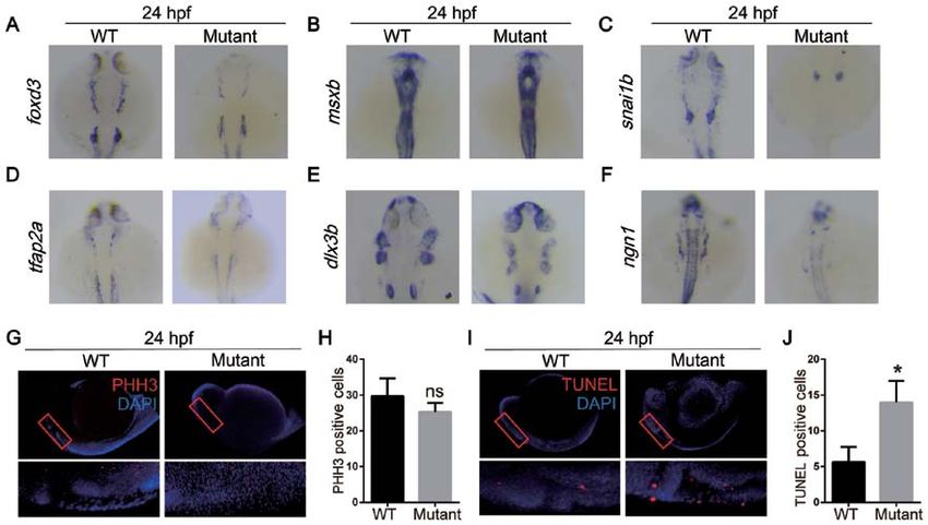
2          ZHAO et al: miR‑1 AFFECTS THE DEVELOPMENT OF THE NCCs VIA THE MITOCHONDRIAL APOPTOSIS PATHWAY

presented in Tables SI and SII and Fig. S1. The embryos were      assay was performed using an In Situ Cell Death Detection kit
grown in Holtfreter's solution (0.05 g/l KCL, 0.1 g/l CaCl2,      (Roche Diagnostics) as previously described (16). The staining
0.2 g/l NaHCO3 and 3.5 g/l Nacl) with a 14 h light/10 h dark      was subsequently observed using laser scanning confocal
cycle at 28.5˚C in the zebrafish facility of the Model Animal     microscopy (magnification, x20) (17).
Research Center, Nanjing University (Nanjing, China). The
zebrafish were fed three times a day with pellet food, and        Culture of NCCs. Mouse embryos were harvested at gesta‑
their health and behavior was monitored daily. All experi‑        tional day E9.5 according to a previously published
ments were approved by the Ethics Committee of the School         protocol (18). The pregnant adult mice (n=3; Model Animal
of Stomatology of Nanjing Medical University, and all             Research Center, Yangzhou University) were sacrificed by
procedures were performed according to the guidelines of the      cervical dislocation following anesthesia with sodium pento‑
Animal Care Committee of Nanjing Medical University.              barbital (50 mg/kg). The first branchial arch of the embryos
                                                                  was separated, washed in PBS and dissociated in
Whole mount in situ hybridization. Whole mount in situ            0.25% trypsin‑EDTA (Gibco; Thermo Fisher Scientific, Inc.)
hybridization was performed using standard procedures with        for 30 min at 37˚C. The separated cells were plated into a dish
slight modifications as previously described (13). Probes were    in Nutrient Mixture F‑12 (1:1)media (Gibco; Thermo Fisher
synthesized using a DIG RNA labeling kit (Roche Diagnostics).     Scientific, Inc.) with 15% FBS (ScienCell Research
The probes of forkhead box D3 ( foxd3), muscle segment            Laboratories, Inc.), supplemented with 100 U/ml peni‑
homeobox‑b (msxb), snail homolog 1b (snai1b), transcription       cillin‑streptomycin (PS), 100 mg/ml L‑glutamate, 0.1 mM
factor AP2a (tfap2a), distal‑less 3 (dlx3b), neurog1 (ngn1)       minimum essential medium non‑essential amino acids (Gibco;
(Table SIII) were designed using Primer 5.0 software (Premier     Thermo Fisher Scientific, Inc.) and 1,000 U/ml leukemia
Biotech, Inc.). Embryos (n=5 per group) at 24 h post‑fertil‑      inhibitory factor (PeproTech, Inc.). The inhibitor (5'‑AUA
ization (hpf) were collected, fixed in 4% paraformaldehyde        CAUACUUCUUUACAUUCCA‑3'; 100 nM) and mimics
overnight at 4˚C and subjected to gradient dehydration with       (forward, 5'‑UGGAAUGUAAAGAAGUAUGUAU‑3'; and
methanol (25, 50, 75 and 100%). The embryos were maintained       reverse, 5'‑AUACAUACUUCUUUACAUUCCA‑3'; 50 nM) of
in 100% methanol at ‑20˚C for subsequent use. Following           miR‑1 and their negative controls (NCs; inhibitor NC, 5'‑CAG
rehydration at gradient dilutions using methanol/PBS, the         UACUUUUGUGUAGUACAAA‑3'; mimics NC, 5'‑UUU
embryos were treated with proteinase K (10 µg/ml; Gibco;          GUACUACACAAAAGUACUG‑3'; 5'‑CAGUACUUUUG
Thermo Fisher Scientific, Inc.) at room temperature for 10 min.   UGUAGUACAAA‑3'; all Guangzhou RiboBio Co., Ltd.) were
Subsequently, embryos were prehybridized with the hybrid‑         transfected into NCCs when cells grew to 50% density using a
ization solution at 65˚C for 2 h. In situ hybridization signals   riboFECT™ CP Transfection kit (Guangzhou RiboBio Co.,
were detected by sheep anti‑digoxigenin‑AP Fab‑fragments          Ltd.) according to the manufacturer's protocol.
(Roche Diagnostics) and the color reaction was carried out by
staining with BCIP/NBT (Roche Diagnostics) as previously          Cell Counting Kit‑8 (CCK‑8) assay. CCK‑8 assay (Dojindo
described (14).                                                   Molecular Technologies, Inc.) was used to evaluate cell
                                                                  proliferation according to the manufacturer's protocol. NCCs
Alcian blue staining. Zebrafish embryos at 96 hpf (n=5 per        were seeded at 2x10³ cells/well in 96‑well plates. The cells
group) were fixed in 4% paraformaldehyde overnight at 4˚C,        were incubated at 37˚C for 12 h (day 0) and transfected with
dehydrated with ethanol (50 and 100%) and stained with            the miR‑1 inhibitor. CCK‑8 reagent in Nutrient Mixture F‑12
Alcian blue (0.15 mg/ml; Sigma‑Aldrich; Merck KGaA) over‑         media was added to each well at the specified time points (days
night at room temperature (15). After the embryonic cartilage     0, 1, 2, 3 and 4). Following 1‑h incubation at 37˚C, the optical
was stained, the embryos were incubated in saturated boron        density was measured at 450 nm using a microplate reader
overnight at room temperature. Subsequently, bleaching was        with three blank controls.
performed in a mixture of 3% H2O2 and 1% KOH at a ratio of
15:85 v/v, and the specimens were digested in 1% trypsin at       Wound healing assay. NCCs were seeded at 1.5x10⁵ cells/well
37˚C until all embryonic tissues were degraded. The digestion     in a 6‑well plate. Following transfection with the miR‑1
mixture was replaced with 1% KOH solution, and subse‑             inhibitor, a wound was created with a 10 µl pipette tip, and
quently the specimens were incubated in a glycerol gradient       the cells were incubated in serum‑free medium at 37˚C for
(25, 50, 75 and 100%). The embryos were observed using a          24 h. Cell migration was observed at 0, 12 and 24 h using
stereomicroscope (magnification, x20).                            a Leica DMIRE2 microscope (magnification, x100; Leica
                                                                  Microsystems GmbH) in phase contrast mode (19).
Histology and immunostaining. For whole mount immu‑
nostaining, the embryos (n=5 per group) were fixed in             Cell cycle and apoptosis analysis. NCCs transfected with the
4% paraformaldehyde overnight at 4˚C pretreated with              miR‑1 inhibitor were cultured for 24 h without PS. Following
proteinase K (10 µg/ml) at 37˚C for 30 min and blocked            an additional 48‑h culture, cell cycle and apoptosis analyses
in 2% BSA solution (BioFroxx) at room temperature                 were performed as previously described (20) using flow cytom‑
for 1 h. Subsequently, the embryos were stained with an           etry (FACSCalibur; BD Biosciences). For cell cycle analysis,
anti‑phospho‑histone H3 (PHH3; 1:100; cat. no. 3377; Cell         the cells (~1x10⁴) were fixed overnight with 75% ethanol at
Signaling Technology, Inc.) antibody overnight at 4˚C and         4˚C in the dark, and then stained with propidium iodide
then with an anti‑rabbit Alexa Fluor@594 (1:400; Jackson          (PI; MultiSciences Biotech Co., Ltd.) containing RNase.
ImmunoResearch Laboratories, Inc.) at 4˚C for 2 h. A TUNEL        Apoptosis was detected by harvesting cells using a centrifuge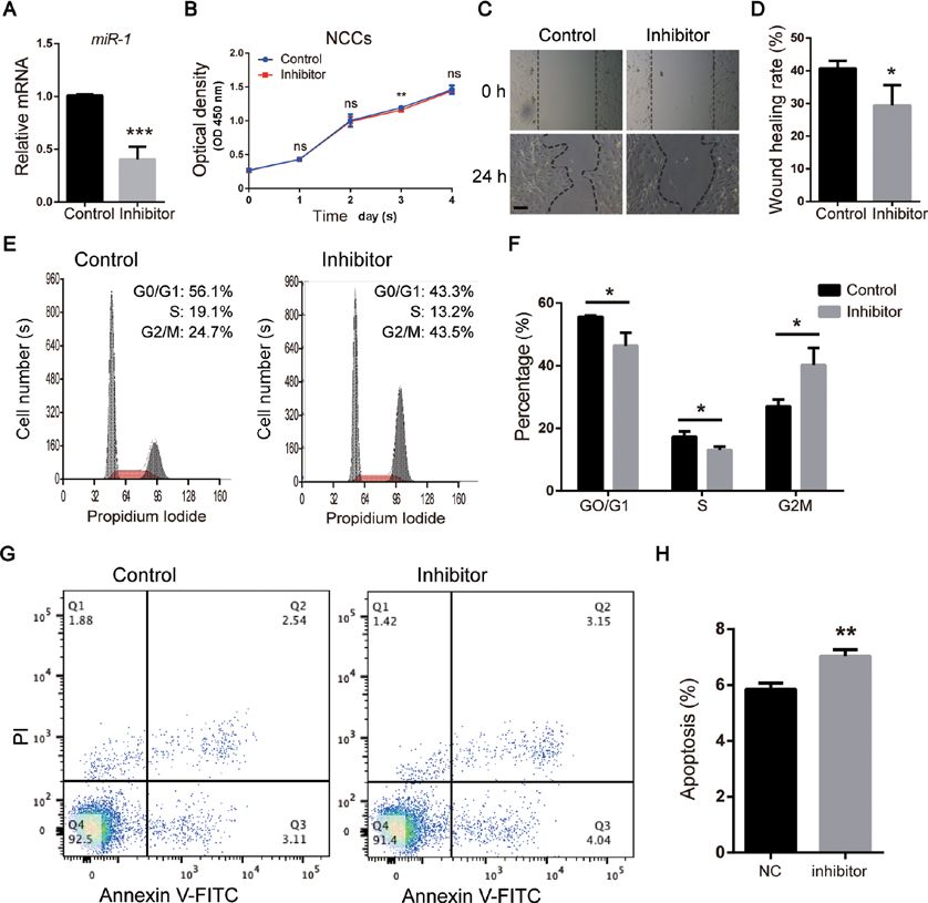
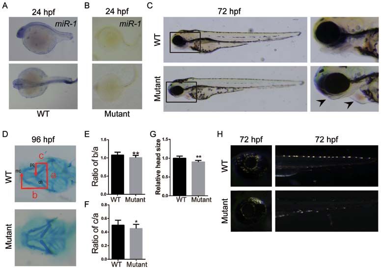
EXPERIMENTAL AND THERAPEUTIC MEDICINE 21: 379, 2021                                                               3

Figure 1. Loss of miR‑1 in zebrafish results in craniofacial, pigment cell and cardiac defects. In situ hybridization of miR‑1 in (A) wild‑type and (B) mutant
zebrafish at 24 hpf. (C) Zebrafish larvae at 72 hpf with mutants exhibiting mandibular retrognathia (left arrow) and edema around the heart (right arrow).
(D) Alcian blue staining at 96 hpf. (E) Quantitative analysis of b/a. (F) Quantitative analysis of c/a. a, width between ch; b, distance between mc and ch; and
c, the distance between pq and ch. (G) Quantitative analysis of head size. (H) Distribution and number of iridophores at 72 hpf. Data are presented as the
mean ± standard deviation (n=5). *P
4            ZHAO et al: miR‑1 AFFECTS THE DEVELOPMENT OF THE NCCs VIA THE MITOCHONDRIAL APOPTOSIS PATHWAY

Figure 2. MicroRNA‑1 knockout affects neural crest cell apoptosis during the migration and differentiation periods of embryonic development. In situ
hybridization of zebrafish at 24 hpf with (A) foxd3, (B) msxb, (C) snai1b, (D) tfap2a, (E) dlx3b and (F) ngn1 probes. (G) Whole mount immunostaining of
zebrafish at 24 hpf with PHH3 (red) and DAPI (blue). (H) Quantitative analysis of immunostaining. (I) TUNEL staining at 24 hpf with TUNEL (red) and
DAPI (blue). (J) Quantitative analysis of TUNEL staining. Data are presented as the mean ± standard deviation (n=5). *P
EXPERIMENTAL AND THERAPEUTIC MEDICINE 21: 379, 2021                                                               5

Figure 3. miR‑1 regulates the function of NCCs in vitro. (A) miR‑1 expression assessed via reverse transcription‑quantitative PCR following transfection
with the miR‑1 inhibitor. (B) Cell proliferation of NCCs analyzed by Cell Counting Kit‑8 assay. (C) Wound healing assay of NCCs. Scale bar, 100 µm.
(D) Quantitative analysis of wound healing assay. (E) Cell cycle analysis of NCCs performed using flow cytometry. (F) Quantitative analysis of cell cycle
analysis. (G) Apoptosis analysis of NCCs evaluated via flow cytometry. (H) Quantitative analysis of apoptosis analysis. Data are presented as the mean ± stan‑
dard deviation (n=3). *P
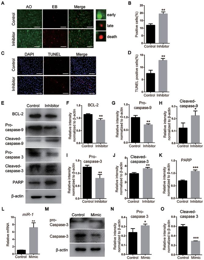
6            ZHAO et al: miR‑1 AFFECTS THE DEVELOPMENT OF THE NCCs VIA THE MITOCHONDRIAL APOPTOSIS PATHWAY

Figure 4. miR‑1 affects the apoptosis of neural crest cells via the mitochondrial apoptosis pathway and caspase‑3. (A) Detection of apoptosis on day 3
following miR‑1 inhibitor transfection using AO/EB staining. Scale bar, 100 µm. (B) Quantitative analysis of AO/EB staining. (C) TUNEL staining of
NCCs. Scale bar, 100 µm. (D) Quantitative analysis of TUNEL staining. (E) Protein levels of the mitochondrial apoptosis pathway markers BCL‑2, pro‑ and
cleaved‑caspase‑3, pro‑ and cleaved‑caspase‑9 and PARP detected via western blotting. (F‑K) Quantification of the protein levels of (F) BCL‑2, (G) pro‑cas‑
pase‑9, (H) cleaved‑caspase‑9, (I) pro‑caspase‑3, (J) cleaved‑caspase‑3 and (K) PARP, which are presented in (E). (L) miR‑1 expression assessed by reverse
transcription‑quantitative PCR. (M) Expression levels of pro‑ and cleaved‑caspase‑3 after transfection with the miR‑1 mimic. (N and O) Quantification of
the protein levels. Data are presented as the mean ± standard deviation (n=3). *P
EXPERIMENTAL AND THERAPEUTIC MEDICINE 21: 379, 2021                                             7

with miR‑1 mimics compared with that in the control group                 Caspase‑3 has been reported to be a key protein in the mito‑
(Fig. 4M‑O), suggesting that miR‑1 inhibited the activation of       chondrial apoptosis pathway, and RARP is one of the principal
caspase‑3 and affected the apoptosis of NCCs.                        targets of caspase‑3 (37). Also, pro‑caspase‑9 requires to be
    Collectively, these results suggested that miR‑1 regulated       activated to subsequently cleave and activate caspase‑3 (38).
the apoptosis of NCCs via the mitochondrial apoptosis                Western blotting results suggested that miR‑1 may regulate the
pathway.                                                             mitochondrial apoptosis pathway.
                                                                          The biogenesis of miRNAs is a complex process. After
Discussion                                                           several processing steps (39), mature single‑stranded miRNAs
                                                                     have been indicated to enter the miRNA‑induced silencing
In a previous study, a latitudinal expression of miR‑1 was           complex, which silences the expression of target genes
observed in WT zebrafish via in situ hybridization, and it was       primarily at the post‑transcriptional level (40). The target
demonstrated that the expression of miR‑1 increased during           genes to be silenced are selected via base‑pairing interactions
embryonic development at 24 hpf (11).                                between the miRNA and the target mRNA that contains a
    In the present study, in situ hybridization was used to detect   partial or complete complementary sequence, generally local‑
the expression of miR‑1 in WT zebrafish embryos. The expres‑         ized in 3'‑untranslated region (41). The results of the current
sion of miR‑1 was demonstrated to be primarily localized in the      study suggested that caspase‑3 may be targeted by miR‑1.
head, pharyngeal region and pectoral fin of zebrafish at 24 hpf.     However, the regulatory pathways and mechanisms of this
Previous findings have indicated that the injection of miR‑1         interaction require additional studies.
morpholino oligomers in zebrafish resulted in impaired cranio‑            In conclusion, the present study demonstrated an impor‑
facial chondrogenesis, severe maxillofacial malformations and        tant role of miR‑1 in regulating the apoptosis of NCCs during
an abnormal function of NCCs within 4 days post‑fertilization.       embryonic development via modulating the mitochondrial
However, it was not clear whether these defects were also            apoptosis pathway. A normal expression of miR‑1 was iden‑
affected by the injection of the morpholino (11). Therefore, a       tified to be essential for the development of NC derivatives,
miR‑1 knockout zebrafish model was generated in the present          including the pharyngeal cartilage, mandible and hyoid bone.
study to elucidate the role of miR‑1 in NC development.              A thorough understanding of molecular pathway by which
    NCCs have been reported to contribute to the development         miR‑1 regulates its target genes may aid the prevention and
of bones, cartilage, cranial ganglia and connective tissues in the   treatment of miR‑1‑associated developmental malformations.
face and neck (1). Previous studies have demonstrated that the
abnormal expression of genes associated with NC may result           Acknowledgements
in craniofacial, bone and cartilage dysplasia due to abnormal
cell migration, proliferation and differentiation (26‑29). For       Not applicable.
example, dlx3b has been associated with the differentiation
of the pharyngeal arch in zebrafish (30). Also, Ngn1 has             Funding
been reported to regulate the differentiation of the dorsal
root ganglion in zebrafish development (31). In the current          The current study was funded by the National Natural Science
study, in situ hybridization evidence collectively suggested         Foundation of China (grant no. 81771029), the Natural Science
that miR‑1 serve an important role during the migration and          Fund for Colleges and Universities in Jiangsu Province, China
differentiation periods of NCCs.                                     (grant no. 18KJA320004), the Southeast University‑Nanjing
    miRNAs are a class of highly conserved, noncoding RNA            Medical University Cooperative Research Project (grant
molecules with a length of 18‑25 nucleotides. The mature             no. JX218GSP20180705) and the Priority Academic Program
sequence of miR‑1 is highly conserved among different verte‑         Development of Jiangsu Higher Education Institutions (grant
brates, including zebrafish, mice and humans, which suggests         no. 2014‑037).
that miR‑1 may exhibit similar functions among verte‑
brates (32). The role of miR‑1 in regulating the development         Availability of data and materials
of heart, cartilage and liver, among other organs, has been
reported (33‑35). In the present study, the following abnormal       All data generated or analyzed during this study are included
phenotypes were observed in the miR‑1‑knockout model:                in this published article.
Mandibular retraction, shortening of the inferior dental arch
and lingual arch, and atypical neural crest‑derived tissues,         Authors' contributions
such as abnormal pericardial edema and pigment cells.
    Li et al (36) reported that miR‑1 decreased cardiomyocyte        NZ, WQ, DW, AGR and LY performed the experiments and
apoptosis via mediating the expression of apoptosis‑related          analyzed the data. NZ, YM, CM, ZX and JM designed the
genes in the heart. In the current study, TUNEL staining             study. NZ, AGR, ZX and JM wrote the manuscript. All authors
revealed an increase in apoptosis of pharyngeal NCCs in miR‑1        read and approved the final manuscript.
mutant compared with WT zebrafish, which was consistent
with previous reports (36). By contrast, PHH3 immunofluores‑         Ethics approval and consent to participate
cence staining did not demonstrate any differences between
the WT and mutant groups; additional time points may be              The present study was approved by the Ethics Committee of
required to determine whether alterations in NCC prolifera‑          the School of Stomatology of Nanjing Medical University
tion were caused by miR‑1 knockout.                                  (approval no. 1403049), and all procedures were performed
8 ZHAO et al: miR‑1 AFFECTS THE DEVELOPMENT OF THE NCCs VIA THE MITOCHONDRIAL APOPTOSIS PATHWAY
according to the guidelines of the Animal Care Committee of 20. Lu M, Guo S, Hong F, Zhang Y, Yuan L, Ma C and Ma J: Pax2
is essential for proliferation and osteogenic differentiation of
Nanjing Medical University. mouse mesenchymal stem cells via Runx2. Exp Cell Res 371:
342‑352, 2018.
Patient consent for publication 21. Ono W, Sakagami N, Nishimori S, Ono N and Kronenberg HM:
Parathyroid hormone receptor signalling in osterix‑expressing
mesenchymal progenitors is essential for tooth root formation.
Not applicable. Nat Commun 7: 11277, 2016.
22. Livak KJ and Schmittgen TD: Analysis of relative gene expression
data using real‑time quantitative PCR and the 2(‑Delta Delta
Competing interests C(T)) Method. Methods 25: 402‑408, 2001.
23. Barrallo‑Gimeno A, Holzschuh J, Driever W and Knapik EW:
The authors declare that they have no competing interests. Neural crest survival and differentiation in zebrafish depends on
mont blanc/tfap2a gene function. Development 131: 1463‑1477,
2004.
References 24. Nelms BL, Pfaltzgraff ER and Labosky PA: Functional
interaction between Foxd3 and Pax3 in cardiac neural crest
1. Mayor R and Theveneau E: The neural crest. Development 140: development. Genesis 49: 10‑23, 2011.
2247‑2251, 2013. 25. Medeiros DM and Crump JG: New perspectives on pharyngeal
2. Trainor PA: Specification and patterning of neural crest cells dorsoventral patterning in development and evolution of the
during craniofacial development. Brain Behav Evol 66: 266‑280, vertebrate jaw. Dev Biol 371: 121‑135, 2012.
2005. 26. Lake JI, Avetisyan M, Zimmermann AG and Heuckeroth RO:
3. Keyte A and Hutson MR: The neural crest in cardiac congenital Neural crest requires Impdh2 for development of the enteric
anomalies. Differentiation 84: 25‑40, 2012. nervous system, great vessels, and craniofacial skeleton. Dev
4. Jones NC, Lynn ML, Gaudenz K, Sakai D, Aoto K, Rey JP, Biol 409: 152‑165, 2016.
Glynn EF, Ellington L, Du C, Dixon J, et al: Prevention of the 27. Lei R, Zhang K, Wei Y, Chen M, Weinstein LS, Hong Y, Zhu M,
neurocristopathy Treacher Collins syndrome through inhibition Li H and Li H: G‑protein α‑subunit Gsα is required for cranio‑
of p53 function. Nat Med 14: 125‑133, 2008. facial morphogenesis. PLoS One 11: e0147535, 2016.
5. Etchevers HC, Amiel J and Lyonnet S: Molecular bases of human 28. Shao R, Liu J, Yan G, Zhang J, Han Y, Guo J, Xu Z, Yuan Z, Liu J,
neurocristopathies. Adv Exp Med Biol 589: 213‑234, 2006. Malumbres M, et al: Cdh1 regulates craniofacial development
6. Chai Y and Maxson RE Jr: Recent advances in craniofacial via APC‑dependent ubiquitination and activation of Goosecoid.
morphogenesis. Dev Dyn 235: 2353‑2375, 2006. Cell Res 26: 699‑712, 2016.
7. Ying SY and Lin SL: Current perspectives in intronic micro 29. Tu CT, Yang TC, Huang HY and Tsai HJ: Zebrafish arl6ip1 is
RNAs (miRNAs). J Biomed Sci 13: 5‑15, 2006. required for neural crest development during embryogenesis.
8. Townley‑Tilson WH, Callis TE and Wang D: MicroRNAs 1, PLoS One 7: e32899, 2012.
133, and 206: critical factors of skeletal and cardiac muscle 30. Alexander C, Piloto S, Le Pabic P and Schilling TF: Wnt
development, function, and disease. Int J Biochem Cell Biol 42: signaling interacts with bmp and edn1 to regulate dorsal‑ventral
1252‑1255, 2010. patterning and growth of the craniofacial skeleton. PLoS
9. Li P, Wei X, Guan Y, Chen Q, Zhao T, Sun C and Wei L: Genet 10: e1004479, 2014.
MicroRNA‑1 regulates chondrocyte phenotype by repressing 31. Cornell RA and Eisen JS: Delta/Notch signaling promotes
histone deacetylase 4 during growth plate development. FASEB formation of zebrafish neural crest by repressing Neurogenin 1
J 28: 3930‑3941, 2014. function. Development 129: 2639‑2648, 2002.
10. Samal E, Evangelista M, Galang G, Srivastava D, Zhao Y and 32. Bartel DP: MicroRNAs: Genomics, biogenesis, mechanism, and
Vedantham V: Premature microRNA‑1 expression causes function. Cell 116: 281‑297, 2004.
hypoplasia of the cardiac ventricular conduction system. Front 33. Fleissner F, Jazbutyte V, Fiedler J, Gupta SK, Yin X, Xu Q,
Physiol 10: 235, 2019. Galuppo P, Kneitz S, Mayr M, Ertl G, et al: Short communication:
11. Wang D, Weng Y, Guo S, Qin W, Ni J, Yu L, Zhang Y, Zhao Q, Asymmetric dimethylarginine impairs angiogenic progenitor
Ben J and Ma J: MicroRNA‑1 regulates NCC migration and cell function in patients with coronary artery disease through
differentiation by targeting sec63. Int J Biol Sci 15: 2538‑2547, a microRNA‑21‑dependent mechanism. Circ Res 107: 138‑143,
2019. 2010.
12. Fishman MC: Genomics. Zebrafish ‑ the canonical vertebrate. 34. Karp X and Ambros V: Developmental biology. Encountering
Science 294: 1290‑1291, 2001. microRNAs in cell fate signaling. Science 310: 1288‑1289, 2005.
13. Liu QY, Wu ZL, Lv WJ, Yan YC and Li YP: Developmental 35. Xu P, Guo M and Hay BA: MicroRNAs and the regulation of cell
expression of cyclin H and Cdk7 in zebrafish: The essential death. Trends Genet 20: 617‑624, 2004.
role of cyclin H during early embryo development. Cell Res 17: 36. Li W, Liu M, Zhao C, Chen C, Kong Q, Cai Z and Li D: miR‑1/133
163‑173, 2007. attenuates cardiomyocyte apoptosis and electrical remodeling in
14. Schulte‑Merker S, Ho RK, Herrmann BG and Nüsslein‑Volhard C: mice with viral myocarditis. Cardiol J 27: 285-294, 2020.
The protein product of the zebrafish homologue of the mouse 37. Perchellet EM, Wang Y, Weber RL, Sperfslage BJ, Lou K,
T gene is expressed in nuclei of the germ ring and the notochord Crossland J, Hua DH and Perchellet JP: Synthetic 1,4‑anthra‑
of the early embryo. Development 116: 1021‑1032, 1992. cenedione analogs induce cytochrome c release, caspase‑9,
15. de Peralta MS, Mouguelar VS, Sdrigotti MA, Ishiy FA, ‑3, and ‑8 activities, poly(ADP‑ribose) polymerase‑1 cleavage
Fanganiello RD, Passos‑Bueno MR, Coux G and Calcaterra NB: and internucleosomal DNA fragmentation in HL‑60 cells by
Cnbp ameliorates Treacher Collins syndrome craniofacial a mechanism which involves caspase‑2 activation but not Fas
anomalies through a pathway that involves redox‑responsive signaling. Biochem Pharmacol 67: 523‑537, 2004.
genes. Cell Death Dis 7: e2397, 2016. 38. Green DR and Reed JC: Mitochondria and apoptosis. Science 281:
16. Ning G, Liu X, Dai M, Meng A and Wang Q: MicroRNA‑92a 1309‑1312, 1998.
upholds Bmp signaling by targeting noggin3 during pharyngeal 39. Lee Y, Ahn C, Han J, Choi H, Kim J, Yim J, Lee J, Provost P,
cartilage formation. Dev Cell 24: 283‑295, 2013. Rådmark O, Kim S, et al: The nuclear RNase III Drosha initiates
17. Babb‑Clendenon S, Shen YC, Liu Q, Turner KE, Mills MS, microRNA processing. Nature 425: 415‑419, 2003.
Cook GW, Miller CA, Gattone VH II, Barald KF and Marrs JA: 40. Lin SL and Ying SY: Gene silencing in vitro and in vivo using
Cadherin‑2 participates in the morphogenesis of the zebrafish intronic microRNAs. Methods Mol Biol 1733: 107‑126, 2018.
inner ear. J Cell Sci 119: 5169‑5177, 2006. 41. Huntzinger E and Izaurralde E: Gene silencing by microRNAs:
18. Guo S, Zhang Y, Zhou T, Wang D, Weng Y, Chen Q, Ma J, Li YP Contributions of translational repression and mRNA decay. Nat
and Wang L: GATA4 as a novel regulator involved in the devel‑ Rev Genet 12: 99‑110, 2011.
opment of the neural crest and craniofacial skeleton via Barx1. This work is licensed under a Creative Commons
Cell Death Differ 25: 1996‑2009, 2018. Attribution-NonCommercial-NoDerivatives 4.0
19. Guo S, Zhang Y, Zhou T, Wang D, Weng Y, Wang L and Ma J: International (CC BY-NC-ND 4.0) License.
Role of GATA binding protein 4 (GATA4) in the regulation of
tooth development via GNAI3. Sci Rep 7: 1534, 2017.You can also read