NF ΚB/IΚBΑ SIGNALING PATHWAYS ARE ESSENTIAL FOR RESISTANCE TO HEAT STRESS INDUCED ROS PRODUCTION IN PULMONARY MICROVASCULAR ENDOTHELIAL CELLS
←
→
Page content transcription
If your browser does not render page correctly, please read the page content below
MOLECULAR MEDICINE REPORTS 24: 814, 2021

                      NF‑κB/IκBα signaling pathways are essential for
                      resistance to heat stress‑induced ROS production
                        in pulmonary microvascular endothelial cells
                              WEIDANG XIE1*, WEI HUANG1,2*, SHUMIN CAI1*, HUI CHEN1,
                                  WEIJUN FU1, ZHONGQING CHEN1 and YANAN LIU1

                  1
                   Department of Critical Care Medicine, Nanfang Hospital, 2The First School of Clinical Medicine,
                            Southern Medical University, Guangzhou, Guangdong 510515, P.R. China

                                       Received May 22, 2021; Accepted September 10, 2021

                                                   DOI: 10.3892/mmr.2021.12454

Abstract. The results of a previous study demonstrated that heat    rat PMVECs, and that it could be a potential therapeutic target
stress (HS) triggered oxidative stress, which in turn induced the   to reduce the mortality and morbidity of heat stroke.
apoptosis of epithelial cells. These results uncovered a novel
mechanism underlying the activation of NF‑κ B in primary            Introduction
human umbilical vein endothelial cells. The present study
aimed to further investigate the role of NF‑κ B/Iκ Bα signaling     Heat stroke is a life threatening illness characterized by extreme
pathways in the inhibition of HS‑induced reactive oxygen            elevations in core temperature reaching >40.5˚C and damage to
species (ROS) generation and cytotoxicity in endothelial cells.     multiple organ systems (1). Previous studies have suggested that
The results of the present study demonstrated that HS triggered     inflammatory injury and severe hypoxemia induced by heat
a significant amount of NF‑κ B and Iκ Bα nuclear translocation      stroke lead to acute lung injury and respiratory failure (2,3).
without Iκ Bα degradation in a time‑dependent manner. Mutant        Clinical and experimental evidence suggested that endothe‑
constructs of Iκ Bα phosphorylation sites (Ser32, Ser36) were       lial cell injury served a vital role in the early acute phase of
employed in rat pulmonary microvascular endothelial cells           heat stroke (4). Heat stress (HS) refers to conditions involving
(PMVECs). Cell Counting Kit‑8 assays demonstrated that              increased core body temperature above the normal level due
both the small interfering (si)RNA‑mediated knockdown of            to thermoregulatory failure, which leads to heat stroke (5).
p65 and Iκ Bα mutant constructs significantly decreased cell        However, the molecular mechanisms underlying the endothe‑
viability and aggravated ROS accumulation in HS‑induced rat         lial cell injury in heat stroke‑induced acute lung injury remain
PMVECs compared with the control. Additionally, western             to be elucidated. Thus, further investigation into the molecular
blot analysis revealed that p65 siRNA attenuated the protein        mechanisms underlying tissue injury caused by direct HS are
expression of Iκ Bα. However, Iκ Bα mutant constructs failed        required to develop clinical strategies for heat stroke.
to attenuate NF‑κ B activation and nuclear translocation, indi‑         HS induces endothelial cell injury through oxidative stress,
cating that Iκ Bα‑independent pathways contributed to NF‑κ B        which stimulates reactive oxygen species (ROS) generation,
activity and nucleus translocation in a time‑dependent manner       lipid peroxidation and DNA damage (6). ROS are by‑products
following HS. Collectively, the results of the present study        of oxygen metabolism, including superoxide anions, hydrogen
suggested that the NF‑κ B/Iκ Bα pathway was essential for           peroxide and hydroxyl radicals (7). ROS target proteins,
resistance to HS‑induced ROS production and cytotoxicity in         polysaccharides, DNA and lipids and increase the rate of cell
                                                                    damage (8). Furthermore, results of our previous study demon‑
                                                                    strated that NF‑κ B activation decreased HS‑induced oxidative
                                                                    stress in HUVECs (9). However, the potential mechanisms
                                                                    underlying NF‑κ B in HS‑induced oxidative stress remain to
Correspondence to: Dr Yanan Liu, Department of Critical             be elucidated.
Care Medicine, Nanfang Hospital, Southern Medical University,
                                                                        The results of previous studies have demonstrated that
1023 South Shatai Road, Baiyun, Guangzhou, Guangdong 510515,
P.R. China                                                          NF‑κ B serves a critical role in regulating cell proliferation,
E‑mail: 123302688@qq.com                                            inflammatory responses, survival and apoptosis (10,11). When
                                                                    bound, NF‑κ B and Iκ B remain inactive in the cytoplasm (12).
*
    Contributed equally                                             A main component of Iκ B is Iκ Bα, the first protein described
                                                                    for the Iκ B family and the most extensively studied Iκ B protein
Key words: heat stress, reactive oxygen species, cytotoxicity,      to date (13,14). NF‑κ B‑activating stimuli activate Iκ B kinase,
NF‑κ B, Iκ Bα, pulmonary microvascular endothelial cells            which induces the phosphorylation at Ser32 and Ser36 on
                                                                    Iκ Bα (15). NF‑κ B is subsequently activated and translocated
                                                                    into the nucleus to control transcription (16). As a dominant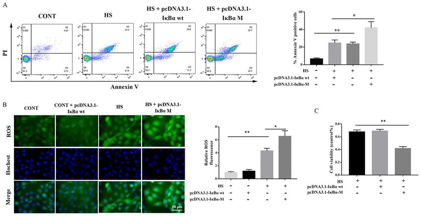
2                                         XIE et al: ROLES OF NF-κ B/Iκ Bα IN HEAT STRESS

regulatory kinase in the classic NF‑κ B signaling pathway,          Thermo Fisher Scientific, Inc). A total of 1x10⁶ cells were
Iκ Bα serves a prominent role in apoptosis (17). In addition,       collected and washed three times in PBS. Subsequently, cells
the Iκ Bα/NF‑κ B signaling pathway is one of the most popular       were incubated in binding buffer containing Annexin V‑FITC
antitumor targets (18). The results of the present study revealed   for at 4˚C 15 min, the buffer was removed by centrifugation at
a novel mechanism for HS‑induced NF‑κ B activation in rat           1,000 x g for 5 min at 4˚C and the cells were resuspended in
pulmonary microvascular endothelial cells (PMVECs) without          buffer containing PI solution. After 5 min, apoptotic cells were
Iκ Bα degradation, which differs from the classic NF‑κ B            detected using a FACSCalibur cytometer (Becton, Dickinson
signaling pathway. However, the precise molecular mechanism         and Company). The data were analyzed using Flowing
underlying the generation of ROS by NF‑κ B without Iκ Bα            version 2.5.3 software (Turku Bioscience) and Origin version 7
degradation and phosphorylation remains to be elucidated.           software (OriginLab). The results of a previous study have
    The present study aimed to determine whether the                suggested that Annexin V‑positive cells in the right quadrant
NF‑κ B/Iκ Bα pathway inhibited HS‑induced ROS genera‑               are apoptotic, including the early and late apoptotic cells (20).
tion and cell death in PMVECs. The association between
HS‑induced NF‑κ B activation and the phosphorylation of             Florescence confocal assay of intracellular ROS. Cells were
upstream Iκ Bα was also investigated.                               treated with 2'‑7'‑dichlorofluorescin diacetate (Beyotime
                                                                    Institute of Biotechnology) at 37˚C for 20 min in the dark as
Materials and methods                                               previously described (21). For live imaging, DNA was stained
                                                                    with 0.2 mg/ml Hoechst 33342 (Thermo Fisher Scientific, Inc.)
Animals. Male Sprague Dawley (SD) rats (n=50; body                  at 37˚C for 10 min and PMVECs were washed with DMEM
weight, 125‑155 g; age, 7 weeks) were purchased from the            Fluoro Brite (Gibco; Thermo Fisher Scientific, Inc.). Cells were
Experimental Animal Center of Southern Medical University.          analyzed using a confocal fluorescent microscope (magnifica‑
Rats were housed at a constant temperature of 22‑25˚C and           tion, x400; LI‑COR Biosciences, Inc.).
50‑60% relative humidity under a controlled 16‑h light/8‑h
dark cycle for at ≥5 days; they had free access to rat chow and     Western blot analysis. Cells were exposed to HS treatment
water. Experimental protocols involving animals followed the        and a nuclear extraction kit (Active Motif, Inc.) was used to
guidelines approved by the Chinese Association of Laboratory        obtain cytoplasmic and nuclear protein extracts, according
Animal Care and approved by the Institutional Animal                to the manufacturer's protocol. Total protein was quanti‑
Care and Use Committee of Nanfang Hospital (approval                fied using an Enhanced BCA Protein Assay kit (Beyotime
no. NFYY‑2019‑176; Guangzhou, China).                               Institute of Biotechnology). Western blotting was performed
                                                                    as previously described (22), the proteins (40 µg per lane) were
Cell culture and treatment. Primary PMVECs were isolated            separated via 12% SDS‑PAGE and further transferred onto
from SD rats as previously described (19). Briefly, after           PVDF membranes. Then, the membranes were blocked with
anesthesia with pentobarbital sodium (50 mg/kg, i.p.), the          5% skimmed milk for 1 h at room temperature, and cultured
distal lung tissues (diameter, 3‑5 mm) were resected from           with the following primary antibodies: p65 (cat. no. ab16502;
rat lungs, cut into small pieces and digested with type I           1:1,000; Abcam), Iκ Bα (cat. no. 4812; 1:1,000; Cell Signaling
collagenase (2 mg/ml) and neutral protease (0.6 U/ml) for           Technology, Inc.), phosphorylated (p)‑Iκ Bα (cat. no. 2859;
1 h. The mixture was centrifuged at 800 x g for 10 min at           1:1,000; Cell Signaling Technology, Inc.), p‑p65 (cat. no. 3303;
37˚C. Subsequently, cells were resuspended and plated with          1:1,000; Cell Signaling Technology, Inc.), Lamin B
Dynabeads (Invitrogen; Thermo Fisher Scientific, Inc.) and          (cat. no. 13435; 1:1,000; Cell Signaling Technology, Inc.) and
rat anti‑mouse platelet endothelial adhesion molecule‑1 anti‑       GAPDH (cat. no. 5174; 1:1,000; Cell Signaling Technology, Inc.)
body (Invitrogen; Thermo Fisher Scientific, Inc.) for 1 h. The      overnight at 4˚C. Following primary incubation, membranes
cell‑bead mixture was separated using a magnetic particle           were incubated with HRP‑conjugated anti‑rabbit IgG anti‑
concentrator (Invitrogen; Thermo Fisher Scientific, Inc.) and       body (cat. no. ZB‑2301; 1:5,000; OriGene Technologies, Inc.)
incubated in EGM‑2 culture media containing 10% FBS with            at room temperature for 1 h. Protein bands were visualized
growth factor bullet kit (Lonza Group, Ltd.). For HS treatment,     using ECL reagents (Pierce; Thermo Fisher Scientific, Inc.)
the bottom of each culture dish was placed into a circulating       with GAPDH and Lamin B as the internal control. ImageJ
water bath maintained at 43±0.5˚C for 2 h and PMVECs were           (version 6.0; National Institutes of Health) was used for the
further incubated at 37˚C for periods as indicated.                 semi‑quantification of protein expression.

Cell Counting Kit‑8 (CCK‑8) assay. Cells were seeded in a           NF‑ κ B dual‑luciferase assay. The luciferase activity was
96‑well plate at a density of 1x10⁵/ml and incubated at 37˚C        determined using the Dual‑Glo luciferase assay system
overnight. Subsequently, cells were cultured at 43˚C for 2 h        (cat. no. E2980; Promega Corporation) following the manu‑
or treated with p65 small interfering (si)RNA or Iκ Bα mutant       facturer's protocol. Briefly, plasmids were purified from
construct, mixed with 100 µl CCK‑8 solution (Dojindo                bacterial cultures using the QIAGENP plasmid Midi kit
Molecular Technologies, Inc.) and incubated for 1 h. Cell           (Qiagen GmbH). Rat PMVECs were transfected with the
viability was assessed following the manufacturer's protocol.       vector constructs using Lipofectamine® 2000 transfection
                                                                    reagent (Invitrogen; Thermo Fisher Scientific, Inc.) and the
Apoptosis assay. Cell apoptosis was detected using an               pGL3‑Basic plasmid without promoter as the negative control.
Annexin V‑FITC apoptosis detection kit and flow cytom‑              The pGl4.74 vector (Promega Corporation) was used as an
etry according to the manufacturer's protocol (Invitrogen;          internal control to determine the efficiency of transfection.
MOLECULAR MEDICINE REPORTS 24: 814, 2021 3
Table I. Sequences of oligonucleotide primers.
Gene Direction Sequence
p65 Forward 5'-GCCCUAUCCCUUUACGUCATT-3'
Reverse 5'-UGACGUAAAGGGAUAGGGCTT-3'
Negative control Forward 5'-UUCUCCGAACGUGUCACGUTT-3'
Reverse 5'-ACGUGACACGUUCGGAGAATT-3'
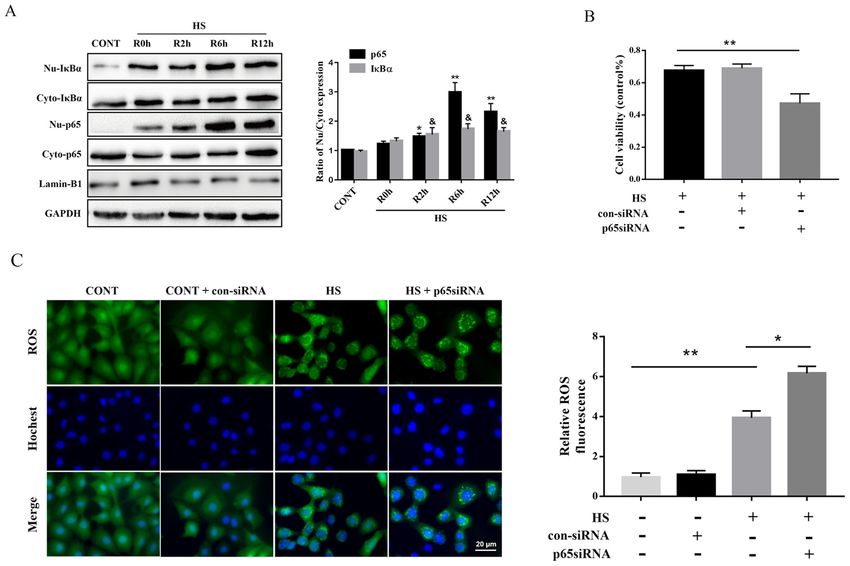
After 24 h of transfection, cells were washed with PBS and P4 XIE et al: ROLES OF NF-κ B/Iκ Bα IN HEAT STRESS Figure 1. Influence of NF‑κ B on HS‑induced ROS accumulation and cytotoxicity in rat PMVECs. (A) Cells were incubated at 37˚C (CONT) or were subjected to HS treatment at 43˚C for 2 h, followed by a recovery period at 37˚C for 0 (R0h), 2 (R2h), 6 (R6h) or 12 h (R12h). Expression levels of p65 and Iκ Bα were detected in cytoplasmic and nuclear fractions of PMVECs using western blotting. (B) PMVECs were subjected to HS treatment at 43˚C for 2 h and incubated at 37˚C for 12 h before cell proliferation was determined using a Cell Counting Kit‑8 assay. (C) Representative images of intracellular ROS visualized using fluorescence microscopy. Sections were co‑stained with ROS (green) and Hoechst (blue). Scale bar, 20 µm. Each value represents the mean ± SD of three inde‑ pendent experiments. *P
MOLECULAR MEDICINE REPORTS 24: 814, 2021                                                                  5

Figure 3. HS mediated NF‑κ B activity and nucleus translocation‑independent Iκ Bα phosphorylation. PMVECs were transfected with the pcDNA3.1‑Iκ Bα wt
(Iκ Bα wt) or pcDNA3.1‑Iκ Bα‑M (Iκ Bα M) constructs. After 48 h, cells were exposed to HS at 43˚C for 2 h and were further incubated at 37˚C for 12 h.
(A) Schematic of Iκ Bα consensus sites (p‑Ser/Ub‑Lys). pcDNA3.1‑Iκ Bα‑M had Ala substitutions at two phosphorylation sites, Ser32 and Ser36. (B) Western
blot analysis of the phosphorylation of Iκ Bα and p65. (C) pcDNA3.1‑Iκ Bα‑M regulated phosphorylation levels of NF‑κ B p65 in both the cytoplasm and nuclei.
(D) Cells were incubated at 37˚C (CONT) or were subjected to HS treatment at 43˚C for 2 h, followed by a recovery period at 37˚C for 0 (R0h), 2 (R2h), 6 (R6h)
or 12 h (R12h). Cells were analyzed for NF‑κ B activation using dual‑luciferase assays. Each value represents the mean ± SD from three independent experi‑
ments. **P
6                                                XIE et al: ROLES OF NF-κ B/Iκ Bα IN HEAT STRESS

Figure 4. Phosphorylation of Iκ Bα mediates HS‑induced apoptosis and ROS accumulation in rat PMVECs. PMVECs were transfected with pcDNA3.1,
pcDNA3.1‑Iκ Bα wt or pcDNA3.1‑Iκ Bα‑M constructs for 48 h. Cells were subsequently subjected to HS treatment at 43˚C for 2 h and were further incubated
at 37˚C for 12 h. (A) Apoptosis of PMVECs was measured with Annexin V‑FITC/PI staining and analyzed by flow cytometry. (B) Representative images
of intracellular ROS visualized using fluorescence microscopy and the quantification of intracellular ROS in all groups. Sections were co‑stained with ROS
(green) and Hoechst (blue). (C) Cell proliferation of PMVECs was determined using Cell Counting Kit‑8 assays. Scale bar, 20 µm. Each value represents
the mean ± SD from three independent experiments. *P
MOLECULAR MEDICINE REPORTS 24: 814, 2021                                                       7

    Collectively, the results of the present study suggested            4. Li L, Tan H, Gu Z, Liu Z, Geng Y, Liu Y, Tong H, Tang Y, Qiu J
                                                                           and Su L: Heat stress induces apoptosis through a Ca²+‑mediated
that the NF‑κ B/Iκ Bα pathway was essential for resistance to              mitochondrial apoptotic pathway in human umbilical vein endo‑
HS‑induced ROS production and cytotoxicity in rat PMVECs.                  thelial cells. PLoS One 9: e111083, 2014.
Further investigation will demonstrate the molecular mecha‑             5. Rodriguez‑Fernandez M, Grosman B, Yuraszeck TM,
                                                                           Helwig BG, Leon LR and Doyle FJ III: Modeling the intra‑ and
nisms underlying the functional role of NF‑κB/IκBα proteins in             extracellular cytokine signaling pathway under heat stroke in the
HS. Moreover, the current study suggested roles for NF‑κB/IκBα             liver. PLoS One 8: e73393, 2013.
as a potential target to modulate treatment of heat stroke.             6. Zhang L, Li Y, Xing D and Gao C: Characterization of mito‑
                                                                           chondrial dynamics and subcellular localization of ROS reveal
                                                                           that HsfA2 alleviates oxidative damage caused by heat stress in
Acknowledgements                                                           Arabidopsis. J Exp Bot 60: 2073‑2091, 2009.
                                                                        7. Das M, Solanki A, Ganesh A and Thakore S: Emerging hybrid
                                                                           biomaterials for oxidative stress induced photodynamic therapy.
Not applicable.                                                            Photodiagnosis Photodyn Ther 34: 102259, 2021.
                                                                        8. Sharma A, Tewari D, Nabavi SF, Nabavi SM and Habtemariam S:
Funding                                                                    Reactive oxygen species modulators in pulmonary medicine.
                                                                           Curr Opin Pharmacol 57: 157‑164, 2021.
                                                                        9. Liu Y, Zhou G, Wang Z, Guo X, Xu Q, Huang Q and Su L: NF‑κ B
The present study was supported by National Natural Science                signaling is essential for resistance to heat stress‑induced early
Foundation of China (grant no. 82172181) and the Southern                  stage apoptosis in human umbilical vein endothelial cells. Sci
                                                                           Rep 5: 13547, 2015.
Medical University Southern Hospital Dean's Fund (grant                10. Kostyuk SV, Porokhovnik LN, Ershova ES, Malinovskaya EM,
no. 2016C016).                                                             Konkova MS, Kameneva LV, Dolgikh OA, Veiko VP, Pisarev VM,
                                                                           Martynov AV, et al: Changes of KEAP1/NRF2 and IKB/NF-κ B
                                                                           Expression Levels Induced by Cell-Free DNA in Different Cell
Availability of data and materials                                         Types. Oxid Med Cell Longev 2018: 1052413, 2018.
                                                                       11. Lalle G, Twardowski J and Grinberg‑Bleyer Y: NF‑κ B in Cancer
The datasets used and/or analyzed during the current study are             Immunity: Friend or Foe? Cells 10: 355, 2021.
                                                                       12. Zhao H, Wang Y, Liu Y, Yin K, Wang D, Li B, Yu H and Xing M:
available from the corresponding author on reasonable request.             ROS‑Induced Hepatotoxicity under Cypermethrin: Involvement
                                                                           of the Crosstalk between Nrf2/Keap1 and NF‑κ B/iκ B‑α Pathways
Authors' contributions                                                     Regulated by Proteasome. Environ Sci Technol 55: 6171‑6183,
                                                                           2021.
                                                                       13. Shen H, Ji Y, Xiong Y, Kim H, Zhong X, Jin MG, Shah YM,
WX, WH, YL and SC performed the study and composed this                    Omary MB, Liu Y, Qi L, et al: Medullary thymic epithelial
manuscript. WX, WF and YL were responsible for primary                     NF‑κ B‑inducing kinase (NIK)/IKKα pathway shapes autoim‑
                                                                           munity and liver and lung homeostasis in mice. Proc Natl Acad
data generation and analysis. HC and ZC participated in cell               Sci USA 116: 19090‑19097, 2019.
culture and transfection. WH, WX and YL performed the                  14. Shen J, Cheng J, Zhu S, Zhao J, Ye Q, Xu Y, Dong H and Zheng X:
western blot analysis. YL was the principal investigators and              Regulating effect of baicalin on IKK/IKB/NF‑κ B signaling
                                                                           pathway and apoptosis‑related proteins in rats with ulcerative
corresponding authors for these studies. WX, WH, YL and SC                 colitis. Int Immunopharmacol 73: 193‑200, 2019.
are responsible for confirming the authenticity of the raw data.       15. Liang W‑J, Yang H‑W, Liu H‑N, Qian W and Chen X‑L: HMGB1
All authors read and approved the final manuscript.                        upregulates NF‑κ B by inhibiting IKB‑ α and associates with
                                                                           diabetic retinopathy. Life Sci 241: 117146, 2020.
                                                                       16. Nelson DE, Ihekwaba AE, Elliott M, Johnson JR, Gibney CA,
Ethics approval and consent to participate                                 Foreman BE, Nelson G, See V, Horton CA, Spiller DG, et al:
                                                                           Oscillations in NF‑kappaB signaling control the dynamics of
                                                                           gene expression. Science 306: 704‑708, 2004.
Experimental protocols involving animals followed the                  17. Amaro‑Leal Â, Shvachiy L, Pinto R, Geraldes V, Rocha I and
guidelines approved by the Chinese Association of Laboratory               Mota‑Filipe H: Therapeutic effects of IkB kinase inhibitor during
Animal Care and approved by the Institutional Animal                       systemic inflammation. Int Immunopharmacol 84: 106509, 2020.
                                                                       18. Fordjour FA, Asiedu E, Larbi A and Kwarteng A: The role of
Care and Use Committee of Nanfang Hospital (approval no.                   nuclear factor kappa B (NF‑κ B) in filarial pathology. J Cell
NFYY‑2019‑176; Guangzhou, China).                                          Commun Signal 15: 185‑193, 2021.
                                                                       19. Huang W, Xie W, Gong J, Wang W, Cai S, Huang Q, Chen Z and
                                                                           Liu Y: Heat stress induces RIP1/RIP3‑dependent necroptosis
Patient consent for publication                                            through the MAPK, NF‑κ B, and c‑Jun signaling pathways in
                                                                           pulmonary vascular endothelial cells. Biochem Biophys Res
Not applicable.                                                            Commun 528: 206‑212, 2020.
                                                                       20. Zhang H, Ji J, Liu Q and Xu S: MUC1 downregulation promotes
                                                                           TNF‑α‑induced necroptosis in human bronchial epithelial cells
Competing interests                                                        via regulation of the RIPK1/RIPK3 pathway. J Cell Physiol 234:
                                                                           15080‑15088, 2019.
                                                                       21. Li L, Tan H, Zou Z, Gong J, Zhou J, Peng N, Su L, Maegele M,
The authors declare that they have no competing interests.                 Cai D and Gu Z: Preventing necroptosis by scavenging ROS
                                                                           production alleviates heat stress‑induced intestinal injury. Int J
                                                                           Hyperthermia 37: 517‑530, 2020.
References                                                             22. Huang Z, Wu SQ, Liang Y, Zhou X, Chen W, Li L, Wu J,
                                                                           Zhuang Q, Chen C, Li J, et al: RIP1/RIP3 binding to HSV‑1 ICP6
 1. Bouchama A and Knochel JP: Heat stroke. N Engl J Med 346:              initiates necroptosis to restrict virus propagation in mice. Cell
    1978‑1988, 2002.                                                       Host Microbe 17: 229‑242, 2015.
 2. Lin C‑H, Tsai C‑C, Chen T‑H, Chang C‑P and Yang H‑H:               23. Peng D, Li J, Deng Y, Zhu X, Zhao L, Zhang Y, Li Z, Ou S,
    Oxytocin maintains lung histological and functional integrity to       Li S and Jiang Y: Sodium para‑aminosalicylic acid inhibits
    confer protection in heat stroke. Sci Rep 9: 18390, 2019.              manganese‑induced N LRP3 inf lammasome‑dependent
 3. Yang Y, Li C, Liu N, Wang M, Zhou X, Kim IH and Wu Z:                  pyroptosis by inhibiting NF‑κ B pathway activation and oxidative
    Ursolic acid alleviates heat stress‑induced lung injury by regu‑       stress. J Neuroinflammation 17: 343, 2020.
    lating endoplasmic reticulum stress signaling in mice. J Nutr      24. Epstein Y and Yanovich R: Heatstroke. N Engl J Med 380:
    Biochem 89: 108557, 2021.                                              2449‑2459, 2019.
8 XIE et al: ROLES OF NF-κ B/Iκ Bα IN HEAT STRESS
25. Dokladny K, Myers OB and Moseley PL: Heat shock response 31. Rasmi RR, Sakthivel KM and Guruvayoorappan C: NF‑κ B
and autophagy ‑ cooperation and control. Autophagy 11: 200‑213, inhibitors in treatment and prevention of lung cancer. Biomed
2015. Pharmacother: 110569, 2020.
26. Li L, Su Z, Zou Z, Tan H, Cai D, Su L and Gu Z: Ser46 phos‑ 32. Hellweg CE: The Nuclear Factor κB pathway: A link to the immune
phorylation of p53 is an essential event in prolyl‑isomerase system in the radiation response. Cancer Lett 368: 275‑289, 2015.
Pin1‑mediated p53‑independent apoptosis in response to heat 33. Kawai T and Akira S: Signaling to NF‑kappaB by Toll‑like
stress. Cell Death Dis 10: 96, 2019. receptors. Trends Mol Med 13: 460‑469, 2007.
27. Liu ZF, Zheng D, Fan GC, Peng T and Su L: Heat stress 34. Alharbi KS, Fuloria NK, Fuloria S, Rahman SB, Al‑Malki WH,
prevents lipopolysaccharide‑induced apoptosis in pulmonary Javed Shaikh MA, Thangavelu L, Singh SK, Rama Raju Allam VS,
microvascular endothelial cells by blocking calpain/p38 MAPK Jha NK, et al: Nuclear factor‑kappa B and its role in inflammatory
signalling. Apoptosis 21: 896‑904, 2016. lung disease. Chem Biol Interact 345: 109568, 2021.
28. Gu ZT, Wang H, Li L, Liu YS, Deng XB, Huo SF, Yuan FF, 35. Cai J, Guan H, Jiao X, Yang J, Chen X, Zhang H, Zheng Y, Zhu Y,
Liu ZF, Tong HS and Su L: Heat stress induces apoptosis Liu Q and Zhang Z: NLRP3 inflammasome mediated pyroptosis
through transcription‑independent p53‑mediated mitochondrial is involved in cadmium exposure‑induced neuroinflammation
pathways in human umbilical vein endothelial cell. Sci Rep 4: through the IL‑1β /IkB‑ α ‑NF‑ κ B‑NLRP3 feedback loop in
4469, 2014. swine. Toxicology 453: 152720, 2021.
29. Yu J, Liu F, Yin P, Zhao H, Luan W, Hou X, Zhong Y, Jia D, Zan J,
Ma W, et al: Involvement of oxidative stress and mitogen‑activated This work is licensed under a Creative Commons
protein kinase signaling pathways in heat stress‑induced injury Attribution-NonCommercial-NoDerivatives 4.0
in the rat small intestine. Stress. 16: 99-113, 2013. International (CC BY-NC-ND 4.0) License.
30. Hop HT, Arayan LT, Reyes AWB, Huy TXN, Min WG, Lee HJ,
Rhee MH, Chang HH and Kim S: Heat‑stress‑modulated
induction of NF‑κ B leads to brucellacidal pro‑inflammatory
defense against Brucella abortus infection in murine macro‑
phages and in a mouse model. BMC Microbiol 18: 44, 2018.You can also read