Ubisol Coenzyme Q10 promotes mitochondrial biogenesis in HT22 cells challenged by glutamate
←
→
Page content transcription
If your browser does not render page correctly, please read the page content below
EXPERIMENTAL AND THERAPEUTIC MEDICINE 22: 1295, 2021

                   Ubisol Coenzyme Q10 promotes mitochondrial
                  biogenesis in HT22 cells challenged by glutamate
  MARY A. ZIMMERMAN1,2, MIA HALL1, QI QI1,3, SURESH L. MEHTA1,4, GUISHENG CHEN3 and P. ANDY LI1

      1
      Department of Pharmaceutical Sciences, Biomanufacturing Research Institute Biotechnology Enterprise (BRITE),
     North Carolina Central University, Durham, NC 27707; 2Department of Biology, University of Wisconsin La Crosse,
          La Crosse, WI 54601, USA; 3Department of Neurology, General Hospital of Ningxia Medical University,
             Ningxia Key Laboratory of Cerebrocranial Diseases, Incubation Base of National Key Laboratory,
                       Yinchuan, Ningxia 750004, P.R. China; 4Department of Neurological Surgery,
                  University of Wisconsin School of Medicine and Public Health, Madison, WI 53792, USA

                                          Received March 20, 2020; Accepted July 15, 2021

                                                     DOI: 10.3892/etm.2021.10730

Abstract. Glutamate‑induced excitotoxicity is a well-                  mitochondrial biogenesis. Taken together, the results described
recognized cause of neuronal cell death. Nutritional                   a novel mechanism of CoQ10‑induced neuroprotection
supplementation with Coenzyme Q10 (CoQ10) has been previ‑              and indicated a central role for mitochondrial biogenesis in
ously demonstrated to serve neuro‑protective effects against           protecting against glutamate‑induced excitotoxicity.
glutamate‑induced excitotoxicity. The aim of the present study
was to determine whether the protective effect of CoQ10                Introduction
against glutamate toxicity could be attributed to stimulating
mitochondrial biogenesis. Mouse hippocampal neuronal                   Glutamate excitotoxicity is a condition in which excessive
HT22 cells were incubated with glutamate with or without               glutamate accumulates in the central nervous system (CNS)
ubisol Q10. The results revealed that glutamate significantly          causing acute neuronal injury and long‑term neurodegenera‑
decreased levels of mitochondrial biogenesis related proteins,         tion (1). Abrupt increases in glutamate are known to accompany
including peroxisome proliferator‑activated receptor gamma             traumatic brain injuries and cerebral ischemia, as well as to
coactivator (PGC)‑1α and nuclear respiratory factor (NRF)2.            contribute to neurodegenerative diseases, such as amyotrophic
Additionally, glutamate reduced mitochondrial biogenesis, as           lateral sclerosis, fibromyalgia, multiple sclerosis, Alzheimer's,
determined using a mitochondrial biogenesis kit. Pretreatment          Parkinson's, and Huntington's diseases. Glutamate‑induced
with CoQ10 prevented decreases in phosphorylated (p)‑Akt,              injury is primarily the result of increased intracellular calcium
p‑cAMP response element‑binding protein, PGC‑1α ,                      levels facilitated by the engagement of glutamate receptors (2).
NRF2 and mitochondrial transcription factor A, increasing              In addition to Ca2+ overload, receptor stimulation also leads
                                                                       to collapse of the electrochemical gradient, and activation of
                                                                       protein kinases and endonucleases. These alterations accelerate
                                                                       cell death through multiple pathways and through degradation
Correspondence to: Professor P. Andy Li, Department of                 of important substances (3). Work in our own lab has shown
Pharmaceutical Sciences, Biomanufacturing Research Institute           that HT22 mouse hippocampal cells exposed to glutamate
Biotechnology Enterprise (BRITE), North Carolina Central University,   experience mitochondrial dysfunction causing formation of
Building 2031, 1801 Fayetteville Street, Durham, NC 27707, USA         the mitochondrial permeability transition pore (mPTP). This
E‑mail: pli@nccu.edu                                                   occurred with increased calcium retention, alteration of the
                                                                       mitochondrial membrane potential (MMP), fragmentation of
Abbreviations: CNS, central nervous system; CREB, cAMP                 the mitochondria, release of apoptosis inducing factor (AIF),
response element‑binding protein; CoQ10, coenzyme Q10; COX‑I,          and breakdown of DNA (4,5).
cyclooxygenase I; ETC, electron transport chain; mPTP, mitochondrial       Much work has been done on how to best limit the effects
permeability transition pore; NAD, nicotinamide adenine
                                                                       of glutamate to prevent overstimulation of its receptors. We
dinucleotide; NRF, nuclear respiratory factor; PGC‑1α, peroxisome
                                                                       have found that supplementation with coenzyme Q10 (CoQ10)
proliferator‑activated receptor gamma coactivator‑1α; ROS, reactive
oxygen species; SDH‑A, succinate dehydrogenase complex flavoprotein    can prevent many of the glutamate‑ or rotenone‑induced
subunit A; TFAM, mitochondrial transcription factor A                  changes within the mitochondria, and improve viability of
                                                                       neurons (4,6‑9).
Key words: coenzyme Q10, excitotoxicity, glutamate, mitochondrial          One of the principal defenses against mitochondrial dysfunc‑
biogenesis                                                             tion is the detoxification of reactive oxygen species (ROS)
                                                                       whose stimulation is both triggered and enhanced when the
                                                                       mitochondrial membrane potential is disrupted. CoQ10 is a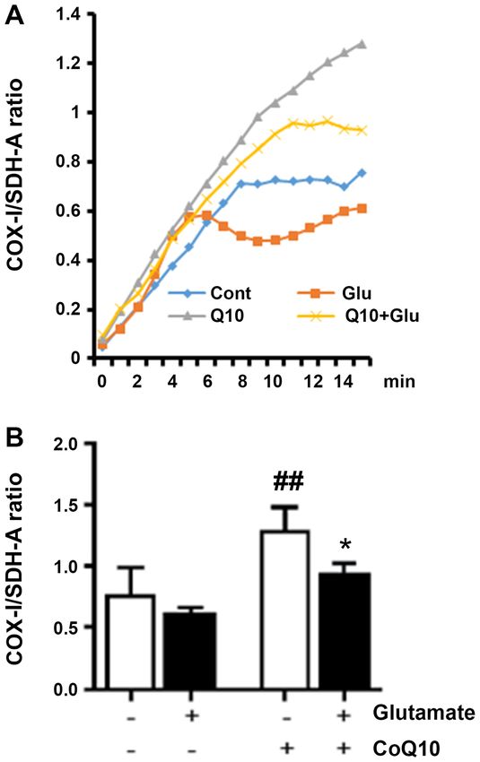
2                               ZIMMERMAN et al: COQ10 PROMOTES MITOCHONDRIAL BIOGENESIS

major cofactor of the electron transport chain (ETC) where          of these transcription factors by PGC‑1α, leads to the synthesis
majority ROS are produced. Within the ETC, CoQ10 plays a            of mitochondrial genes encoded in nuclear and mitochondrial
key role in transporting electrons between complexes I, II and      DNA, promoting generation of new mitochondria.
III. It is also recognized as having antioxidant functions. Thus,       The overall objectives of this work were to determine
when ETC function is disrupted by a mitochondrial insult,           whether glutamate exposure itself affected mitochondrial
CoQ10's location positions itself as a key antioxidant to reduce    biogenesis in hippocampal cells and whether CoQ10 supple‑
oxidative damage (10). However, continuous, high levels             mentation conferred protection against glutamate‑induced
of oxidative stress can lead to CoQ10 depletion and prevent         toxicity by promoting mitochondrial biogenesis. To this end,
adequate detoxification of ROS.                                     we measured mitochondrial biogenesis in hippocampal cell
     Therefore, exogenous antioxidant supplementation               exposed to glutamate, with and without CoQ10 pre‑treatment.
of CoQ10, can improve outcomes after mitochondrial                  We further analyzed mitochondrial biogenesis protein
damage (6‑8), including damage caused by glutamate (4).             expression patterns to determine the molecular mechanisms
CoQ10 is produced naturally within cells as part of the             involved. Given the active role mitochondria play in many
cholesterol pathway, but the cellular content can be further        disease models, including neuronal injury from glutamate
increased through consumption of food sources high in               excitotoxicity, we believe elucidating mitochondrial changes,
CoQ10, such as meat, nuts and green leafy vegetables.               and targeting them for correction, may improve therapeutic
Exogenous ubiquinone (oxidized form of CoQ10), or                   outcomes in patients.
ubiquinol [a reduced and more bioavailable form of
CoQ10 (11)], can also be administered. This additional              Materials and methods
supplementation may particularly benefit aging adults as
CoQ10 levels naturally decline with age (12).                       Cell culture. HT22 is a mouse hippocampal cell line kindly
     The benefits from CoQ10 supplementation have been docu‑        provided by Dr Panee at the University of Hawaii. HT22 cells
mented in a number of clinical trials. For example, in chronic      were cultured as previously described (21). Stock preparations
heart failure patients, CoQ10 decreased mortality, reduced          of glutamate (Sigma‑Aldrich; Merck KGaA) and CoQ10 supple‑
the incidence of hospitalization, and improved patients' func‑      ment, Ubisol‑Q10 (Zymes LLC), dissolved in water, were diluted
tional classification by the New York Heart Association (13).       with cell culture media before being added to the cells. Glutamate
CoQ10 increases total capacity of antioxidant enzymes and           concentrations (1‑8 mM) were tested in pilot study and 4 mM
reduces inflammatory biomarkers in diabetic hemodialysis            was selected for subsequent studies because this concentration
patients (14,15). CoQ10 benefits have also been reported in         resulted in a 30% reduction of cell viability assessed by resazurin
a clinical trial of multiple sclerosis (16). More clinical trials   assay after 24 h exposure. Treatment of Ubisol‑Q10 (25 µg/µl)
are needed to establish the efficacy of CoQ10 in preventing         was initiated 24 h prior to glutamate addition.
neurodegeneration and in preserving mitochondrial function.
Our lab has been interested in the neuroprotective benefits of      Reproductive potential assay. The reproductive potential of
CoQ10 supplementation. We believe this protection can be            HT22 cells after exposure to glutamate and/or CoQ10 treat‑
attributed to the effects of CoQ10 on the mitochondria, beyond      ment was assessed using colony formation assay. Cells were
antioxidant functions. In this study, our goal was to determine     treated as described above for untreated, glutamate, CoQ10,
whether CoQ10 could increase mitochondrial biogenesis to            and glutamate plus CoQ10 experimental groups. After treat‑
improve the outcome against glutamate toxicity.                     ment, the cells were harvested and seeded at a density of 100
     Mitochondrial biogenesis is a process by which new             cells per 10 cm dish, except in the case of the glutamate only
mitochondria are formed through the growth and division of          group, which were seeded at a higher density of 5,000 cells
pre‑existing mitochondria (17). Activation of this process often    per dish. The cells were incubated under standard culture
occurs during an insult to the cell. This is an attempt to coun‑    conditions for 14 days to allow visible colonies to form from
teract the damage process. Essentially, increasing numbers of       individual cells seeded. The colonies were stained with a
mitochondria serves to boost ATP production and to increase         0.05% crystal violet solution. Numbers of colonies were
detoxification of ROS. Unfortunately, this response is usually      counted using ImageJ software version 1.49 (22). Plating
not sufficient to counteract the damage process (18). However,      efficiencies were calculated by taking the actual numbers of
because of its protective effect, enhancing the mitochondrial       colonies formed in each plate and dividing by the number of
biogenesis could be used as a strategy to protect cells against     cells originally seeded and then multiplying by 100 to obtain
various damages including neurological diseases (19).               a percent. The percent of cells surviving was calculated by
     Changes in mitochondrial biogenesis can be determined          taking the individual plates' plating efficiencies and dividing
by examining expression of key proteins mediating mitochon‑         by the average plating efficiency of the untreated, control
drial biogenesis pathways. Peroxisome proliferator‑activated        group and again multiplying by 100. The average percent
receptor gamma coactivator‑1α (PGC‑1α) appears to be                survival for each group was quantified and analyzed for statis‑
the master regulator of mitochondrial biogenesis based on           tical significance using one‑way ANOVA and Bonferroni's
current knowledge (17). Once being activated, either through        multiple comparisons test.
phosphorylation or deacetylation, PGC‑1α activates the mito‑
chondrial transcription factors, nuclear respiratory factor 1       Western blotting. After undergoing experimental treatments
and 2 (NRF1,2). The NRF 1,2 could activate the mitochondrial        for 24 h [untreated, glutamate alone (4 mM), CoQ10 alone
NDA and protein synthesis either directly or through activating     (25 µg/µl), and glutamate plus CoQ10 24 h pre‑treatment],
mitochondrial transcription factor A (TFAM) (20). Activation        cells were harvested and washed with phosphate‑buffered
EXPERIMENTAL AND THERAPEUTIC MEDICINE 22: 1295, 2021 3
saline (PBS) before lysing to obtain either total protein where indicated. P‑values ≤0.05 were considered statistically
extracts, or protein fractions from cytosolic, mitochondrial, and significant.
nuclear compartments as previously described (21,23). Protein
concentrations were measured using standard Bradford assays Results
(Bio‑Rad Laboratories). Western blot analysis was performed
essentially as previously described (21). Equal amount of protein CoQ10 protects against cellular damage caused by glutamate
(20 µg) samples were loaded and separated on 4‑12% Bis‑Tris exposure. Because the toxic effect of glutamate exposure can
NuPAGE gels (Invitrogen; Thermo Fisher Scientific, Inc.) and vary between cell lines, we first looked at glutamate‑induced
then transferred to Polyvinylidene fluoride membranes. The loss of viability in HT22 cells using a range of glutamate
following primary antibodies from Cell Signaling Technology concentrations from 1 to 8 mM. The cells were exposed to
were used at 1:1,000 dilutions: Anti‑AKT, anti‑p‑AKT, these concentrations for 24 h before adding the resazurin dye
anti‑CREB, anti‑p‑CREB, anti‑COX‑IV, and anti‑Histone 3. as described previously (21). We observed a dose‑dependent
Anti‑PGC‑1α (1:1,000) was obtained from Abcam. Anti‑NRF2 decrease in cell viability as glutamate concentration rises.
(1:200) was from Santa Cruz Biotechnology and anti‑TFAM At 4 mM, glutamate decreased viability by roughly 30%
(1:1,000) was from Calbiochem. Both anti‑Histone 3 and (P80% of
β‑actin (1:5,000) were used as protein loading controls. IRDye cells were died (data not shown). As a result, 4 mM glutamate
680LT goat anti‑rabbit, IRDye 800CW goat anti‑mouse, and was selected for subsequent study. CoQ10 (25 µg/µl) treatment
donkey anti‑goat secondary antibodies from Li‑COR, Inc. alone did not affect cell viability. However, CoQ10 pretreat‑
were used at 1:10,000 dilutions for visualization using the ment to glutamate‑exposed cells effectively prevented the
Li‑COR Odyssey Classic Imaging System scanner. Protein decline of cell viability caused by glutamate (data not shown).
bands were analyzed with the Li‑COR Image Studio Software In addition to reducing cell viability directly, cells surviving
version 5.2.5. as previously described (21). Statistical signifi‑ glutamate exposure may lose their proliferative capability even
cance was measured using one‑way ANOVA and Bonferroni's after glutamate has been removed. We assessed the the repro‑
multiple comparisons test. ductive potential of HT22 cells after exposure to glutamate
and/or CoQ10 treatment using clonogenic colony forming
Measurement of mitochondrial biogenesis. Different levels of assay, an indication that they have retained their reproductive
mitochondrial biogenesis among experimental groups were ability and are still capable of cellular division. Our experi‑
assessed using a MitoBiogenesis™ In‑Cell ELISA Kit (Abcam) ments assessed this ability in cells that survived the initial 24 h
according to the manufacturer's protocol. Briefly, 20,000 cells glutamate exposure, CoQ10 treatment alone, or glutamate plus
per well were seeded in 96‑well plates and allowed to adhere CoQ10 pretreatment. Surviving cells were harvested, washed
overnight. The cells were then fixed with 4% paraformalde‑ and then seeded at low densities to allow individual cells to
hyde, briefly permeabilized with Triton X‑100, blocked, and form visible colonies.
incubated overnight at 4˚C with primary antibodies. Primary After staining colonies with crystal violet, the plating
antibodies were specific against mitochondrial DNA‑encoded efficiency (PE) for each group was calculated by taking the
protein, COX‑I, and nuclear DNA‑encoded protein, SDH‑A. number of colonies formed and dividing it by the number of
The cells were washed and incubated for 1 h at room tempera‑ cells seeded and then multiplying by 100 to obtain a percent.
ture with a solution of secondary antibodies containing an We found that in untreated cells, the average PE was 68%. Our
AP‑labeled antibody specific for SDH‑A and an HRP‑labeled results showed the PE was reduced in glutamate‑exposed cells
antibody specific for COX‑I. The reactions were sequentially dropping to 13%, but was not significantly altered from control
developed; first with an AP development solution, and then in the CoQ10 alone, or the glutamate plus CoQ10 group. To
an HRP development solution. Fifteen minute kinetic reac‑ find the surviving fraction, we divided the PE of the treated
tions with 1 min intervals were recorded using a PHERAstar sample by the PE of the control cells and multiplied by 100.
microplate reader (BMG Labtech) to measure optical density Value in untreated control group was set at 100% survival and
at 405 nm for AP development and 600 nm for HRP develop‑ our experimental groups were reported relative to this value.
ment. Whole cell staining with Janus Green was also done to Glutamate exposure dropped survival to 19% (P4 ZIMMERMAN et al: COQ10 PROMOTES MITOCHONDRIAL BIOGENESIS Figure 1. CoQ10 prevents the reduced cell survival induced by gluta‑ mate. The surviving fraction of cells was calculated as follows: PE of the treated sample/PE of the control cells x100. ###P
EXPERIMENTAL AND THERAPEUTIC MEDICINE 22: 1295, 2021                                                               5

Figure 3. CoQ10 pretreatment prevents the reduction of PGC‑1α protein expression induced by glutamate. (A) Representative western blotting images for
PGC‑1α protein, with β‑actin as a loading control. (B) Bar graph presenting the average protein expression ratio of PGC‑1α over β‑actin. ###P
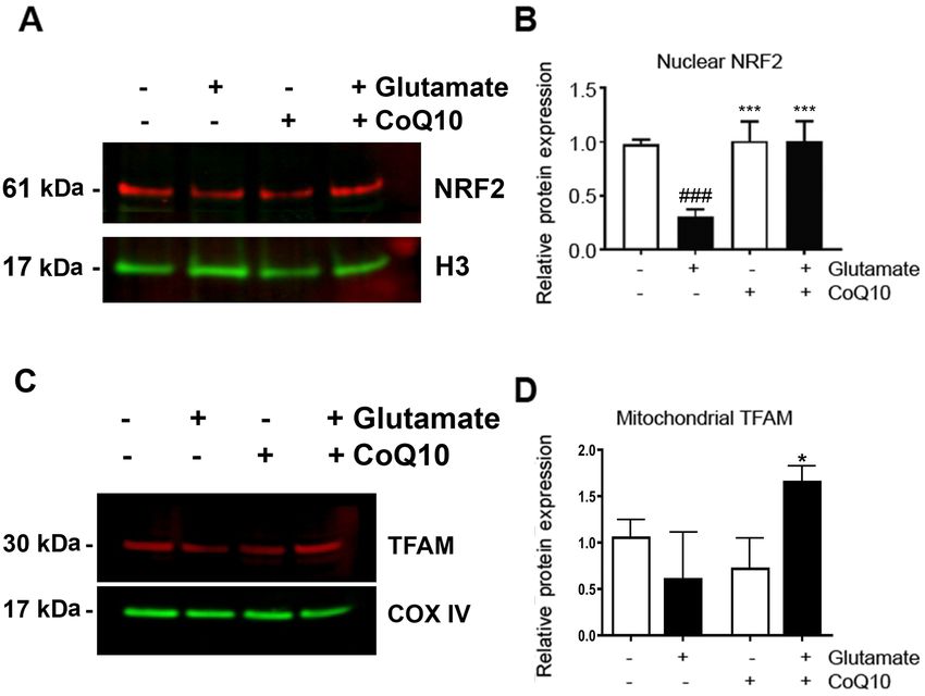
6                                    ZIMMERMAN et al: COQ10 PROMOTES MITOCHONDRIAL BIOGENESIS

Figure 5. CoQ10 increased NRF2 and TFAM. (A) Representative western blotting images of NRF2 in the nuclear fraction. The results demonstrated that
glutamate reduced the protein expression of NRF2, which was restored by CoQ10. (B) Average nuclear protein expression of NRF2 relative to an untreated
control. Protein content was normalized to H3. (C) Representative western blotting images of TFAM in the mitochondrial fraction. CoQ10 treatment increased
the expression of TFAM. (D) Average mitochondrial protein expression of TFAM relative to an untreated control. Protein content was normalized to
COX‑IV. Results from at least three separate experiments are presented. *P
EXPERIMENTAL AND THERAPEUTIC MEDICINE 22: 1295, 2021                                                      7

Availability of data and materials                                          13. Mortenson SA, Rosenfeldt F, Kumar A, Dolliner P, Filipiak KJ,
                                                                                Pella D, Alehagen U, Steurer G and Littarru GP: Q‑SYMBIO
                                                                                study investigators: The effect of coenzyme Q10 on morbidity
The datasets used and/or analyzed during the current study are                  and mortality in chronic heart failure: Results from Q‑SYMBIO:
available from the corresponding author on reasonable request.                  A randomized double‑blind trial. JACC Heart Fail 2: 641‑649,
                                                                                2014.
                                                                            14. Folkers K and Simonsen R: Two successful double‑blind trials
Authors' contributions                                                          with coenzyme Q10 (vitamin Q10) on muscular dystrophies and
                                                                                neurogenic atrophies. Biochim Biophys Acta 1271: 281‑286,
                                                                                1995.
GC and PAL conceived and designed the experiments of the                    15. Fallah M Askari G, Soleimani A, Feizi A and Asemi Z:
current study. MAZ, MH, QQ and SLM performed the experi‑                        Clinical trial of the effects of coenzyme Q10 supplementation
ments. MAZ, GC, PAL wrote the manuscript. All authors read                      on biomarkers of inflammation and oxidative stress in diabetic
                                                                                hemodialysis patients. Int J Prev Med 10: 12, 2019.
and approved the final manuscript.                                          16. Sanoobar M, Dehghan P, Khalili M, Azimi A and Seifar F:
                                                                                Coenzyme Q10 as a treatment for fatigue and depression in
Ethics approval and consent to participate                                      multiple sclerosis patients: A double blind randomized clinical
                                                                                trial. Nutr Neurosci 19: 138‑143, 2016.
                                                                            17. Valero T: Mitochondrial biogenesis: Pharmacological
Not applicable.                                                                 approaches. Curr Pharm Des 20: 5507‑5509, 2014.
                                                                            18. Uittenbogaard M and Chiaramello A: Mitochondrial biogenesis:
                                                                                A therapeutic target for neurodevelopmental disorders and
Patient consent for publication                                                 neurodegenerative diseases. Curr Pharm Des 20: 5574‑5593,
                                                                                2014.
Not applicable.                                                             19. Procaccio V, Bris C, Chao de la Barca JM, Oca F, Chevrollier A,
                                                                                Amati‑Bonneau P, Bonneau D and Reynier P: Perspectives of
                                                                                drug‑based neuroprotection targeting mitochondria. Rev Neurol
Competing interests                                                             (Paris) 170: 390‑400, 2014.
                                                                            20. Li PA, Hou X and Hao S: Mitochondrial biogenesis in neurode‑
                                                                                generation. J Neurosci Res 95: 2025‑2029, 2017.
The authors declare that they have no competing interests.                  21. Zimmerman MA, Biggers CD and Li PA: Rapamycin treatment
                                                                                increases hippocampal cell viability in an mTOR‑independent
                                                                                manner during exposure to hypoxia mimetic, cobalt chloride.
References                                                                      BMC Neurosci 19: 82, 2018.
                                                                            22. Guzman C, Bagga M, Kaur A, Westermarck J and Abankwa D:
 1. Olivares‑Banuelos TN, Chí‑Castañeda D and Ortega A: Glutamate               ColonyArea: an ImageJ plugin to automatically quantify colony
    transporters: Gene expression regulation and signaling properties.          formation in clonogenic assays. PLoS ONE 9:e92444, 2014.
    Neuropharmacology 161: 107550, 2019.                                    23. Andrews NC and Faller DV: A rapid micro‑preparation technique
 2. Amantea D and Bagetta G: Excitatory and inhibitory amino acid               for extraction of DNA‑binding proteins from limiting numbers of
    neurotransmitters in stroke: From neurotoxicity to ischemic toler‑          mammalian cells. Nucleic Acids Res 19: 2499, 1991.
    ance. Curr Opin Pharmacol 35: 111‑119, 2017.                            24. Mehta SL, Kumari S, Mendelev N and Li PA: Selenium preserves
 3. You J, Feng L, Xin M, Ma D and Feng J: Cerebral ischemic post‑              mitochondrial function, stimulates mitochondrial biogenesis,
    conditioning plays a neuroprotective role through regulation of             and reduces infarct volume after focal cerebral ischemia. BMC
    central and peripheral glutamate. Biomed Res Int 2018: 6316059,             Neurosci 13: 79, 2012.
    2018.                                                                   25. Ma YM, Ibeanu G, Wang LY, Zhang JZ, Chang C, Dong JD,
 4. Kumari S, Mehta SL, Milledge GZ, Huang X, Li H and Li PA:                   Li PA and Jing L: Selenium suppresses glutamate‑induced cell
    Ubisol‑Q10 prevents glutamate‑induced cell death by blocking                death and prevents mitochondrial morphological dynamic altera‑
    mitochondrial fragmentation and permeability transition pore                tions in hippocampal HT22 neuronal cells. BMC Neurosci 18:
    opening. Int J Biol Sci 12: 688‑700, 2016.                                  15, 2017.
 5. Kumari S, Mehta SL and Li PA: Glutamate induces mitochondrial           26. Wang X, Li H and Ding S: The effects of NAD+ on apoptotic
    dynamic imbalance and autophagy activation: Preventive effects              neuronal death and mitochondrial biogenesis and function after
    of selenium. PLoS One 7: e39382, 2012.                                      glutamate excitotoxicity. Int J Mol Sci 15: 20449‑20468, 2014.
 6. Jing L, Kumari S, Mendelev N and Li PA: Coenzyme Q10 amelio‑            27. Dinkova‑Kostova AT and Abramov AY: The emerging role
    rates ultraviolet B irradiation induced cell death through inhibition       of Nrf2 in mitochondrial function. Free Radic Biol Med 88:
    of mitochondrial intrinsic cell death pathway. Int J Mol Sci 12:            179‑188, 2015.
    8302‑8315, 2011.                                                        28. Hayashi G, Jasoliya M, Sahdeo S, Saccà F, Pane C, Filla A,
 7. Li H, Chen G, Ma W and Li PA: Water‑soluble coenzyme Q10                    Marsili A, Puorro G, Lanzillo R, Brescia Morra V and
    inhibits nuclear translocation of apoptosis inducing factor and cell        Cortopassi G: Dimethyl fumarate mediates Nrf2‑dependent
    death caused by mitochondrial complex I inhibition. Int J Mol               mitochondrial biogenesis in mice and humans. Hum Mol
    Sci 15: 13388‑13340, 2014.                                                  Genet 26: 2864‑2873, 2017.
 8. Jing L, He MT, Chang Y, Mehta SL, He QP, Zhang JZ and Li PA:            29. Bernard K, Logsdon NJ, Miguel V, Benavides GA, Zhang J,
    Coenzyme Q10 protects astrocytes from ROS‑induced damage                    Carter AB, Darley‑Usmar VM and Thannickal VJ: NADPH
    through inhibition of mitochondrial‑mediated cell death pathways.           oxidase 4 (Nox4) suppresses mitochondrial biogenesis and bioen‑
    Int J Biol Sci 11: 59‑66, 2015.                                             ergetics in lung fibroblasts via a nuclear factor erythroid‑derived
 9. Li HN, Zimmerman M, Milledge GZ, Hou XL, Cheng J, Wang ZH                   2‑like 2 (Nrf2)‑dependent pathway. J Biol Chem 292: 3029‑3038,
    and Li PA: Water soluble coenzyme Q10 reduced rotenone‑induced              2017.
    mitochondrial fission. Neurochem Res 42: 1096‑1103, 2017.               30. Chaturvedi RK and Beal MF: Mitochondrial diseases of the
10. Niyazov DM, Kahler SG and Frye RE: Primary mitochondrial                    brain. Free Radic Biol Med 63: 1‑29, 2013
    disease and secondary mitochondrial dysfunction: Importance of          31. Khatua TN, Dinda AK, Putcha UK and Banerjee SK: Diallyl
    distinction for diagnosis and treatment. Mol Syndromol 7: 122‑137,          disulfide ameliorates isoproterenol induced cardiac hypertrophy
    2016.                                                                       acting mitochondrial biogenesis via eNOS‑Nrf2‑Tfam pathway
11. Failla ML, Chitchumroonchokchai C and Aoki F: Increased                     in rats. Biochem Biophys Rep 5: 77‑88, 2015.
    bioavailability of ubiquinol compared to that of ubiquinone is          32. Kang I, Chu CT and Kaufman BA: The mitochondrial transcrip‑
    due to more efficient micellarization during digestion and greater          tion factor TFAM in neurodegeneration: Emerging evidence and
    GSH‑dependent uptake and basolateral secretion by Caco‑2 cells.             mechanisms. FEBS Lett 592: 793‑811, 2018.
    J Agric Food Chem 62: 7174‑7182, 2014.                                                   This work is licensed under a Creative Commons
12. Kalen A, Appelkvist EL and Dallner G: Age‑related changes in the
    lipid compositions of rat and human tissues. Lipids 24: 579‑584,                         Attribution-NonCommercial-NoDerivatives 4.0
    1989.                                                                                    International (CC BY-NC-ND 4.0) License.
You can also read