IN VITRO CYTOTOXICITY AND IN VIVO ACUTE ORAL TOXICITY EVALUATION OF COPTIS CHINENSIS AQUEOUS EXTRACT
←
→
Page content transcription
If your browser does not render page correctly, please read the page content below

WCRJ 2021; 8: e1971

IN VITRO CYTOTOXICITY
AND IN VIVO ACUTE ORAL TOXICITY
EVALUATION OF COPTIS CHINENSIS
AQUEOUS EXTRACT
C. T. KUMARAPPAN1,2, M. J. CINI3
1
 Department of Pharmacology and Toxicology, College of Pharmacy, King Khalid University, Abha, Asir
 Province, Saudi Arabia
2
  Department of Pharmacology and Lifescience, School of Pharmacy, Taylor’s University, Subang Jaya,
 Selangor, Malaysia
3
  Pharmacology and Toxicology Research Laboratory, Faculty of Pharmacy, University Technology MARA
 (UiTM), Bandar Puncak Alam, Malaysia

    Abstract – Objective: Rhizoma Coptidis (Coptis chinensis, Hunaglian) is most widely used Tradi-
tional Chinese Medicine (TCM) in daily life to treat inflammatory diseases and other various clinical
conditions in Malaysia and China. Our aim was to evaluate in vitro cytotoxicity and in vivo acute
oral toxicity of aqueous extract of Coptidis Rhizoma (AECR).
    Materials and Methods: The toxicology profile includes in vivo acute oral toxicity study and in
vitro cytotoxicity against non-cancerous human embryonic hepatic cell lines (WRL68 cells). The in vivo
study aims to test the acute toxicity in Wistar rats with a fixed oral dose of 2,000 mg/kg for 14 days.
Signs of acute oral toxicity in terms of behavior changes and mortality were noted for few hours till
14 days. Concurrently, the in vitro cytotoxicity, cell cycle arrest and apoptosis induction assays were
performed against WRL68 cells using, MTS cell viability and flow cytometry assays, respectively.
    Results: In acute oral toxicity study, there was no lethality and acute toxic signs were observed
up to 2,000 mg/kg b.w. for the 14 days of observed duration. However, AECR demonstrated low lev-
el of in vitro cytotoxicity (IC50= 7.167 ± 2.57 µg/ml) and moderate apoptosis inducing effect (p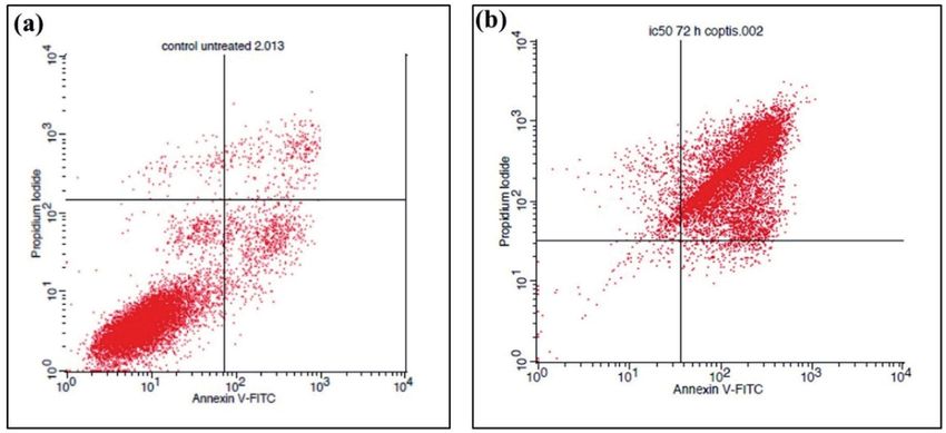
and studied for their medicinal properties and poten-           WRL68 cells for the rhizomes of C. chinensis to develop
tial bioactivities, but there are still many plants to be       an effective natural anticancer drug, which has been re-
investigated for their safety and toxicities2. Although         ported in the literature to some extent.
traditional herbal medicines are used by an increasing
number of Malaysians, little scientific information is
available regarding the safety and effectiveness3. Most         MATERIALS AND METHODS
medicinal plants used by the Malaysian have not been
tested in terms of their possible toxicity. The abundant        Chemicals and reagents
use of herbal medicines by numerous Malaysian pa-
tients may offer opportunities to ethnopharmacology             Gallic acid and Folin-Ciocalteu’s reagent were pur-
researchers to evaluate the safety and efficacy of many         chased from Merck (Malaysia). Sodium bicarbonate
herbs. It is an essential approach needed early in the          was purchased from Fluka Chemicals. Phosphate
drug discovery and development of natural products              buffered saline (PBS) pellet was purchased from MP
to determine their likely cytotoxic effects.                    Biomedicals (MP Biomedicals Inc, Santa Ana, CA,
    Coptidis Rhizoma (CR), a member of the Ranun-               USA). Sodium bicarbonate, Dulbecco’s Modified Ea-
culaceae family, derives from the dried rhizomes of             gle’s Medium (DMEM), fetal bovine serum (FBS),
Coptis chinensis. The dried rhizome of C. chinensis             RPMI, penicillin, streptomycin and trypsin were pur-
Franch. (Huanglian) has been widely used in Asian               chased from Sigma-Aldrich (Sigma-Aldrich Co, St
traditional medicine4. It was reported that Coptis Rhi-         Louis, MO, USA). The aqueous solution was obtained
zomes have anti-inflammatory, immune-modulatory,                from the research laboratory. Annexin-V and propidi-
antidiabetic, cardioprotective and neuroprotective              um iodide (PI) were obtained from Becton Dickinson
activities5-9. Based on current scientific literature, the      (Brea, CA, USA). Tetrazolium compound(3-(4,5-di-
major pharmacologically active compounds contrib-               methylthiazol-2-yl)-5-(3-carboxymethoxyphenyl)-2-
uting to Coptis’s bioactive properties are berberine,           (4-sulfophenyl)-2H tetrazolium, inner salt; MTS) was
coptisine, palmatine, epiberberine, jatorrhizine and            purchased from Promega (Promega, Madison, WI,
magnoflorine and worenine10. It also contains antisep-          USA). A dried sample of Coptidis Rhizoma was pur-
tic alkaloids and thus its rhizomes and preparations are        chased from a local Chinese Medicine supplier. All
widely used for the treatment of fish diseases. Today,          other reagents were of analytical grade and obtained
CR is still extensively consumed in TCM for the treat-          from common sources.
ment and management of numerous disorders. In Chi-
na, it is a common component in traditional medicines
used to treat cardiovascular associated problems11.             Preparation of aqueous extract Coptidis
    Recently the importance of CR has been increased            Rhizoma (AECR)
due to new biological and pharmacological investigations
about its therapeutic applications. In contrast to its ben-     Coptidis Rhizoma (50.0 g) was coarsely powdered for
eficial effect, CR is also found to be toxic in human and       the preparation of solvent extraction. The dried Coptidis
animals12. Given the intensive therapeutic applications of      Rhizoma was extracted with 250.0 ml of aqueous solvent
CR in the TCM, growing concerns have arisen about its           at room temperature. The aqueous extract of Coptidis
unintentional health impacts. There were very few clini-        Rhizoma (AECR) was concentrated in a water bath incu-
cal reports of adverse reaction attributable to CR in oral      bator (Memmert, Buchenbach, Germany) at 60°C-70°C.
TCM formulation. It has been banned in Singapore for            The extract was obtained and kept in a 4°C refrigerator
the past three decades due to the implication of berber-        in the sterile conical flask for further in vitro pharma-
ine in aggravating jaundice and kernicterus in neonates         cological studies. The yield of the extracted sample was
with glucose-6-phosphate dehydrogenase deficiency13.            calculated by the following equation:
The results from a new toxicological study revealed that
C. chinensis extracts appeared to demonstrate weak em-                        weight of Coptidis rhizoma extract (g)
                                                                Extracted =                                          x 100%
bryotoxicity14. Even though, the toxicity of this medicinal                   weight of dried Coptidis rhizoma (g)
plant is poorly understood. A review of available liter-
ature indicates that there has been a considerably lack
of research on its cytotoxic and apoptotic activities. The      Cytotoxic assay (MTS)
toxicological studies of AECR were initiated by the in vi-
tro evaluation of mammalian cell cytotoxicity performed         Cytotoxic activity of AECR against Human Nor-
with rat human embryonic normal liver cells (WRL68              mal Embryonic Liver Cell Line (WRL68) was deter-
cell line), as hepatic cells are playing a key role in detox-   mined by using standard MTT [(3-(4, 5-dimethylthi-
ification reactions and general metabolism control. Thus,       azol-2-yl)-2,5-diphenyltetrazolium bromide] viability
we conducted in vivo acute toxicity, in vitro cytotoxic,        assay as described previously15. Briefly, the WRL-68
cell cycle arrest and induction of apoptosis assays in          cells were counted and seeded (2x10⁴ cells/mL) in a 96-

2
SAFETY AND CYTOTOXICITY STUDY OF C. CHINENSIS EXTRACT

well flat-bottom microplate followed by 24-h incuba-        cycle). All the animals were acclimatized in the labo-
tion. Cells were incubated with various concentrations      ratory for at least 7 days before the toxicity study, with
of the AECR (2-500 µg/mL) at 37°C in a CO2 environ-         free access to water and food. The experimental pro-
ment for 72 h. Wells containing DMEM or RPMI 1640           tocol was approved by the Institutional Animal Care
was used as the negative controls. After the incubation     and Use Committee, Faculty of Medicine, University
periods, 20 µL of MTT solution (5 mg/mL) was added          of Malaya, Malaysia.
to each well and incubated for 4 h. To dissolve the for-
mazan crystals formed, 100 µL of DMSO was added to
each well. The absorbance for each well was measured        Ethical approval
at 540 nm in a micro-titer plate reader (Bio-Rad, Her-
cules, CA, USA). The dose-response curve was plotted        The in vivo toxicity study and its protocol were ap-
and the concentration which gave 50% of cell growth         proved by the Institutional Animal Ethical Commit-
(IC50) was calculated. All treatments, including negative   tee, Faculty of Medicine, University of Malaya. The
control, were carried out in triplicate experiments.        in vivo experiments and procedures were performed
                                                            by adopting international ethical guidelines of the Na-
                                                            tional Institutes of Health on the Care and Use of Lab-
Apoptosis analysis                                          oratory Animals and the Organization for Economic
                                                            Co-operation and Development (OECD, 2000) guide-
Flow cytometry analysis was carried out to investigate      lines.
the proportion of cancer cells undergoing apoptosis
using annexin-V conjugated with fluorescein isothio-
cyanate (FITC) detection kit according to the manu-         The acute oral toxicity study
facturer’s instructions. Cells (1×10⁶/well) were seeded
onto 24-well plates and incubated at 37°C overnight.        The acute oral toxicity test was carried out according
The next day, cells were treated with various concen-       to the OECD test guideline 425 (Up and Down Proce-
trations of AECR and incubated for 72 h. After this,        dure), with slight modifications16. The limit test was
cells were detached using trypsin for 5 min. Cells were     performed at 2000 mg/kg p.o. as a single dose and rats
collected and centrifuged at 1000 rpm for 5 min. The        were kept without food for 3-4 h before dosing but
cell pellet was washed with PBS and resuspended in          had free access to water ad libitum. The animals were
100 mL binding buffer. Subsequently, 2.5 mL annexin         divided into two groups (Vehicle control and ACER
V-FITC and 3 mL propidium iodide were added and             treated), each comprising 6 animals. The dose was ad-
kept in the dark at room temperature for 15 min. An-        ministered to a single female rat from the test group
nexin V-FITC/PI stained cells were analyzed using           according to body weight. The animal was closely
flow cytometry (BD Biosciences LSR II FACS, San             observed for the first 30 min, then for 4 h. Food was
Francisco, CA, USA).                                        provided after 1–2 h of dosing. After the survival of
                                                            the treated rat, 5 more rats were administered with the
                                                            same dose under similar conditions. The equal proce-
Cell Cycle Analysis by flow cytometry                       dure was followed for the vehicle-treated (0.1 % car-
                                                            boxymethyl cellulose) control group of 6 rats with the
Cells were treated as described in flow cytometry anal-     same volume as that of the AECR treated group. Sur-
ysis. After treatment and incubation for 72 h, cells were   vived animals from both groups were observed closely
harvested and fixed with ice-cold 70% ethanol (1 mL)        for any acute toxic effects within the first 4 h and then
at -20°C for 2 h. Ethanol was then removed (1000 rpm,       at regular intervals for a total period of 14 days for oth-
5 min) and the cells were washed twice with cold PBS.       er toxic effects. Surviving animals were further visu-
Subsequently, cells were resuspended in 425 μL of PBS,      ally observed for mortality, behavioral pattern, chang-
25 μL propidium iodide (1 mg/mL), and 50 μL RNaseA          es in physical appearance, injury, pain, and other signs
(1 mg/mL) and incubated for 30 min at room tempera-         of illness were recorded daily for 14 days.
ture. Distribution of the cell cycle was measured at 585
nm by flow cytometer and data analysis was carried out
with ModFitLT software (version 4)17.                       Statistical analysis

                                                            Data were subjected to analysis of variance (ANOVA)
Experimental Animals                                        using Dunnett’s test and were used to assess differ-
                                                            ences between means. A significant difference was
Six Male and Six female (nulliparous and nonpregnant)       considered at a level of p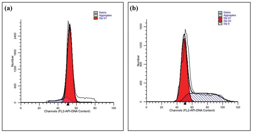
RESULTS significantly (p
SAFETY AND CYTOTOXICITY STUDY OF C. CHINENSIS EXTRACT
dose of 2000 mg/kg thus establishing its safety in use.
All the behavioral and other visual observations were
found to be normal for the AECR (2000 mg/kg) and
vehicle-treated groups.
DISCUSSION
The rhizomes of C. chinensis is traditionally used in
Chinese folk medicine for treating various ailments
and reported to have numerous pharmacological activi-
ties18,19. The best known biologically active compounds
are the alkaloids identified in the rhizome of the plant20.
Despite the various traditional claim, phytochemical
and pharmacological activities, little is known about its
effect on cell survival, growth, and apoptosis to define
its cytotoxic nature. Safety assessment is a part of ex-
perimental research on the extensive therapeutic effects
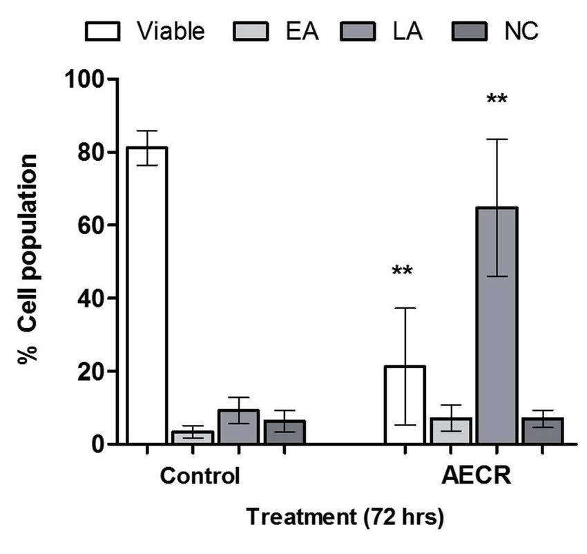
Fig. 2. Flow cytometric analysis of Annexin V and FITC/ besides the major biological activity. On the other hand,
propidium iodide staining of WRL68 cells exposed to the issue of the safety of traditional herbal preparations
AECR. Three independent experiments are performed in worldwide has been constantly questioned. Moreover,
triplicate. **p 2000 mg/kg). Our results are following previ-
were observed in Wistar rats treated with AECR at a ously published works and the LD50 value of AECR by
Fig. 3. Effect of AECR on cell cycle progression in WRL68 cells. (a) Control cells (b) Cells (1×10⁶ /well) were treated with 7.16
µg/ml of AECR for 72 h at 37˚C. Data are representative of three independent experiments.
5TABLE 1. Effect of oral administration of AECR (2000 mg/kg) on behavioral response and general appearance.
Parameters Control 2 ml/kg AECR 2000 mg/kg
Alertness Normal Normal
Touch and Pain Response Normal Normal
Food and water Intake Normal Normal
Tremors/Epilepsy/Sedation/Coma Not observed Not observed
Gripping/Corneal/Righting reflex Normal Normal
Salivation, Urination Normal Normal
Skin color and pupils Normal Normal
General physique Normal Normal
Temperature Normal Normal
Faeces consistency Normal Normal
Mortality Not observed Not observed
oral route was more than 2000 mg/kg, thereby suggest- 7.167 µg/ml concentrations caused an increase in the
ing that AECR is a non-toxic drug25. It is indicated that proportion of cells at the S phase and a decrease in the
the AECR can be used for further investigation of any proportion of cells at the G1 phase of the cell cycle. For
preclinical and other pharmacological effects on the WRL68 cells, after 72 hrs treatment, a significant de-
above-mentioned dose of extract. However, a detailed crease in the G1 phase coupled with the accumulation
subacute and chronic experimental analysis of its long- of cells in the S phase was observed. This effect sug-
term toxicity is essential for further clinical support of gests that the reduction in cell viability and prolifera-
this drug. tion of WRL68 caused by the induction of apoptosis.
The primary aim of cancer chemotherapy is to In this study, we showed that AECR efficiently
kill cancer cells selectively while limiting toxicity in lowered the proliferation of a WRL 68 cell line, by
healthy cells. This is a downside to the use of many signifying the roles of C. rhizome’s phytochemical
chemotherapeutic agents; as a result, selective toxici- in decreasing the number of cells, in the induction of
ty must be factored when exploring cancer treatment apoptosis, and in interfering with cell cycle arrest at
leads. Cytotoxicity screening models provide import- the S and G1 phases of the cell cycle. As experimen-
ant preliminary data to help select plant extracts with tal noted above, alterations in cell growth measured
potential cytotoxic or bioactivities for future work 26. in AECR treated cells may be due to the induction of
The criteria of cytotoxicity activity for the crude ex- the apoptosis process in corresponding to cell cycle
tracts, as established by the American National Cancer arrest. This outcome showed that the AECR might be
Institute is an IC50 < 30 µg/ml in the preliminary as- capable of inducing DNA fragmentation which is the
say following incubation time between 48 and 72 h27. hallmark for programmed cell death. Taken together,
An important point is that AECR shows cytotoxic re- our results suggest that there are potentially multiple
sponse on WRL68 cells for 72 hrs with an IC50 value mechanisms by which CR can inhibit the growth of
of 7.167 µg/ml. The IC50 value was found to be lower WRL68 cells. Our preliminary study potentially of-
than that specified by the American National Cancer fers valuable information that may be used to expand
Institute for categorization of developing as a promis- the safety application and chemotherapeutic investiga-
ing chemotherapeutic agent. The reduction of cell via- tion of CR to the field of TCM. Remarkably, several
bility as compared with untreated control cell may be recent investigations reveal that CR induces apoptosis
contributed by its apoptosis-inducing or proliferation in various cancer cells, suggesting that tumour cells
inhibiting effects. This investigation provides confir- may be more susceptible to CR than normal healthy
mation for cytotoxicity in WRL68 which may be due cells30-33. The limitation of the current study is that de-
to existing phytochemicals in the aqueous extract C. tection of key molecules related to apoptosis such as
chinensis as mentioned previously28. BCL2, BAX, PRPP and caspase cascades was not per-
Apoptosis also seems to be a reliable biomarker for formed to confirm the cell apoptosis as additional con-
the preliminary study of cytotoxic potential of natural founding information due to economic constraint. Our
compounds29. Our current study concerning AECR’s current study also recommends that care should be
ability to induce apoptosis may provide valuable infor- taken in terms of overdose and sustained treatments,
mation for the possible toxic effect in normal human which causes accumulation of the CR in the normal
embryonic liver cells. The cytotoxic activity was fur- organism34. Long-term in vivo and in vitro studies of
ther confirmed by measuring the different phases of cytotoxic, cell cycle arrest and apoptotic effects of
cell cycle progression of AECR treated WRL68 cells AECR on other normal and cancerous cells are now
using flow cytometry assay. It appears that AECR at in progress.
6SAFETY AND CYTOTOXICITY STUDY OF C. CHINENSIS EXTRACT
CONCLUSIONS 4. Linn YC, Lu J, Lim LC, Sun H, Sun J, Zhou Y, Ng HS. Ber-
berine-induced haemolysis revisited: safety of Rhizoma
coptidis and Cortex phellodendri in chronic haemato-
Acute in vivo oral administration of aqueous extract logical diseases. Phytother Res 2012; 26: 682-686.
of C. chinensis rhizome is non-toxic nature in healthy 5. Kim E, Ahn, S, Rhee HI, Lee DC. Coptis chinensis Franch
animals. However, the in vitro cytotoxicity and apop- extract up-regulate type I helper T-cell cytokine through
tosis analysis revealed the cytotoxic potential of the MAPK activation in MOLT-4 T cell. J Ethnopharmacol
rhizome extracts on WRL-68 cells. Additional re- 2016; 189: 126-131.
6. Wu J, Zhang H, Hu B, Yang L, Wang P, Wang F, Meng
search into subacute toxicity in healthy animals and X. Coptisine from Coptis chinensis inhibits production
chemoprevention mechanisms in various cancer cells of inflammatory mediators in lipopolysaccharide-stim-
is strongly needed. ulated RAW 264.7 murine macrophage cells. Eur J
Pharmacol 2016; 780: 106-114.
Acknowledgement: 7. Cui L, Liu M, Chang X, Sun K. The inhibiting effect of
the Coptis chinensis polysaccharide on the type II dia-
The authors would like to express their apprecia- betic mice. Biomed Pharmacother 2016; 81: 111-119.
tion to Taylors University’s Division of Research 8. Kaufmann D, Kaur Dogra A, Tahrani A, Herrmann F,
and Enterprise for their technical and financial Wink M. Extracts from traditional Chinese medicinal
assistance with the Emerging Research Fund plants inhibit acetylcholinesterase, a known Alzhei-
Scheme (ERFS/May/2017). mer’s disease target. Molecules 2016, 21: 1161.
9. Friedemann T, Ying Y, Wang W, Kramer ER, Schumach-
er U, Fei J. Neuroprotective effect of Coptis chinensis
Funding: in MPP and MPTP-induced parkinson’s disease models.
The study was financially supported by King Kha- Am J Chinese Med 2016; 44: 907-925.
lid University’s Small Research Grant Program 10. Kamath S, Skeels M, Pai A. Significant differences in
(SRP/275/1442) in the Kingdom of Saudi Arabia. alkaloid content of Coptis chinensis (Huanglian), from
its related American species. Chin Med 2009; 4: 17.
11. Tan HL, Chan KG, Pusparajah P, Duangjai A, Saokaew
Authors’ contributions: S, Mehmood Khan T. Rhizoma Coptidis: a potential
KC primarily designed the whole investigation cardiovascular protective agent. Front Pharmacol 2016;
and protocols, conducted the in vivo toxicologi- 7: 362.
cal experiments, performed Behavioural analysis, 12. Lee YH, Kim D, Lee MJ, Kim MJ, Jang HS, Park SH.
Subchronic toxicity study of Coptidis rhizoma in rats. J
measured acute toxicity signs, and secured fund- Ethnopharmacol 2014; 152: 457-463.
ing. KC had a chief role in the scientific writing of 13. Chen YN, Yuan L. Acute Toxicity of Coptis chinensis
the manuscript. KC and CMJ involved an active Rhizome Extracts to Daphnia carinata. Huan Jing Ke
role in the arrangement of research facilities and Xue 2015; 36: 3892-3895.
served as the project’s supervisor. CMJ complete- 14. Li LY, Cao FF, Su ZJ, Zhang QH, Dai XY1, Xiao X. As-
sessment of the embryotoxicity of four Chinese herbal
ly carried out the in vitro cytotoxicity, apoptosis extracts using the embryonic stem cell test. Mol Med
analysis and wrote the comments on in vitro part. Rep 2015; 12: 2348-2354.
KC actively involved in the scientific analysis of 15. Malich G, Markovic B, Winder C. The sensitivity and
the results, assisting to draft the manuscript, sta- specificity of the MTS tetrazolium assay for detecting
tistical study. Finally, both authors reviewed the the in vitro cytotoxicity of 20 chemicals using human
cell lines. Toxicology 1997; 124: 179-192.
whole manuscript and agreed to submit the paper 16. Saleem U, Amin S, Ahmad B, Azeem H, Anwar F, Mary
for scientific communication. S. Acute oral toxicity evaluation of aqueous ethanolic
extract of Saccharum munja Roxb. Roots in albino mice
Conflict of interest: as per OECD 425 TG. Toxicol Rep 2017; 4: 580-585.
The author declares that there are no conflicts of 17. Armania N, Yazan LS, Musa SN, Ismail IS, Foo JB, Chan
KW. Dillenia suffruticosa exhibited antioxidant and
interest. cytotoxic activity through induction of apoptosis and
G2/M cell cycle arrest. J Ethnopharmacol 2013; 146:
525-535.
18. Jung HA, Min BS, Yokozawa T, Lee JH, Kim YS, Choi
JS. Anti-Alzheimer and antioxidant activities of Coptidis
REFERENCES Rhizoma alkaloids. Biol Pharm Bull 2009; 32: 1433-
1438.
1. Yuan H, Ma Q, Ye L, Piao G. The traditional medicine 19. Kim JM, Jung HA, Choi JS, Lee NG. Identification of
and modern medicine from natural products. Mole- anti-inflammatory target genes of Rhizoma coptidis
cules 2016; 21: 1-18. extract in lipopolysaccharide-stimulated RAW264.7
2. Farooqui M, Hassali MA, Shatar AK, Farooqui MA, murine macrophage-like cells. J Ethnopharmacol 2010;
Saleem F, Haq NU, Othman CN. Use of complementary 130: 354-362.
and alternative medicines among Malaysian cancer 20. Li KL, Sheu SJ. Determination of flavonoids and al-
patients: a descriptive study. J Tradit Complement Med kaloids in the scute-coptis herb couple by capillary
2016; 6: 321-326. electrophoresis. Anal Chim Acta 1995; 313: 113-120.
3. Saranyaa S, Low BS. Current public awareness on 21. Taraphdar AK, Roy M, Bhattacharya RK. Natural prod-
the safety of traditional and complementary medicines ucts as inducers of apoptosis: Implication for cancer
(T&CM) in Malaysia. Eur J Integr Med 2015; 7: 184-189. therapy and prevention. Curr Sci 2001; 80: 1387-1396.
722. Efferth T, Sch0ttler U, Krishna S, Schmiedek P, Wenz F, 28. Choi JS, Ali MY, Jung HA, Oh SH4, Choi RJ5, Kim EJ.
Giordano FA. Hepatotoxicity by combination treatment Protein tyrosine phosphatase 1B inhibitory activity of
of temozolomide, artesunate and Chinese herbs in a alkaloids from Rhizoma Coptidis and their molecular
glioblastoma multiforme patient: case report review of docking studies. J Ethnopharmacol 2015; 171: 28-36.
the literature. Arch Toxicol 2016; 91: 1833-1846. 29. Amit KT, Madhumita R, Bhattacharya RK. Natural prod-
23. Wang H, Bai J, Chen G, Li W, Xiang R, Su G, Pei Y. A ucts as inducers of apoptosis: Implication for cancer
metabolic profiling analysis of the acute hepatotoxicity therapy and prevention. Curr Sci 2001; 80: 1387-1396.
and nephrotoxicity of Zhusha Anshen Wan compared 30. Wang N, Tan HY, Li L, Yuen MF, Feng Y. Berberine
with cinnabar in rats using (1)H NMR spectroscopy. J and Coptidis Rhizoma as potential anticancer agents:
Ethnopharmacol 2013; 146: 572-580. recent updates and future perspectives. J Ethnophar-
24. Nema R, Khare S, Jain P, Pradhan, A, Gupta A, Singh macol 2015; 176: 35-48.
D. Natural products potential and scope for modern 31. Liu J, He C, Zhou K, Wang J, Kang JX. Coptis extracts
cancer research. Am J Plant Sci 2013; 4: 1270-1277. enhance the anticancer effect of estrogen receptor
25. Martin AC, Johnston E, Xing C, Hegeman AD. Mea- antagonists on human breast cancer cells. Biochem
suring the chemical and cytotoxic variability of com- Biophys Res Commun 2009; 378: 174-78.
mercially available kava (Piper methysticum G. Forster). 32. Park HJ, Kim YJ, Leem K, Park SJ, Seo JC, Kim HK,
PLoS One 2014; 9: e111572. Chung JH. Coptis japonica root extract induces apop-
26. Lai X, Pei Q, Lei C, Peng L, Yin Z, Jia R. Acute and tosis through caspase3 activation in SNU-668 human
subchronic toxicity as well as evaluation of safety phar- gastric cancer cells. Phytother Res 2005; 19: 189-192.
macology of Coptis Chinensis Franch solution. Int J Clin 33. Chen HY, Ye XL, Cui XL, He K, Jin YN, Chen Z, Li XG.
Exp Med 2016; 9: 7668-7679. Cytotoxicity and antihyperglycemic effect of minor
27. Suffness M, Pezzuto JM. Assays related to cancer drug constituents from Rhizoma Coptis in HepG2cells. Fito-
discovery. In: Hostettmann K (Editor) Methods in Plant terapia 2012; 83: 67-73.
Biochemistry: Assays for Bioactivity. Academic Press, 34. Ning N, Wang YZ, Zou ZY, Zhang DZ, Wang DZ, Li XG.
London, U.K. 1990, pp. 71-133. Pharmacological and safety evaluation of fibrous root of
Rhizoma Coptidis. Environ Toxicol Pharmacol 2015; 39:
53-69.
8You can also read