TROPISM, REPLICATION COMPETENCE, AND INNATE IMMUNE RESPONSES OF THE CORONAVIRUS SARS-COV-2 IN HUMAN RESPIRATORY TRACT AND CONJUNCTIVA: AN ANALYSIS ...
←
→
Page content transcription
If your browser does not render page correctly, please read the page content below
Articles

Tropism, replication competence, and innate immune
responses of the coronavirus SARS-CoV-2 in human
respiratory tract and conjunctiva: an analysis in ex-vivo and
in-vitro cultures
Kenrie P Y Hui, Man-Chun Cheung, Ranawaka A P M Perera, Ka-Chun Ng, Christine H T Bui, John C W Ho, Mandy M T Ng, Denise I T Kuok,
Kendrick C Shih, Sai-Wah Tsao, Leo L M Poon, Malik Peiris, John M Nicholls, Michael C W Chan

Summary
Background Severe acute respiratory syndrome coronavirus 2 (SARS-CoV-2) emerged in December 2019, causing a                          Lancet Respir Med 2020
respiratory disease (coronavirus disease 2019, COVID-19) of varying severity in Wuhan, China, and subsequently                       Published Online
leading to a pandemic. The transmissibility and pathogenesis of SARS-CoV-2 remain poorly understood. We evaluate                     May 7, 2020
                                                                                                                                     https://doi.org/10.1016/
its tissue and cellular tropism in human respiratory tract, conjunctiva, and innate immune responses in comparison
                                                                                                                                     S2213-2600(20)30193-4
with other coronavirus and influenza virus to provide insights into COVID-19 pathogenesis.
                                                                                                                                     See Online/Comment
                                                                                                                                     https://doi.org/10.1016/
Methods We isolated SARS-CoV-2 from a patient with confirmed COVID-19, and compared virus tropism and                                S2213-2600(20)30227-7
replication competence with SARS-CoV, Middle East respiratory syndrome-associated coronavirus (MERS-CoV), and                        School of Public Health
2009 pandemic influenza H1N1 (H1N1pdm) in ex-vivo cultures of human bronchus (n=5) and lung (n=4). We                                (K P Y Hui PhD, M-C Cheung MSc,
assessed extrapulmonary infection using ex-vivo cultures of human conjunctiva (n=3) and in-vitro cultures of human                   R A P M Perera PhD, K-C Ng BSc,
                                                                                                                                     C H T Bui PhD, J C W Ho PhD,
colorectal adenocarcinoma cell lines. Innate immune responses and angiotensin-converting enzyme 2 expression                         M M T Ng BSc, D I T Kuok PhD,
were investigated in human alveolar epithelial cells and macrophages. In-vitro studies included the highly pathogenic                Prof L L M Poon DPhil,
avian influenza H5N1 virus (H5N1) and mock-infected cells as controls.                                                               Prof M Peiris FRCPath,
                                                                                                                                     Prof J M Nicholls FRCPA,
                                                                                                                                     M C W Chan PhD), Department
Findings SARS-CoV-2 infected ciliated, mucus-secreting, and club cells of bronchial epithelium, type 1 pneumocytes                   of Ophthalmology
in the lung, and the conjunctival mucosa. In the bronchus, SARS-CoV-2 replication competence was similar to                          (K C Shih MBBS), School of
MERS-CoV, and higher than SARS-CoV, but lower than H1N1pdm. In the lung, SARS-CoV-2 replication was similar                          Biomedical Science
to SARS-CoV and H1N1pdm, but was lower than MERS-CoV. In conjunctiva, SARS-CoV-2 replication was greater                             (Prof S-W Tsao PhD), and
                                                                                                                                     Department of Pathology,
than SARS-CoV. SARS-CoV-2 was a less potent inducer of proinflammatory cytokines than H5N1, H1N1pdm, or                              Queen Mary Hospital
MERS-CoV.                                                                                                                            (Prof J M Nicholls), Li Ka Shing
                                                                                                                                     Faculty of Medicine,
                                                                                                                                     The University of Hong Kong,
Interpretation The conjunctival epithelium and conducting airways appear to be potential portals of infection for
                                                                                                                                     Pokfulam, Special
SARS-CoV-2. Both SARS-CoV and SARS-CoV-2 replicated similarly in the alveolar epithelium; SARS-CoV-2 replicated                      Administrative Region of
more extensively in the bronchus than SARS-CoV. These findings provide important insights into the transmissibility                  Hong Kong, China
and pathogenesis of SARS-CoV-2 infection and differences with other respiratory pathogens.                                           Correspondence to:
                                                                                                                                     Dr Michael C W Chan, School of
Funding US National Institute of Allergy and Infectious Diseases, University Grants Committee of Hong Kong Special                   Public Health, Li Ka Shing Faculty
                                                                                                                                     of Medicine, The University of
Administrative Region, China; Health and Medical Research Fund, Food and Health Bureau, Government of Hong Kong                      Hong Kong, Pokfulam, Special
Special Administrative Region, China.                                                                                                Administrative Region of
                                                                                                                                     Hong Kong, China
                                                                                                                                     mchan@hku.hk
Copyright © 2020 Elsevier Ltd. All rights reserved.

Introduction                                                             especially within health-care facilities. To date, within
Several coronaviruses infect the human respiratory tract,                health-care facilities, with 2519 cases, with 866 deaths
and usually cause mild disease; however, the beta                        across 27 countries, have been confirmed as of January,
coronaviruses severe acute respiratory syndrome-                         2020.1
associated coronavirus (SARS-CoV) and Middle East                          In December, 2019, the novel coronavirus SARS-CoV-2
respiratory syndrome-associated coronavirus (MERS-CoV)                   caused an outbreak of respiratory illness (coronavirus
cause severe zoonotic respiratory disease. SARS emerged                  disease 2019; COVID-19) in Wuhan, China. Within
in 2002 in Guangdong province, China, and caused an                      5 months, the disease burden and fatalities have
epidemic leading to 8096 cases and 774 deaths globally in                surpassed both SARS and MERS, with more than
more than 25 countries across five continents, but was                   2 million confirmed cases and more than 150 000 deaths
contained through public health interventions. MERS-CoV                  reported globally, as of April 19, 2020.2 WHO declared
transmits from dromedary camels to humans, sometimes                     this outbreak a pandemic on March 11, 2020. Although
leading to clusters of human-to-human transmission,                      the virus appears to be more transmissible than either

www.thelancet.com/respiratory Published online May 7, 2020 https://doi.org/10.1016/S2213-2600(20)30193-4                                                            1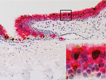
Articles
Research in context
Evidence before this study infection of SARS-CoV-2. Both SARS-CoV and SARS-CoV-2
We searched PubMed without language restriction for studies replicated comparably in the alveolar epithelium, but
published from database inception until March 9, 2020, with SARS-CoV-2 replicated more extensively than SARS-CoV in
the terms “SARS-CoV-2” or ”novel coronavirus” and “virus bronchial epithelium, which might explain the increased
tropism” or “respiratory tract” or “ocular” or “conjunctiva” or transmissibility of the virus. SARS-CoV-2 was a less potent
“innate immunity” or “cytokine”, and found no relevant articles inducer of proinflammatory cytokines than H5N1,
pertaining to severe acute respiratory syndrome coronavirus 2 MERS-CoV, or H1N1pdm.
(SARS-CoV-2). To our knowledge, there have been no reports
Implications of all the available evidence
on infection, replication competence, tropism, and
SARS-CoV-2 replicates better than other human
pathogenesis of the novel coronavirus SARS-CoV-2, in
coronaviruses, such as SARS-CoV, but not as well as
comparison with other respiratory pathogens including
H1N1pdm in ex-vivo cultures of the human bronchus.
SARS-CoV, Middle East respiratory syndrome coronavirus
The conjunctiva is an additional portal of infection. These
(MERS-CoV), 2009 pandemic influenza H1N1 virus (H1N1pdm),
findings are relevant to understanding transmission for
and highly pathogenic avian influenza H5N1 virus (H5N1), in
infection prevention and control. Extrapulmonary routes of
human respiratory tract or extrapulmonary organs.
infection by SARS-CoV-2 should be further studied and
Added value of this study validated in animal models.
We report that the conjunctival epithelium and the
conducting airways appear to be potential portals of
SARS or MERS, disease severity is variable—from We aimed to compare virus tropism and replication
asymptomatic to fatal—and case fatality appears to be competence of SARS-CoV-2 virus with SARS-CoV,
substantially lower than both SARS and MERS.3 MERS-CoV, and H1N1pdm viruses in ex-vivo cultures of
Modes of transmission and pathogenesis have been human bronchus and lung. The potential for SARS-CoV-2
key knowledge gaps. The virus is assumed to be to infect extrapulmonary tissues was also assessed using
primarily transmitted by large respiratory droplets, but ex-vivo cultures of human conjunctiva and in-vitro
there has been no direct evidence for this hypothesis. cultures of human colorectal adenocarcinoma cell lines.
Identifying the organs and cell types that are permissive The innate immune responses to SARS-CoV-2 were
to implantation and virus replication will help to investigated in infected human alveolar epithelial cells
understand the portals by which the infection can be and macrophages, and compared with those of highly
established. To our knowledge, to date there has been pathogenic avian influenza (HPAI) H5N1 virus.
only one autopsy study reporting a patient dying after
testing positive for SARS-CoV-2 using needle core Methods
samples and one histopathological study on two patients SARS-CoV-2 isolation
undergoing lobectomy.4,5 Although these studies have VeroE6 cells were used for virus isolation and were
provided information on the histopathology of cultured in DMEM and 10% FCS. Cultured cell
COVID-19, they did not include characterisation of monolayers were maintained in their respective medium.
SARS-CoV-2 virus tropism by immunohistochemistry, The original clinical specimen was collected from the
and therefore do not shed light on virus tropism in the nasopharyngeal aspirate and throat swab of a patient
early stages of the infection process.4,5 Thus, data from (young adult male) with confirmed COVID-19 in
experimental infection of ex-vivo cultures of the Hong Kong in January, 2020, and was diluted (1:10) with
respiratory tract are needed. DMEM supplemented with 2% FCS before adding to
We have previously used ex-vivo cultures of the human cells. After incubation at 37°C for 1·5 h, medium was
lung, conducting airways, and ocular conjunctiva to topped-up to 1 mL with fresh culture medium. The cells
investigate tropism of epidemic viruses such as the 2009 were incubated at 37°C and observed daily for cytopathic
pandemic influenza H1N1 (H1N1pdm) virus and effect (CPE). The culture supernatant was examined for
MERS-CoV.6,7 Furthermore, when compared with presence of virus RNA by rtPCR.
patients with COVID-19 who were not in the intensive All experiments were done in a biosafety level 3 facility
care unit (ICU), patients in the ICU had higher plasma at the School of Public Health, Li Ka Shing Faculty of
concentrations of proinflammatory cytokines such as Medicine, The University of Hong Kong. Ethics approval
interferon gamma-induced protein 10 (IP-10), monocyte of the use of human tissues was granted by the institutional
chemoattractant protein 1 (MCP-1), and tumour necrosis review board of the University of Hong Kong and the
factor α (TNF-α).8 It is therefore important to investigate hospital authority (Hong Kong West; institutional review
the role of innate host responses in pathogenesis. board approval numbers UW 20–167 and UW19–802).
2 www.thelancet.com/respiratory Published online May 7, 2020 https://doi.org/10.1016/S2213-2600(20)30193-4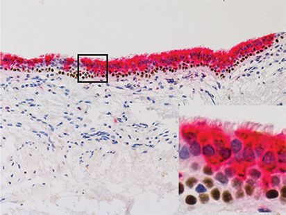
Articles
Viruses In-vitro culture and infection of alveolar epithelial cells,
In addition to the SARS-CoV-2 isolate (BetaCoV/ macrophage, and colorectal carcinoma cells
Hong Kong/VM20001061/2020), we used: SARS-CoV Primary human alveolar epithelial cells (AECs) and
(strain HK39849), isolated from a patient admitted to peripheral blood monocyte-derived macrophages were
hospital with SARS infection in Hong Kong in 2003, isolated from three donors and used for infection, as
MERS-CoV (prototype human MERS-CoV EMC strain, previously described,9 and human colorectal carcinoma
provided by Prof R Fouchier, Erasmus University Medical cells (Caco-2; ATCC HTB-37) purchased from American
Center, Rotterdam, the Netherlands); the HPAI H5N1 Type Culture Collection were cultured in MEM with
virus (A/Hong Kong/483/1997), isolated from a fatal 10% FCS. AECs, macrophages, and Caco-2 cells were
human infection in Hong Kong; and the 2009 H1N1pdm infected with SARS-CoV-2, SARS-CoV, MERS-CoV,
virus (A/Hong Kong/415742/2009), isolated from a H1N1pdm, and H5N1 viruses, either at a multiplicity of
patient in Hong Kong. SARS-CoV-2, SARS-CoV, and infection (MOI) of 0·1 for viral replication kinetics, or at an
MERS-CoV viruses were passaged in VeroE6 cells and MOI of 2 for the analysis of cytokines (TNF-α and
virus stock was aliquoted and titrated to determine interleukin 6 [IL-6]), chemokines (IP-10, regulated on
50% tissue culture infection dose (TCID50) in VeroE6 activation, normal T cell expressed and secreted [RANTES],
cells. Influenza viruses were passaged in Madin-Darby and MCP-1), and angiotensin-converting enzyme 2 (ACE2)
canine kidney (MDCK) cells and virus stock was aliquoted expression. Mock-infected cells served as negative controls.
and titrated to determine TCID50 in MDCK cells. Viral titres in supernatant were determined by TCID50
assay. Cell lysates were collected at 24 hpi or 48 hpi, or both,
Ex-vivo cultures and infection of human respiratory for mRNA expression of SARS-CoV ORF1b,10 MERS-CoV
tract and conjunctiva UpE,7 influenza matrix gene, cytokines, chemokines, and
Fresh non-tumour conjunctiva (n=3), bronchus (n=5), ACE2 using rtPCR. Methods of culture, infection, and
and lung (n=4) tissues were obtained from patients analysis are detailed in the appendix (pp 1–3).
aged 44–85 years undergoing elective surgery in
Department of Ophthalmology and Surgery of Queen Immunohistochemistry staining
Mary Hospital (Pok Fu Lam, Hong Kong, China) from The fixed paraffin-embedded ex-vivo cultures of human
January to March, 2020, and were removed as part of tissues were stained with cell type-specific markers
clinical care but surplus for routine diagnostic (MUC5AC, ThermoFisher, Waltham, MA, USA; acetylated
requirements, as described previously.6,9 Sampling of α-tubulin, Santa Cruz, Dallas, TX, USA; CC10, Protein-
tissues was defined by convenience by the pathologists. tech, Rosemont, IL, USA; p63a, Cell Signaling Technology,
The donor characteristics are listed in the appendix Danvers, MA, USA; pan cytokeratin AE1/AE3 and CD68, See Online for appendix
(p 7). Fragments of human tissues were infected with Dako, Agilent Technologies, Santa Clara, CA, USA),
each virus at 5 × 10⁵ TCID50 per mL for 1 h at 37°C for SARS-CoV-2 and SARS-CoV nucleoprotein (4D11),11
bronchus and lung tissues or 33°C for conjunctival MERS-CoV nucleoprotein,7,12 and influenza nucleoprotein
tissues. Bronchus and lung tissues were infected with (HB65, EVL anti-influenza nucleoprotein, subtype A).9,13
SARS-CoV-2, SARS-CoV, MCoV and H1N1pdm, where Methods of immunohistochemistry staining are detailed in
as conjunctival tissues were infected with SARS-CoV-2 the appendix (p 3).
and SARS-CoV because of the small amount of tissue
available. Mock-infected tissue—ie, tissue samples Statistical analysis
from the same specimens infected with medium Experiments with the human ex-vivo cultures and in-vitro
without virus served as negative controls. The explants cultures of AECs and Caco-2 cells were done independently
were washed three times with PBS and placed in using tissue from least three different donors, each in
culture medium (F-12K nutrient mixture with L-glut duplicate or triplicate. Results shown in figures are mean
amine, and antibiotics) with or without a sterile surgical (SD). Area-under-curve (AUC) was calculated by integ
pathology sponge to establish an air–liquid interface rating infectious virus titres at 24–96 hpi in ex-vivo
condition in 24-well culture plates in a 37°C or 33°C bronchus tissues, lung tissues, AECs, and macrophages;
incubator with 5% CO2. Infectious viral titres in culture 24–48 hpi in conjunctiva tissues and 24–72 hpi in Caco-2
supernatants were assessed at 1, 24, 48, 72 and 96 hours cells. The differences in log10-transformed viral titres and
post-infection (hpi) by titration in VeroE6 or MDCK quantitative cytokine and chemokine mRNA between
cells. Bronchus and lung tissues were fixed at 96 hpi viruses and over time were compared using two-way
and conjunctival tissues were fixed at 48 hpi in ANOVA followed by a Bonferroni multiple-comparison
10% formalin and processed for immunohistochemistry test using GraphPad Prism, version 7.0. Differences were
staining. considered significant at a p value of less than 0·05.
Viral titres in tissue culture wells without tissues were
harvested at 1, 24, 48, and 72 hpi for titration by TCID50 Role of the funding source
assay to define the thermal inactivation of the virus in the The funders had no role in study design, data collection,
absence of replication. analysis, or interpretation of the data, or in the writing of
www.thelancet.com/respiratory Published online May 7, 2020 https://doi.org/10.1016/S2213-2600(20)30193-4 3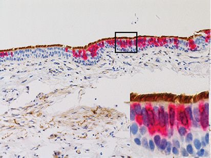
Articles
Bronchus Lung Conjunctiva
(figure 2). Similar tissue tropism was observed with the
bronchus infected with MERS-CoV, SARS-CoV, and
H1N1pdm immunohistochemical staining, although the
viral antigen staining was more extensive with SARS-CoV-2
(figure 1). In lung tissues, similar viral antigen staining
Mock
was observed with SARS-CoV-2 as for SARS-CoV and
H1N1pdm, but all were less extensive than that with
MERS-CoV. In the lung parenchyma, there was positive
antigen staining for SARS-CoV-2 in the spindled,
morphologically epithelial type 1 pneumocytes (figures 1, 2).
Double staining showed no colocalisation of viral antigen
in macrophages (figure 2). There was no evidence of
SARS-CoV-2
infection of vascular endothelium in the blood vessels of
the lung, as was previously seen with MERS-CoV.12
SARS-CoV-2 replicated in ex-vivo cultures of the human
bronchus with a two log10 increase in TCID50 from 24 h to
96 h (figure 3). SARS-CoV-2 replicated similarly to
MERS-CoV at all-timepoints, had lower titres than the
pandemic H1N1pdm virus in bronchus at 24 hpi and
48 hpi, and replicated to significantly higher titres than
SARS-CoV
SARS-CoV at 72 and 96 hpi (figure 3). In the absence of
cells for virus replication, thermal inactivation of all
viruses with input titres of 5 × 10² TCID50 per mL led to
negligible residual infectious virus after 24 h incubation
at 37˚C (appendix p 6). AUC from 24 h to 96 h post
infection was calculated from the data in figure 3, and is
shown in the appendix (appendix p 5). The AUC of
MERS-CoV
SARS-CoV-2 in bronchus explants was lower than that of
H1N1pdm and a higher, but non-significant, AUC was
observed when compared with SARS-CoV (appendix p 5).
In lung explants, SARS-CoV-2 titres were similar to
H1N1pdm and SARS-CoV, but had a lower replication
competence than MERS-CoV at 48 hpi and 72 hpi in the
lung (figure 3), supported further by the AUC of
H1N1pdm
SARS-CoV-2 in lung explants compared with other
viruses (appendix p 5).
SARS-CoV-2 can be detected in patients’ tears,14
conjunctiva, and anal swabs;15 thus, it is important to
study the potential for experimental infection of
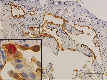
Figure 1: Tissue tropism of SARS-CoV-2 and SARS-CoV viruses in ex-vivo cultures of human respiratory tract
extrapulmonary tissues. Human conjunctival explant
and conjunctiva cultures were more extensively infected by SARS-CoV-2
Ex-vivo cultures of human bronchus and lung were infected with mock, SARS-CoV-2, SARS-CoV, MERS-CoV, and than by SARS-CoV, as shown in immunohistochemical
H1N1pdm viruses and the tissues were fixed with formalin at 96 hpi. Conjunctiva tissues were infected with mock, staining (figure 1) and higher infectious viral titres at
SARS-CoV-2, and SARS-CoV, and the tissues were fixed with formalin at 48 hpi. Paraffin-embedded sections were
subjected to immunohistochemical staining with a monoclonal antibody against the SARS-CoV nucleoprotein, 48 hpi (figure 3; appendix p 5). Thermal inactivation of
MCoV nucleoprotein, and influenza nucleoprotein. Positive cells are brown. Inset images are 200x magnification. the two coronaviruses showed that there are negligible
SARS-CoV=severe acute respiratory syndrome-associated coronavirus. MERS-CoV=Middle East respiratory infectious viruses after 24 h incubation at 33°C with a
syndrome-associated coronavirus. H1N1pdm=2009 pandemic influenza H1N1. remaining viral titre of 5 × 10² TCID50 per mL (appendix
p 6). We also infected human colorectal carcinoma
the report. The corresponding author had full access to (Caco-2) cells with MERS-CoV, SARS-CoV, SARS-CoV-2,
all of the data and the final responsibility to submit for H1N1pdm and H5N1. Infectious viral titres of all viruses
publication. increased by more than four log10 differences from
1 h to 72 h incubation in the Caco-2 cells. SARS-CoV-2
Results had a similar replication competence to SARS-CoV,
Immunohistochemistry staining showed that SARS-CoV-2 H1N1pdm and H5N1 but was lower than MERS-CoV
extensively infected bronchial epithelium (figure 1), with (figure 3; appendix p 5).
infection observed in ciliated cells, non-ciliated mucus We assessed viral replication, proinflammatory
secreting (goblet) cells, and club cells, but not basal cells cytokine responses, and chemokine responses in AECs,
4 www.thelancet.com/respiratory Published online May 7, 2020 https://doi.org/10.1016/S2213-2600(20)30193-4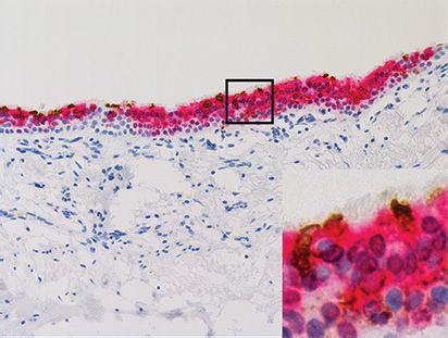
Articles
human peripheral blood-derived macrophages, and
A
Caco-2 cells; we compared the response elicited by Ciliated cell NP/Ac-Tub Club cell NP/CC10
SARS-CoV-2 to that of SARS-CoV, MERS-CoV, H1N1pdm,
and H5N1 (figure 4). High levels of viral gene mRNA
expression were detected in AECs, macrophages at
24 hpi, and Caco-2 cells at 48 hpi (figure 4), with
differences between the viruses. However, productive
viral replication of SARS-CoV-2 was detected only in
Caco-2 cells (figure 3; appendix p 5) with no robust virus 100 µm 100 µm
replication observed in AECs or macrophages (appendix
Goblet cell NP/MUC5AC Basal cell NP/p63a
p 5). There was less induction of proinflammatory
cytokines and chemokines (TNF-α, IP-10, RANTES, IL-6,
and MCP-1) by SARS-CoV-2, SARS-CoV, and MERS-CoV
in AECs and macrophages than by H5N1. In Caco-2 cells,
both SARS-CoV-2 and SARS-CoV induced less intense
cytokine responses than did H1N1pdm and H5N1,
whereas MERS-CoV induced modest level of most 100 µm 100 µm
cytokines and chemokines except for high levels for IP-10
in Caco-2 cells (figure 4). B
We detected upregulation of ACE2 receptor mRNA in Macrophage NP/CD68 Epithelial cell NP/AE1/3
H1N1pdm and H5N1 influenza-infected AECs (figure 4);
however, in human macrophages, ACE2 expression was
low and this remained similar after infection with
influenza and all tested coronaviruses (data not shown).
Discussion 100 µm 100 µm
We report the replication competence and cellular
tropism of SARS-CoV-2 in the human respiratory tract
and in extrapulmonary explant tissue and cells. The Figure 2: Cellular tropism of SARS-CoV-2 in ex-vivo cultures of human
bronchus and lung
conjunctival epithelium and conducting airways appear
Ex-vivo explant cultures of (A) human bronchus and (B) lung were infected with
to be potential portals of infection of SARS-CoV-2. Both SARS-CoV-2. At 96 hpi the tissues were fixed in formalin, embedded in paraffin,
SARS-CoV and SARS-CoV-2 replicated similarly in and immunohistochemically stained (brown) for indicated cell markers:
the alveolar epithelium, but SARS-CoV-2 replicated (A) Ac-Tub-positive for ciliated cells, MUC5AC-positive for secretory goblet cells,
CC10-positive for club cells, p63a-positive for basal cells; and (B) AE1/3 for
extensively in bronchial epithelium, which might
epithelial cells (brown) and CD68 for macrophages (red), and a monoclonal
explain the robust transmission of this pandemic antibody against the SARS-CoV-2 nucleoprotein (red). SARS-CoV-2-infected cells
coronavirus. SARS-CoV-2 was a less potent inducer of are identified with brown arrows, and CD68-positive cells are identified with red
proinflammatory cytokines than H5N1, H1N1pdm, or arrows. The images are representative of three individual donors.
NP=nucleoprotein. Ac-Tub=acetyl-α-tubulin.MUC5AC=mucin 5AC.
MERS-CoV viruses.
SCGB1A1=secretoglobin family 1A member 1. CC10=club cell protein 10.
SARS-CoV-2 replicated more efficiently than SARS-CoV p63a=p63-alpha. AE1/3= cytokeratin AE1/AE3. CD68=cluster of differentiation 68.
in ex-vivo cultures of human bronchus, with peak viral
titres at 96 hpi (figures 1, 3). Replication of SARS-CoV-2 controlling the spread of SARS-CoV-2 by public health
was similar to MERS-CoV and less efficient than interventions—much more so than with SARS-CoV.
H1N1pdm. The lower AUC of SARS-CoV-2 might be Immunohistochemistry of ex-vivo cultures of the
partly attributable to differences at 24 hpi, at which time human bronchus showed SARS-CoV-2 infection of
H1N1pdm already showed high virus titres although ciliated, non-ciliated mucus secreting (goblet), and club
SARS-CoV-2 titres were low and only increased at 48 hpi. cells. Because there were no reliable antibodies for
MERS-CoV can be transmitted among humans, mainly double staining which could distinguish between type 1
in health-care settings. The robust replication competence and 2 cells in the alveoli, we used the morphology of
of SARS-CoV-2 in human bronchus might explain its cells expressing virus antigen together with epithelial
efficient transmission efficiency among humans. In cell markers to conclude that type 1-like alveolar
human lung, SARS-CoV-2 and H1N1pdm replicated to epithelium was being infected, similar to observations
similar titres, but both were lower than that of MERS-CoV. in patients infected with SARS-CoV11 and consistent
The viral titres of SARS-CoV-2 at 24 hpi and 48 hpi were with a study in macaques infected with SARS-CoV-2.18,19
significantly lower than that of H1N1pdm. This observ Type 1 pneumocytes, flattened in shape, are crucial in
ation is compatible with the longer incubation period of the process of gas exchange between the alveoli and the
SARS-CoV-2 than of H1N1pdm. Virus shedding before capillaries; their intercellular connections via occluding
symptom onset16,17 probably contributes to the difficulty of junctions prevent the leakage of tissue fluid into the
www.thelancet.com/respiratory Published online May 7, 2020 https://doi.org/10.1016/S2213-2600(20)30193-4 5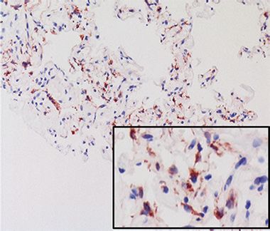
Articles

                  A       Bronchus                                                                                                                         B    Lung
                                                   p=0·0017                                                                                                                                                                                SARS-CoV-2
                                                p=0·047                                                                                                                                            p=0·044                                 SARS-CoV
                 107                                          p=0·0232                                                                                 106                                     p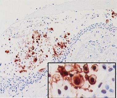
Articles

                                      A    Viral gene                                                                              B    Viral gene                                                                              C     Viral gene
                                                                       AECs                                                                                    Macrophage                                                                                   Caco-2
                                                       p=0·0113                                                                                                     p=0·0028                                                                                 p
Articles
the insert sequence of SerProArgArg in the S1/S2 protease virus and the infecting dose (MOI) cannot be precisely
cleavage site in SARS-CoV-2, but not in SARS-CoV, controlled, we compared the innate immune response to
causes the formation of an extended loop, which is more SARS-CoV-2 infection with that to H5N1, H1N1pdm,
suitable for protease recognition than in SARS-CoV.24 SARS-CoV, and MERS-CoV infections in Caco-2 cells
Because there is a strong positive staining of TMPRSS2 (the only in-vitro system in which productive virus
and HAT in human bronchial tissues,22 and the evidence replication was observed). There was little induction of
that trypsin treatment can overcome low-level expression proinflammatory cytokines in Caco-2 cells by SARS-CoV-2
of ACE2 receptor on target cells to activate spike protein or SARS-CoV, even less so than MERS-CoV, whereas
of SARS-CoV,22 HAT might play a more important role H1N1pdm and H5N1 viruses induced even higher levels
than TMPRSS2 in upper respiratory replication of of proinflammatory cytokine than MERS-CoV (with the
SARS-CoV-2. This finding is in line with the higher exception of IP-10). The insufficiency of productive
replication competence of SARS-CoV-2 than SARS-CoV replication of SARS-CoV-2 in AECs is in line with lack of
in human ex-vivo bronchus explants, and suggests that replication in human adenocarcinoma cells (A549).27 In
protease inhibitors of TMPRSSs are potential therapeutic summary, proinflam matory cytokine responses might
candidates for COVID-19. contribute modestly to the pathogenesis and severity of
We showed that ACE2 mRNA expression was human COVID-19, as was seen with MERS-CoV,28 and
significantly upregulated in alveolar epithelial cells after contrast with those observed in H5N1.29
influenza A virus infection, with H5N1 having a more The absence of robust virus replication in AECs in
pronounced effect than H1N1pdm in vitro. If replicated vitro, despite detection of infection in AECs in ex-vivo
in a larger sample, this upregulation could suggest that lung explant cultures, is reminiscent of observations
recent exposure to influenza virus might worsen the during previous research into SARS-CoV.30 In-vitro
outcome of COVID-19 through upregulation of the ACE2 culture of primary human lung epithelium is associated
receptor in human respiratory epithelium. By contrast, with a loss of permissiveness to both SARS-CoV and
ACE2 expression might also offer protective effects SARS-CoV-2, possibly via downregulation of the ACE2
during acute lung injury as shown for SARS.25 Therefore, receptor, which was ten times lower than that in Caco-2
the role of ACE2 expression during influenza infection cells (data not shown), or the absence of crucial proteases
should be defined, and its implications on susceptibility for the cleavage of the spike protein during isolation and
to and severity of SARS-CoV-2 infection should be in-vitro culture, or both. The experimental findings from
investigated. the Caco-2 model could not be directly compared with
Infection of extrapulmonary sites by SARS-CoV-2 has clinical observation; however, the human gastrointestinal
been supported by detection of the virus in tears and in tract comprises a complex mixture of cell types with
anal swabs14,15 and the detection and isolation of differing gene expression profiles between intestinal
SARS-CoV-2 in tears, anal swabs, and stool specimens.15,26 epithelium, mucosal epithelium, and the crypt–villus
We showed infection and productive replication of axis. Nevertheless, Caco-2 cells are readily available to
SARS-CoV-2 in conjunctiva and in a colorectal carcinoma most research laboratories as a simple and reproducible
epithelial cell line, which might support these reports. experimental tool allowing interlaboratory comparisons
Similar replication competence was observed in of findings.
SARS-CoV-infected Caco-2 cells compared with SARS- A limitation of our study is the absence of data on
CoV-2. The demonstration of infection of conjunctiva explant cultures of nasopharyngeal tissues because of the
suggests that the eye might be an additional route of unavailability of such tissues for ex-vivo infection
infection, an observation that is crucially relevant for experiments. The nasopharynx is crucial for virus for
infection prevention and control. Productive infection of SARS-CoV-2 replication and one that is highly relevant
the colorectal carcinoma epithelial cell line hints at virus for virus transmission. Restricted availability of con
infection and replication in the gastrointestinal tract, and junctival tissue allowed infection with only two of the
might indicate the possibility of oral-faecal transmission coronaviruses. Additionally, cytokines were analysed only
as an additional route. These multiple routes of at 24 hpi (AECs and macrophages) and 48 hpi (Caco-2
transmission by SARS-CoV-2 might explain the rapid cells). Replication of our study with increased sample
global spread of COVID-19. size, more cytokines, and detection of secreted cytokine
Higher plasma concentrations of proinflammatory proteins would shed light on the pathogenesis of
cytokines and chemokines, including IP-10, MCP-1, and SARS-CoV-2.
TNF-α, have been found in patients with COVID-19 in Our findings support the notion that SARS-CoV-2 can
the ICU when compared with non-ICU patients.8 transmit between humans via droplets being deposited
However, such studies cannot clarify whether these in the airways or eyes and via fomite transmission when
elevated cytokines and chemokines are a major driver of infectious virus is introduced to the eyes via contaminated
pathology or simply a reflection of the more severe lung hands. Replication of SARS-CoV-2 in colorectal cells
damage that has taken place. Because ex-vivo cultures are suggests that the virus might also spread via the oral-
heterogeneous in cell type and because susceptibility to faecal route.
8 www.thelancet.com/respiratory Published online May 7, 2020 https://doi.org/10.1016/S2213-2600(20)30193-4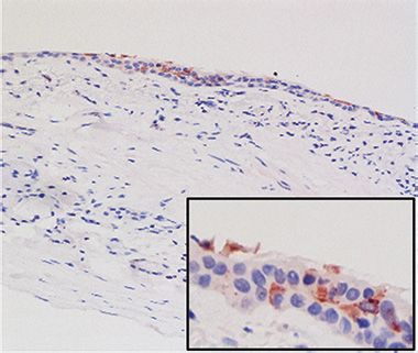
Articles

Contributors                                                                    11   Nicholls JM, Butany J, Poon LL, et al. Time course and cellular
KPYH isolated SARS-CoV-2, and designed and coordinated the study,                    localization of SARS-CoV nucleoprotein and RNA in lungs from
analysed and interpreted the results, did experiments and wrote the                  fatal cases of SARS. PLoS Med 2006; 3: e27.
Article. M-CC, K-CN, CHTB, JCWH, MMTN, and DITK did                             12   Mok CK, Lee HH, Lestra M, et al. Amino acid substitutions in
experiments, and analysed and interpreted results. RAPMP isolated                    polymerase basic protein 2 gene contribute to the pathogenicity of
SARS-CoV-2. KCS provided human conjunctiva tissue. S-WT provided                     the novel A/H7N9 influenza virus in mammalian hosts. J Virol
and technically supported the human respiratory epithelial cells. LLMP               2014; 88: 3568–76.
developed quantitative PCR assays. MP designed the study, analysed and          13   Hui KPY, Ching RHH, Chan SKH, et al. Tropism, replication
interpreted results and critically reviewed the manuscript. JMN designed             competence, and innate immune responses of influenza virus: an
                                                                                     analysis of human airway organoids and ex-vivo bronchus cultures.
the study, analysed and interpreted results from immunohistochemical
                                                                                     Lancet Respir Med 2018; 6: 846–54.
staining and critically reviewed the manuscript. MCWC designed and
                                                                                14   Xia J, Tong J, Liu M, Shen Y, Guo D. Evaluation of coronavirus in
was responsible for overall coordination of the study, analysed and
                                                                                     tears and conjunctival secretions of patients with SARS-CoV-2
interpreted the results, and wrote the Article.                                      infection. J Med Virol 2020; 92: 589–94.
Acknowledgments                                                                 15   Zhang W, Du RH, Li B, et al. Molecular and serological
Rachel Ching, Eric Lau, Rita Lai, Cassia Lin, Hin-Wo Yeung, Daniel Chu               investigation of 2019-nCoV infected patients: implication of
(School of Public Health, University of Hong Kong, Hong Kong, China),                multiple shedding routes. Emerg Microbes Infect 2020; 9: 386–89.
and Dora Kwong (Department of Clinical Oncology, University of                  16   Zou L, Ruan F, Huang M, et al. SARS-CoV-2 Viral load in upper
Hong Kong, Hong Kong, China) provided the human upper respiratory                    respiratory specimens of infected patients. N Engl J Med 2020;
tract tissues. Kevin Fung (Department of Pathology, The University of                382: 1177–79.
Hong Kong, Hong Kong, China) provided technical support, and Ron                17   Pan Y, Zhang D, Yang P, Poon LLM, Wang Q. Viral load of
Fouchier (Erasmus University Medical Center, Rotterdam, Netherlands)                 SARS-CoV-2 in clinical samples. Lancet Infect Dis 2020; 20: 411–12.
provided the MERS-CoV. We acknowledge research funding from the                 18   Munster VJ, Feldmann F, Williamson BN, et al. Respiratory disease
US National Institute of Allergy and Infectious Diseases (NIAID) under               and virus shedding in rhesus macaques inoculated with
Centers of Excellence for Influenza Research and Surveillance (CEIRS)                SARS-CoV-2. bioRxiv 2020; published online March 21.
                                                                                     DOI:10.1101/2020.03.21.001628 (preprint).
contract no. HHSN272201400006C and the Theme Based Research
                                                                                19   Rockx B, Kuiken T, Herfst S, et al. Comparative pathogenesis
Scheme (Ref: T11–705/14N, T11-712/19-N), Hong Kong Special
                                                                                     of COVID-19, MERS and SARS in a non-human primate model.
Administrative Region and a Commissioned Grant from the Health and
                                                                                     Science 2020; published online April 17. DOI:10.1126/science.abb7314.
Medical Research Fund (Ref: HKS-18-B03), Food and Health Bureau,
                                                                                20   Zhou P, Yang XL, Wang XG, et al. A pneumonia outbreak
Government of Hong Kong Special Administrative Region. The funding
                                                                                     associated with a new coronavirus of probable bat origin. Nature
sources had no roles in the writing of the manuscript or the decision to             2020; 579: 270–73.
submit it for publication. The authors have not been paid to write this
                                                                                21   Hoffmann M, Kleine-Weber H, Schroeder S, et al. SARS-CoV-2 cell
article by a pharmaceutical company or other agency. The authors had                 entry depends on ACE2 and TMPRSS2 and is blocked by a clinically
full access to all the data in the study and had final responsibility for the        proven protease inhibitor. Cell 2020; 181: 271–80.
decision to submit for publication.                                             22   Bertram S, Glowacka I, Müller MA, et al. Cleavage and activation of
Declaration of interests                                                             the severe acute respiratory syndrome coronavirus spike protein by
We declare no competing interests.                                                   human airway trypsin-like protease. J Virol 2011; 85: 13363–72.
                                                                                23   Bertram S, Heurich A, Lavender H, et al. Influenza and SARS-
References                                                                           coronavirus activating proteases TMPRSS2 and HAT are expressed
1    WHO. MERS monthly summary, November 2019. 2019 (accessed                        at multiple sites in human respiratory and gastrointestinal tracts.
     Feb 24, 2020).                                                                  PLoS One 2012; 7: e35876.
2    WHO. COVID-19 dashboard. 2019. https://covid19.who.int/                    24   Tong Meng HC, Zhang H, Kang Z, et al. The insert sequence in
     (accessed April 19, 2020).                                                      SARS-CoV-2 enhances spike protein cleavage by TMPRSS. bioRxiv
3    Chen J. Pathogenicity and transmissibility of 2019-nCoV—a quick                 2020; published online Feb 16. DOI:10.1101/2020.02.08.926006
     overview and comparison with other emerging viruses.                            (preprint).
     Microbes Infect 2020; 22: 69–71.                                           25   Imai Y, Kuba K, Rao S, et al. Angiotensin-converting enzyme 2
4    Xu Z, Shi L, Wang Y, et al. Pathological findings of COVID-19                   protects from severe acute lung failure. Nature 2005; 436: 112–16.
     associated with acute respiratory distress syndrome.                       26   Zhang Y, Chen C, Zhu S, et al. Isolation of 2019-nCoV from a stool
     Lancet Respir Med 2020; 8: 420–22.                                              specimen of a laboratory-confirmed case of the coronavirus disease
5    Tian S, Hu W, Niu L, Liu H, Xu H, Xiao SY. Pulmonary pathology of               2019 (COVID-19). 2020. http://weekly.chinacdc.cn/en/article/id/
     early-phase 2019 novel coronavirus (COVID-19) pneumonia in                      ffa97a96-db2a-4715-9dfb-ef662660e89d (accessed Apr 3, 2020).
     two patients with lung cancer. J Thorac Oncol 2020; published              27   Harcourt J, Tamin A, Lu X, et al. Severe acute respiratory syndrome
     online Feb 28. DOI:10.1016/j.jtho.2020.02.010.                                  coronavirus 2 from patient with 2019 novel coronavirus disease,
6    Chan MC, Chan RW, Yu WC, et al. Tropism and innate host                         United States. Emerg Infect Dis 2020; published online March 11.
     responses of the 2009 pandemic H1N1 influenza virus in ex vivo                  DOI:10.3201/eid2606.200516.
     and in vitro cultures of human conjunctiva and respiratory tract.          28   Chan RW, Chan MC, Agnihothram S, et al. Tropism of and innate
     Am J Pathol 2010; 176: 1828–40.                                                 immune responses to the novel human betacoronavirus lineage C
7    Chu DKW, Hui KPY, Perera RAPM, et al. MERS coronaviruses from                   virus in human ex vivo respiratory organ cultures. J Virol 2013;
     camels in Africa exhibit region-dependent genetic diversity.                    87: 6604–14.
     Proc Natl Acad Sci USA 2018; 115: 3144–49.                                 29   Cheung CY, Poon LL, Lau AS, et al. Induction of proinflammatory
8    Huang C, Wang Y, Li X, et al. Clinical features of patients infected            cytokines in human macrophages by influenza A (H5N1) viruses:
     with 2019 novel coronavirus in Wuhan, China. Lancet 2020;                       a mechanism for the unusual severity of human disease? Lancet
     395: 497–506.                                                                   2002; 360: 1831–37.
9    Hui KP, Chan LL, Kuok DI, et al. Tropism and innate host                   30   Mossel EC, Wang J, Jeffers S, et al. SARS-CoV replicates in primary
     responses of influenza A/H5N6 virus: an analysis of ex vivo and                 human alveolar type II cell cultures but not in type I-like cells.
     in vitro cultures of the human respiratory tract. Eur Respir J 2017;            Virology 2008; 372: 127–35.
     49: 1601710.
10 Chu DKW, Pan Y, Cheng SMS, et al. Molecular diagnosis of a novel
     coronavirus (2019-nCoV) causing an outbreak of pneumonia.
     Clin Chem 2020; 66: 549–55.

www.thelancet.com/respiratory Published online May 7, 2020 https://doi.org/10.1016/S2213-2600(20)30193-4                                                                9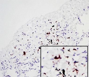
You can also read