MiR-142-3p suppresses apoptosis in spinal cord-injured rats
←
→
Page content transcription
If your browser does not render page correctly, please read the page content below
Translational Neuroscience 2020; 11: 105–115
Research Article
Jun Zheng*, Jing Kuang, Xianyu Zhang, Daya Luo, Weijing Liao
miR-142-3p suppresses apoptosis in spinal
cord-injured rats
https://doi.org/10.1515/tnsci-2020-0105 The findings suggested that miR-142-3p may inhibit the
received January 03, 2020; accepted March 30, 2020 MAP by inhibiting the expression of cleaved-caspase-3/-9
Abstract and Bax in SCI rats.
Introduction ‒ Spinal cord injury (SCI) leads to abnormal Conclusion ‒ This study concludes that miR-142-3p
expression of miRs, leading to secondary responses such as may attenuate the functional recovery and decrease
oxidative stress, inflammation and apoptosis. In the present apoptosis in neuronal cells via inhibiting the MAP in the
work, we screened the miRs involved and the associated spinal cord-injured rats, confirming miR-142-3p as a
pathway. potential therapeutic target in treating SCI.
Methods ‒ In a rat model of SCI, the microarray Keyword: Bax, caspase-3, caspase-9, miR-142-3p, spinal
analysis for expression of miRs at various time points cord injury
post-SCI was done. The locomotor analysis was done by
Basso, Beattie and Bresnahan score, and Cresyl violet
staining was done for lesion volume and TUNEL assay
was done for apoptosis in neuronal cells. The expression
of apoptotic proteins was done by the western blot study. 1 Introduction
Results ‒ It was evidenced that the expression of the
number of miRs was altered on the 14th day post-SCI, and Spinal cord injury (SCI) is one of the common injuries of
miR-142-3p was found to be the most significantly the spine causing permanent disability such as paralysis,
suppressed miR. The results suggested that overexpression loss of sensitivity and loss of movement [1]. Every year, a
of miR-142-3p by its agomir-attenuated functional recovery large number of patients are reported to be affected with
decreased lesion size and apoptosis of neuronal cells in SCI [2]. Currently, SCI has been the biggest challenge for
rats subjected to SCI. The luciferase assay indicated that neuro physicians [3]. Apoptosis plays a crucial role in
miR-142-3p blocked the levels of Bax, which is a significant functional and physical deficits in SCI, and it is a major
activator of the mitochondrial apoptotic pathway (MAP) via hurdle in treating SCI [4,5]. Therefore, developments of new
targeting the 3′UTR region of BV-2 cells, and in addition, therapeutic approaches, which can target and suppress
pc-DNA-Bax restored Bax and inhibited the correcting apoptosis for countering SCI, are needed. Apoptosis, which
role of miR-142-3p in hydrogen peroxide-treated BV-2 cells. is also defined as programmed cell death, is a key feature
in neuronal tissue damage after SCI [6]. In the previous
study, two pathways, namely, the mitochondrial and death
* Corresponding author: Jun Zheng, Department of receptor pathways, have been identified to play an
Neurorehabilitation, Zhongnan Hospital of Wuhan University, important role in inducing apoptosis [7]. In the mitochon-
Wuhan, Hubei, 430071, China, e-mail: blatfish@163.com, drial pathway, B-cell lymphoma-2 also called Bcl-2 is a
tel: +86-027-67813386, fax: +86-027-67813386 group of genes that regulate apoptosis via antiapoptotic
Jing Kuang: Department of Plastic Surgery, Jinan Central Hospital
proteins (Bcl-2) and proapoptotic proteins (Bcl-2 homo-
Affiliated to Shandong University, Jinan, Shandong, 250013, China,
e-mail: miaoer9999@sina.com logous, Bax and Bcl-2 agonist) [8]. The collapse of the
Xianyu Zhang: Orthopedics Department, ShangRao People’s Hospital, mitochondrial membrane potential is identified as the
Shangrao, Jiangxi, 334000, China, e-mail: zmcppg@163.com important process in the mitochondrial apoptosis cascade,
Daya Luo: Department of Biochemistry and Molecular Biology, which leads to the transfer of cytochrome C (CYT-C) from
School of Basic Medical Sciences, Nanchang University, Nanchang,
the mitochondria to the cytosol [9]. Then, CYT-C along with
Jiangxi, 330006, China, e-mail: aazykx@163.com
Weijing Liao: Department of Neurorehabilitation, Zhongnan
dATP and apoptotic protease-activating factor 1 (Apaf-1) in
Hospital of Wuhan University, Wuhan, Hubei, 430071, China, cytosol leads to the cleavage of caspase-9 and p-caspase-9
e-mail: weijingliao@sina.com [10]. Caspase-9 further leads to the cleavage of caspase-7,
Open Access. © 2020 Jun Zheng et al., published by De Gruyter. This work is licensed under the Creative Commons Attribution 4.0 Public
License.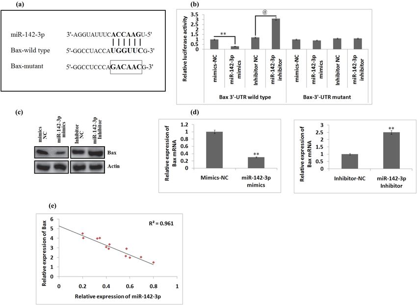
106 Jun Zheng et al.
caspase-6 and caspase-3 [11]. Hence, the release of CYT-C is The animal study protocols were sanctioned by the Animal
a very important step for the activation of p-caspase-9 in Ethical Review Board of Zhongnan Hospital of Wuhan
the apoptosis-induced cell death. University, China. The rats were housed under pathogen-
MiRNAs (miRs) comprise 21–23 nucleotides, and they free standard lab conditions at 25℃ with a relative humidity
can modulate genes at the posttranscriptional level via of 50–60% in 12 h dark and light cycle. The rats were
suppressing translation or inducing degradation [12,13]. miRs provided free access to food and water. The rats were
have been found to regulate at least 65% of genes in the divided into four groups (n = 6/group), namely, the SCI-
human genome [14]. So far studies have identified multiple induced group, sham-operated group, miR-142-3p agomir
miRs in the spinal cord and the brain, which are believed to group and miR-142-3p antagomir group (NC group). The
regulate functioning and plasticity [15,17]. In addition, miRs sham-operated rats were subjected to laminectomy at T10
have been identified to play a key role in the development of without the weight-drop injury; similarly, the SCI group rats
the nervous system and may be crucial in the process of cell underwent T10 laminectomy by the New York spinal
differentiation in specific organs [16]. Earlier studies have segment impactor, and miR-142-3p antagomir-(NC) and
demonstrated that the spinal injury may lead to abnormal agomir-treated rats were subjected to SCI and were given
expression of miRs, which is a response against a number of dose intrathecally (1 µL/h, 20 nmol/mL). The agomir and
secondary injuries, including oxidative stress, apoptosis and antagomir were obtained from RiboBio (Guangzhou, China).
inflammation; this also regulates the expression of some
specific genes [18,19]. Recently, growing evidence suggest the Ethical approval: The research related to animals use
involvement of a number of miRs in regulating apoptosis has been complied with all the relevant national
involving mitochondrial cascade in various disorders [20–22]. regulations and Animal Ethical Review Board of
Hence, in the present work, we speculated that the spinal Zhongnan Hospital of Wuhan University, China, for the
injury-mediated miRs may encourage apoptosis by activating care and use of animals.
the mitochondrial apoptosis pathway. Here, we developed a
rat model of the SCI followed by the microarray analysis for
identifying the expression profiles of miRs in injured spinal
tissues. Subsequently, we evaluated the role of miR-142-3p in 2.3 Establishment of SCI model
the SCI-mediated apoptosis and the mechanisms involved.
For inducing SCI, the rats were subjected to anesthesia
by intraperitoneal injection of 50 mg/kg sodium pento-
barbital followed by laminectomy at the T9–T10 by
2 Material and methods opening the cord below without disturbing the dura.
Clamping of T8 and T11 disc was done for stabilizing the
spinal cord, and then, the dorsal side of the cord was
2.1 Cell lines and treatment
submitted to weight drop injury as described in the
New York impactor process [23]. In sham rats, the
In this work, we used immortalized murine BV2 cell lines
laminectomy of T10 was performed by removing the weights.
provided by the Zhongnan Hospital of Wuhan University,
China, and were incubated in the modified DMEM media
(ThermoFisher, USA). The medium was supplemented
2.4 miRNA microarray analysis
with fetal bovine serum (10%) and 100 U/mL each of
streptomycin and penicillin (10%) and maintained at 37℃
For studying the expression of miRs in the spinal tissue of
under humid conditions with 5% CO2. The BV2 cell lines
rats, we sacrificed rats with the pentobarbital injection
were exposed to various concentrations (50, 100, 200 and
(n = 2 from each group). From each animal, the spinal
400 µM) of H2O2 (30%; Merck, USA) for 10 h for the
cord tissue segment (10 mm) along with the injury
process of inducing cell injury.
epicenter was isolated and freezed in liquid nitrogen.
The total RNA was separated from the spinal tissue with the
help of TRIzol reagent (ThermoFisher, USA) following the
2.2 Animals and treatment supplied instructions, and the RNAs were purified using
the RNeasy cleanup kit (Qiagen, Germany). The absorbance
We used Sprague-Dawley rats aged between 6 and 7 weeks was measured using Shimadzu 1800 spectrophotometer
(n = 36 rats) weighing approximately between 225 and 250 g. (Shimadzu). The miRs were isolated using the microarray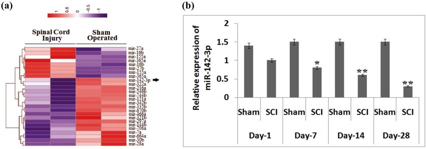
miR-142-3p improves functional recovery in SCI 107
labeling kit (ThermoFisher, USA) and were then hybridized mixture of 4% paraformaldehyde in 0.1 M phosphate-
using the Exiqon miRCURY LNA array (Biocompare, USA). buffered saline for 20 min. Then, the spinal segment
The slides were scanned using the SureScan microarray measuring 1 cm at the injured site was harvested and fixed
scanner (Agilent, USA). The miRs having an intensity of in the mixture of 4% paraformaldehyde in 0.1 M phosphate-
more than 50 were selected for further study, and the buffered saline for 12 h. The tissues were then fixed in
expression of miRs was normalized by median normal- paraffin, and transverse sections having a thickness of
ization. The miRs were evaluated by using volcano plots. 10 µm were produced using a microtome. The sections were
The final clustering was utilized for determining the mounted on slides. These sections of spinal tissue at the
variations in the expression profiles of miRs. injured site were subjected to staining with Cresyl-violet
acetate (0.5%) for 45 min at 37°C and viewed under a
microscope at 200× magnification. The images of sections
were processed to outline the lesion part and the spared
2.5 Isolation of RNA and RT-PCR areas with the help of the image analysis software. The
spared tissues were regarded as the remaining area with no
The total RNA was isolated from the spinal tissue segment injured part with normal features. The portion of the section
at the injured site with the help of TRIzol reagent following with minimal spared tissue was regarded as the injury
the supplied instructions. The reverse transcription was epicenter.
carried out using the TaqMan miR reverse transcription kit
in accordance to supplied instructions. The PCR was done
with the help of the TaqMan miR assay kit (ThermoFisher,
USA) on a real-time PCR system (ThermoFisher, USA) 2.8 Terminal deoxynucleotidyl-transferase-
following the manufacturer’s instructions. The primers used mediated dUTP nick-end labeling
in the study were miR-142-3p, 5′-GTCGTATCCAGTGCAGGG (TUNEL) assay
TCCGAGGTATTCGCACTGGATACGACTCCATA-3′, and Bax,
5′-GGCTGGACACTGGACTTCCT-3′, U6, 5′-CTCGCTTCGGCA To study the extent of apoptosis in the spinal tissues, we
GCACA-3′. The expression of miR was observed relative subjected the tissues sections for TUNEL staining. The
to U6. All the experiments were done in triplicate. study was performed using the TUNEL apoptosis assay
kit (Biovision, USA). The TUNEL staining showed the
apoptotic cells in the spinal tissues, following the
supplied instructions. Briefly, the sections were immersed
2.6 Behavioral study in the TUNEL reagent for 45 min at room temperature, the
nuclei were stained with DAPI (1 µg/mL) for 10 min. The
The impact of SCI on behavioral parameters was sections were quantified for a number of DAPI-positive
evaluated by the Basso, Beattie and Bresnahan (BBB) cells and observed in 15 random areas form each slide
scoring system for locomotor activity done on the 1st, with the help of a fluorescent microscope.
7th, 14th and the 28th day after SCI [26]. The experiment
evaluated the locomotor activity for 4 min. The evalua-
tion was done by a trained person. We selected the open
field test for locomotion coupled with BBB scoring as 2.9 Luciferase reporter assay
described earlier [26]. For minimizing the differences in
the study, the experiments were performed by two We performed bioinformatics analysis by TargetScan for
independent investigators, and the mean was recorded. finding the favorable binding site between miR-142-3p
and Bax. For the luciferase assay, we used inhibitor,
mimics and negative control. The mutant (mut) and
wild-type (wt) 3′UTR region of Bax was amplified and
2.7 Evaluation of lesion volume was cloned in the pmiR reporter luciferase vector. The
Phusion Site-Directed Mutagenesis Kit (ThermoFisher,
To mark the lesion volume due to SCI or defined treatments USA) was used for the site-directed mutagenesis of Bax
(antagomir and agomiR), the animals were anesthetized by 3′UTR in the potential binding site of miR-142-3p. The
injecting pentobarbital (50 mg/kg). The rats were perfused BV-2 cells (2 × 10⁶ cells/well) were incubated in 96-well
with isotonic saline solution (0.9%, 250 mL) and then by the plates followed by co-transfection with 3′UTR-pMIR-Bax108 Jun Zheng et al.
or 3′UTR-pMIR-Bax-mut, miR-142-3p inhibitor, mimics 2.12 Flow cytometry analysis for apoptosis
and respective negative control using Lipofectamine-
2000 reagent. The BV-2 cells were washed using PBS (ice cold) and
were then fixed in ethanol (70%, ice-cold) in PBS for
20 min. We used the FITC Annexin V Apoptosis detection
kit for evaluating apoptosis. The cells were washed
2.10 Western blot analysis using PBS and were then resuspended in Annexin V
buffer and incubated in Annexin V-FITC and propidium
The spinal cord tissue portion at the lesion site was iodide. Cells receiving staining were evaluated with the
isolated followed by homogenization, and total proteins flow cytometer (BD Biosciences, USA).
were isolated. The BV-2 cells were also processed to
isolate proteins. The samples were centrifuged (10,000g)
at 4℃ for 30 min; the supernatant was subjected to the
bicinchoninic acid assay for studying the protein expres- 2.13 Evaluation of caspase-3 activity
sion levels. The proteins (30 µg) were subjected to
electrophoresis followed by the transfer to Millipore The activity of caspase-3 was evaluated by the colori-
PVDF membranes. The membranes were blocked by metric assay kit following the supplied instructions. The
nonfat milk (5%) at 4℃ for 12 h. The membranes were BV-2 cells isolated by centrifugation at 5,000 rpm for
then incubated with Iry antibodies Bax (1:1,000), Bcl-2 5 min at 4℃ and were incubated in lysis buffer for
(1:1,000), cleaved-caspase-9 (1:1,000) and pro-caspase-3 10 min. Then, the lysates were subjected to centrifuga-
(1:1,000), and Actin was opted as a loading control. The tion (10,000g for 20 min) at 4℃. The lysates were
membranes were incubated along with anti-mouse IgG screened for protein levels using the protein estimation
horse radish peroxidase-conjugated IIry antibody (1:200) kit. The lysates were incubated with 0.2 mM DEVD-pNA
for 60 min. The proteins were detected by quantifying the buffer at room temperature. The samples were analyzed
protein bands with the help of ImageQuant TL 8.2 image using a microplate reader at 405 nm.
analysis software (GE health care and life sciences, USA).
2.14 Statistics
2.11 Immunohistochemistry studies
The statistics was done using Graphpad Prism software.
For immunohistochemistry, the spinal tissues were All the experiments were done in triplicate, and the
subjected to intracardiac perfusion using isotonic sodium results are presented as mean ± standard deviation.
chloride solution and then by PFA (4%) in 0.1 M PBS at 4℃ Spearman’s rank correlation coefficient was done for
for 20 min. Subsequently, a 10 mm portion of the spinal analyzing the difference between Bax and miR-142-3p.
cord tissue at the injured site was isolated and was fixed in One-way ANOVA along with Tukey’s post hoc analysis
a paraffin block, and 5 µm thick section was prepared using was done for establishing the differences between
a microtome. The paraffinized sections were treated with groups. Student’s t-test was done to establish differences
xylene for deparaffinization at room temperature for 2 min between two groups. P < 0.05 was considered as
and hydrated using alcohol. The sections were incubated for statistically significant.
15 min in H2O2 (3%) at room temperature, followed by
washing by 0.01 PBS. The sections were then blocked with
fetal bovine serum (10%) in PBS for 20 min at room
temperature and incubated at 4℃ for 12 h with anticleaved
3 Results
caspase-3 antibodies (1:1,000). Then, the sections were
incubated along with anti-mouse IgG horseradish perox- 3.1 Aberrant expression of miRs after SCI
idase IIry antibodies (1:1,000) at room temperature for
20 min. Finally, 3,3′-diaminobenzidine staining was done to To study the possible involvement of miRs in spinal
study the immunoreactivity. The images were viewed under injury, we created an animal model of SCI followed by
the Olympus light microscope (Olympus Corp.). The images the microarray analysis for identifying the expression of
were quantified using Image scope-9 software. miRs in spinal tissues. We observed multiple miRsmiR-142-3p improves functional recovery in SCI 109
Figure 1: Expression analysis of miR expression in rats subjected to SCI. (a) The results of the heat map analysis showing changes in
expression levels of miRNA at the end of the 14th day after submitting them to SCI. Violet color shows downregulation, and red color
shows overexpression of miRs. (b) Quantitative results of RT-PCR for showing the expression of miR-142-3p in spinal tissues after SCI at
day 1, day 7, day 14 and day 28 after SCI. The results are presented as mean ± SD. *P < 0.05, **P < 0.01 compared to sham-operated rats.
showing altered expression at 14 days after SCI, and of agomir group of rats showed a significant improvement in
them, miR-142-3p was the most prominently down- the motor function form the 7th day against the SCI rats (P <
regulated compared with sham-operated rats (Figure 1a). 0.05, Figure 2b). Subsequently, the results of Cresyl staining
Previously, miR-142-3p has been found to be over- suggested that the SCI + miR-142-3p agomir-treated SCI rats
expressed in dorsal root ganglion (DRG) neurons [23]. showed an increased amount of the spared tissue compared
Hence, we performed qRT-PCR to further confirm the to the SCI rats, suggesting that miR-142-3p agomir decreased
expression levels of miR-142-3p in the spinal cord tissues the lesion volume in spinal tissues after SCI. In addition, we
on day 1, day 7, day 14 and day 28 after SCI. We observed studied whether miR-142-3p regulates the expression of
that the levels of miR-142-3p were suppressed signifi- apoptosis-involved proteins such as cleaved-caspase-3 by
cantly (P < 005) in the spinal cord-injured group of rats immunohistochemistry analysis in the spinal cord tissues
compared to that in the sham group between the 7th and after injury. As shown in Figure 2c, SCI caused a significant
the 28th day (Figure 1b). These findings confirmed that overexpression of cleaved-caspase-3 in the spinal cord
SCI leads to aberrant levels of miRs in the spinal tissues tissues against the sham group, whereas the treatment of
and the possible involvement of miR-142-3p in the SCI. miR-142-3p agomir resulted in the inhibition of cleaved-
caspase-3 (P < 0.05, Figure 2c). In addition, the neuronal
cell apoptosis was evaluated by the TUNEL assay. It was
found that the number of TUNEL-positive cells increased
3.2 Upregulation of miR-142-3p attenuates significantly (P < 0.01) in the SCI rats compared to the
functional recovery and inhibits sham-operated group; however, miR-142-3p agomir signifi-
apoptosis after SCI. cantly decreased the TUNEL-positive cells in the SCI rats
treated with miR-142-3p agomir against the SCI rats (P <
To find the role of miR-142-3p in the rats subjected to the 0.01, Figure 2d). These findings suggest that miR-142-3p
SCI, we developed a rat model and treated the rats with agomir improved the functional recovery, suppressed
miR-142-3p agomir via the intrathecal route. qRT-PCR was apoptosis and decreased the lesion volume in rats after SCI.
done to evaluate the effect of overexpression by miR-142-3p
agomir in the spinal tissues. We found that the relative
levels of miR-142-3p were significantly overexpressed
(Figure 2a) between the 7th and the 28th day compared to 3.3 miR-142-3p causes downregulation of
the rats treated with agomir-NC (P < 0.05) with the Bax via targeting the 3′UTR in BV-2
maximum levels on the 14th day. Furthermore, in our cells
study, we utilized the BBB rating score for evaluating the
motor function in the spinal cord-injured rats after treating Previously, it has been reported that miR-142-3p inhibits
them with miR-142-3p agomir. The results suggested that apoptosis and promotes the neuronal cell cycle [23],
the upregulation of miR-142-3p in the SCI + miR-142-3p and in addition, a report confirmed the involvement of110 Jun Zheng et al. Figure 2: miR-142-3p agomir increases SCI. (a) Expression of miR-142-3p relative to agomir-NC detected at various days in rats submitted to spinal injury following the dose of miR-142-3p agomir. *P < 0.05, **P < 0.01 compared to agomiR-NC. (b) Results of BBB score for assessing the locomotor activity at various time intervals after spinal injury following the treatment of mir-142-3p. (c) Immunohistochemistry analysis for detecting the expression of cleaved-caspase-3 in the spinal tissues after SCI (magnification at 200×). *P < 0.05, **P < 0.01 compared to sham-operated rats, P < 0.05 compared to spinal injury rats. (d) Results of the TUNEL assay for identifying extent of apoptosis of neuronal cells on the 14th day after spinal injury (magnification at 200×). The results are presented as mean ± SD. miR-142 in apoptosis via downregulation of pro-apoptosis the increased luciferase activity against the inhibitor (NC; proteins caspase-3 and Bax [25]. Therefore, we speculated P < 0.01, Figure 3b). In addition, miR-142-3p did not that miR-142-3p inhibits the apoptosis in neuronal cells of suppress the activity of luciferase in the vector bearing rats subjected to the SCI via suppressing the levels of Bax. 3′-UTR of Bax having mutations in the miR-142-3p- Subsequently, the bioinformatics analysis was done to binding site (Figure 3b). predict the potential targets of miR-142-3p, and it was To further evaluate whether Bax is regulated noticed that Bax may be a potential target of miR-142-3p negatively by miR-142-3p, we performed western blot having the potential binding site in the 3′UTR (Figure 3a). and qRT-PCR analyses to determine protein and mRNA We further validated the bioinformatics data, and the levels, respectively. It was noticed that upregulated miR- Bax-3′-UTR constructs (wild type (wt) and mutant (mut)) 142-3p decreased the levels of Bax as both protein and were inserted in the firefly luciferase-expressing vector mRNA levels in BV-2 cells (Figure 3c and d). Further- pmiR-reporter. To evaluate the pathologic parameters more, the RT-PCR study was performed to find the mRNA after SCI, we used the BV-2 cells, which are reported to levels of Bax in the spinal cord tissue, and we evidenced have various features of Iry microglia [25]. The plasmids that the levels of mRNA of Bax were significantly received co-transfection of mir-142-3p mimics/inhibitor or overexpressed in the spinal cord tissue compared to NC mimics/inhibitor in the BV-2 cells followed by that in sham-operated rats (P < 0.01, Figure 3d). The evaluation of luciferase activity. We observed that miR- overall analysis suggested a significant negative correla- 142-3p-mimics caused the inhibition of the luciferase tion between miR-142-3p and Bax in the spinal cord activity significantly compared to NC mimics in the tissue (Figure 3e). Overall, the findings of the experiment presence of 3′UTR (wt), whereas the inhibitor resulted in established that miR-142-3p suppressed the levels of Bax
miR-142-3p improves functional recovery in SCI 111
Figure 3: Bax is the potential target of miR-142-3p in BV-2 cells. (a) TargetScan predicted potential binding sites on the 3′UTR of Bax mRNA.
(b) Quantitative results of luciferase activity of Bax-wt or mut 3′UTR in cells transfected with miR-142-3p mimics/inhibitor and NC.
**P < 0.01, and P < 0.01 compared to NC. (c) Protein expression by western blot analysis. (d) Quantitative results of mRNA expression for
Bax done by western blot analysis and qRT-PCR. **P < 0.01 compared to a negative control. (e) Quantitative results showing a negative
correlation between expression of miR-142-3p and Bax (r = 0.961). The results are presented as mean ± SD.
via targeting the 3′UTR site in BV-2 cells, hence concentrations (50–400 µM) for 10 h after which the
suggesting Bax as a possible target of miR-142-3p in levels of miR-142-3p were studied using RT-PCR. The
spinal cord tissues. results (Figure 4a) suggested that hydrogen peroxide
caused a significant downregulation of miR-142-3p in
BV-2 cells in a concentration-dependent manner between
concentrations 50 and 200 µM (P < 0.05). Subsequently,
3.4 Upregulation of Bax blocks the western blot and RT-PCR analyses were carried out to
protective activity of miR-142-3p in study the upregulation of miR-142-3p or Bax. The
hydrogen peroxide-exposed BV-2 cells outcomes suggested that miR-142-3p and Bax were
overexpressed in Bv-2 cells treated with miR-142-3p
Studies earlier have demonstrated that reactive oxygen mimics pc-DNA-Bax (Figure 4b and c). Also, it was found
species play an important role in the SCI as they activate that the upregulation of miR-142-3p downregulated the
multiple pathways of apoptosis, and hydrogen peroxide- protein expression of hydrogen peroxide-induced BV-2
exposed BV-2 cells are established as a cellular model of cells, whereas the pc-DNA-Bax inhibited the restoration
SCI for evaluating the pathologic factors after the SCI of Bax. miR-142-3p reduced the expression of Bax in
[26]. In the present work, we used murine BV-2 cell lines hydrogen peroxide-treated cells (Figure 4d). The results
that were exposed with hydrogen peroxide of varied of the present experiment suggested that the upregulation112 Jun Zheng et al.
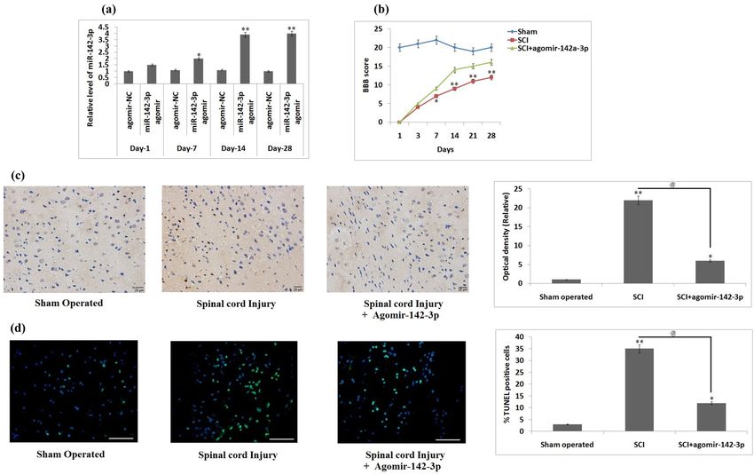
Figure 4: Restitution of Bax blocks the protective effect of miR-142-3p in hydrogen peroxide-exposed BV-2 cells. (a) The BV-2 cells were
exposed to hydrogen peroxide of varied concentrations (50–400 μM) for 10 h followed by qRT-PCR evaluation of miR-142-3p expression.
*P < 0.05, **P < 0.01 compared to the control group of cells. (b) qRT-PCR analysis for expression of miR-142-3p in BV-2 cells after
transfection with miR-142-3p mimics or NC. **P < 0.01 compared to NC mimics. (c) BV-2 cells received transfection of pc-DNA-Bax or
pc-DNA-vector followed by western blot analysis for expression of Bax. (d) Results of western blot analysis for expression of Bax in
hydrogen peroxide-treated cells, which received transfection of miR-142-3p mimics or miR-142-3p and pc-DNA-Bax. After treatment with
hydrogen peroxide, the cells were transfected with miR-142-3p-mimics or miR-142-3p-mimics and pc-DNA-Bax. (e) Quantitative results of
% apoptotic cells. (f) Quantitative results of capase-3 activity. **P < 0.01, @P < 0.01. The results are presented as mean ± SD.
of miR-142-3p suppressed the number of apoptotic cells in molecule is able to build up homodimers on the
hydrogen peroxide-exposed Bv-2 cells against the control mitochondrial membranes and lead to the opening of
cells significantly, but the protective effect of miR-142-3p permeability pores on it for releasing CYT-C from the
decreased significantly with the overexpression of Bax mitochondria in the cytoplasm [33]. In the MAP, CYT-C
(Figure 4e, P < 0.01). Furthermore, the overexpression of may activate the caspase-9-induced reaction, which
Bax halted the effect of miR-142-3p-blocked caspase-3 converts pro-caspase-3 to release caspase-3 [34]. To
activity in hydrogen peroxide-treated cells (P < 0.01, find whether miR-142-3p is involved in the regulation of
Figure 4f). Taken together, the outcomes suggested that MAP by inhibiting the levels of apoptosis-associated
miR-142-3p inhibits apoptosis of cells via inhibiting Bax protein in rats post SCI, the immunoblotting analysis
in hydrogen peroxide-treated Bv-2 cells. was performed to assess the expression levels of pro-
caspase-9, cleaved-caspase-9, pro-caspase-3, cleaved-
caspase-3, Bcl-2 and Bax in the isolated spinal cord
tissues. It was noticed that the expressions of cleaved-
3.5 Upregualtion of miR-142-3p inhibits the caspase-3/-9 and Bax were overexpressed significantly,
mitochondrial apoptosis cascade and the levels of pro‑caspase‑3/-9 and Bcl-2 were found
to be downregulated in the SCI + NC agomiR group
Bax, a protein belonging to Bcl-2 family, has been against the sham-operated group. However, the upre-
reported to show the pro-apoptotic effect and also leads gulation of miR-142-3p significantly decreased the
to the release of CYT-C [27–29]. In the process of expression of cleaved-caspase-3/-9 and Bax and sig-
apoptosis, the overexpression of Bax may encourage nificantly elevated the expression of pro-caspase-3/-9
apoptosis via blocking Bcl-2, which is an antiapoptotic and Bcl-2 in SCI + miR-142-3p agomir group against the
protein [30]. It had been reported earlier that both the SCI + NC-agomiR group (P < 0.01, Figure 5). These
death receptor and the mitochondrial apoptosis findings suggested that the upregulation of miR-142-3p
pathway (MAP) are the important pathways [31,32]. can inhibit the MAP via suppressing the levels of Bax in
Bax is an important regulator in the MAP, and the rats after SCI.miR-142-3p improves functional recovery in SCI 113
Figure 5: miR-142-3p inhibits the mitochondrial apoptotic pathway. The rats submitted to spinal injury followed by the treatment of
miR-142-3p agomir, agomir-NC followed by western blot analysis for expression of calaved-caspase-9/-3, pro-caspase-9/-3, Bax, Bcl-2
in the spinal injured tissue. The results are presented as mean ± SD. **P < 0.01 compared to the spinal cord-injured + agomir NC rats.
##P < 0.01, #P < 0.05 compared to sham-operated rats.
4 Discussion nerve transection in the first 24 h, and in addition, it was
also identified in apoptosis and cell growth after sciatic
The SCI causes a number of biochemical and molecular injury [23]. miR-142-3p has also been reported to play an
changes in the body, which are associated with the important therapeutic target in SCI [39]. Although these
production of free radicals, triggering inflammatory studies have established the role of miR-142-3p in
response, neuronal cell death and plasticity of axions neuronal development, sciatic nerve injury and functional
[35,36]. Previous studies have established that SCI can recovery in SCI, its specific role and the involved pathway
lead to aberrant expression of miRNA, and deregulated in SCI remain unclear. Here, we studied the expression
miRs can affect the pathophysiology of SCI and its profile of miRs in the animal model of SCI in the post-
functional outcomes [37]. However, studies confirming injury phase. We evidenced that miR-142-3p was the most
the specific role of miRs in the SCI are missing. In the downregulated miR, which was consistent with the
present work, an animal model of SCI was developed, and findings of the earlier study [39].
microarray analysis was done to screen the expression Mitochondria play a significant role in the process of
profiles of miRs after SCI. It was noticed that SCI caused apoptosis by releasing the molecules responsible for
deregulation in the expression of miRs, among which apoptosis such as CYT-C [41]. Bax also called as BCL2-
miR-142-3p was one of the most significantly deregulated associated X protein is a pro-apoptotic protein, which
in the spinal tissues. In addition, it was evidenced that can open the transition pores present on the mitochon-
the upregulation of miR-142-3p improved the SCI by dria membrane, facilitating the release of CYT-C from the
attenuating functional recovery, decreasing the size of mitochondria [33]. CYT-C can promote the amplification
lesions and apoptosis. Furthermore, it was found that reaction of the caspase‑9‑molulated pathway in the
miR-142-3p inhibited the levels of Bax via 3′UTR region in MAP, which in turn forces pro-caspase-3 to form
the BV-2 cells exposed to hydrogen peroxide. However, caspase-3 [34]. Here, it was evidenced that miR-142-3p
the present outcomes suggested that miR-142-3p shows decreases the levels of Bax via targeting the 3′UTR
its protective action on the SCI via inhibiting the MAP. region in the BV-2 cells. In addition, our results
miR-142-3p is identified to play an important role in suggested a negative link between expression of miR-142-3p
inflammation, viral infection, cancer, and immunity [38]. and Bax in the spinal cord tissues of the spinal cord-injured
miR-142-3p is associated to be linked with neuronal rats, suggesting Bax as a favorable target of miR-142-3p
disorders, and it has also been identified to be a key in vivo. Hence, we speculated that miR-142-3p may
target in repairing of sensory function in neurons through modulate the MAP via blocking the expression of Bax
adenylyl cyclase [39]. miR-142-3p miR-142-3p is respon- in rats subjected to SCI. The present findings suggested
sible for regulating the proinflammatory function in lupus that the increased expression of miR-142-3p suppressed
erythematosus [40]. miR-142-3p has also been found to be the cleaved-caspase-3/-9 and Bax and increased the
overexpressed in the rat dorsal route ganglion after sciatic expression of pro-caspase-3/-9 and Bcl-2 in the spinal114 Jun Zheng et al.
cord tissues of SCI-induced rats. These findings sug- [10] Li P, Nijhawan D, Wang X. Mitochondrial activation of
gested that miR-142-3p may show its therapeutic effect in apoptosis. Cell. 2004;116(2 Suppl):S57–61.
SCI via blocking the MAP. However, the present study [11] Nicholson DW, Thornberry NA. Caspases: killer proteases.
Trends Biochem Sci. 1997;22(8):299–306.
has some shortfalls, i.e., the study focused on only the
[12] Croce CM. Causes and consequences of microRNA dysregula-
most overexpressed miR-142-3p, and we do not screen tion in cancer. Nat Rev Genet. 2009;10(10):704–14.
other miRs for their role in SCI. [13] Bartel DP. MicroRNAs: target recognition and regulatory
In conclusion, the present work showed that SCI functions. Cell. 2009;136(2):215–33.
leads to aberrant expression of miRs in the rats subjected [14] Friedman RC, Farh KK, Burge CB, Bartel DP. Most mammalian
mRNAs are conserved targets of microRNAs. Genome Res.
to SCI, and among miRs, miR-142-3p was the most
2009;19(1):92–105.
significantly decreased miR in the spinal cord. Also, it [15] Krichevsky AM. MicroRNA profiling: from dark matter to white
was evidenced that the increased levels of miR-142-3p matter, or identifying new players in neurobiology.
improved the functional recovery, decreased the lesion ScientificWorldJournal. 2007;7:155–66.
size and also suppressed the apoptosis of neuronal cells [16] Kosik KS. The neuronal microRNA system. Nat Rev Neurosci.
2006;7(12):911–20.
post-SCI. This work also confirmed that miR-142-3p
[17] Bak M, Silahtaroglu A, Møller M, Christensen M, Rath MF,
targets Bax in the BV-2 cells and may show its protective
Skryabin B, et al. MicroRNA expression in the adult mouse
effect in SCI via inhibiting the MAP, suggesting miR-142- central nervous system. RNA. 2008;14(3):432–44.
3p can be an important therapeutic target for treating SCI. [18] Liu NK, Wang XF, Lu QB, Xu XM. Altered microRNA expression
following traumatic spinal cord injury. Exp Neurol.
Acknowledgments: This work was supported by the Natural 2009;219(2):424–9.
[19] Liu G, Keeler BE, Zhukareva V, Houlé JD. Cycling exercise
Science Fund of Hubei Province Fund NO. 2019CFB528.
affects the expression of apoptosis-associated microRNAs after
spinal cord injury in rats. Exp Neurol. 2010;226(1):200–6.
Conflict of interest: Authors state no conflict of interest. [20] Sun Y, Su Q, Li L, Wang X, Lu Y, Liang J. MiR-486 regulates
cardiomyocyte apoptosis by p53-mediated BCL-2 associated
mitochondrial apoptotic pathway. BMC Cardiovasc Disord.
2017;17(1):119.
[21] Makhdoumi P, Roohbakhsh A, Karimi G. MicroRNAs regulate
References mitochondrial apoptotic pathway in myocardial ischemia-
reperfusion-injury. Biomed Pharmacother. 2016;84:1635–44.
[1] Chen Y, Cao S, Xu P, Han W, Shan T, Pan J, et al. Changes in [22] Wang J, Jiao Y, Cui L, Jiang L. miR-30 functions as an oncomiR
the expression of miR-34a and its target genes following in gastric cancer cells through regulation of P53-mediated
spinal cord injury in rats. Med Sci Monit. 2016;22:3981–93. mitochondrial apoptotic pathway. Biosci Biotechnol Biochem.
[2] Hu W, Wang H, Liu Z, Liu Y, Wang R, Luo X, et al. 2017;81(1):119–26.
Neuroprotective effects of lycopene in spinal cord injury in [23] Wu DM, Wen X, Han XR, Wang S, Wang YJ, Shen M, et al. MiR-
rats via antioxidative and anti-apoptotic pathway. Neurosci 142-3p enhances cell viability and inhibits apoptosis by
Lett. 2017;642:107–12. targeting CDKN1B and TIMP3 following sciatic nerve injury.
[3] Thuret S, Moon LD, Gage FH. Therapeutic interventions after Cell Physiol Biochem. 2018;46(6):2347–57.
spinal cord injury. Nat Rev Neurosci. 2006;7(8):628–43. [24] Gao YF, Zhang QJ, Yu Z, Liu SH, Liang J. miR142 supresses
[4] Blight AR. Miracles and molecules-progress in spinal cord proliefartion and induces apoptosis of osteosarcoma cells by
repair. Nat Neurosci. 2002;5(Suppl 1):S1051–S1054. upregulating Rb. Oncol Lett. 2018;16(1):733–40.
[5] Rabchevsky AG, Patel SP, Springer JE. Pharmacological [25] Yu DS, Lv G, Mei XF, Cao Y, Wang YF, Wang YS, et al. MiR-200c
interventions for spinal cord injury: where do we stand? how regulates ROS-induced apoptosis in murine BV-2 cells by
might we step forward? Pharmacol Ther. 2011;132(1): targeting FAP-1. Spinal Cord. 2015;53(3):182–9.
15–29. [26] Hu F, Min J, Cao X, Liu L, Ge Z, Hu J, et al. MiR-363-3p inhibits
[6] Kawabata H, Setoguchi T, Yone K, Souda M, Yoshida H, the epithelial-to-mesenchymal transition and suppresses
Kawahara K, et al. High mobility group box 1 is upregulated metastasis in colorectal cancer by targeting Sox4. Biochem
after spinal cord injury and is associated with neuronal cell Biophys Res Commun. 2016;474(1):35–42.
apoptosis. Spine (Phila Pa 1976). 2010;35(11):1109–15. [27] Nishimura R, Tabata K, Arakawa M, Ito Y, Kimura Y, Akihisa T,
[7] Li J, Huang C-Y, Zheng RL, Cui KR, Li JF. Hydrogen peroxide et al. Isobavachalcone, a chalcone constituent of Angelica
induces apoptosis in human hepatoma cells and alters cell keiskei, induces apoptosis in neuroblastoma. Biol Pharm Bull.
redox status. Cell Biol Int. 2000;24(1):9–23. 2007;30(10):1878–83.
[8] Reuter S, Eifes S, Dicato M, Aggarwal BB, Diederich M. [28] He J, Xiao Y, Casiano CA, Zhang L. Role of mitochondrial
Modulation of anti-apoptotic and survival pathways by cytochrome c in cocaine-induced apoptosis in coronary artery
curcumin as a strategy to induce apoptosis in cancer cells. endothelial cells. J Pharmacol Experimental Ther.
Biochem Pharmacol. 2008;76(11):1340–51. 2000;295(3):896–903.
[9] Balaban RS, Nemoto S, Finkel T. Mitochondria, oxidants, and [29] Liu H, Qin CK, Han GQ, Xu HW, Ren WH, Qin CY. Synthetic
aging. Cell. 2005;120(4):483–95. chenodeoxycholic acid derivative, HS-1200, inducesmiR-142-3p improves functional recovery in SCI 115
apoptosis of human hepatoma cells via a mitochondrial [36] De Biase A, Knoblach SM, Di Giovanni S, Fan C, Molon A,
pathway. Cancer Lett. 2008;270(2):242–9. Hoffman EP, et al. Gene expression profiling of experimental
[30] Noguchi K, Kitanaka C, Yamana H, Kokubu A, Mochizuki T, traumatic spinal cord injury as a function of distance from
Kuchino Y. Regulation of c-Myc through phosphorylation at impact site and injury severity. Physiol Genomics.
Ser-62 and Ser-71 by c-Jun N-terminal kinase. J Biol Chem. 2005;22(3):368–81.
1999;274(46):32580–7. [37] Hu JZ, Huang JH, Zeng L, Wang G, Cao M, Lu HB. Anti-apoptotic
[31] Klapsinou E, Argyri E, Panotopoulou E, Daskalopoulou D, effect of microRNA-21 after contusion spinal cord injury in
Patsouris E, Nonni A, et al. Bax and Bak expression in cervical rats. J Neurotrauma. 2013;30(15):1349–60.
smears of women with low-and high-risk HPV types: a study [38] Shrestha A, Mukhametshina RT, Taghizadeh S, Vásquez-
of 120 cases. J Cytol. 2015;32(4):223–9. Pacheco E, Cabrera-Fuentes H, Rizvanov A, et al. MicroRNA-
[32] Su CC, Lee KI, Chen MK, Kuo CY, Tang CH, Liu SH. Cantharidin 142 is a multifaceted regulator in organogenesis, homeostasis
induced oral squamous cell carcinoma cell apoptosis via the and disease. Dev Dyn. 2017;246(4):285–90.
JNK-regulated mitochondria and endoplasmic reticulum stress- [39] Wang T, Yuan W, Liu Y, Zhang Y, Wang Z, Chen X, et al. miR-
related signaling pathways. PLoS One. 2016;11(12):e0168095. 142-3p is a potential therapeutic target for sensory function
[33] Lu Z, Chen H, Zheng XM, Chen ML. Experimental study on the recovery of spinal cord injury. Med Sci Monit. 2015;21:
apoptosis of cervical cancer Hela cells induced by juglone 2553–6.
through c-Jun N-terminal kinase/c-Jun pathway. Asian Pac J [40] Wang Y, Liang J, Qin H, Ge Y, Du J, Lin J, et al. Elevated
Trop Med. 2017;10(6):572–5. expression of miR-142-3p is related to the pro-inflammatory
[34] Zou H, Li Y, Liu X, Wang X. An APAF-1.cytochrome c multimeric function of monocyte-derived dendritic cells in SLE. Arthritis
complex is a functional apoptosome that activates procas- Res Ther. 2016;18(1):263.
pase-9. J Biol Chem. 1999;274(17):11549–56. [41] Malhotra R, Lin Z, Vincenz C, Brosius III FC. Hypoxia induces
[35] Giovanni SD, Knoblach SM, Brandoli C, Aden SA, Hoffman EP, apoptosis via two independent pathways in Jurkat cells:
Faden AI. Gene profiling in spinal cord injury shows role of cell differential regulation by glucose. Am J Physiol Cell Physiol.
cycle neuronal death. Ann Neurol. 2003;53(4):454–68. 2001;281(5):C1596–C1603.You can also read