Potential Role of PUMA in Delayed Death of Hippocampal CA1 Neurons After Transient Global Cerebral Ischemia
←
→
Page content transcription
If your browser does not render page correctly, please read the page content below
Potential Role of PUMA in Delayed Death of Hippocampal
   CA1 Neurons After Transient Global Cerebral Ischemia
                      Kuniyasu Niizuma, MD; Hidenori Endo, MD; Chikako Nito, MD, PhD;
                                   D. Jeannie Myer, PhD; Pak H. Chan, PhD

Background and Purpose—p53-upregulated modulator of apoptosis (PUMA), a BH3-only member of the Bcl-2 protein
  family, is required for p53-dependent and -independent forms of apoptosis. PUMA localizes to mitochondria and
  interacts with antiapoptotic Bcl-2 and Bcl-XL or proapoptotic Bax in response to death stimuli. Although studies have
  shown that PUMA is associated with pathomechanisms of cerebral ischemia, clearly defined roles for PUMA in
  ischemic neuronal death remain unclear. The purpose of this study was to determine potential roles for PUMA in
  cerebral ischemia.
Methods—Five minutes of transient global cerebral ischemia (tGCI) were induced by bilateral common carotid artery
  occlusion combined with hypotension.
Results—PUMA was upregulated in vulnerable hippocampal CA1 neurons after tGCI as shown by immunohistochemistry.
  In Western blot and coimmunoprecipitation analyses, PUMA localized to mitochondria and was bound to Bcl-XL and
  Bax in the hippocampal CA1 subregion after tGCI. PUMA upregulation was inhibited by pifithrin-␣, a specific inhibitor
  of p53, suggesting that PUMA is partly controlled by the p53 transcriptional pathway after tGCI. Furthermore, reduction
  in oxidative stress by overexpression of copper/zinc superoxide dismutase, which is known to be protective of
  vulnerable ischemic hippocampal neurons, inhibited PUMA upregulation and subsequent hippocampal CA1 neuronal
  death after tGCI.
Conclusions—These results imply a potential role for PUMA in delayed CA1 neuronal death after tGCI and that it could
  be a molecular target for therapy. (Stroke. 2009;40:618-625.)
         Key Words: PUMA 䡲 cerebral ischemia, global 䡲 apoptosis 䡲 superoxide dismutase 䡲 oxidative stress

T    he Bcl-2 protein family is a principal regulator of
     mitochondrial membrane integrity and function and is
classified into 3 subgroups according to structural homol-
                                                                         activating caspases-9 and -3.3–5 However, the roles of PUMA in
                                                                         cerebral ischemia remain unclear.
                                                                            To determine these roles, we used as our model 5
ogy (Bcl-2 homology [BH] domains): the antiapoptotic                     minutes of transient global cerebral ischemia (tGCI),
proteins (Bcl-2, Bcl-XL, Mcl-1, BCL-W), the proapoptotic pro-            which induces delayed neuronal death in the hippocampal
teins (Bax, Bak), and the BH3-only proteins (Bim, Bad, Bid,              CA1 subregion in rats.6 We investigated expression of
Bik, p53-upregulated modulator of apoptosis [PUMA],                      PUMA and the interaction between PUMA and Bcl-XL,
NADPH oxidase activator, Hrk). BH3-only proteins share                   Bcl-2, and Bax in the hippocampal CA1 subregion after
sequence homology with other Bcl-2 proteins in the BH3                   tGCI. To investigate the regulation of PUMA by p53 after
region only1 and are involved in the mechanisms of cyto-                 tGCI, we administered pifithrin-␣ (PFT), a specific inhib-
chrome c release in neuronal apoptosis.2                                 itor of p53. To demonstrate the effects of oxidative stress
   According to a recent hierarchy model, BH3-only pro-                  on PUMA expression after tGCI, we used copper/zinc
teins are subdivided into activator or inactivator proteins.3            superoxide dismutase (SOD1) transgenic (Tg) rats, which
Activator BH3-only proteins can interact with both anti-                 have neuroprotection against ischemia because of reduced
apoptotic Bcl-2 proteins and proapoptotic Bcl-2 proteins.                oxidative stress.7
Among these activator proteins, PUMA was initially iden-
tified as a gene activated by p53 in cells undergoing                                     Materials and Methods
p53-induced apoptosis.4,5 In p53-induced cell death, PUMA                Global Cerebral Ischemia
was shown to localize to mitochondria; interact with Bcl-2,              Five minutes of tGCI was induced by bilateral common carotid
Bcl-XL, and Bax; and induce cytochrome c release, thereby                artery occlusion combined with hypotension according to a

  Received April 28, 2008; final revision received June 26, 2008; accepted July 15, 2008.
  From the Departments of Neurosurgery, Neurology and Neurological Sciences, and the Program in Neurosciences, Stanford University School of
Medicine, Stanford, Calif.
  Correspondence to Dr Pak H. Chan, Neurosurgical Laboratories, Stanford University, 1201 Welch Road, MSLS #P314, Stanford, CA 94305-5487.
E-mail phchan@stanford.edu
  © 2009 American Heart Association, Inc.
  Stroke is available at http://stroke.ahajournals.org                                              DOI: 10.1161/STROKEAHA.108.524447

                                                                  618
                                 Downloaded from http://stroke.ahajournals.org/ by guest on July 24, 2015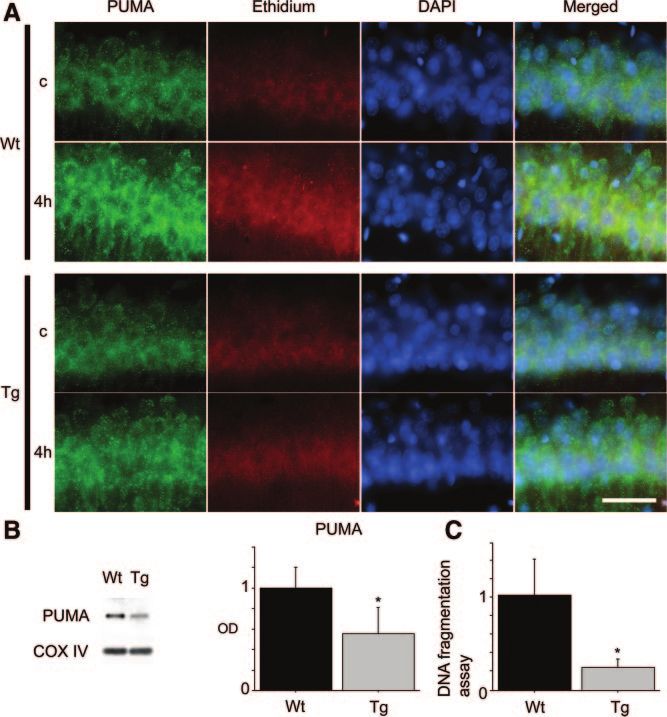
Niizuma et al Role of PUMA in Neuronal Death After Ischemia 619
method described previously,7 with some modifications.6 Male anti-Bax (#2772, Cell Signaling Technology) and protein G–Sepharose
Sprague-Dawley rats (300 to 350 g) were anesthetized with 2.0% for 2 hours at 4°C. The negative control was prepared with protein
isoflurane in 70% nitrous oxide and 30% oxygen via face mask. G–Sepharose without an antibody. Whole-brain extract was included as
Rectal temperature was controlled at 37°C during surgery with a a positive control. The 14 000g pellets were washed 3 times and
homeothermic blanket. The femoral artery was catheterized with analyzed as the samples bound to each antibody by Western blotting
a PE-50 catheter to allow continuous recording of arterial blood with anti-PUMA (1:1000, #4976; Cell Signaling Technology), anti–
pressure. After heparinization, blood was quickly withdrawn via Bcl-XL (1:1000), anti–Bcl-2 (1:1000), or anti-Bax (1:1000).
the jugular vein. When the mean arterial blood pressure became
30 mm Hg, both common carotid arteries were clamped with Cresyl Violet Staining and Immunohistochemistry
surgical clips. Blood pressure was maintained at 30 mm Hg
during the ischemic period. After 5 minutes of ischemia, the clips
of PUMA
were removed and the blood was reinfused. Regional cerebral Anesthetized animals were perfused with 10 U/mL heparin saline
blood flow was monitored by laser Doppler flowmetry as previ- and subsequently with 4% formaldehyde in PBS after 1, 4, 24, or
ously described.7 Sham-operated animals underwent exposure of 72 hours of reperfusion. Brains were removed, postfixed for 24
the vessels without blood withdrawal or clamping of the carotid hours, and sectioned at 50 m with a Vibratome. For histologic
arteries. The animals were maintained at 20°C with ad libitum assessment, the sections were stained with cresyl violet. For
access to food and water. All animals were treated in accordance immunohistochemistry of PUMA, sections were reacted with
with Stanford University guidelines, and the animal protocols anti-PUMA (1:50, #4976; Cell Signaling Technology). Immuno-
were approved by Stanford University’s Administrative Panel on histochemistry was performed with the avidin-biotin technique,
Laboratory Animal Care. and nuclei were counterstained with methyl green solution.
Drug Treatment Immunofluorescence Staining
To examine the effect of a specific p53 inhibitor on PUMA To evaluate colocalization of PUMA and COX IV with neuron-
expression after tGCI, we administered PFT (P4359; Sigma-Aldrich, specific nuclear protein (NeuN), Bcl-XL, or Bax, we performed
St. Louis, Mo), dissolved in dimethyl sulfoxide and phosphate- double immunofluorescence. For double immunofluorescence of
buffered saline (PBS). This drug (4 mg/kg in dimethyl sulfoxide in PUMA and COX IV or Bax, the sections were reacted with
PBS) or vehicle (dimethyl sulfoxide in PBS) was injected via the left anti–COX IV (1:100, #4844; Cell Signaling Technology) or
jugular vein just after reperfusion as described previously.8 anti-Bax (1:50, sc-526; Santa Cruz Biotechnology), followed by
fluorescein isothiocyanate– conjugated anti-rabbit monovalent
SOD1-Tg Rats Fab fragments of a secondary antibody (Jackson ImmunoRe-
search, West Grove, Pa) for labeling and blocking of COX IV or
Heterozygous SOD1-Tg rats with a Sprague-Dawley background
carrying human SOD1 genes were derived from founder stock and Bax. Then the sections were incubated with anti-PUMA (1:50,
were further bred with wild-type (Wt) Sprague-Dawley rats to #4976; Cell Signaling Technology) followed by Texas Red–
generate heterozygous rats, as previously described. The phenotype conjugated anti-rabbit IgG (Jackson ImmunoResearch). For dou-
of the SOD1-Tg rats was identified by isoelectric focusing gel ble immunofluorescence of PUMA and NeuN or Bcl-XL, sections
electrophoresis as described.7 There were no observable phenotypic were immunostained with anti-PUMA (Cell Signaling Technol-
differences in brain vasculature between the SOD1-Tg rats and their ogy) followed by Texas Red– conjugated anti-rabbit IgG. The
Wt littermates.7 sections were then incubated with anti-NeuN (1:50, MAB377;
Chemicon International, Temecula, Calif) or anti–Bcl-XL (1:50,
#610209; BD Biosciences), followed by fluorescein isothiocya-
Western Blot Analysis nate– conjugated antimouse IgG (Jackson ImmunoResearch). The
The hippocampal CA1 subregion was removed after 1, 4, 24, or sections were covered with Vectashield mounting medium with
72 hours of reperfusion. Protein extraction of the cytosolic,
4⬘,6-diamidino-2-phenylindole (DAPI; Vector Laboratories, Bur-
mitochondrial, and nuclear fractions was performed with a
lingame, Calif) and examined under an LSM510 confocal laser
multiple centrifugation method as described previously.9 Equal
scanning microscope or an Axioplan 2 microscope (Carl Zeiss,
amounts of samples were loaded per lane and analyzed by sodium
Thornwood, NY).
dodecyl sulfate–polyacrylamide-gel electrophoresis on a 10% to
20% Tris-glycine gel (Invitrogen, Carlsbad, Calif) and then
immunoblotted. Anti-PUMA (1:1000, #4976; Cell Signaling Tech- In Situ Detection of Superoxide Anion Production
nology, Beverly, Mass), anti–p-53 (1:1000, #554147; BD Bio- Early production of superoxide anions after tGCI was investi-
sciences, San Jose, Calif), anti– cytochrome c (1:1000, #556433; BD gated with the use of hydroethidine as previously described.10
Biosciences), anti– caspase-9 (1:1000, sc-8355; Santa Cruz Biotech- Hydroethidine is diffusible into the central nervous system
nology, Santa Cruz, Calif), anti–-actin (1:10 000, A5441; Sigma- parenchyma after intravenous injection and is selectively oxidized
Aldrich), anti-cytochrome oxidase subunit IV (COX IV; 1:5000, to ethidium by superoxide anions. Hydroethidine solution (200
A21348; Invitrogen), or anti-TFIID (transcription factor II D; 1:200, L of 1 mg/mL in 1% dimethyl sulfoxide with saline) was
sc-204; Santa Cruz Biotechnology) primary antibody was used. After administered intravenously 15 minutes before ischemia induction.
incubation with horseradish peroxidase– conjugated secondary anti- A sample was prepared as described in the immunohistochemistry
body, the antigen was detected by chemiluminescence Western method. For fluorescent double staining of the ethidium signal
blotting detection reagents (Amersham, Buckinghamshire, UK). The and PUMA, sections were incubated with anti-PUMA (1:50,
image was scanned with a GS-700 imaging densitometer (Bio-Rad #4976; Cell Signaling Technology), followed by fluorescein
Laboratories, Hercules, Calif), and the results were quantified by isothiocyanate– conjugated anti-rabbit IgG (Jackson ImmunoRe-
Multi-Analyst software (Bio-Rad). search). Slides were covered with DAPI (Vector Laboratories)
and observed with a fluorescence microscope.
Coimmunoprecipitation
A sample of mitochondrial fractions was prepared as described in the Cell Death Assay
Western blotting method. The procedure for precipitation was per- For quantification of apoptosis-related DNA fragmentation, we used
formed as described previously.8 After protein extraction, the mitochon- a commercial enzyme immunoassay to determine cytoplasmic his-
drial samples were incubated with protein G–Sepharose (Amersham) tone–associated DNA fragments (1774425; Roche Molecular Bio-
for 1 hour at 4°C, and this mixed sample was then centrifuged. The chemicals, Mannheim, Germany) and to detect apoptotic but not
supernatant was incubated with 2 g of anti–Bcl-XL (#2762, Cell necrotic cell death.11 A sample was prepared as described in the
Signaling Technology), anti–Bcl-2 (#610538, BD Biosciences), or Western blotting method. A cytosolic volume containing 20 g of
Downloaded from http://stroke.ahajournals.org/ by guest on July 24, 2015
620 Stroke February 2009
Figure 1. A, Representative photomicro-
graphs of immunohistochemistry of
PUMA after tGCI. Cresyl violet staining
shows no neuronal degeneration in the
hippocampal CA1 subregion 1, 4, and 24
hours after tGCI. Seventy-two hours after
tGCI, ⬎80% of the CA1 neurons degen-
erated, although neurons in the hip-
pocampal CA3 subregion were spared.
Immunoreactivity for PUMA increased,
peaked at 4 hours, and declined by 24
hours. At 72 hours, the CA1 neurons
degenerated, and immunoreactivity could
not be evaluated. C indicates control.
Scale bar⫽300 m (hippocampus),
50 m (CA1). B, Representative photomi-
crographs of fluorescent double staining
of PUMA (red) and NeuN (green) in the
hippocampal CA1 subregion 4 hours after
tGCI. Nuclei were counterstained with
DAPI (blue). NeuN immunoreactivity
showed the distribution of neurons. Over-
lapped image demonstrates that PUMA-
positive cells in the hippocampal CA1
subregion colocalized with neurons.
Scale bar⫽50 m.
protein was used for the ELISA, according to the manufacturer’s confirmed by cresyl violet staining (Figure 1A). However,
protocol. ⬎80% of the CA1 neurons were degenerated 72 hours after
tGCI, which was compatible with our previous reports.6,7 In
Statistical Analysis contrast, neurons in the hippocampal CA3 subregion were
Comparisons among multiple groups were performed with ANOVA
followed by a Scheffé post hoc analysis (SigmaStat; Systat Software, spared even at 72 hours. PUMA immunoreactivity increased
San Jose, Calif). Comparisons between 2 groups were achieved with a after tGCI, peaked at 4 hours, and then started to decline at 24
Student unpaired t test. Data are expressed as mean⫾SD, and signifi- hours. At 72 hours, the CA1 neurons degenerated and
cance was accepted with P⬍0.05. immunoreactivity could not be evaluated (Figure 1A). Double
immunofluorescence for PUMA and NeuN demonstrated that
Results PUMA-positive cells in the hippocampal CA1 subregion
PUMA Induction and Selective Neuronal Death colocalized with neurons 4 hours after tGCI (Figure 1B).
After tGCI These results indicate that PUMA is upregulated in the
One, 4, or 24 hours after 5 minutes of tGCI, there was no hippocampal CA1 neurons after tGCI, which precedes CA1
neuronal degeneration in the hippocampal CA1 subregion, as neuronal death.
Downloaded from http://stroke.ahajournals.org/ by guest on July 24, 2015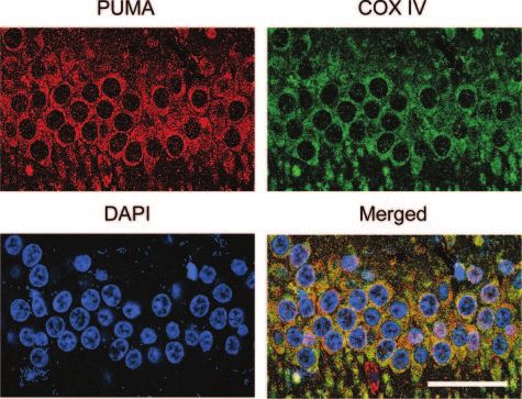
Niizuma et al Role of PUMA in Neuronal Death After Ischemia 621
Figure 2. Mitochondrial upregulation of
PUMA preceded cytochrome c release
and caspase-9 activation. A, Western blot
analysis showed that immunoreactivity of
PUMA in the cytosolic fraction was only
slightly detectable at any time point. In
contrast, PUMA immunoreactivity signifi-
cantly increased in the mitochondrial
fraction 4 and 24 hours after tGCI (n⫽4,
*P⬍0.05). -Actin and COX IV analyses
are shown as internal controls. C indicates
control; OD, optical density. B, Western
blot analysis showed that nuclear p53 was
significantly increased 4 hours after tGCI
(n⫽4, *P⬍0.05). Transcription factor II D
(TFIID) analysis is shown as an internal
control. C, Western blot analysis revealed
that cytosolic cytochrome c and cleaved
caspase-9 were significantly increased in
the hippocampal CA1 subregion 24 hours
after tGCI (n⫽4, *P⬍0.05). -Actin analysis
is shown as an internal control.
Mitochondrial Localization of PUMA and and 20 kDa, respectively, and showed no significant change
Subsequent Cytochrome c Release in the at any time point (data not shown). PUMA expression
Hippocampal CA1 Subregion After tGCI precipitated by Bcl-XL increased time-dependently and sig-
Western blotting showed that PUMA immunoreactivity was nificantly increased at 24 and 72 hours (Figure 4A, n⫽4,
evident as a single band of molecular mass of 19 kDa (Figure P⬍0.05). PUMA expression precipitated by Bax also in-
2A). In cytosolic samples from the hippocampal CA1 subre- creased 24 hours after tGCI (Figure 4B, n⫽4, P⬍0.01). In
gion, PUMA immunoreactivity was slightly detectable and contrast, PUMA expression precipitated by Bcl-2 showed no
showed no significant change after tGCI. In contrast, PUMA significant difference at any time point, although it tended to
expression was significantly increased in mitochondrial sam- increase after tGCI (data not shown).
ples, peaking at 4 hours and then decreasing by 72 hours after For further investigation of direct interaction between
tGCI. It was barely detectable in the sham-operated brains PUMA and Bcl-XL or Bax, we performed double immuno-
(Figure 2A, n⫽4, P⬍0.05). fluorescence, which demonstrated that PUMA-positive cells
To confirm the upstream pathway of PUMA, we investi- colocalized with Bcl-XL–positive cells (Figure 4C) or Bax-
gated nuclear p53 upregulation. Western blotting showed that positive cells (Figure 4D) in the hippocampal CA1 subregion
nuclear p53 significantly increased 4 hours after tGCI (Figure 24 hours after tGCI. In combination with the coimmunopre-
2B), presenting a pattern similar to that of mitochondrial cipitation data, these results indicate that PUMA directly
PUMA upregulation. To confirm activation of the mitochon- interacts with Bcl-XL and Bax in the hippocampal CA1
drial apoptotic pathway after tGCI, we examined cytochrome subregion after tGCI.
c release and caspase-9 activation. Cytosolic cytochrome c
and cleaved caspase-9 significantly increased at 24 hours
(Figure 2C, n⫽4, P⬍0.05), which suggests cytochrome c
release to the cytosol and subsequent caspase chain reaction
after tGCI. These results indicate that PUMA increases in the
mitochondria before cytochrome c release and caspase-9
activation.
For further investigation of mitochondrial localization of
PUMA after ischemia, we performed double immunofluores-
cence for PUMA and COX IV, which was used as a
mitochondrial marker. Double immunofluorescence demon-
strated that PUMA colocalized with COX IV in the hip-
pocampal CA1 subregion 4 hours after tGCI (Figure 3).
Interaction Between PUMA and Bcl-XL or Bax
After tGCI
To investigate potential direct interactions between PUMA
and Bcl-XL, Bcl-2, or Bax, we performed coimmunoprecipi- Figure 3. Double immunofluorescence of PUMA (red) and COX
tations in the mitochondrial fraction from the hippocampal IV (green) demonstrated that PUMA-positive cells colocalized
with COX IV–positive cells in the hippocampal CA1 subregion 4
CA1 subregion. With Western blot analysis, Bcl-XL, Bcl-2, hours after tGCI. Nuclei were counterstained with DAPI (blue).
and Bax immunoreactivity was evident as bands of 30, 26, Scale bar⫽50 m.
Downloaded from http://stroke.ahajournals.org/ by guest on July 24, 2015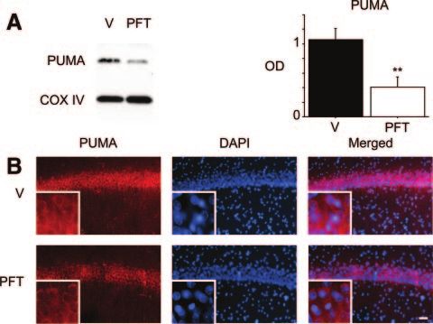
622 Stroke February 2009
Figure 5. The effect of PFT on PUMA expression in the hip-
pocampal CA1 subregion after tGCI. A, Western blot analysis
showed that PUMA expression was significantly decreased in
the PFT-treated animals (PFT) compared with the vehicle-
treated animals (V) 4 hours after tGCI (n⫽4, **P⬍0.01). COX IV
analysis is shown as an internal control. OD indicates optical
density. B, In an immunofluorescence study, PUMA (red)
expression was decreased in the PFT-treated animals compared
with the vehicle-treated animals 4 hours after tGCI. Nuclei were
counterstained with DAPI (blue). Scale bar⫽50 m.
hippocampal CA1 subregion was significantly decreased in
PFT-treated animals compared with vehicle-treated animals 4
hours after tGCI (Figure 5A, n⫽4, P⬍0.01). Bax, Bcl-2, and
Bcl-XL expression levels showed no differences between
vehicle-treated and PFT-treated rats (data not shown). An
immunofluorescence study showed that PUMA expression
was decreased in the hippocampal CA1 subregion of the
PFT-treated animals compared with the vehicle-treated ani-
mals 4 hours after tGCI, which was compatible with the result
Figure 4. Interaction between PUMA and Bcl-XL or Bax in the of the Western blot study (Figure 5B). These results indicate
mitochondrial fraction of the hippocampal CA1 subregion after that PFT administration inhibits PUMA upregulation after
tGCI. A, Coimmunoprecipitation analysis of PUMA immunoreac- tGCI.
tivity precipitated by Bcl-XL showed a significant increase 24
and 72 hours after tGCI (n⫽4, *P⬍0.05). Bcl-XL immunoreactiv-
ity precipitated by Bcl-XL was used to show equal precipitation. SOD1 Overexpression
C indicates control; IP, immunoprecipitation; IB, immunoblotting; To confirm that superoxide production is associated with
OD, optical density. B, Coimmunoprecipitation analysis of
PUMA immunoreactivity precipitated by Bax showed a signifi- PUMA induction, we performed double immunofluorescence
cant increase 24 hours after tGCI (n⫽4, **P⬍0.01). Bax immu- of ethidium and PUMA. In the hippocampal CA1 pyramidal
noreactivity precipitated by Bax was used to show equal precip- neurons, ethidium signals were shown as small particles in
itation. C, Representative fluorescent double staining of PUMA
(red) and Bcl-XL (green) in the hippocampal CA1 subregion 24 the cytosol of the nonischemic brains in both the Wt and
hours after tGCI. Nuclei were counterstained with DAPI (blue). SOD1-Tg rats (Figure 6A). Four hours after ischemia, the
Overlapped image demonstrates that PUMA-positive cells colo- hippocampal CA1 neurons showed a marked increase in
calized with Bcl-XL–positive cells. D, Representative fluorescent
double staining of PUMA (red) and Bax (green) in the hippocam- punctate and diffuse signals for both ethidium and PUMA in
pal CA1 subregion 24 hours after tGCI. Nuclei were counter- the Wt rats. However, the increase in signal was less
stained with DAPI (blue). Overlapped image demonstrates that noticeable in the SOD1-Tg rats. The Western blot analysis
PUMA-positive cells colocalized with Bax-positive cells. Scale
bars⫽50 m. indicated that PUMA expression was significantly decreased
4 hours after tGCI in the SOD1-Tg rats compared with the Wt
rats (Figure 6B, n⫽4, P⬍0.05). Bax, Bcl-2, and Bcl-XL
PFT Administration expression levels showed no differences between the Wt and
To investigate the regulation of PUMA by p53, we intrave- Tg rats (data not shown).
nously administered 4 mg/kg of PFT just after reperfusion. We then examined apoptosis-related DNA fragmentation
Our previous study indicated that this dose was effective in after tGCI to investigate neuroprotection of SOD1 overex-
inhibiting PUMA expression.8 Western blot analysis showed pression. DNA fragmentation in the hippocampal CA1 sub-
that PUMA expression in the mitochondrial fraction from the region at 72 hours was significantly decreased in the
Downloaded from http://stroke.ahajournals.org/ by guest on July 24, 2015Niizuma et al Role of PUMA in Neuronal Death After Ischemia 623
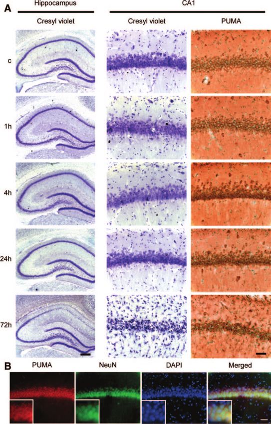
Figure 6. Effect of SOD1 overexpression
on PUMA expression, ethidium signals,
and DNA fragmentation after tGCI. A,
Representative photomicrographs show
fluorescent double staining of PUMA
(green) and ethidium (red) in the hip-
pocampal CA1 subregion. Nuclei were
counterstained with DAPI (blue). Ethidium
signals were seen as small particles in
the cytosol in nonischemic brains of both
the Wt and Tg rats. Four hours after
tGCI, hippocampal CA1 neurons showed
a marked increase in ethidium signals in
the Wt rats. However, the signal increase
was less noticeable in the Tg rats 4 hours
after tGCI. In the Wt rats, the signals for
PUMA increased dramatically at 4 hours
compared with the sham-operated rats
and overlapped with the ethidium signals.
In the Tg rats, PUMA expression was less
strong than in the Wt rats at 4 hours. C
indicates control. Scale bar⫽50 m. B,
Western blot analysis showed that PUMA
expression decreased significantly in the
Tg rats 4 hours after tGCI (n⫽4,
*P⬍0.05). COX IV analysis is shown as an
internal control. C, Apoptosis-related
DNA fragmentation assay. DNA fragmen-
tation at 72 hours was significantly
decreased in the hippocampal CA1 sub-
region of the Tg rats compared with the
Wt rats (n⫽4, *P⬍0.05).
SOD1-Tg rats compared with the Wt rats at the same time decreased not only PUMA upregulation but also neuronal
point, which was compatible with the results of a counting death in the hippocampal CA1 subregion after tGCI. Our
study of cells positive for terminal deoxynucleotidyl trans- findings are supported by studies reporting that PUMA is
ferase–mediated dUTP nick end-labeling (Figure 6C, n⫽4, extremely effective in inducing apoptosis. In an in vitro
P⬍0.05).7 In combination with the results of immunofluores- study, PUMA expression induced rapid apoptosis,5 and
cence and Western blotting, these results indicate that SOD1 PUMA suppression by an antisense oligonucleotide re-
overexpression reduces superoxide production, PUMA up- duced apoptosis.4 Furthermore, PUMA induces apoptosis
regulation, and subsequent hippocampal CA1 neuronal death through cytochrome c release and caspase activation.4,5
after tGCI. PUMA also plays an important role in neuronal apoptosis.
PUMA-nullizygous neurons are protected against araC-
induced apoptosis,14 and forced expression of PUMA was
Discussion sufficient to induce apoptosis in primary neurons.15 PUMA
Important roles for PUMA in apoptosis have been explored regulated oxidative stress–induced neuronal apoptosis
under various conditions. Although the role of PUMA in through cytochrome c release and caspase activation in a
cerebral ischemia is unresolved, our results suggest an primary mouse neuron culture.16 It was also necessary for
important role, through cytochrome c release and caspase camptothecin-induced neuronal death in a primary culture of
activation. We base this conclusion on the following mouse neurons.17 In our study, PUMA expression was up-
findings. First, PUMA increased in mitochondria of vul- regulated after tGCI as previously described.18 PUMA ex-
nerable hippocampal CA1 neurons after tGCI. Second, pression was inhibited by PFT, which can inhibit p53 tran-
PUMA induction temporally preceded cytochrome c re- scriptional activity and prevent DNA damage–induced
lease and caspase-9 activation. Third, it localized to apoptosis.19 Its expression was also inhibited in SOD1-Tg
mitochondria and interacted with Bcl-XL and Bax. Fourth, rats, resulting in significant neuroprotection. Finally, these
PUMA upregulation was inhibited by PFT administration results indicate that PUMA has important roles in delayed
or SOD1 overexpression, both of which have neuroprotec- and selective CA1 neuronal death after tGCI.
tive effects against cerebral ischemia through inhibition of Although our results of PFT administration demonstrated
cytochrome c release and caspase activation.7,12,13 Finally, that PUMA was regulated at least in part by p53, this finding
reduction in oxidative stress by SOD1 overexpression is controversial. PUMA was first identified as a direct target
Downloaded from http://stroke.ahajournals.org/ by guest on July 24, 2015624 Stroke February 2009
of the p53 oncogene with 2 putative p53 binding sites.4,5 Acknowledgments
Gene-knockout studies revealed that DNA damage–induced We thank Liza Reola and Bernard Calagui for technical assistance,
p53-dependent apoptosis was severely diminished in PUMA- Cheryl Christensen for editorial assistance, and Elizabeth Hoyte for
deficient cells in vitro.20,21 In neuronal cell death, PUMA was figure preparation.
shown to be associated with p53. PUMA-deficient neurons
are resistant to p53-induced neuronal apoptosis.15 However, Source of Funding
This study was supported by National Institutes of Health grants P50
several studies have reported that PUMA could also be NS014543, R01 NS025372, R01 NS036147, and R01 NS038653.
induced by a p53-independent mechanism.18,20,22 PUMA
mRNA was induced by p53-independent apoptotic stimuli, Disclosures
including dexamethasone treatment of thymocytes and serum None.
deprivation of tumor cells.22 Moreover, PUMA induction
directly links the endoplasmic reticulum stress response to the References
mitochondrial apoptosis pathway in neurons after tGCI.18 1. Puthalakath H, Strasser A. Keeping killers on a tight leash: transcriptional
In our study, PUMA induction after ischemia was signifi- and post-translational control of the pro-apoptotic activity of BH3-only
proteins. Cell Death Differ. 2002;9:505–512.
cantly inhibited by administration of PFT. Although we 2. Ward MW, Kögel D, Prehn JHM. Neuronal apoptosis: BH3-only proteins
cannot exclude the possibility that upregulation of PUMA the real killers? J Bioenerg Biomembr. 2004;36:295–298.
after ischemia is facilitated by other mechanisms, such as 3. Kim H, Rafiuddin-Shah M, Tu H-C, Jeffers JR, Zambetti GP, Hsieh JJ-D,
endoplasmic reticulum stress after ischemia,18 these results Cheng EH-Y. Hierarchical regulation of mitochondrion-dependent apo-
ptosis by BCL-2 subfamilies. Nat Cell Biol. 2006;8:1348 –1358.
suggest that PUMA is controlled at least in part by the p53 4. Nakano K, Vousden KH. PUMA, a novel proapoptotic gene, is induced
transcriptional pathway in CA1 neurons after tGCI. by p53. Mol Cell. 2001;7:683– 694.
Recent reports have demonstrated that the potency of 5. Yu J, Zhang L, Hwang PM, Kinzler KW, Vogelstein B. PUMA induces
the rapid apoptosis of colorectal cancer cells. Mol Cell. 2001;7:673– 682.
PUMA in apoptosis induction is related to its interaction with
6. Sugawara T, Lewén A, Noshita N, Gasche Y, Chan PH. Effects of global
anti- or pro-apoptotic proteins.3–5,16,23 The BH3 domain of ischemia duration on neuronal, astroglial, oligodendroglial, and
PUMA can promiscuously interact with multiple antiapoptot- microglial reactions in the vulnerable hippocampal CA1 subregion in rats.
ic Bcl-2 family members.23 PUMA was shown to localize to J Neurotrauma. 2002;19:85–98.
7. Chan PH, Kawase M, Murakami K, Chen SF, Li Y, Calagui B, Reola L,
mitochondria and to bind to Bcl-2 or Bcl-XL through a BH3
Carlson E, Epstein CJ. Overexpression of SOD1 in transgenic rats
domain.4,5 Furthermore, PUMA could interact with Bax as protects vulnerable neurons against ischemic damage after global cerebral
well as Bcl-2 or Bcl-XL.3,16 In the present study, coimmuno- ischemia and reperfusion. J Neurosci. 1998;18:8292– 8299.
precipitation showed that PUMA bound to Bcl-XL and Bax in 8. Endo H, Kamada H, Nito C, Nishi T, Chan PH. Mitochondrial translo-
cation of p53 mediates release of cytochrome c and hippocampal CA1
the mitochondrial fraction after tGCI. A double immunofluo- neuronal death after transient global cerebral ischemia in rats. J Neurosci.
rescence study demonstrated that these protein interactions 2006;26:7974 –7983.
occurred in vulnerable CA1 neurons. Binding of PUMA to 9. Fujimura M, Morita-Fujimura Y, Murakami K, Kawase M, Chan PH.
Bcl-2 also tended to increase, but not significantly. These Cytosolic redistribution of cytochrome c after transient focal cerebral
ischemia in rats. J Cereb Blood Flow Metab. 1998;18:1239 –1247.
results suggest that the interaction between PUMA and 10. Murakami K, Kondo T, Kawase M, Li Y, Sato S, Chen SF, Chan PH.
Bcl-XL and Bax has roles in delayed CA1 neuronal death after Mitochondrial susceptibility to oxidative stress exacerbates cerebral
tGCI. However, how this interaction is associated with infarction that follows permanent focal cerebral ischemia in mutant mice
ischemic neuronal death requires further investigation. with manganese superoxide dismutase deficiency. J Neurosci. 1998;18:
205–213.
Reactive oxygen species play important roles in the patho- 11. Leist M, Kühnle S, Single B, Nicotera P. Differentiation between apo-
genesis of central nervous system injury. We have reported ptotic and necrotic cell death by means of the BM Cell Death Detection
that SOD1 is a crucial endogenous enzyme responsible for ELISA or annexin V staining. Biochemica. 1998;2:25–28.
eliminating superoxide and that overexpression of SOD1 12. Culmsee C, Zhu X, Yu Q-S, Chan SL, Camandola S, Guo Z, Greig NH,
Mattson MP. A synthetic inhibitor of p53 protects neurons against death
reduces superoxide production and protects neurons from induced by ischemic and excitotoxic insults, and amyloid -peptide.
death after transient focal cerebral ischemia24 and tGCI.7 J Neurochem. 2001;77:220 –228.
Thus, SOD1-Tg animals are very useful tools for investigat- 13. Leker RR, Aharonowiz M, Greig NH, Ovadia H. The role of p53-induced
ing the relation between oxidative stress and ischemic neu- apoptosis in cerebral ischemia: effects of the p53 inhibitor pifithrin ␣. Exp
Neurol. 2004;187:478 – 486.
ronal death. In our study, superoxide production and neuronal 14. Wyttenbach A, Tolkovsky AM. The BH3-only protein PUMA is both
death in the hippocampal CA1 subregion after tGCI were necessary and sufficient for neuronal apoptosis induced by DNA damage
prevented in the SOD1-Tg rats, results consistent with those in sympathetic neurons. J Neurochem. 2006;96:1213–1226.
15. Cregan SP, Arbour NA, MacLaurin JG, Callaghan SM, Fortin A, Cheung
of our previous report in the same tGCI model.25 Further-
ECC, Guberman DS, Park DS, Slack RS. p53 activation domain 1 is
more, PUMA upregulation after ischemia was significantly essential for PUMA upregulation and p53-mediated neuronal cell death.
decreased in the SOD1-Tg rats compared with the Wt rats, J Neurosci. 2004;24:10003–10012.
suggesting that reduction in oxidative stress by SOD1 over- 16. Steckley D, Karajgikar M, Dale LB, Fuerth B, Swan P, Drummond-Main
C, Poulter MO, Ferguson SSG, Strasser A, Cregan SP. PUMA is a
expression could modulate PUMA upregulation.
dominant regulator of oxidative stress induced Bax activation and
In conclusion, PUMA has potential roles in delayed CA1 neuronal apoptosis. J Neurosci. 2007;27:12989 –12999.
neuronal death after tGCI and can hypothetically be a 17. Uo T, Kinoshita Y, Morrison RS. Apoptotic actions of p53 require
molecular target for therapy, although our study may lack transcriptional activation of PUMA and do not involve a direct mitochon-
drial/cytoplasmic site of action in postnatal cortical neurons. J Neurosci.
direct evidence. To confirm the role of PUMA, PUMA- 2007;27:12198 –12210.
knockout mice or an RNA interference technique should be 18. Reimertz C, Kögel D, Rami A, Chittenden T, Prehn JHM. Gene
used in future studies. expression during ER stress–induced apoptosis in neurons: induction of
Downloaded from http://stroke.ahajournals.org/ by guest on July 24, 2015Niizuma et al Role of PUMA in Neuronal Death After Ischemia 625
the BH3-only protein Bbc3/PUMA and activation of the mitochondrial by diverse cell death and survival signals. Proc Natl Acad Sci U S A. 2001;98:
apoptosis pathway. J Cell Biol. 2003;162:587–597. 11318–11323.
19. Komarov PG, Komarova EA, Kondratov RV, Christov-Tselkov K, Coon 23. Chen L, Willis SN, Wei A, Smith BJ, Fletcher JI, Hinds MG, Colman
JS, Chernov MV, Gudkov AV. A chemical inhibitor of p53 that protects PM, Day CL, Adams JM, Huang DCS. Differential targeting of pro-
mice from the side effects of cancer therapy. Science. 1999;285: survival Bcl-2 proteins by their BH3-only ligands allows complementary
1733–1737. apoptotic function. Mol Cell. 2005;17:393– 403.
20. Jeffers JR, Parganas E, Lee Y, Yang C, Wang J, Brennan J, MacLean KH,
24. Kinouchi H, Epstein CJ, Mizui T, Carlson E, Chen SF, Chan PH. Atten-
Han J, Chittenden T, Ihle JN, McKinnon PJ, Cleveland JL, Zambetti GP.
uation of focal cerebral ischemic injury in transgenic mice overexpressing
PUMA is an essential mediator of p53-dependent and -independent apo-
ptotic pathways. Cancer Cell. 2003;4:321–328. CuZn superoxide dismutase. Proc Natl Acad Sci U S A. 1991;88:
21. Villunger A, Michalak EM, Coultas L, Müllauer F, Böck G, Ausser- 11158 –11162.
lechner MJ, Adams JM, Strasser A. p53- and drug-induced apoptotic 25. Sugawara T, Noshita N, Lewén A, Gasche Y, Ferrand-Drake M, Fujimura
responses mediated by BH3-only proteins Puma and Noxa. Science. M, Morita-Fujimura Y, Chan PH. Overexpression of copper/zinc
2003;302:1036 –1038. superoxide dismutase in transgenic rats protects vulnerable neurons
22. Han J-w, Flemington C, Houghton AB, Gu Z, Zambetti GP, Lutz RJ, Zhu L, against ischemic damage by blocking the mitochondrial pathway of
Chittenden T. Expression of bbc3, a pro-apoptotic BH3-only gene, is regulated caspase activation. J Neurosci. 2002;22:209 –217.
Downloaded from http://stroke.ahajournals.org/ by guest on July 24, 2015Potential Role of PUMA in Delayed Death of Hippocampal CA1 Neurons After Transient
Global Cerebral Ischemia
Kuniyasu Niizuma, Hidenori Endo, Chikako Nito, D. Jeannie Myer and Pak H. Chan
Stroke. 2009;40:618-625; originally published online December 18, 2008;
doi: 10.1161/STROKEAHA.108.524447
Stroke is published by the American Heart Association, 7272 Greenville Avenue, Dallas, TX 75231
Copyright © 2008 American Heart Association, Inc. All rights reserved.
Print ISSN: 0039-2499. Online ISSN: 1524-4628
The online version of this article, along with updated information and services, is located on the
World Wide Web at:
http://stroke.ahajournals.org/content/40/2/618
Permissions: Requests for permissions to reproduce figures, tables, or portions of articles originally published
in Stroke can be obtained via RightsLink, a service of the Copyright Clearance Center, not the Editorial Office.
Once the online version of the published article for which permission is being requested is located, click
Request Permissions in the middle column of the Web page under Services. Further information about this
process is available in the Permissions and Rights Question and Answer document.
Reprints: Information about reprints can be found online at:
http://www.lww.com/reprints
Subscriptions: Information about subscribing to Stroke is online at:
http://stroke.ahajournals.org//subscriptions/
Downloaded from http://stroke.ahajournals.org/ by guest on July 24, 2015You can also read