Sudachinoid- and Ichangensin-Type Limonoids from Citrus junos Downregulate Pro-Inflammatory Cytokines - MDPI
←
→
Page content transcription
If your browser does not render page correctly, please read the page content below
Article

Sudachinoid- and Ichangensin-Type Limonoids
from Citrus junos Downregulate
Pro-Inflammatory Cytokines
Jihun Shin, Hwa Young Song and Mina Lee *
 College of Pharmacy and Research Institute of Life and Pharmaceutical Sciences,
 Sunchon National University, Suncheon 57922, Korea; wlgns0551@s.scnu.ac.kr (J.S.);
 blueocean33@s.scnu.ac.kr (H.Y.S.)
 * Correspondence: minalee@scnu.ac.kr; Tel.: +82-61-750-3764; Fax: +82-61-750-3708

 Received: 29 August 2020; Accepted: 19 September 2020; Published: 22 September 2020

 Abstract: Limonoids, a dominant group of phytochemicals in the Rutaceae family, are known to
 exhibit several pharmacological activities. To identify natural products having efficacy against
 inflammatory bowel disease (IBD), we isolated 13 limonoids including a new compound, methyl
 sudachinoid A, from the seeds of Citrus junos and investigated their anti-inflammatory effects by
 assessing the expression of pro-inflammatory cytokines in lipopolysaccharide-stimulated RAW
 264.7 mouse macrophages and HT-29 human colon epithelial cells. Our findings revealed that
 limonoids significantly downregulated the pro-inflammatory cytokines, such as interleukin (IL)-1β,
 IL-6, IL-8, tumor necrosis factor-α, and nuclear transcription factor κB. In particular, sudachinoid-
 type compounds, methyl sudachinoid A and sudachinoid B, and ichangensin-type compound, 1-O-
 methyichangensin downregulated the expression of pro-inflammatory cytokines more potently
 than other limonoids, nomilin and limonin, which have been previously reported to exhibit anti-
 inflammatory activities in other cells; nomilin and limonin were therefore employed as positive
 controls in this study. Herein, we reveal that the anti-inflammatory activities of limonoids including
 a new compound methyl sudachinoid A from C. junos were mediated via the downregulation of
 pro-inflammatory cytokines and these limonoids can be employed as potential therapeutic
 phytochemicals for IBD.

 Keywords: Citrus junos; limonoids; inflammation; interleukin-1β; nuclear transcription factor κB;
 inflammatory bowel disease

1. Introduction
      Inflammation is a biological response to stimuli, such as pathogen infection, which serves as a
major obstacle in the maintenance of a high quality of life [1]. Inflammatory reactions within the colon
that might be caused by bacteria or viruses may initiate or accelerate the development or progression
of colon cancer [2]. Inflammatory bowel disease (IBD) is characterized by chronic inflammation of
the gastrointestinal tract. There are two principal types of IBDs: ulcerative colitis (UC) and Crohn’s
disease (CD) [3]. To develop new treatments against these diseases, the mechanisms underlying the
initiation, modulation, and progression of intestinal mucosal inflammation must be better
understood. The chronic immune reaction in IBD may be regulated via increased secretion of pro-
inflammatory cytokines caused by an improper response to the initial stimulating effect or impaired
downregulation of cytokine secretion [4]. Inflammatory cytokines are rapidly induced and expressed
in the early stages of a disease or injury in an antigen-independent manner [5]. The primary
inflammatory cytokines are interleukin (IL)-1β, IL-6, and tumor necrosis factor (TNF)-α. IL-1β and
TNF-α are pleiotropic cytokines that can alter the physiological and immunological responses and

Int. J. Mol. Sci. 2020, 21, 6963; doi:10.3390/ijms21186963                        www.mdpi.com/journal/ijms
Int. J. Mol. Sci. 2020, 21, 6963 2 of 13
mediate the pathophysiological responses in different health conditions [6]. In addition, the
activation of nuclear transcription factor κB (NF-κB) is noticeably stimulated in IBD patients and
strongly affects the course of mucosal inflammation by inducing the expression of various pro-
inflammatory genes [7].
The aberrant production of pro-inflammatory factors, such as chemokines (e.g., IL-8), often
results in chronic inflammation [8]. The release of these cytokines results in the development of many
inflammatory diseases, such as rheumatoid arthritis and IBD. As IL-8 functions as a significant
regulatory factor within the tumor microenvironment [9], abrogating its activity represents a
candidate therapeutic strategy for chronic inflammatory diseases. In the present study, the anti-
inflammatory response of IL-8 was assessed in HT-29 human colon carcinoma cells.
Recently, several studies have sought to identify biologically active compounds from natural
resources. Limonoids are plant-derived, highly-oxygenated, modified triterpenoids that have various
pharmacological activities, such as antibacterial, antifungal, antimalarial, and anticancer effects [10].
Thus, there is increased interest in research on limonoids. The Citrus species from the Rutaceae family
include coumarins, flavonoids, limonoids and carotenoids that have various pharmacologic activities
[11]. Previously, we identified extracts with anti-inflammatory activity and fractions of coumarins
using Citrus junos seeds. Additionally, we also attempted to discover novel anti-inflammatory
phytochemicals from extracts that contain limonoids as their bitter constituents [12]. It was reported
that some limonoids, such as obacunone, a limonoid abundantly distributed in citrus fruits, have
anti-inflammatory activities [13]. In the present study, we sought to isolate limonoids and investigate
their potential anti-inflammatory response by measuring the levels of inflammatory mediators, such
as IL-1β and IL-6, and the activation of TNF-α, in RAW 264.7 mouse macrophage cells, and the level
of IL-8 in HT-29 human colon carcinoma cells.
2. Results
Compound 1 was isolated as a whitish amorphous powder from the EtOAc fraction of the C.
junos seed extract (Figure 1). Based on positive HRESIMS, we determined that its molecular formula
was C27H36O9 with m/z 505.2444 [M+H]+ (calculated for C27H37O9, 505.2422). Further, by normalizing
the peak areas detected via ultra-performance liquid chromatography-photodiode array (UPLC-
PDA) analysis, we calculated that its purity was 95%. Its 1H-NMR spectrum revealed the following
results: one olefin proton signal [δH 7.61 (1H, d, J = 1.6 Hz, H-22)], three methines attached to the
oxygen proton signal [δH 6.08 (1H, d, J = 1.6 Hz), 5.32 (1H, s, H-17), and 4.42 (1H, s, H-15)], two
methoxy groups [δH 3.43 (3H, s, H-23) and 3.18 (3H, s, H-1)], two methine proton signals [δH 2.87 (1H,
dd, J = 11.2, 6.4 Hz, H-9) and 2.54 (1H, m, H-5)], five methylene proton signals [δH 2.74 (1H, d, J = 14.8
Hz, H-6a), 2.20 (1H, dd, J = 14.6, 2.2 Hz, H-6b), 1.74 (1H, m, H-12), 1.71 (2H, m, H-11), and 1.23 (1H,
m, H-12)], and six methyl groups [δH 1.23 (3H, s, H-2), 1.19 (3H, s, H-24), 1.16 (3H, s, H-25), 1.13 (3H,
s, H-18), 1.11 (3H, s, H-19), and 1.03 (3H, s, H-26)].Int. J. Mol. Sci. 2020, 21, 6963 3 of 13
Figure 1. Isolation of limonoids from C. junos seeds. Twelve limonoids were isolated from the ethyl
acetate fraction while three were isolated from the n-butanol fraction.
The 13C-NMR spectrum of compound 1 contained resonances corresponding to three carbonyl
carbon groups [δc 208.4 (C-7), 169.0 (C-21) and 166.9 (C-16)], one double bond that might be the
conjugated enone [δc 148.7 (C-22) and 135.5 (C-20)], two acetal carbon signals [δc 108.1 (C-1) and 103.5
(C-23)], three oxygenated methane carbon signals [δc 103.5 (C-23), 75.1 (C-17), and 55.6 (C-15)], two
methoxy groups [δc 56.0 (C-OCH3) and 48.2(C-OCH3)], and six methyl carbon signals [δc 31.6 (C-25),
23.6 (C-26), 18.9 (C-24), 18.4 (C-18), 17.6 (C-2), and 14.5 (C-19)]. In the H-C multiple bond correlation
(HMBC) spectrum, the correlation of δH 1.13 (H-18) with δc 69.3 (C-14) and 75.1 (C-17); δH 3.43 (OCH3)
with δc 103.5 (C-23); δH 7.61 (H-22) with δc 103.5 (C-23), 135.5 (C-20), and 169.0 (C-21); δH 4.42 (H-15)
with δc 166.9 (C-16); δH 1.19 (H-24) with δc 69.3 (C-14); δH 1.11 (H-19), 1.23 (H-2), and 3.18 (OCH3) with
δc 108.1 (C-1); and δH 1.16 (H-25) and 1.03 (H-26) with δc 79.4 (C-4) confirmed the structure of
compound 1. Further validation of the structure was achieved with the 1H-1H correlation
spectroscopy (COSY) correlations of δH 7.61 (H-22) with δH 6.08 (H-23) and δH 2.87 (H-9) with δH 1.71
(H-11) (Figure 2). Based on the above spectroscopic data, compound 1 was determined to be methyl
sudachinoid A, a new compound isolated from a natural source.
By comparing these spectroscopic data to those obtained in previously reported studies, the
following 12 known compounds were identified: sudachinoid B (2), ichangensin (3), 1-O-
methyichangensin (4), nomilin (5), deacetylnomilin (6), methylnomilinate (7),
methyldeacetylnomilinate (8), deacetylnomilinic acid-17-O-glucopyranoside (9), nomilinate A ring
lactone (10), obacunone (11), limonin (12), and ichangin (13) (Figure 3) [14–21].Int. J. Mol. Sci. 2020, 21, 6963 4 of 13
Figure 2. Key 2D NMR correlations for compound 1.
RAW 264.7 mouse macrophages were pretreated with the 13 limonoids at different
concentrations (1, 10, and 100 µM) for 1 h before stimulation with LPS (1 µg/mL) for 18 h. The control
group was not treated with LPS or limonoids. The potential viability of the cells treated with the
isolated compounds was measured via MTT assay. Subsequently, the inhibitory effect of the 13
compounds on the viability of cells was analyzed. With the exception of compound 9, none of the
limonoids isolated from C. junos seeds were cytotoxic (Figure 4A). Cellular exposure to LPS is known
to result in the secretion of different inflammatory cytokines, such as IL-1β, IL-6, and TNF-α, leading
to the amplification and initiation of inflammatory responses. To investigate the potential inhibitory
effect of the inflammatory signaling in LPS-treated macrophages, we measured the production of IL-
1β, IL-6, and TNF-α by ELISA. Based on our findings, all limonoids inhibited the LPS-activated IL-
1β production in a concentration-dependent manner (Figure 4B). Among them, compounds 1–4, 6,7,
and 11 dramatically suppressed the production of IL-1β to less than 50% at concentrations of 10 and
100 µM. Especially at a very low concentration of 1 µM, compound 1 and 2 had the most potent effect
on inhibition of IL-1β production in the LPS-stimulated mouse macrophages. Additionally, these
compounds showed better inhibitory activities against IL-6 and TNF-α production than the other
limonoids (Figure 4C,D). As shown in Figure 4C, 100 µM of compound 4 decreased the production
of IL-6 to 73.5%; however, the anti-inflammatory effect of this compound on LPS-induced TNF-α
production was not observed. Compounds 1 and 2 potently reduced the pro-inflammatory cytokines
in LPS-stimulated macrophages at all concentrations.
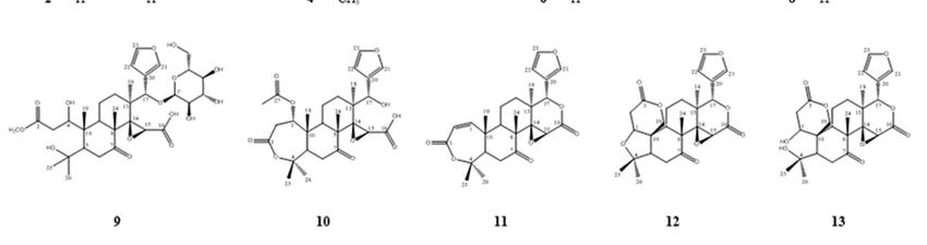
Figure 3. Structures of the limonoids isolated from C. junos seeds. Methyl sudachinoid A (1),
sudachinoid B (2), ichangensin (3), 1-O-methylichangensin (4), nomilin (5), deacetylnomilin (6),Int. J. Mol. Sci. 2020, 21, 6963 5 of 13
methylnomilinate (7), methyldeacetylnomilinate (8), deacetylnomilinic acid-17-O-glucopyranoside
(9), nomilinate A ring lactone (10), obacunone (11), limonin (12), and ichangin (13).
Figure 4. Effect of limonoids on the cell viability (A) and LPS-induced pro-inflammatory cytokine
production (B–D). RAW 264.7 cells pretreated with compounds 1–13 (1, 10, and 100 µM) for 2 h were
stimulated with 1 µg/mL LPS for 18 h. Cell viability was determined via MTT assay. The amount ofInt. J. Mol. Sci. 2020, 21, 6963 6 of 13
IL-1β (A), IL-6 (B), and TNF-α (C) in the culture medium was measured with ELISA kits. The values
are expressed as mean ± standard deviation of three individual experiments. * p < 0.05, ** p < 0.01, ***
p < 0.001, compared to the control group.
According to the results, compounds 1, 2, and 4 markedly affect the inflammatory cytokines.
Western blot analysis was conducted at the treatment concentration of 10 µM for each compound to
elucidate the anti-inflammatory effects via NF-κB signaling. All of these compounds downregulated
the phosphorylation of NF-κB, stimulated by LPS (Figure 5). Compound 4 showed a greater
inhibitory effect on LPS-induced pNF-κB activation than compounds 1 and 2.
We assessed the effect of the 13 limonoids (1 and 10 µM) on the viability of LPS-stimulated HT-
29 human colon epithelial cells. Our findings show that none of the limonoids exhibited any
cytotoxicity. As a result, the 13 limonoids were employed in the subsequent experiments to
investigate their anti-inflammatory activity (Figure 6A). IL-8 is a major chemoattractant and an
activator of the neutrophils involved in mediating the immune response and promoting
inflammation. The inhibitory effects of compounds 1–13 on IL-8 production were evaluated using
ELISA kits. Compared to those in the untreated cells, the levels of IL-8 increased in colon epithelial
cells subjected to LPS stimulation (100 ng/mL). Compounds 1, 2, 4, and 5 inhibited IL-8 production
in a concentration-dependent manner; the level of IL-8 in these cells was lower than that in LPS-
stimulated cells (Figure 6B). In particular, 10 µM of compounds 1 and 2 resulted in potent anti-
inflammatory activities (35.6 pg/mL and 22.8 pg/mL, respectively).
Figure 5. Effect of limonoids on NF-κB activation. RAW 264.7 cells were cultured in the presence of
compounds 1, 2, and 4 (10 µM) for one hour and stimulated with LPS (1 µg/mL) for one hour. The
levels of pNF-κB were detected by Western blot analysis. Relative density was calculated as the ratio
of the expression levels of each protein with β-actin. The data are expressed as the mean ± SD (n = 3).
** p < 0.01 and *** p < 0.001, compared with LPS-stimulated group.Int. J. Mol. Sci. 2020, 21, 6963 7 of 13
Figure 6. Effects of limonoids on the LPS-induced expression of IL-8 in HT-29 cells. HT-29 cells were
treated with compounds 1–13 (1 and 10 µM) for 2 h and stimulated with 100 ng/mL LPS for 18 h. The
viability of cells was then determined using an MTT assay (A) and the level of IL-8 in the culture
media was measured with an ELISA kit (B). The values are expressed as mean ± standard deviation
of three individual experiments. # p < 0.01, compared to the control group; * p < 0.05, ** p < 0.01, *** p
< 0.001, compared to the LPS-treated group.
3. Discussion
Inflammation is a major global health problem that requires urgent management through the
development of novel and efficacious therapeutic strategies. In recent years, there has been renewed
interest in studying the mechanisms underlying inflammation as a basis for drug development [22].
Among the inflammatory diseases, IBDs, which are chronic disorders characterized by inflammation
of the gastrointestinal tract, are being increasingly reported around the world [23]. An analysis of the
inflamed mucosa from patients with UC and CD revealed that the pathogenesis of IBDs is related to
the enhanced expression of pro-inflammatory cytokines such as IL-1 β, IL-6, TNF-α and IL-8, which
induce other mediators that act on the inflammatory tissue, ultimately enhancing the inflammatory
response [24,25]. Currently, anti-TNF therapies such as biosimilar anti-TNF monoclonal antibodies
are widely used to treat IBD; however, these treatments are limited by factors such as adverse effects
[26]. Therefore, we attempted to identify a novel agent capable of inhibiting the expression of pro-
inflammatory cytokines, from a natural source.
Limonoids are highly oxygenated, secondary metabolites of the terpenoid class that are
dominant in the Meliaceae and Rutaceae families [9]. Citrus fruits have an abundance of limonoids,
and these compounds are mainly bitter in taste. Their prototypical structure either contains or is
derived from a precursor possessing a 4,4,8-trimethyl-17-furanylsteroid skeleton. Citrus limonoids
contain a furan ring and oxygen-containing functional group at C-3, C-4, C-7, C-16, and C-17, thereby
displaying structural variations [27]. Limonoids also exhibit a wide range of biological properties
such as antibacterial, antifungal, antimalarial, anti-viral, and anticancer activities [6]. Many studies
have reported the chemical properties and pharmacological activities of limonoids, including their
structure–activity relationship [27]. Recently, the anti-inflammatory activities of limonoids andInt. J. Mol. Sci. 2020, 21, 6963 8 of 13
limonoid glucosides were reported [28]. Therefore, we sought to determine the effects of the
limonoids isolated from C. junos seeds on inflammatory responses using two in vitro models, RAW
264.7 mouse macrophage cells and HT-29 colon epithelial cells, which can simulate the characteristics
of intestinal epithelial cells in IBD.
RAW 264.7 cells were treated with limonoids for 2 h before stimulation with 1 µg/mL LPS for 18
h. With the exception of compound 9, none of the limonoids exerted any significant cytotoxicity at
concentrations of 1, 10, and 100 µM (Figure 4A). However, the LPS-activated macrophages produced
enhanced levels of pro-inflammatory cytokines such as IL-1β, IL-6 and TNF-α. The increased
secretion of IL-1β, IL-6 and TNF-α has been demonstrated in experimental colitis models and in
patients with IBD [29]. A reduction in IL-1β, IL-6 and TNF-α signaling was found to be effective at
inhibiting macrophages in chronic intestinal inflammation, thereby indicating that IL-1β, IL-6 and
TNF-α are potential therapeutic targets in IBD [30,31]. Pro-inflammatory cytokine levels are also
enhanced in IBD intestinal cells [25]. To confirm the anti-inflammatory activity of limonoids, we
assessed the LPS-induced production of IL-1β, IL-6 and TNF-α using ELISA kits. IL-1β plays a pivotal
role in the regulation of immunity and inflammatory response. Additionally, it might play an
important role in the pathogenesis of IBD by virtue of its pro-inflammatory and immunological
activities [24]. Compounds 1 and 2, which are sudachinoid-type limonoids, inhibited IL-1β
production in a concentration-dependent manner (Figure 4B). This inhibition was found to be more
potent than that induced by nomilin (5) and limonin (12), which were previously reported to have
anti-inflammatory activities and were thus employed as the positive controls in this study [32,33].
Methyl sudachinoid (1; 1 µM), a new compound, dramatically suppressed IL-1β production to 60.3%.
As compounds 3 and 4, which are ichangensin-type limonoids, participated in the downregulation
of IL-1β (71.2% and 83.8% at the high concentration of 100 µM), they might serve as therapeutic
candidates for the treatment of IBD.
High mucosal secretion of IL-6 and TNF-α in IBD could be caused by infiltrating macrophages,
which have been found to migrate in large numbers into the stimulated mucosal and intestinal lumen
during UC and CD [4]. Similar to IL-1β production, compounds 1-4 reduced IL-6 production (47.7%,
63.9%, 36.7%, and 73.5%, respectively) at a concentration of 100 µM. Additionally, the ichangin (13),
seco-limonin, significantly decreased IL-6 production in LPS-induced RAW 264.7 cells (Figure 4C).
Compounds 1–3, 5, 12, and 13 weakly attenuated TNF-α expression (Figure 4D). Such findings
demonstrate that the citrus limonoids evaluated in this study markedly affect the downregulation of
the pro-inflammatory cytokines in the order, IL-1β, IL-6 and TNF-α, and sudachinoid-type limonoids
exert more potent anti-inflammatory activities than other limonoids containing nomilin and limonin.
NF-κB is an important regulator of gene transcription involved in anti-inflammatory signaling
pathways in the gut [34]. In IBD patients, NF-κB signaling is often dysregulated resulting in
inflammation and its activation is associated with the rapid, acute production of various
proinflammatory mediators, such as IL-1β, and IL-6 [35]. Sudachinoid-type limonoids (compounds 1
and 2) and ichangensin-type limonoid (compound 4) most potently suppressed the production of IL-
1β and IL-6, respectively (Figure 4). Therefore, among limonoids, we investigated whether inhibitory
activities of proinflammatory cytokines by compounds 1, 2, and 4 are related to NF-κB signaling in
LPS-stimulated macrophages. As shown in Figure 5, compounds 1, 2, and 4 attenuated the
phosphorylation of NF-κB at a low concentration of 10 µM. The results showed that compounds 1, 2,
and 4 have anti-inflammatory activity via inhibition of NF-κB-mediated pro-inflammatory cytokines
and Citrus limonoids can effectively treat IBD.
Herein, we assessed the effects of the limonoids isolated from C. junos by treating HT-29 cells
with 1 and 10 µM (i.e., the low concentrations) of the compounds for 2 h followed by incubation with
0.1 µg/mL LPS for 18 h before the MTT assay. Based on our results, none of the limonoids affected
the viability of the cells (Figure 6A). When exposed to LPS, HT-29 cells secreted substantial amounts
of IL-8. Standard intestinal epithelial cells can secrete the potent chemoattractant, IL-8, and could
contribute to inflammation as opposed to level mucosa [36]. As the inhibitors of the pro-inflammatory
chemokine, IL-8, may be used to treat immune-associated diseases, such as IBD, we measured the
LPS-stimulated expression of IL-8 by treating HT-29 cells with limonoids. Ten limonoids (1–7, 9, 12,Int. J. Mol. Sci. 2020, 21, 6963 9 of 13
and 13) significantly suppressed the LPS-stimulated expression of IL-8 at the low concentrations of 1
and 10 µM. However, only compounds 1, 2, 4, and 5 inhibited IL-8 expression in a concentration-
dependent manner; the remaining six compounds resulted in similar or increased expression with an
increase in concentration (Figure 6B). The sudachinoid-type limonoids 1 and 2 effectively decreased
the expression level of IL-8 in the LPS-stimulated human colon epithelial cells, a finding similar to
that of the anti-inhibitory effect of pro-inflammatory cytokines in LPS-induced mouse macrophages.
It was recently shown that obacunone (11) has an effect on bowel disease in mice via downregulating
inflammatory signaling and restoring disrupted epithelial barriers [13]. Therefore, we considered that
citrus limonoids, newly elucidated in our study, are more potent against IBD than previously
reported limonoids and more in vivo studies need to be carried out to confirm the therapeutic effects.
In conclusion, we attempted to discover novel phytochemicals for the treatment of IBD.
Accordingly, we evaluated the anti-inflammatory effects of limonoids isolated from C. junos by
measuring the production of pro-inflammatory cytokines in LPS-stimulated cell lines. During the
investigation, a new limonoid, methyl sudachinoid (1), was isolated from the EtOAc fraction of C.
junos seeds in addition to 12 known limonoids. Among the different limonoids, the sudachinoid-type
compounds 1 and 2, and ichangesin-type compound 4 potently suppressed the expression of pro-
inflammatory cytokines, IL-1 β, IL-6, TNF-α and IL-8. Herein, we revealed the prospects of other
limonoids, such as ichangin, for medicinal and/or nutraceutical use in the treatment and/or protection
against IBD. According to our results, limonoids, including the new compound, isolated from C.
junos, can be employed as potential therapeutic phytochemicals against IBD. In this study, we
revealed the anti-inflammatory activity of citrus limonoids, which is underpinned by the
downregulation of pro-inflammatory cytokines.
4. Materials and Methods
4.1. Plant Extract Preparation
C. junos seeds were collected from Goheung, Jeollanam-do, Korea, in November 2017.
Subsequently, the essential oil was extracted from pulverised C.junos seeds (7.7 kg) by supercritical
extraction. Following the removal of the essential oil, C.junos seeds were extracted with methanol to
obtain the total extract (886 g), which was divided into n-hexane (171.4 g), EtOAc (13.0 g), n-BuOH
(20.9 g), and distilled water (318.8 g) fractions [11].
4.2. Isolation of Limonoids from the Fractions
The EtOAc fraction was separated into 16 subfractions (EA1 to EA16) by silica gel column
chromatography using a gradient solvent (n-hexane: EtOAc = 5:1 → 100% MeOH). Compounds 3 (tR
17.88, 23.2 mg) and 11 (tR 41.12, 1.8 mg) were obtained from EA1 by reverse phase (RP) high
performance liquid chromatography (HPLC) (YMC-Triart, C18 column, 250 × 10 mm, CH3CN:H2O =
50:50 → 0:100). The EA2 fraction yielded compounds 2 (tR 32.92, 1.1 mg) and 4 (tR 48.04, 2.2 mg) when
subjected to RP HPLC (YMC-Triart, C18 column, 250 × 10 mm, CH3CN:H2O = 5:95 → 100:0). The EA3
fraction yielded compounds 7 (tR 54.82, 7.6mg), 8 (tR 46.96, 2.3mg), and 10 (tR 51.56, 1.7 mg) when
subjected to RP HPLC (YMC-Triart, C18 column, 250 × 10 mm, CH3CN:H2O = 50:50 → 100:0). The EA4
fraction yielded compound 1 (tR 49.15, 0.8mg) when subjected to RP HPLC (YMC-Triart, C18 column,
250 × 10 mm, CH3CN:H2O = 8:92 → 100:0). The EA5 fraction yielded purified compound 12 through
crystallization (MeOH), and compounds 5 (tR 33.84, 8.9mg) and 13 (tR 29.94, 2.7 mg) when subjected
to RP HPLC (YMC-Triart, C18 column, 250 × 10 mm, CH3CN:H2O = 5:95 → 100:0). The EA10 fraction
yielded compound 6 through crystallization (MeOH). The n-BuOH fraction was separated into 12
subfractions (B1-B12) by Diaion HP20 column chromatography using 0%, 20%, 40%, 60%, 80%, and
100% MeOH. The B5 fraction was separated into 10 subfractions (B5-1 to B5-10) by silica gel column
chromatography using a gradient solvent (chloroform:MeOH:H2O = 200:4:1 → 100% MeOH).
Compound 9 (tR 24.64, 4.5mg) was obtained from B5-7 by RP HPLC (YMC-Triart, C18 column, 250 ×
10 mm, CH3CN:H2O = 10:90 → 100:0). Compounds 4 (tR 15.81, 2.4 mg) and 5 (tR 29.11, 2.5 mg) were
obtained from the B9 fraction via RP HPLC (YMC-Triart, C18 column, 250 × 10 mm, CH3CN:H2O =Int. J. Mol. Sci. 2020, 21, 6963 10 of 13
40:60 → 100:0). The basic schemes depicting the isolation process and structure are shown in Figures
1 and 2, respectively.
25
Methyl sudachinoid A (1): whitish amorphous powder; [α]D: 23.7 (c 0.4 MeOH); 1H-NMR (400
MHz, DMSO-d6): δH 7.61(1H, d, J = 1.6 Hz, H-22), 6.08(1H, d, J = 1.6Hz, H-23), 5.32(1H, s, H-17),
4.42(1H, s, H-15), 3.43(3H, s, OCH3), 3.18(3H, s, OCH3), 2.87(1H, dd, J = 11.2, 6.4 Hz, H-9), 2.74(1H, d,
J = 14.8Hz, H-6a), 2.54(1H, m, H-5), 2.20(1H, dd, J = 14.6, 2.2Hz, H-6b), 1.74(1H, m, H-12), 1.71(2H, m,
H-11), 1.23(3H, s, H-2), 1.23(1H, m, H-12), 1.19(3H, s, H-24), 1.16(3H, s, H-25), 1.13(3H, s, H-18),
1.11(3H, s, H-19), 1.03(3H, s, H-26); 13C-NMR (100 MHz, DMSO-d6): δC 208.4(s, C-7), 169.0(s, C-21),
166.9(s, C-16), 148.7(d, C-22), 135.5(s, C-20), 108.1(s, C-1), 103.5(d, C-23), 79.4(s, C-4), 75.1(d, C-17),
69.3(s, C-14), 56.0(q, OCH3), 55.6(d, C-15), 52.5(d, C-5), 49.5(s, C-8), 48.9(s, C-10), 48.2(q, OCH3), 40.5(s,
C-13), 39.7(d, C-9), 36.6(t, C-6), 31.6(q, C-25), 26(t, C-12), 23.6(q, C-26), 18.9(q, C-24), 18.4(q, C-18),
17.6(q, C-2), 15.6(t, C-11), 14.5(q, C-19). High-resolution electrospray ionization mass spectrometry
(HRESIMS) m/z 505.2444 [M+H]+ (calcd. C27H37O9, 505.2422). The spectra are available as
Supplementary Materials (Figure S1–S7).
4.3. Cell Culture
HT-29 human colon epithelial cells and RAW 264.7 mouse macrophage cells were obtained from
the Korean Cell Line Bank (Seoul, Korea) and cultured in Dulbecco’s modified Eagle’s medium
(DMEM) (Hyclone, Logan, UT, USA) containing 10% heat-inactivated fetal bovine serum (FBS)
(Hyclone, Logan, U.S.A.), 100 µg/mL streptomycin, and 100 IU/mL penicillin. The cells were
incubated as monolayers for 72 h at 37 ℃ in a humidified environment containing 5% CO2.
4.4. Determination of Cytotoxicity by MTT (3-[4,5-Dimethyl-2-Thiazolyl]-2,5-Diphenyl Tetrazolium
Bromide) Assay
The cells were seeded in 96-well plates (105 cells/well) and maintained in DMEM containing 10%
FBS for 24 h. Then, cells were treated with different concentrations of limonoids for 1 h prior to
stimulation with lipopolysaccharide (1 µg/mL; LPS; Sigma-Aldrich, St. Louis, MO, USA) for 18 h. Cell
viability was measured by an MTT assay, which involved the incubation of the cultured cells with
MTT (0.05 mg/mL) at 37 °C for 4 h. After removal of the supernatants, the absorbance of the formazan
solution was measured at 570 nm using a microplate reader.
4.5. Measurement of Pro-Inflammatory Cytokine Expression by Enzyme-Linked Immunosorbent Assay
(ELISA)
RAW 264.7 and HT-29 cells were seeded in 96-well plates at a density of 1 × 10⁵ and 2 × 10⁴
cells/well, respectively, for 24 h. Thereafter, the cells were treated with the compounds for 2 h before
stimulation with 1 µg/mL of LPS, followed by a 20 h incubation at 37 °C. We detected IL-
1β(Invitrogen, Waltham, MA, USA), IL-6 and TNF-α (BD OptEIATM, San Diego, CA, USA) levels in
RAW 264.7 mouse macrophage cells, and the level of IL-8 (BD OptEIATM, San Diego, CA, USA) in
HT-29 cells using ELISA kits, as per the manufacturer’s protocol. Relative production (%) was
calculated as ratio of the production levels of LPS treated and control group.
Relative production (%) = 100 × (sample treated group-control)/(LPS treated group-control)
4.6. Western Blot Analysis
Mouse macrophage were seeded at the density of 1 × 10⁶ cells/well in 6-well plates in culture
medium for 24 h. Seeded cells were treated with compounds 1, 2, and 4 for 1 h, then stimulated with
LPS (1 µg/mL). After 1 h, the cells were washed two times with cold phosphate-buffered saline (PBS)
and whole cell lysates were extracted with protein extraction solution (proprep, iNtRON,
Biotechnology, Daejeon, Korea). The protein concentration was determined by the Bradford reagent
and Western blot analysis was done as described previously [37]. The primary (pNF-κB, β-actin) and
secondary antibodies were diluted at 1:1000 and 1:2000, respectively.Int. J. Mol. Sci. 2020, 21, 6963 11 of 13
4.7. Statistical Analysis
The data are presented as mean ± standard deviation (n = 3). Data analysis was carried out using
one-way analysis of variance (ANOVA). p-values < 0.05 were considered to indicate significant
differences.
Supplementary Materials: The following are available online at www.mdpi.com/1422-0067/21/18/6963/s1,
Figure S1: 1H-NMR spectra of compound 1; Figure S2: 13C-NMR spectra of compound 1; Figure S3: 1H-1H COSY
spectrum of compound 1; Figure S4: HMQC spectrum of compound 1; Figure S5: HMBC spectrum of compound
1; Figure S6: DEPT spectrum of compound 1; Figure S7: HRESIMS of compound 1.
Author Contributions: J.S. and H.Y.S. performed the experiment, data analysis. J.S. and M.L. drafted the
manuscript. M.L. conceived the study and were also involved in the coordination of the study and interpretation
of the data. All authors have read and agreed to the published version of the manuscript.
Funding: This research was supported by the Ministry of Trade, Industry & Energy (MOTIE), Korea Institute
for Advancement of Technology (KIAT) through the Encouragement Program for the Industries of Regional
Innovation Cluster (Community business) (P0002383) and National Research Foundation of Korea (NRF) grant
funded by the Korea government (MSIP) (No. NRF-2020R1A2C1101252).
Conflicts of interest: The authors declare that there are no conflict of interest.
References
1. Sung, M.J.; Davaatseren, M.; Kim, S.H.; Kim, M.J.; Hwang, J. Boehmeria Nivea Attenuates LPS-Induced
Inflammatory Markers by Inhibiting p38 and JNK Phosphorylations in RAW264. 7 Macrophages. Pharm.
Biol. 2013, 51, 1131–1136.
2. Karin, M.; Greten, F.R. NF-κB: Linking Inflammation and Immunity to Cancer Development and
Progression. Nat. Rev. Immunol. 2005, 5, 749–759.
3. Verma, N.; Ahuja, V.; Paul, J. Profiling of ABC Transporters during Active Ulcerative Colitis and in Vitro
Effect of Inflammatory Modulators. Dig. Dis. Sci. 2013, 58, 2282–2292.
4. Reinecker, H.; Steffen, M.; Witthoeft, T.; Pflueger, I.; Schreiber, S.; MacDermott, R.; Raedler, A. Enhanced
Secretion of Tumour Necrosis factor-alpha, IL-6, and IL-1β by Isolated Lamina Propria Mononuclear Cells
from Patients with Ulcerative Colitis and Crohn’s Disease. Clin. Exp. Immunol. 1993, 94, 174–181.
5. Goeddel, D.V.; Aggarwal, B.B.; Gray, P.W.; Leung, D.W.; Nedwin, G.E.; Palladino, M.A.; Patton, J.S.;
Pennica, D.; Shepard, H.M.; Sugarman, B.J. Tumor Necrosis Factors: Gene Structure and Biological
Activities. Cold Spring Harb. Symp. Quant. Biol. 1986, 51, 597–609.
6. Pauli, U.; Beutler, B.; Peterhans, E. Porcine Tumor Necrosis Factor Alpha: Cloning with the Polymerase
Chain Reaction and Determination of the Nucleotide Sequence. Gene 1989, 81, 185–191.
7. Atreya, I.; Atreya, R.; Neurath, M. NF-κB in Inflammatory Bowel Disease. J. Intern. Med. 2008, 263, 591–596.
8. Penna, G.; Mondaini, N.; Amuchastegui, S.; Degli Innocenti, S.; Carini, M.; Giubilei, G.; Fibbi, B.; Colli, E.;
Maggi, M.; Adorini, L. Seminal Plasma Cytokines and Chemokines in Prostate Inflammation: Interleukin
8 as a Predictive Biomarker in Chronic prostatitis/chronic Pelvic Pain Syndrome and Benign Prostatic
Hyperplasia. Eur. Urol. 2007, 51, 524–533.
9. Waugh, D.J.; Wilson, C. The Interleukin-8 Pathway in Cancer. Clin. Cancer Res. 2008, 14, 6735–6741.
10. Tundis, R.; Loizzo, M.R.; Menichini, F. An Overview on Chemical Aspects and Potential Health Benefits of
Limonoids and their Derivatives. Crit. Rev. Food Sci. Nutr. 2014, 54, 225–250.
11. Zibaee, E.; Kamalian, S.; Tajvar, M.; Amiri, M.S.; Ramezani, M.; Moghadam, A.T.; Emami, S.A.; Sahebkar,
A. Citrus Species: A Review of Traditional Uses, Phytochemistry and Pharmacology. Curr. Pharm. Des.
2020, 26, 44–97.
12. Song, H.Y.; Jo, A.; Shin, J.; Lim, E.H.; Lee, Y.E.; Jeong, D.E.; Lee, M. Anti-Inflammatory Activities of Isogosferol,
a Furanocoumarin Isolated from Citrus Junos Seed Shells through Bioactivity-Guided Fractionation. Molecules
2019, 24, 4088.
13. Luo, X.; Yue, B.; Yu, Z.; Ren, Y.; Zhang, J.; Ren, J.; Wang, Z.; Dou, W. Obacunone Protects Against Ulcerative
Colitis in Mice by Modulating Gut Microbiota, Attenuating TLR4/NF-κB Signaling Cascades, and
Improving Disrupted Epithelial Barriers. Front. Microbiol. 2020, 11, 497.
14. Nakagawa, H.; Duan, H.; Takaishi, Y. Limonoids from Citrus Sudachi. Chem. Pharm. Bull. (Tokyo) 2001, 49,
649–651.Int. J. Mol. Sci. 2020, 21, 6963 12 of 13
15. Cho, E.; Piao, X.; Piao, L.; Piao, H.; Park, M.; Kim, B.; Park, J. Chemical Constituents of the Fruit of Citrus
Junos. Nat. Prod. Sci. 2000, 6, 179–182.
16. Bennett, R.D.; Herman, Z.; Hasegawa, S. Ichangensin: A New Citrus Limonoid. Phytochemistry 1988, 27, 1543–
1545.
17. Liu, J. Two New Limonoids from Polygonum Orientale L. Indian J. Chem. 2001, 40B, 644–646.
18. Khalil, A.T.; Maatooq, G.T.; El Sayed, K.A. Limonoids from Citrus Reticulata. Zeitschrift Für
Naturforschung C. J. Biosci. 2003, 58, 165–170.
19. Bennett, R.D.; Hasegawa, S.; Herman, Z. Glucosides of Acidic Limonoids in Citrus. Phytochemistry 1989, 28,
2777–2781.
20. Zukas, A.A.; Breksa III, A.P.; Manners, G.D. Isolation and Characterization of Limonoate and Nomilinoate
A-Ring Lactones. Phytochemistry 2004, 65, 2705–2709.
21. Bennett, R.; Hasegawa, S. Limonoids of Calamondin Seeds. Tetrahedron 1981, 37, 17–24.
22. Misko, T.P.; Trotter, J.L.; Cross, A.H. Mediation of Inflammation by Encephalitogenic Cells: Interferon γ
Induction of Nitric Oxide Synthase and Cyclooxygenase 2. J. Neuroimmunol. 1995, 61, 195–204.
23. Ng, S.C.; Shi, H.Y.; Hamidi, N.; Underwood, F.E.; Tang, W.; Benchimol, E.I.; Panaccione, R.; Ghosh, S.; Wu,
J.C.; Chan, F.K. Worldwide Incidence and Prevalence of Inflammatory Bowel Disease in the 21st Century:
A Systematic Review of Population-Based Studies. Lancet 2017, 390, 2769–2778.
24. Schreiber, S.; Heinig, T.; Thiele, H.; Raedler, A. Immunoregulatory Role of Interleukin 10 in Patients with
Inflammatory Bowel Disease. Gastroenterology 1995, 108, 1434–1444.
25. Mitselou, A.; Grammeniatis, V.; Varouktsi, A.; Papadatos, S.S.; Katsanos, K.; Galani, V. Proinflammatory
Cytokines in Irritable Bowel Syndrome: A Comparison with Inflammatory Bowel Disease. Intest. Res. 2020,
18, 115–120.
26. Shivaji, U.N.; Sharratt, C.L.; Thomas, T.; Smith, S.C.; Iacucci, M.; Moran, G.W.; Ghosh, S.; Bhala, N.
Managing the Adverse Events Caused by anti-TNF Therapy in Inflammatory Bowel Disease. Aliment.
Pharmacol. Ther. 2019, 49, 664–680.
27. Roy, A.; Saraf, S. Limonoids: Overview of Significant Bioactive Triterpenes Distributed in Plants Kingdom.
Biol. Pharma. Bull. 2006, 29, 191–201.
28. Chen, Y.; Ruan, J.; Sun, F.; Wang, H.; Yang, S.; Zhang, Y.; Yan, J.; Yu, H.; Guo, Y.; Zhang, Y. Anti-
Inflammatory Limonoids from Cortex Dictamni. Front. Chem. 2020, 8, 73.
29. Soufli, I.; Toumi, R.; Rafa, H.; Touil-Boukoffa, C. Overview of Cytokines and Nitric Oxide Involvement in
Immuno-Pathogenesis of Inflammatory Bowel Diseases. World J. Gastrointest. Pharmacol. Ther. 2016, 7, 353–
360.
30. Jo, A.; Yoo, H.J.; Lee, M. Robustaflavone Isolated from Nandina Domestica using Bioactivity-Guided
Fractionation Downregulates Inflammatory Mediators. Molecules 2019, 24, 1789.
31. Zhang, H.; Kovacs-Nolan, J.; Kodera, T.; Eto, Y.; Mine, Y. Γ-Glutamyl Cysteine and γ-Glutamyl Valine
Inhibit TNF-α Signaling in Intestinal Epithelial Cells and Reduce Inflammation in a Mouse Model of Colitis
Via Allosteric Activation of the Calcium-Sensing Receptor. Biochim. Biophys. Acta (BBA) Mol. Basis Dis. 2015,
1852, 792–804.
32. Pratheeshkumar, P.; Kuttan, G. Nomilin Inhibits Tumor-Specific Angiogenesis by Downregulating VEGF,
NO and Proinflammatory Cytokine Profile and also by Inhibiting the Activation of MMP-2 and MMP-9.
Eur. J. Pharmacol. 2011, 668, 450–458.
33. Matsuda, H.; Yoshikawa, M.; Kubo, M. Antinociceptive and Anti-Inflammatory Activities of Limonin
Isolated from the Fruits of Evodia Rutaecarpa Var. Bodinieri. Planta Med. 1998, 64, 339–342.
34. Aggarwal, B.B.; Takada, Y.; Shishodia, S.; Gutierrez, A.M.; Oommen, O.V.; Ichikawa, H.; Baba, Y.; Kumar, A.
Nuclear Transcription Factor NF-Kappa B: Role in Biology and Medicine. Indian J. Exp. Biol. 2004, 42, 341–353.
35. McDaniel, D.K.; Eden, K.; Ringel, V.M.; Allen, I.C. Emerging Roles for Noncanonical NF-κB Signaling in
the Modulation of Inflammatory Bowel Disease Pathobiology. Inflamm. Bowel Dis. 2016, 22, 2265–2279.Int. J. Mol. Sci. 2020, 21, 6963 13 of 13
36. Lee, M.; Shim, S.; Sung, S.H. Triterpenoids Isolated from Alnus Japonica Inhibited LPS-Induced
Inflammatory Mediators in HT-29 Cells and RAW264. 7 Cells. Biol. Pharma. Bull. 2017, 40, 1544–1550.
37. Jo, A.; Kim, C.E.; Lee, M. Serratane Triterpenoids Isolated from Lycopodium Clavatum by Bioactivity-
Guided Fractionation Attenuate the Production of Inflammatory Mediators. Bioorg. Chem. 2020, 96, 103632.
© 2020 by the authors. Licensee MDPI, Basel, Switzerland. This article is an open access
article distributed under the terms and conditions of the Creative Commons Attribution
(CC BY) license (http://creativecommons.org/licenses/by/4.0/).You can also read