Human giant larvae 1 promotes migration and invasion of malignant glioma cells by regulating N cadherin
←
→
Page content transcription
If your browser does not render page correctly, please read the page content below
ONCOLOGY LETTERS 21: 167, 2021

            Human giant larvae‑1 promotes migration and invasion
              of malignant glioma cells by regulating N‑cadherin
        YAN WANG1‑3, YU ZHANG1,2, BEN SANG1, XIANLONG ZHU1, RUTONG YU2,3 and XIUPING ZHOU2,3

1
    The Graduate School and 2Institute of Nervous System Diseases, Xuzhou Medical University; 3Department of Neurosurgery,
                    Affiliated Hospital of Xuzhou Medical University, Xuzhou, Jiangsu 221002, P.R. China

                                     Received August 3, 2020; Accepted December 1, 2020

                                                   DOI: 10.3892/ol.2021.12428

Abstract. Human giant larvae‑1 (Hugl‑1) is a human homo‑           Introduction
logue of Drosophila tumor suppressor lethal (2)‑giant larvae
and has been reported to be involved in the development of         Glioblastoma (GBM) is the most common malignant primary
human malignancies. Previous studies performed by our              central nervous system tumor, with an average survival time
group demonstrated that Hugl‑1 inhibits glioma cell prolifera‑     of 12‑15 months (1). The failure of conventional treatments
tion in an intracranial model of nude mice. However, the exact     is attributed to its highly invasive and diffusely infiltrative
molecular mechanisms underlying the participation of Hugl‑1        nature (2). Thus, the identification of novel therapeutic targets
in glioma invasion and migration, and in the depolarizing          and strategies to improve the efficacy of existing forms of
process remain largely unknown. Utilizing the U251‑MG cells        treatment is urgently required.
with stable expression of Hugl‑1, the present study used wound         Cell polarity and intercellular adhesion play a key role
healing, Transwell invasion and western blot assays to explore     in regulating normal tissue structure and function (3). The
the role and specific mechanism of Hugl‑1 in glioma invasion       disruption of cell polarity and cell adhesion is usually associ‑
and migration. The results of the present study demonstrated       ated with tumor formation (4). Lethal (2)‑giant larvae (Lgl) is
that overexpression of Hugl‑1 decreased cell‑cell adhesion and     a cortical cytoskeletal protein, which was initially identified in
increased cell‑cell extracellular matrix adhesion. In addition,    Drosophila and exhibits notable effects in the establishment
overexpression of Hugl‑1 promoted pseudopodia formation,           and maintenance of apical‑basal epithelial polarity, asym‑
glioma cell migration and invasion. The molecular mecha‑           metric cell division, tissue integrity and cell proliferation (5).
nism of action involved the negative regulation of N‑cadherin      The human homologues of Lgl1 and Lgl2 are termed human
protein levels by Hugl‑1. Overexpression or knockdown of           giant larvae (Hugl)‑1 and Hugl‑2. Mutations that cause loss
N‑cadherin partially suppressed or enhanced the effects of         of function of Lgl have been demonstrated to result in tissue
Hugl‑1 on glioma cell migration and invasion, respectively.        overgrowth and neoplastic tumor formation (6,7). The Hugl‑1
Furthermore, Hugl‑1 inhibited cell proliferation, while            protein shares 62.5% similarity with Lgl (8‑10). A previous
promoting cell migration, which suggests that it may serve a       study indicated that hepatocellular carcinoma (HCC) contains
two‑sided biological role in cellular processes. Taken together,   frequent mutations of Hugl‑1, whereas overexpression of
these results suggest that Hugl‑1 promotes the migration and       HCC‑derived aberrant Hugl‑1 variants significantly promote
invasion of malignant glioma cells by decreasing N‑cadherin        HCC cell migration and invasion (11). In addition, Hugl‑1
expression. Thus, Hugl‑1 may be applied in the development of      expression is downregulated in different types of human
targeted and personalized treatment.                               cancer, including colorectal cancer, melanoma, prostate
                                                                   cancer, breast cancer, endometrial cancer, lung cancer and
                                                                   esophageal carcinoma (12‑15). Hugl‑1 expression is positively
                                                                   associated with a higher survival rate in patients with pancre‑
                                                                   atic carcinoma, suggesting its use as a reliable prognostic
Correspondence      to: Professor Xiuping Zhou, Institute          marker (16). The majority of previous studies have focused
of Nervous System Diseases, Xuzhou Medical University,             on epithelial‑derived tumors (11‑15), thus the role of Hugl‑1 in
84 West Huai‑hai Road, Xuzhou, Jiangsu 221002, P.R. China          gliomas (glia‑derived tumors) has not yet been fully elucidated.
E‑mail: xpzhou@xzhmu.edu.cn                                        A previous study performed by our group has demonstrated
Professor Rutong Yu, Department of Neurosurgery, Affiliated
                                                                   that Hugl‑1 protein levels decrease in human glioma tissues,
Hospital of Xuzhou Medical University, 99 West Huai‑hai Road,      whereas overexpression of Hugl‑1 attenuates glioma cell prolif‑
Xuzhou, Jiangsu 221002, P.R. China                                 eration in an intracranial model of nude mice; however, it does
E‑mail: yu.rutong@163.com                                          not affect glioma cell proliferation in vitro (17). As a regulator
                                                                   of cell polarity, Hugl‑1 exhibits important properties that are
Key words: glioma, Hugl‑1, migration, invasion, N‑cadherin         closely associated with cell adhesion and cytoskeletal func‑
                                                                   tion and structure (18). However, the role of Hugl‑1 in glioma
                                                                   migration and invasion has not yet been fully investigated.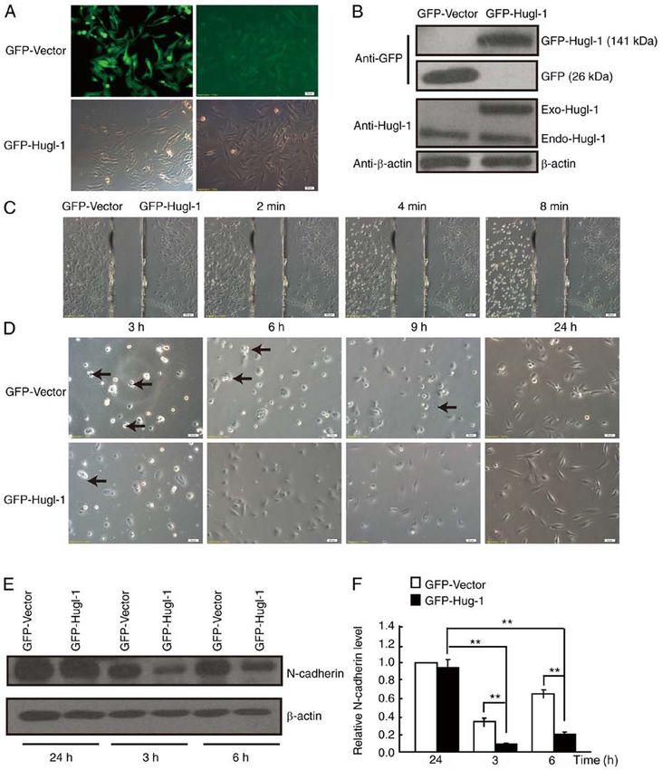
2                                WANG et al: N-cadherin MEDIATES THE PROMOTING EFFECT OF Hugl-1

    Cell surface adhesion molecules are the main mediators of         at room temperature for 8 min and observed at designated
cell‑cell interactions, which are essential for tumor malignant       time points (0, 2, 4 and 8 min) under an inverted light micro‑
biological behaviors. Reorganization of the cell cytoskeleton         scope during trypsinization at x200 magnification (Olympus
and alteration of cell‑cell adhesion are required prior to cell       Corporation; IX71).
migration (19,20). These processes are mainly mediated
by cadherin family members. It is reported that E‑cadherin            Attachment assay. The attachment assay was performed using
is essential for the normal migration of cranial neural crest         12‑well plates. The cell suspension was added into the plates
cells in vivo, while P‑cadherin, also known as placental              and cell images were obtained at 3, 6, 9 and 24 h using an
cadherin, is associated with malignant invasion of esophageal         inverted light microscope at x400 magnification (Olympus
squamous cells (21‑24). In most tumors, N‑cadherin expres‑            Corporation; IX71).
sion is often upregulated and can be used as a promoter of
tumor invasion (25,26). N‑cadherin expression in epithe‑              Wound healing assay. Cell migration was assessed via the
lial cells can induce morphological changes of fibroblast             wound healing assay, as previously described (31). Briefly, cells
phenotype and orchestrate cell‑cell communication during              were seeded into 6‑well plates and cultured until they reached
cell movement (27). N‑cadherin is also known as an epithe‑            ~80% confluence. The cell monolayers were scratched using a
lial‑to‑mesenchymal transition marker and exhibits several            10 µl sterile pipette tip. Cells were subsequently washed twice
functions according to the cell environment that can promote          with PBS to remove floating cells and serum‑free DMEM/F‑12
adhesion or induce migration (28,29). However, increasing             medium (Gibco; Thermo Fisher Scientific, Inc.) was added.
evidence suggests that N‑cadherin exhibits tumor‑inhibitory           Cell wound healing was observed at 0, 24 or 48 h using an
roles in non‑epithelial derived neoplasms, such as osteosar‑          inverted light microscope at x200 magnification (Olympus
coma and glioma (27,30). Thus, the functions of N‑cadherin            Corporation; IX71).
may be tumor‑type specific (27).
    The present study aimed to investigate the role and molecular     Transwell invasion assay. The cell invasion assay was
mechanism of Hugl‑1 on the motility of malignant glioma cells.        performed as previously described (31). Briefly, Transwell
                                                                      membranes were precoated with DMEM‑diluted Matrigel®
Materials and methods                                                 (BD Biosciences) for 3 h at 37˚C. Cells (2x10⁴) were plated in
                                                                      the upper chambers of Transwell plates in 200 µl serum‑free
Cell culture. The U251‑MG glioma cell line was purchased              culture DMEM/F‑12 medium (Gibco; Thermo Fisher Scientific,
from the Shanghai Cell Bank, Type Culture Collection                  Inc.). A total of 500 µl DMEM/F‑12 medium supplemented
Committee, Chinese Academy of Sciences. Cells were                    with 10% FBS was plated in the lower chambers. Following
maintained in DMEM/F‑12 media (Gibco; Thermo Fisher                   incubation at room temperature for 24 h, the invasive cells
Scientific, Inc.) supplemented with 10% fetal bovine serum            were fixed in methanol for 15 min at room temperature and
(FBS; Biological Industries), at 37˚C in 5% CO2.                      subsequently stained for 15 min at room temperature with
                                                                      0.1% crystal violet. Invasive cells were viewed and counted
Stable transfection of Hugl‑1 into U251‑MG cells. The                 under an inverted light microscope at x200 magnification
pEGFP‑C1 vector alone or the pEGFP‑C1‑Hugl‑1 construct                (Olympus Corporation; IX71).
(provided by Professor Zhengjun Chen, Shanghai Institute
of Biochemistry and Cell Biology, Chinese Academy of                  Western blotting. U251‑MG cells were lysed with RIPA
Sciences) was transfected into U251‑MG cells (GFP‑Vector or           lysis buffer (50 mM Tris‑HCl, 150 mM NaCl, 0.5% sodium
GFP‑Hugl‑1 cells, respectively) using Lipofectamine® 2000             deoxycholate and 0.1% SDS) supplemented with protease
(Invitrogen; Thermo Fisher Scientific, Inc.), according to the        inhibitor cocktail, and total proteins were quantified using a
manufacturer's instructions. Briefly, 9 µl Lipofectamine® 2000        BCA kit (Beyotime Institute of Biotechnology) according to
and 3 µg of the Hugl‑1 expression plasmid were added to 1 ml          the manufacturer's protocol. Western blotting was performed
Opti‑MEM (Invitrogen; Thermo Fisher Scientific, Inc.) and             as previously described (32). Briefly, equal amounts of protein
incubated for 10 min at room temperature. The plasmid and             (20 µg/lane) were separated by SDS‑PAGE on 8 or 10% gels,
Lipofectamine were mixed together and incubated for 30 min            transferred onto polyvinylidene fluoride membranes and
before adding them to the U251‑MG cells. The transfectants            blocked using 3% BSA (Sangon Biotech Co., Ltd.) for 2 h at
were subsequently selected using G418 (1,200 µg/ml), and              room temperature. The membranes were then incubated with
single‑cell clones were obtained following 3‑4 weeks of               primary antibodies against Hugl‑1 (1:500) [kindly gifted by
growth for expansion. The G418‑resistant cells were used for          Dr ZG Luo from the Institute of Neuroscience, Shanghai
subsequent experiments. DsRed‑C1 or DsRed‑N‑cadherin                  Institutes for Biological Sciences, Chinese Academy of
plasmids were kindly provided by the Laboratory of Cell               Sciences (33)], N‑cadherin (1:2,000; cat. no. ab76011; Abcam),
Biology of Northeast Normal University (Changchun, China).            β ‑catenin (1:5,000; cat. no. ab32572; Abcam), integrin β1
DsRed‑C1 or DsRed‑N‑cadherin plasmids were transfected                (1:1,000; cat. no. ab134179; Abcam) and β ‑actin (1:1,000;
into Hugl‑1 overexpressing U251‑MG cells. The specific trans‑         cat. no. MABT523; EMD Millipore). Following the primary
fection procedure was the same as that of Hugl‑1.                     antibody incubation at 4˚C overnight, membranes were
                                                                      probed with HRP-labelled goat anti-rabbit or anti‑mouse IgG
Digestion assay. Cultured GFP‑Vector or GFP‑Hugl‑1 cells              secondary antibodies (1:4,000; cat. nos. sc2004 and sc2005;
were digested with trypsin simultaneously (Gibco; Thermo              Santa Cruz Biotechnology, Inc.) at room temperature for 2 h.
Fisher Scientific, Inc.). Briefly, cells were digested with trypsin   The signal was detected using the Pierce ECL Plus Western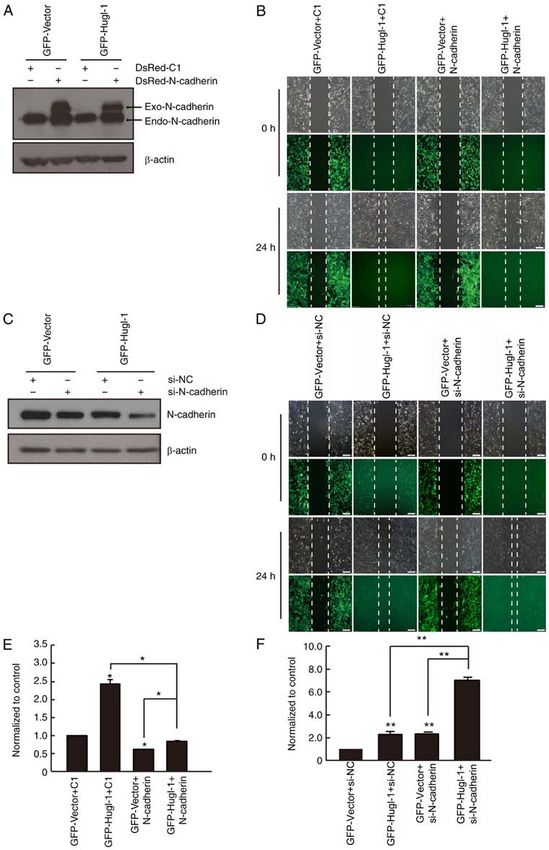
ONCOLOGY LETTERS 21: 167, 2021 3
Blotting Substrate (Pierce; Thermo Fisher Scientific, Inc.) and N‑cadherin is a member of the calcium‑dependent adhesion
exposed to ChemiDoc Touch (Bio‑Rad Laboratories, Inc.). molecule family, which mediates adhesion between homotypic
Finally, gray analysis was performed using ImageJ 1.48V cells (34). Thus, N‑cadherin protein expression was detected at
(National Institutes of Health) to compare the level of each 3, 6 and 24 h following plating. The results demonstrated that
protein. N‑cadherin expression was lower in Hugl‑1 overexpressing
cells compared with GFP cells at 3 and 6 h following plating
Phalloidin staining. U251‑MG cells were incubated for 24 h at (Fig. 1E and F).
37˚C and cultured in DMEM/F‑12 medium supplemented with Previous studies have demonstrated that adhesion
10% FBS for 30 min at 37˚C. Subsequently, cells were fixed molecules play an important role in the early stage of cell
with 4% paraformaldehyde for 10 min at room temperature, adhesion (35,36), which gradually decreases overtime (37).
washed twice with PBS, and 0.5% Triton X‑100 was added In the present study, no significant difference was observed
for 5 min at room temperature. Finally, 200 µl of the diluted in N‑cadherin expression between the two groups 24 h after
phalloidin (cat. no. 94072; Sigma‑Aldrich; Merck KGaA) plating. Taken together, these results suggest that overexpres‑
was added and incubated at room temperature in the dark sion of Hugl‑1 decreases cell‑cell adhesion, while increasing
for 30 min. Actin filaments were observed using an inverted cell‑extracellular matrix adhesion by regulating N‑cadherin
fluorescence microscope at x400 magnification (Olympus expression.
Corporation; IX71).
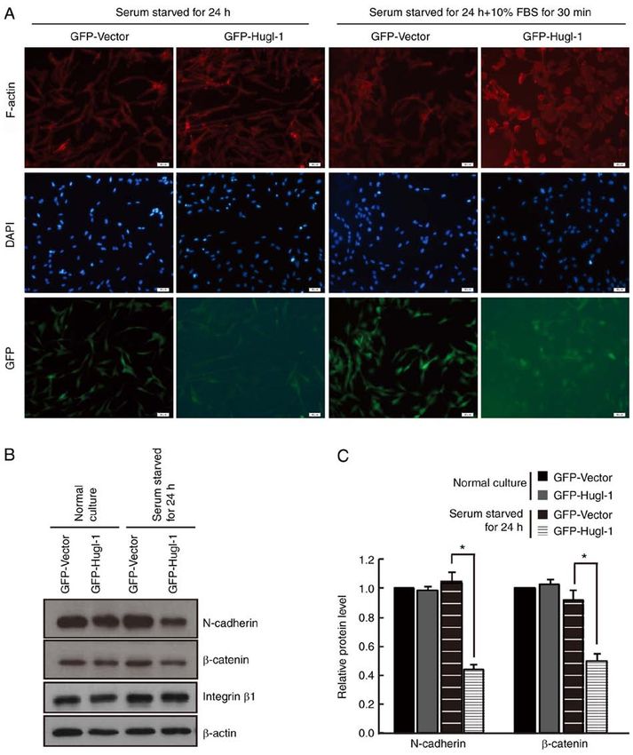
Hugl‑1 accelerates cytoskeletal remodeling. To fully charac‑
Statistical analysis. Statistical analysis was performed terize the intercellular adhesion defects observed in Hugl‑1
using SPSS 13.0 software (SPSS, Inc.). All experiments overexpressing cells, the intracellular organization of the
were performed in triplicate and data are presented as the cytoskeleton was assessed. Cells were incubated for 24 h and
mean ± standard error of the mean. Student's unpaired t‑test cultured in media supplemented with 10% FBS for 30 min.
was used to compare differences between two groups, while Subsequently, cells were stained with phalloidin‑conjugated
one‑way AVONA followed by Tukey's post hoc test were used actin to assess actin reassembling. The results demonstrated
to compare differences between multiple groups. P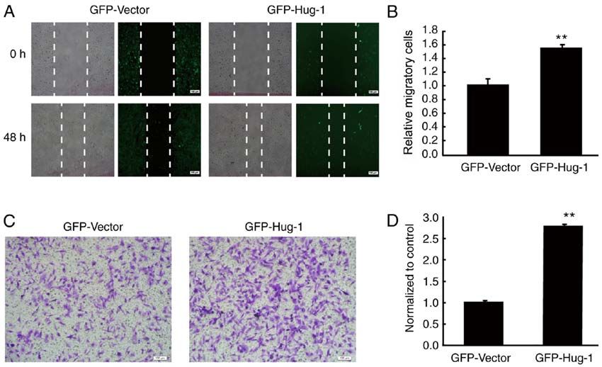
4                                     WANG et al: N-cadherin MEDIATES THE PROMOTING EFFECT OF Hugl-1

Figure 1. Hugl‑1 affects cell adhesion. (A) GFP‑Hugl‑1 or GFP‑Vector plasmids were transfected into U251‑MG glioma cells, followed by G418 selection. The
transfection efficiency was assessed via GFP fluorescence (scale bar, 50 µm). (B) Western blot analysis was performed to detect Hugl‑1 protein expression.
(C) Representative digital images obtained at 0, 2, 4 and 8 min during trypsinization (scale bar, 100 µm). (D) Representative digital images obtained at 3, 6,
9 and 24 h following plating. Black arrowheads indicate the cell aggregates (scale bar, 50 µm). (E) N‑cadherin protein levels were detected at 3, 6 and 24 h
following cell attachment. (F) Quantification results of (E). **P
ONCOLOGY LETTERS 21: 167, 2021                                                                 5

Figure 2. Hugl‑1 accelerates cytoskeletal remodeling. (A) Following serum starvation for 24 h, GFP and Hugl‑1 overexpressing cells were stimulated with
10% FBS for 30 min and stained with conjugated phalloidin (red). The images indicated that more dot or fan‑like protrusions were detected at the cell
periphery in Hugl‑1 overexpressing cells. (B) Western blot analysis was performed to detect the protein expression levels of N‑cadherin, β ‑catenin and
integrin β1 in GFP and Hugl‑1 overexpressing cells. (C) Quantification results of (B). Scale bar, 50 µm. *P
6                                     WANG et al: N-cadherin MEDIATES THE PROMOTING EFFECT OF Hugl-1

Figure 3. Hugl‑1 promotes migration and invasion of glioma cells. (A) Representative digital images of the wound healing assay taken at 0 and 48 h following
scratching. (B) Quantification results of (A). (C) The invasive ability of glioma cells was assessed via the Transwell assay. (D) Quantification results of (C).
Scale bar, 100 µm. **P
ONCOLOGY LETTERS 21: 167, 2021                                                                   7

Figure 4. N‑cadherin partially mediates the effect of Hugl‑1 expression on glioma cell migration. (A) The overexpression efficiency of N‑cadherin in U251‑MG
cells was detected via western blot analysis. (B) The wound healing assay was performed to assess cell migration. (C) The downregulation efficiency of
N‑cadherin was detected via western blot analysis. (D) The wound healing assay was performed to assess cell migration. (E) Quantification results of (B).
(F) Quantification results of (D). Scale bar, 100 µm. *P
8                                 WANG et al: N-cadherin MEDIATES THE PROMOTING EFFECT OF Hugl-1

Acknowledgements                                                         10. Vasioukhin V: Lethal giant puzzle of Lgl. Dev Neurosci 28:
                                                                             13‑24, 2006.
                                                                         11. Lu X, Feng X, Man X, Yang G, Tang L, Du D, Zhang F, Yuan H,
Not applicable.                                                              Huang Q, Zhang Z, et al: Aberrant splicing of Hugl‑1 is associ‑
                                                                             ated with hepatocellular carcinoma progression. Clin Cancer
                                                                             Res 15: 3287‑3296, 2009.
Funding                                                                  12. Kuphal S, Wallner S, Schimanski CC, Bataille F, Hofer P,
                                                                             Strand S, Strand D and Bosserhoff AK: Expression of Hugl‑1 is
The present study was supported by the National Natural Science              strongly reduced in malignant melanoma. Oncogene 25: 103‑110,
                                                                             2006.
Foundation of China (grant nos. 81672489, 81872053 and                   13. Schimanski CC, Schmitz G, Kashyap A, Bosserhoff AK,
81902526) and the Postgraduate Research & Practice Innovation                Bataille F, Schäfer SC, Lehr HA, Berger MR, Galle PR, Strand S
Program of Jiangsu Province (grant no. KYCX20_2460).                         and Strand D: Reduced expression of Hugl‑1, the human homo‑
                                                                             logue of Drosophila tumour suppressor gene lgl, contributes to
                                                                             progression of colorectal cancer. Oncogene 24: 3100‑3109, 2005.
Availability of data and materials                                       14. Tsuruga T, Nakagawa S, Watanabe M, Takizawa S, Matsumoto Y,
                                                                             Nagasaka K, Sone K, Hiraike H, Miyamoto Y, Hiraike O, et al:
                                                                             Loss of Hugl‑1 expression associates with lymph node metastasis
The datasets used and/or analyzed during the current study are               in endometrial cancer. Oncol Res 16: 431‑435, 2007.
available from the corresponding author on reasonable request.           15. Matsuzaki T, Takekoshi S, Toriumi K, Kitatani K, Nitou M,
                                                                             Imamura N, Ogura G, Masuda R, Nakamura N and Iwazaki M:
                                                                             Reduced expression of Hugl 1 contributes to the progression
Authors' contributions                                                       of lung squamous cell carcinoma. Tokai J Exp Clin Med 40:
                                                                             169‑177, 2015.
YW wrote the manuscript and contributed to data analysis.                16. Biesterfeld S, Kauhausen A, Kost C, Gockel I, Schimanski CC
                                                                             and Galle PR: Preservation of HUGL‑1 expression as a favour‑
YZ, BS and XZ performed the experiments. RY contributed to                   able prognostic factor in pancreatic carcinoma. Anticancer
the study design. XZ contributed to the study design, reviewed               Res 32: 3153‑3159, 2012.
and edited the manuscript. All authors have read and approved            17. Liu X, Lu D, Ma P, Liu H, Cao Y, Sang B, Zhu X, Shi Q, Hu J,
                                                                             Yu R and Zhou X: Hugl‑1 inhibits glioma cell growth in intracra‑
the final manuscript.                                                        nial model. J Neurooncol 125: 113‑121, 2015.
                                                                         18. Massimi P, Narayan N, Thomas M, Gammoh N, Strand S,
Ethics approval and consent to participate                                   Strand D and Banks L: Regulation of the hDlg/hScrib/Hugl‑1
                                                                             tumour suppressor complex. Exp Cell Res 314: 3306‑3317, 2008.
                                                                         19. Zhong XL and Rescorla FJ: Cell surface adhesion molecules
Not applicable.                                                              and adhesion‑initiated signaling: Understanding of anoikis resis‑
                                                                             tance mechanisms and therapeutic opportunities. Cell Signal 24:
                                                                             393‑401, 2012.
Patient consent for publication                                          20. Fife CM, McCarroll JA and Kavallaris M: Movers and shakers:
                                                                             Cell cytoskeleton in cancer metastasis. Brit J Pharmacol 171:
Not applicable.                                                              5507‑5523, 2014.
                                                                         21. Glousha n kova NA, Rubtsova SN a nd Z h it nya k I Y:
                                                                             Cadherin‑mediated cell‑cell interactions in normal and cancer
Competing interests                                                          cells. Tissue Barriers 5: e1356900, 2017.
                                                                         22. Pal M, Bhattacharya S, Kalyan G and Hazra S: Cadherin
                                                                             profiling for therapeutic interventions in Epithelial Mesenchymal
The authors declare that they have no competing interests.                   Transition (EMT) and tumorigenesis. Exp Cell Res 368: 137‑146,
                                                                             2018.
References                                                               23. Nair KS, Naidoo R and Chetty R: Expression of cell adhesion
                                                                             molecules in oesophageal carcinoma and its prognostic value.
                                                                             J Clin Pathol 58: 343‑351, 2005.
 1. Gould J: Breaking down the epidemiology of brain cancer.             24. Huang C, Kratzer MC, Wedlich D and Kashef J: E‑cadherin is
    Nature 561: S40‑S41, 2018.                                               required for cranial neural crest migration in Xenopus laevis.
 2. Meyer MA: Malignant gliomas in adults. N Engl J Med 359:                 Dev Biol 411: 159‑171, 2016.
    1850, 2008.                                                          25. Bremmer F, Schallenberg S, Jarry H, Küffer S, Kaulfuss S,
 3. Osswald M and Morais‑de‑Sá E: Dealing with apical‑basal                  Burfeind P, Strauß A, Thelen P, Radzun HJ, Ströbel P, et al: Role
    polarity and intercellular junctions: A multidimensional chal‑           of N‑cadherin in proliferation, migration, and invasion of germ
    lenge for epithelial cell division. Curr Opin Cell Biol 60: 75‑83,       cell tumours. Oncotarget 6: 33426‑33437, 2015.
    2019.                                                                26. Mrozik KM, Blaschuk OW, Cheong CM, Zannettino ACW and
 4. Martin‑Belmonte F and Perez‑Moreno M: Epithelial cell polarity,          Vandyke K: N‑cadherin in cancer metastasis, its emerging role
    stem cells and cancer. Nat Rev Cancer 12: 23‑38, 2011.                   in haematological malignancies and potential as a therapeutic
 5. Hariharan IK and Bilder D: Regulation of imaginal disc growth            target in cancer. BMC Cancer 18: 939, 2018.
    by tumor‑suppressor genes in Drosophila. Annu Rev Genet 40:          27. Ca ma nd E, Pegl ion F, Osma n i N, Sa nson M a nd
    335‑361, 2006.                                                           Etienne‑Manneville S: N‑cadherin expression level modulates
 6. Kashyap A, Zimmerman T, Ergül N, Bosserhoff A, Hartman U,                integrin‑mediated polarity and strongly impacts on the speed and
    Alla V, Bataille F, Galle PR, Strand S and Strand D: The human           directionality of glial cell migration. J Cell Sci 125: 844‑857, 2012.
    Lgl polarity gene, Hugl‑2, induces MET and suppresses Snail          28. Gheldof A and Berx G: Cadherins and epithelial‑to‑mesenchymal
    tumorigenesis. Oncogene 32: 1396‑1407, 2013.                             transition. Prog Mol Biol Transl Sci 116: 317‑336, 2013.
 7. Zimmermann T, Kashyap A, Hartmann U, Otto G, Galle PR,               29. Kourtidis A, Lu R, Pence LJ and Anastasiadis PZ: A central role
    Strand S and Strand D: Cloning and characterization of the               for cadherin signaling in cancer. Exp Cell Res 358: 78‑85, 2017.
    promoter of Hugl‑2, the human homologue of Drosophila                30. Kashima T, Kawaguchi J, Takeshita S, Kuroda M, Takanashi M,
    lethal giant larvae (lgl) polarity gene. Biochem Biophys Res             Horiuchi H, Imamura T, Ishikawa Y, Ishida T, Mori S, et al:
    Commun 366: 1067‑1073, 2008.                                             Anomalous cadherin expression in osteosarcoma. Possible
 8. Grifoni D, Garoia F, Bellosta P, Parisi F, De Biase D, Collina G,        relationships to metastasis and morphogenesis. Am J Pathol 155:
    Strand D, Cavicchi S and Pession A: aPKCzeta cortical loading            1549‑1555, 1999.
    is associated with Lgl cytoplasmic release and tumor growth in       31. Han ZX, Wang XX, Zhang SN, Wu JX, Qian HY, Wen YY,
    Drosophila and human epithelia. Oncogene 26: 5960‑5965, 2007.            Tian H, Pei DS and Zheng JN: Downregulation of PAK5 inhibits
 9. Ohshiro T, Yagami T, Zhang C and Matsuzaki F: Role of                    glioma cell migration and invasion potentially through the
    cortical tumour‑suppressor proteins in asymmetric division of            PAK5‑Egr1‑MMP2 signaling pathway. Brain Tumor Pathol 31:
    Drosophila neuroblast. Nature 408: 593‑596, 2000.                        234‑241, 2014.
ONCOLOGY LETTERS 21: 167, 2021 9
32. Li F, Jin D, Guan L, Zhang CC, Wu T, Wang YJ and Gao DS: 45. Wan S, Meyer AS, Weiler SME, Rupp C, Tóth M, Sticht C,
CEP55 promoted the migration, invasion and neuroshpere forma‑ Singer S, Thomann S, Roessler S, Schorpp‑Kistner M, et al:
tion of the glioma cell line U251. Neurosci Lett 705: 80‑86, 2019. Cytoplasmic localization of the cell polarity factor scribble
33. Wang T, Liu Y, Xu XH, Deng CY, Wu KY, Zhu J, Fu XQ, supports liver tumor formation and tumor cell invasiveness.
He M and Luo ZG: Lgl1 activation of rab10 promotes axonal Hepatology 67: 1842‑1856, 2018.
membrane trafficking underlying neuronal polarization. Dev 46. Bhattacharya S: Cell polarity: A link to epithelial‑mesenchymal
Cell 21: 431‑444, 2011. transition and vascular mimicry. Crit Rev Eukar Gene 28:
34. Wicki A, Lehembre F, Wick N, Hantusch B, Kerjaschki D and 101‑105, 2018.
Christofori G: Tumor invasion in the absence of epithelial‑ 47. Song J, Peng XL, Ji MY, Ai MH, Zhang JX and Dong WG:
mesenchymal transition: Podoplanin‑mediated remodeling of the Hugl‑1 induces apoptosis in esophageal carcinoma cells both
actin cytoskeleton. Cancer Cell 9: 261‑272, 2006. in vitro and in vivo. World J Gastroenterol 19: 4127‑4136, 2013.
35. Li N, Chen G, Liu J, Xia Y, Chen H, Tang H, Zhang F and Gu N: 48. Ke LD, Shi YX and Yung WK: VEGF(121), VEGF(165) overex‑
Effect of surface topography and bioactive properties on early pression enhances tumorigenicity in U251 MG but not in NG‑1
adhesion and growth behavior of mouse preosteoblast MC3T3‑E1 glioma cells. Cancer Res 62: 1854‑1861, 2002.
cells. ACS Appl Mater Interfaces 6: 17134‑17143, 2014. 49. Painter KJ, Armstrong NJ and Sherratt JA: The impact of adhe‑
36. Lewczuk Ł, Pryczynicz A and Guzińska‑Ustymowicz K: Cell sion on cellular invasion processes in cancer and development.
adhesion molecules in endometrial cancer‑A systematic review. J Theor Biol 264: 1057‑1067, 2010.
Adv Med Sci 64: 423‑429, 2019. 50. Cavallaro U and Christofori G: Cell adhesion and signalling by
37. McKeown SJ, Wallace AS and Anderson RB: Expression cadherins and Ig‑CAMs in cancer. Nat Rev Cancer 4: 118‑132,
and function of cell adhesion molecules during neural crest 2004.
migration. Dev Biol 373: 244‑257, 2013. 51. Grifoni D, Garoia F, Schimanski CC, Schmitz G, Laurenti E,
38. Zhan T, Rindtorff N and Boutros M: Wnt signaling in cancer. Galle PR, Pession A, Cavicchi S and Strand D: The human
Oncogene 36: 1461‑1473, 2017. protein Hugl‑1 substitutes for Drosophila lethal giant larvae
39. Taciak B, Pruszynska I, Kiraga L, Bialasek M and Krol M: tumour suppressor function in vivo. Oncogene 23: 8688‑8694,
Wnt signaling pathway in development and cancer. J Physiol 2004.
Pharmacol 69, 2018. 52. Yu FX, Zhao B and Guan KL: Hippo pathway in organ size
40. Li ZH, Zhou Y, Ding YX, Guo QL and Zhao L: Roles of inte‑ control, tissue homeostasis, and cancer. Cell 163: 811‑828, 2015.
grin in tumor development and the target inhibitors. Chin J Nat 53. Asano K, Duntsch CD, Zhou Q, Weimar JD, Bordelon D,
Med 17: 241‑251, 2019. Robertson JH and Pourmotabbed T: Correlation of N‑cadherin
41. Li JC, Cheng LC and Jiang HY: Cell shape and intercellular expression in high grade gliomas with tissue invasion.
adhesion regulate mitotic spindle orientation. Mol Biol Cell 30: J Neurooncol 70: 3‑15, 2004.
2458‑2468, 2019. 54. Jossin Y, Lee M, Klezovitch O, Kon E, Cossard A, Lien WH,
42. Mack NA and Georgiou M: The interdependence of the Fernandez TE, Cooper JA and Vasioukhin V: Llgl1 connects cell
Rho GTPases and apicobasal cell polarity. Small GTPases 5: 10, polarity with cell‑cell adhesion in embryonic neural stem cells.
2014. Dev Cell 41: 481‑495.e5, 2017.
43. Bonastre E, Brambilla E and Sanchez‑Cespedes M: Cell adhe‑
sion and polarity in squamous cell carcinoma of the lung. This work is licensed under a Creative Commons
J Pathol 238: 606‑616, 2016. Attribution-NonCommercial-NoDerivatives 4.0
44. Waghmare I and Kango‑Singh M: Loss of cell adhesion increases International (CC BY-NC-ND 4.0) License.
tumorigenic potential of polarity deficient scribble mutant cells.
PLoS One 11: e0158081, 2016.You can also read