Evaluating T- cell cross-reactivity between tumors and immune-related adverse events with TCR sequencing: pitfalls in interpretations of ...
←
→
Page content transcription
If your browser does not render page correctly, please read the page content below
Open access Case report
Evaluating T-cell cross-reactivity
J Immunother Cancer: first published as 10.1136/jitc-2021-002642 on 6 July 2021. Downloaded from http://jitc.bmj.com/ on July 15, 2021 by guest. Protected by copyright.
between tumors and immune-related
adverse events with TCR sequencing:
pitfalls in interpretations of
functional relevance
Tricia Cottrell,1,2,3 Jiajia Zhang,2,4,5 Boyang Zhang,6 Genevieve J Kaunitz,7
Poromendro Burman,2,4,5 Hok-Yee Chan,2,4,5 Franco Verde,8 Jody E Hooper,1
Hans Hammers,4,9 Mohamad E Allaf,2,4 Hongkai Ji,6 Janis Taube,1,2,4,10
Kellie N Smith2,4,5,10
To cite: Cottrell T, Zhang J, ABSTRACT require delineation of etiological mechanisms.
Zhang B, et al. Evaluating T-cell T-cell receptor sequencing (TCRseq) enables tracking of One hypothesis is that irAEs are the result of
cross-reactivity between tumors T-cell clonotypes recognizing the same antigen over time
and immune-related adverse
cross-reactivity of an antigen-specific antitumor
and across biological compartments. TCRseq has been used immune response. Supporting this hypothesis,
events with TCR sequencing:
to test if cross-reactive antitumor T cells are responsible improvements in response rates and survival in
pitfalls in interpretations of
for development of immune-related adverse events (irAEs)
functional relevance. Journal patients who develop irAEs have been observed
for ImmunoTherapy of Cancer following immune checkpoint blockade. Prior studies have
in studies across tumor types.3–6 T-cell reper-
2021;9:e002642. doi:10.1136/ interpreted T-cell clones shared among the tumor and irAE
jitc-2021-002642 as evidence supporting this, but interpretations of these toire profiling with T-cell receptor sequencing
findings are challenging, given the constraints of TCRseq. (TCRseq) enables tracking of individual T-cell
►► Additional supplemental Here we capitalize on a rare opportunity to understand the clones recognizing the same antigen.7 Multiple
material is published online only. impact of potential confounders, such as sample size, tissue case series have identified shared T-cell clones
To view, please visit the journal compartment, and collection batch/timepoint, on the relative in tumor and irAE tissues, thereby providing
online (http://dx.d oi.org/10. proportion of shared T-cell clones between an irAE and tumor a foundation for shared antigen specificity
1136/j itc-2021-0 02642). specimens. TCRseq was performed on tumor-involved and between the tumor and irAE.8–10 Unfortu-
-uninvolved tissues, including an irAE, that were obtained nately, these types of analyses may be subject to
TC and JZ contributed equally.
throughout disease progression and at the time of rapid several sources of confounding that are rarely
Accepted 02 June 2021 autopsy from a patient with renal cell carcinoma treated with
considered and are often difficult to address in
programmed death-1 (PD-1) blockade. Our analyses show
the context of human immune-oncology.
significant effects of these confounders on our ability to
understand T-cell receptor overlap, and we present mitigation The current study evaluates notable sources
strategies and study design recommendations to reduce of confounding in the analysis of T-cell receptor
these errors. Implementation of these strategies will enable (TCR) repertoire overlap between tumor-
more rigorous TCRseq-based studies of immune responses involved and irAE specimens in a patient who
in human tissues, particularly as they relate to antitumor T- developed a refractory irAE (dermatitis) while
cell cross-reactivity in irAEs following checkpoint blockade. receiving PD-1 blockade for metastatic renal
cell carcinoma (RCC). We demonstrate how
these analytical pitfalls could lead to erroneous
INTRODUCTION interpretation of TCRseq data obtained from
PD(L)-1 checkpoint blockade is complicated distinct biological compartments and time-
© Author(s) (or their by the development of immune-related adverse points within the same patient. These factors
employer(s)) 2021. Re-use events (irAEs) in 5%–20% of treated patients.1 have previously been considered as potentia-
permitted under CC BY-NC. No tors of confoundment in large-scale genomic
commercial re-use. See rights Severe irAEs have been reported in up to 10%
and permissions. Published by of patients and can result in hospitalization, datasets11 but have not been evaluated or
BMJ. interruption or discontinuation of therapy, and implemented in TCR studies.
For numbered affiliations see rarely, death.1 Notably, irAEs increase in preva-
end of article. lence and severity with combination immuno-
Correspondence to
therapy, an approach likely required to improve CASE REPORT
Dr Kellie N Smith; disappointingly low response rates.2 Predic- A woman in her early 70s underwent radical
ksmit228@jhmi.edu tion, prevention, and treatment of irAEs will nephrectomy for clear cell RCC followed by
Cottrell T, et al. J Immunother Cancer 2021;9:e002642. doi:10.1136/jitc-2021-002642 1Open access
systemic therapy, including anti-PD-1. The patient’s clin- previously published functional assay20 21 and found that
ical course, including development of an irAE in the form viral-
specific T- cell clones showed notable clonotype
J Immunother Cancer: first published as 10.1136/jitc-2021-002642 on 6 July 2021. Downloaded from http://jitc.bmj.com/ on July 15, 2021 by guest. Protected by copyright.
of a lichenoid dermatitis (LD),12–16 and biospecimen sharing across multiple tissue compartments, including
collection are shown in figure 1. The immune-related LD the tumor, in a patient with non-small cell lung cancer. A
(irAE) persisted despite two treatment breaks and systemic similar pattern was previously observed with neoantigen-
prednisone (figure 1). Anti- PD-1 therapy was discon- specific T-cell clones,20 21 suggesting that T cells can traffic
tinued because of the severity of the irAE. Progressing across tumor involved/uninvolved compartments regard-
metastases in the small bowel and a new brain metastasis less of the presence of antigen (online supplemental
were confirmed by biopsy. The patient died approxi- figure S4A,B). Indeed, after mapping the TCRs from our
mately 2 months after cessation of anti-PD-1 therapy and present study to a public TCR database with annotated
a rapid autopsy was performed. Findings included meta- antigen specificity (vdjdb, https://vdjdb.cdr3.net/), we
static RCC involving the brain (specimen TM1), jejunum found an Epstein- Barr Virus (EBV)- specific clone that
(specimen TM2), and mesentery (specimen TM3). Of the was detected in tumor-involved tissues from our study
three mediastinal lymph nodes (LNs) sampled, one was participant (online supplemental figure S4C–D). This
histologically unremarkable (LN1); one showed multiple emphasizes that clonotype sharing alone is not neces-
large fibrotic nodules (LN2); and one showed multiple sarily associated with biological relevance. Additional
small subcapsular fibrotic nodules (LN3). An inflamed abundance measurement and antigen specificity analyses
seborrheic keratosis (benign skin lesion) was sampled are warranted to assist further interpretation. The SK
from the skin (SK) as well as uninvolved normal tissues and LN outliers in online supplemental figure S1D (red
from the left kidney and normal small bowel (NSB). arrows) also indicate increased T-cell repertoire sharing
To test for possible T-cell cross-reactivity among the between specimens from the same tissue compartment,
tumor and irAE (figure 1D), TCR Vβ CDR3 sequencing even when collected at different locations and timepoints
was performed on all specimens (online supplemental (ie, the irAE and SK). Tissue compartment confounding—a
table S1).17 18 The irAE shared 147 unique clonotypes greater degree of T- cell repertoire overlap between
(4.7%) with the pretreatment primary tumor (Tp) and specimens collected from the same tissue site—is illus-
118 unique clonotypes (3.7%) with jejunal metastasis trated with pairwise comparisons in online supplemental
(TM2) (online supplemental figure S1A,B). In total, 127 figure S5A,B. While T-cell repertoire sharing is reduced
unique T-cell clones present in the irAE (4.0%) were also in samples from different tissue compartments, shared
found in at least one tumor specimen and absent in all clones are still detected between seemingly unrelated
healthy, non-lymphoid specimens (online supplemental specimens. These could reflect circulating clones at the
figure S1C). We next tested if library size (the total number time of tissue collection and are not necessarily reflective
of productive sequencing reads) influences T-cell reper- of biologically meaningful clonal sharing, (ie, batch effect
toire overlap among specimens. The number of clones confounding). We used the Morisita Overlap Index (MI),
shared with a given specimen was highly correlated with which incorporates relative clonal abundance and is not
library size, illustrated for the irAE (Spearman’s rho, influenced by library size in our dataset (online supple-
R=0.7, p=0.031; online supplemental figure S1D, left) mental figure S6A), to calculate the overlap between the
and LN2 (R=0.78, p=0.012; online supplemental figure irAE and all other specimens. The relative clonal sharing
S1D, right). Random subsampling weighted by clonal among all specimens is illustrated in a chord diagram
abundance within each specimen was used to equalize (online supplemental figure S6B), in which the width of
library sizes to eliminate this confounding (online supple- the bands is proportional to the MI values. The highest
mental figure S1E).7 11 Not surprisingly, a strong correla- MI was observed between specimens collected from
tion was observed between library size and the number the same batch and from the same tissue compartment
of unique clonotypes (R=0.93, pOpen access
J Immunother Cancer: first published as 10.1136/jitc-2021-002642 on 6 July 2021. Downloaded from http://jitc.bmj.com/ on July 15, 2021 by guest. Protected by copyright.
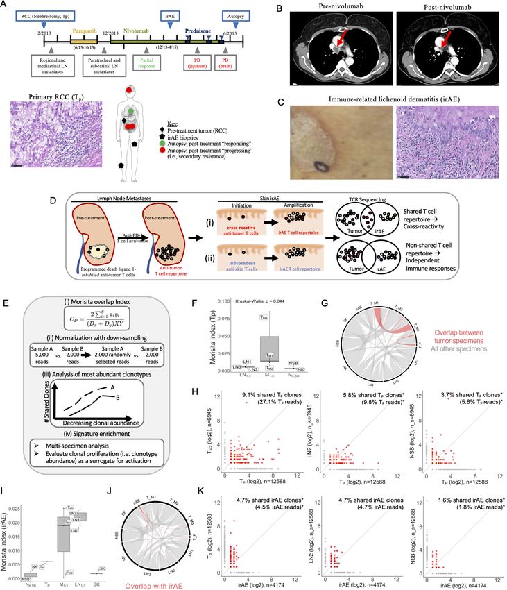
Figure 1 Clinical course and assessment of TCR repertoire overlap among tumor specimens and the irAE. (A) Timeline
shows the clinical course from RCC resection to autopsy. Therapies included pazopanib (yellow bar), nivolumab (green bar),
and prednisone (blue bars), with treatment dates shown below the bars. Radiographical assessments (gray boxes) included
mediastinal metastases with partial response to nivolumab followed by PD in the bowel and brain. Tissue specimens collected
(blue boxes) included the resected RCC (photomicrograph shown), biopsies of the immune-related lichenoid dermatitis (irAE),
and multiple specimens collected at the time of rapid autopsy (see online supplemental table S1). The anatomical sites of the
collected specimens are illustrated, including the primary renal tumor (black diamond), two sites of irAE (black pentagons),
mediastinal LNs at the site of tumor regression (green circles) and progressing lesions in the jejunum, mesentery, and brain (red
circles). (B) Intravenous contrast-enhanced CT of the chest demonstrated right lower paratracheal adenopathy which resolves
after nivolumab treatment (red arrow). (C) A photograph (left) and photomicrograph (right) of the irAE, the latter showing the
brisk lichenoid lymphocytic infiltrate and necrotic keratinocytes. (D) Diagram illustrating two hypotheses for the development
of irAEs following immune checkpoint blockade: (i) the cross-reactivity hypothesis proposed that T cells activated as part of
the antitumor immune response cross-react at the site of the irAE; this would be supported by detection of overlapping TCR
repertoire signatures between the tumor and irAE; (ii) in contrast, if the irAE and antitumor immune responses were independent
of each other, it was unlikely significant TCR repertoire overlap between the two sites would be detected. All photomicrographs
Figure 1 (Continued)
Cottrell T, et al. J Immunother Cancer 2021;9:e002642. doi:10.1136/jitc-2021-002642 3Open access
at ×400 magnification, scale bars 50 um. (E) Proposed approaches to maximize interpretive value and minimize confounding in
TCR sequencing data analysis, including (i) the MI for quantifying global TCR repertoire sharing among multiple specimens in a
J Immunother Cancer: first published as 10.1136/jitc-2021-002642 on 6 July 2021. Downloaded from http://jitc.bmj.com/ on July 15, 2021 by guest. Protected by copyright.
sample size-independent manner, (ii) proportional downsampling for library size normalization to enable relative interpretations
of clonal sharing, and (iii) normalization to relative clonal abundance in each specimen to assess relative sharing among the
most abundant T-cell clones across multiple specimens. (F) The Morita index demonstrates that the three metastatic lesions
consistently showed a greater degree of sharing with the Tp (MI median 0.014, range 0.012–0.088) relative to the normal control
tissues (NSB MI 0.004, NK MI 0.003). (G) Chord diagram illustrates TCR repertoire overlap among the Tp and multiple post-
treatment progressing metastases (red). Non-tumor specimens are shown in gray. (H) Following library size normalization, a
metastasis (TM2, left), LN (LN2, middle), and NSB (right) were evaluated for T-cell repertoire overlap with the primary tumor
(TP). Clonal expansion of clones shared with TM2 is suggested by 9.1% of shared TP clonotypes representing 27.1% of TP
reads. Subsampled comparisons are indicated (*) and the 95% CIs for shared TP clones were 5.2% to 6.8% for LN2 (middle,
representing 4.9%–15.8% total TP reads) and 3.3% to 4.3% for NSB (right, representing 3.5%–14.7% total TP reads). (I) The
Morita Index demonstrates an overall low degree of TCR repertoire between the irAE and the other specimens, although the
relative sharing with two of the three metastatic lesions (MI median 0.02, range 0.0003–0.026) and the three regression site
LNs (MI median 0.025, range 0.021–0.027) was higher than with the normal tissues (NSB MI 0.00004, NK MI 0.003). (J) Chord
diagram highlighting sharing with the irAE as assessed by the MI in red (sharing among other specimens shown in gray for
reference). (K) Following library size normalization, pairwise quantification of shared clones shows that while 4.7% of irAE clones
were shared with the primary tumor and a regression site LN (LN2, 95% CI 4.6% to 5.9% irAE clones), the shared clones were
not expanded in the irAE (representing 4.5% and 4.7%, 95% CI 4.7% to 6.4% of the total irAE reads, respectively). Sharing
between the irAE and NSB shown for comparison, 1.6% shared clones (95% CI 1.3% to 2.2%) represent 1.8% total irAE reads
(95% CI 1.3% to 2.2%). Subsampled comparisons are indicated (*). (L) The GLIPH2 clustering algorithm was used to detect and
quantify potential specificity clusters based on TCR CDR3 sequencing information. Motif clusters were included in downstream
analysis if there were ≥3 unique CDR3s, ≥10 reads for each CDR3, a vb scoreOpen access
from four healthy donors (range: 0.03%–1.67%, online TCRs and non-irAE skin TCRs. We recognize that we are
supplemental figure S8).25 Though the human leukocyte limited in our ability to comprehensively dissect all poten-
J Immunother Cancer: first published as 10.1136/jitc-2021-002642 on 6 July 2021. Downloaded from http://jitc.bmj.com/ on July 15, 2021 by guest. Protected by copyright.
antigen (HLA) information is unknown, three of the tial sources of confounding owing to limited sample avail-
four healthy donors had common clonotypes shared with ability. Lastly, the data-driven recommendations made in
the patient in our study, indicating that at least one HLA this study highlight the scientific value of rapid autopsy to
allele was shared among them. By querying additional answer complex questions using human tissue specimens.
published skin/tumor- reactive TCR data, the specific
motif SSQD was reported in a T-cell clone recognizing an
epitope derived from Maspin, which functions as a tumor METHODS
suppressor gene in epithelial cells.26 Collectively, this Case selection
indicates that, though analyses of the total TCR reper- Specimens from the underlying primary tumor and/or
toire and a subset of high abundance clones do not show metastatic site and from skin affected by the cutaneous
a signature of enriched sharing between the irAE and irAE were collected from the Johns Hopkins Hospital
tumor specimens relative to non-tumor specimens at the surgical pathology archives and the Rapid Autopsy
clonotype level, more ‘antigen-driven’ approaches may be program and Franklin Square Hospital. Overall patient
useful to identify potential specificity clusters, especially response to anti-PD-1 therapy was classified according to
when coupled with functional assays to confirm antigen Response Evaluation Criteria in Solid Tumors V.1.1.
specificity and cross-reactivity between irAEs and tumors.
TCRseq and bioinformatic analysis
DNA extraction from formalin-fixed paraffin-embedded
DISCUSSION (FFPE)- preserved tumor and skin biopsy specimens
As immune checkpoint blocking agents become first-line was performed using the DNeasy Blood and Tissue Kit
and second-line therapies for a growing number of tumor (Qiagen). The TCR-B locus was amplified and sequenced
types, we are faced with an increasing number of diverse using the ImmunoSEQ assay (Adaptive Biotechnologies).
irAEs that may develop during or after treatment. The Non-productive TCR CDR3 sequences (premature stop
association of cutaneous irAEs with clinical benefit in some or frameshift), sequences with amino acid length less
patients suggests that there may be a common antigen than 7, and sequences not starting with ‘C’ or ending with
that may underlie both durable antitumor responses and ‘F/W’ were excluded from the final analyses. Specimens
clinically significant irAEs. It is conceivable that T cells with at least 1000 reads were included in the final anal-
with a common TCR could mediate both tumor regres- ysis. To focus on T cells recognizing the same antigen, we
sion and irAE development and progression, as has been analyzed amino acid clonotypes exclusively.
evidenced by prior studies evaluating clonal overlap of The degree of clonality for each specimen was assessed
TCR clonotypes between tumor and irAE tissues8–10 and by the productive clonality matrix, which is defined as
that expansion of peripheral blood T-cell clones prior to 1-Pielou’s evenness.28 Values near one represent samples
irAE onset positively correlates with irAE severity during with one or a few predominant clones (monoclonal or
checkpoint blockade treatment.27 oligoclonal samples), whereas values near 0 represent a
The large number and circulating nature of T cells polyclonal population.
predispose these studies to detecting false positive signals, A random subsampling approach weighted by clonal
that is, detection of differential or statistically signifi- abundance was used to equalize library sizes for relative
cant clonal overlap that is not necessarily of pathogenic comparisons of TCR repertoire overlap. For subsampling,
relevance. Biological differences exacerbate this issue, each clonotype at amino acid level was treated as a sample
including variation in T-cell numbers and clonality in and specimens were randomly sampled with replacement
different tissue types. In addition, due to differences in and weighted by clonal abundance (or frequency) until
sampling, clonotype detection can be limited, particularly the total read count equaled that of the comparator
for rare/low-frequency clonotypes. The analysis pitfalls library. To account for subsampling variation, the proce-
and mitigation strategies identified in this study are dure was repeated 100 times and the 95%CIs for all subsa-
summarized in table 1, and we present considerations for mpled comparisons are reported.
prospective specimen collection in online supplemental The degree of T cell clone overlap at the species level
figure S9. Many of these factors are already considered was evaluated using the Morisita overlap index.29 30 This
as a standard part of large-scale genomic analyses, but measurement accounts for differences in library size and
they are not yet routinely applied to immune receptor diversity per specimen, values near one the species occur
sequencing datasets and, to date, no studies have demon- in the same proportion in both samples, whereas values
strated the differential outcomes when these important near 0 implies the two samples do not overlap in terms of
sources of confounding are not acknowledged. Strengths species. Clonotypic sharing at the individual clone level
of this study include the rare opportunity to analyze the was assessed in pairwise biological compartments before
TCR repertoire in the same patient across time, tissue and after normalization to the same library size. Clones
compartments, and disease states, and the ability to that were copresented in any of the compartment pairs are
compare with published tumor- reactive/skin-reactive defined as shared clones. Based on the clonal frequency
Cottrell T, et al. J Immunother Cancer 2021;9:e002642. doi:10.1136/jitc-2021-002642 5Open access
Table 1 Mitigating pitfalls and approaches for interpretation of TCRseq data
J Immunother Cancer: first published as 10.1136/jitc-2021-002642 on 6 July 2021. Downloaded from http://jitc.bmj.com/ on July 15, 2021 by guest. Protected by copyright.
Potential
confounders Pitfall Mitigation
Batch effect Circulating T-cell clones may be ‘shared’ by Control normal tissue(s) collected at the same timepoint can
multiple specimens collected at the same be used to identify these background clones.
time point.
Blood During active immune responses,* both Functional assays enable identification of disease-relevant
relevant and non-relevant clones circulate clones (vs batch background).
in blood.
Tissue Specimens from the same organ share Clonotype sharing with ‘paired’ normal tissue does not
compartment effect tissue resident T cells, including antitumor preclude biological relevance. Measurements such as
clones.19 abundance and antigen specificity (antigen-driven clustering/
functional assays) are needed for further discernment.
Library size Increased read count→more clones Analyses must correct for sample size variation (eg, Morisita
variation sampled→a larger proportion of shared Overlap Index, normalization, etc)
clones
LNs/lymphoid-rich Increased probability of repertoire overlap Avoid analysis of background lymphoid tissue in LN
tissues given large, diverse T-cell populations metastases; interpret LN data with caution.
Interpretation Definition Approach
Clonal abundance Relative proportion of sequencing reads Assess for signals of clonal proliferation in relevant tissues to
(relative read count) for a unique clonotype (surrogate for clonal suggest functional relevance.†
proliferation)
Low abundance Meaningful threshold for exclusion Exclude specimens withOpen access
Acknowledgements We thank the patient and the patient’s family for participation tumours treated with NIVO or NIVO+IPI: a systematic review and
in this study, members of our research and administrative teams who contributed meta-analysis. J Immunother Cancer 2019;7.
J Immunother Cancer: first published as 10.1136/jitc-2021-002642 on 6 July 2021. Downloaded from http://jitc.bmj.com/ on July 15, 2021 by guest. Protected by copyright.
to this study, and also Fiamma Berner and Lukas Flatz for generous and prompt 7 Rosati E, Dowds CM, Liaskou E, et al. Overview of methodologies for
tumor-reactive/skin-reactive T-cell receptor sequencing data sharing. T-cell receptor repertoire analysis. BMC Biotechnol 2017;17:61.
8 Läubli H, Koelzer VH, Matter MS, et al. The T cell repertoire in tumors
Contributors TC, GJK, and H-YC conceived of and conducted the experiments. overlaps with pulmonary inflammatory lesions in patients treated with
KNS and JT oversaw the study design, data interpretation, and manuscript checkpoint inhibitors. Oncoimmunology 2018;7:e1386362.
preparation. JZ, BZ, PB, and HJ led the bioinformatic analyses. FV, JEH, HH, and 9 Johnson DB, Balko JM, Compton ML, et al. Fulminant myocarditis
MEA oversaw the clinical care of the patient and led the specimen acquisition. All with combination immune checkpoint blockade. N Engl J Med
2016;375:1749–55.
authors contributed to and edited the manuscript. 10 Berner F, Bomze D, Diem S, et al. Association of checkpoint
Funding KNS was supported by the Lung Cancer Foundation of America, the Inhibitor–Induced toxic effects with shared cancer and tissue
IASLC Foundation, Swim Across America, and The Commonwealth Foundation. KNS, antigens in Non–Small cell lung cancer. JAMA Oncol 2019;5:1043.
JT, JZ, and BZ were supported by the Mark Foundation for Cancer Research. HJ 11 Zhang J, Ji Z, Smith KN. Analysis of TCR β CDR3 sequencing
data for tracking anti-tumor immunity. Methods Enzymol
was partially supported by the National Institutes of Health (NIH)/National Human 2019;629:443–64.
Genome Research Institute (grant R01HG009518). TC was supported by NIH (T32 12 Chou S, Hwang SJE, Carlos G, et al. Histologic assessment
CA193145). KNS and HJ were supported by R37 CA251447. This research was of lichenoid dermatitis observed in patients with advanced
funded in part through the Bloomberg-Kimmel Institute for Cancer Immunotherapy, malignancies on Antiprogramed cell death-1 (anti-PD-1) therapy with
Bloomberg Philanthropies, and P30CA006973. or without ipilimumab. Am J Dermatopathol 2017;39:23–7.
13 Curry JL, Tetzlaff MT, Nagarajan P, et al. Diverse types of
Competing interests HH has received clinical research funding from Bristol-Myers dermatologic toxicities from immune checkpoint blockade therapy. J
Squibb and Merck and serves in an advisory role for Pfizer, Merck, and Bristol- Cutan Pathol 2017;44:158–76.
Myers Squibb. JT receives research funding from Bristol-Myers Squibb and serves 14 Hwang SJE, Carlos G, Wakade D, et al. Cutaneous adverse events
a consulting/advisory role for Bristol-Myers Squibb, Merck, and Astra Zeneca. KNS (AEs) of anti-programmed cell death (PD)-1 therapy in patients
has received travel support/honoraria from Illumina, Inc., receives research funding with metastatic melanoma: A single-institution cohort. J Am Acad
from Bristol-Myers Squibb, Enara Bio, and Astra Zeneca, and owns founder’s equity Dermatol 2016;74:455–61.
15 Schaberg KB, Novoa RA, Wakelee HA, et al. Immunohistochemical
in manaT Bio. The terms of all these arrangements are being managed by the
analysis of lichenoid reactions in patients treated with anti-PD-L1
investigators’ respective institutions in accordance with their conflict of interest and anti-PD-1 therapy. J Cutan Pathol 2016;43:339–46.
policies. 16 Tetzlaff MT, Nagarajan P, Chon S, et al. Lichenoid dermatologic
Patient consent for publication Not required. toxicity from immune checkpoint blockade therapy: a detailed
examination of the clinicopathologic features. Am J Dermatopathol
Ethics approval This study was approved by the institutional review board (IRB) 2017;39:121–9.
at Johns Hopkins University (JHU) and was conducted in accordance with the 17 Robins HS, Campregher PV, Srivastava SK, et al. Comprehensive
Declaration of Helsinki and the International Conference on Harmonization Good assessment of T-cell receptor beta-chain diversity in alphabeta T
Clinical Practice guidelines. The patient described in this study provided written cells. Blood 2009;114:4099–107.
18 Carlson CS, Emerson RO, Sherwood AM, et al. Using synthetic
informed consent as approved by the IRB of JHU.
templates to design an unbiased multiplex PCR assay. Nat Commun
Provenance and peer review Not commissioned; externally peer reviewed. 2013;4:2680.
19 Zhang J, Ji Z, Caushi JX, et al. Compartmental analysis of T-cell
Supplemental material This content has been supplied by the author(s). It has clonal dynamics as a function of pathologic response to neoadjuvant
not been vetted by BMJ Publishing Group Limited (BMJ) and may not have been PD-1 blockade in resectable non-small cell lung cancer. Clin Cancer
peer-reviewed. Any opinions or recommendations discussed are solely those Res 2020;26:1327–37.
of the author(s) and are not endorsed by BMJ. BMJ disclaims all liability and 20 Danilova L, Anagnostou V, Caushi JX, et al. The mutation-associated
responsibility arising from any reliance placed on the content. Where the content neoantigen functional expansion of specific T cells (MANAFEST)
includes any translated material, BMJ does not warrant the accuracy and reliability assay: a sensitive platform for monitoring antitumor immunity. Cancer
Immunol Res 2018;6:888–99.
of the translations (including but not limited to local regulations, clinical guidelines,
21 Forde PM, Chaft JE, Smith KN, et al. Neoadjuvant PD-1 blockade in
terminology, drug names and drug dosages), and is not responsible for any error resectable lung cancer. N Engl J Med 2018;378:1976–86.
and/or omissions arising from translation and adaptation or otherwise. 22 Zhang J, Ji Z, Caushi JX, et al. Compartmental analysis of T-cell
Open access This is an open access article distributed in accordance with the clonal dynamics as a function of pathologic response to neoadjuvant
Creative Commons Attribution Non Commercial (CC BY-NC 4.0) license, which PD-1 blockade in resectable non-small cell lung cancer. Clin Cancer
Res 2019
permits others to distribute, remix, adapt, build upon this work non-commercially, 23 Huang H, Wang C, Rubelt F, et al. Analyzing the Mycobacterium
and license their derivative works on different terms, provided the original work is tuberculosis immune response by T-cell receptor clustering with
properly cited, appropriate credit is given, any changes made indicated, and the use GLIPH2 and genome-wide antigen screening. Nat Biotechnol
is non-commercial. See http://c reativecommons.org/licenses/by-nc/4.0 /. 2020;38:1194–202.
24 Glanville J, Huang H, Nau A, et al. Identifying specificity groups in the
T cell receptor repertoire. Nature 2017;547:94–8.
25 Cheuk S, Schlums H, Gallais Sérézal I, et al. CD49a expression
defines tissue-resident CD8+ T cells poised for cytotoxic function in
REFERENCES human skin. Immunity 2017;46:287–300.
1 Martins F, Sofiya L, Sykiotis GP, et al. Adverse effects of immune- 26 Berner F, Bomze D, Diem S, et al. Association of checkpoint inhibitor-
checkpoint inhibitors: epidemiology, management and surveillance. induced toxic effects with shared cancer and tissue antigens in non-
Nat Rev Clin Oncol 2019;16:563–80. small cell lung cancer. JAMA Oncol 2019;5:1043–7.
2 Haslam A, Prasad V. Estimation of the percentage of US patients 27 Subudhi SK, Aparicio A, Gao J, et al. Clonal expansion of CD8 T cells
with cancer who are eligible for and respond to checkpoint inhibitor in the systemic circulation precedes development of ipilimumab-
immunotherapy drugs. JAMA Netw Open 2019;2:e192535. induced toxicities. Proc Natl Acad Sci U S A 2016;113:11919–24.
3 Weber JS, Kähler KC, Hauschild A. Management of immune-related 28 Kirsch I, Vignali M, Robins H. T-cell receptor profiling in cancer. Mol
adverse events and kinetics of response with ipilimumab. J Clin Oncol 2015;9:2063–70.
Oncol 2012;30:2691–7. 29 Wolda H. Similarity indices, sample size and diversity. Oecologia
4 Freeman-Keller M, Kim Y, Cronin H, et al. Nivolumab in resected 1981;50:296–302.
and unresectable metastatic melanoma: characteristics of immune- 30 Morisita M. Measuring of the dispersion and analysis of distribution
related adverse events and association with outcomes. Clin Cancer patterns. In: Memoires of the faculty of science. Kyushu University,
Res 2016;22:886–94. 1959.
5 Das S, Johnson DB. Immune-Related adverse events and anti-tumor 31 Gu Z, Gu L, Eils R, et al. circlize implements and enhances circular
efficacy of immune checkpoint inhibitors. J Immunother Cancer visualization in R. Bioinformatics 2014;30:2811–2.
2019;7:306. 32 Nazarov VI, Pogorelyy MV, Komech EA, et al. tcR: an R package for T
6 Xing P, Zhang F, Wang G, et al. Incidence rates of immune-related cell receptor repertoire advanced data analysis. BMC Bioinformatics
adverse events and their correlation with response in advanced solid 2015;16:175.
Cottrell T, et al. J Immunother Cancer 2021;9:e002642. doi:10.1136/jitc-2021-002642 7You can also read